Development of a Polarized Helium-3 Ion Source
for RHIC using the Electron Beam Ion Source
by
Charles Samuel Epstein
Submitted to the Department of Physics
in partial fulfillment of the requirements for the degree of
Bachelor of Science
at the
MASSACHUSETTS INSTITUTE OF TECHNOLOGY
MASSA6HUSETTS INsTmffE OFTECHNOLOGY
SEP
04
2013
I
LIBRARIES
June 2013
©
Charles Samuel Epstein, MMXIII. All rights reserved.
The author hereby grants to MIT permission to reproduce and to
distribute publicly paper and electronic copies of this thesis document
in whole or in part in any medium now known or hereafter created.
A u th o r ...
Department of Physics
May 10, 2013
C ertified by ...
Professor 4ichard G. Milner
Professor of Physics, Director of Laboratory for Nuclear Science
Thesis Supervisor
A ccepted by ... ...-...
Professor Nergis Mavalvala
Senior Thesis Coordinator, Department of Physics
Development of a Polarized Helium-3 Ion Source for RHIC
using the Electron Beam Ion Source
by
Charles Samuel Epstein
Submitted to the Department of Physics on May 10, 2013, in partial fulfillment of the
requirements for the degree of Bachelor of Science
Abstract
This thesis presents my work on the design and development of a source of polarized Helium-3 ions for the Relativistic Heavy Ion Collider at Brookhaven National Lab, Upton, NY. The 3He atoms will be polarized using the technique of metastability-exchange optical pumping (MEOP), and will then be flowed into the newly commis-sioned Electron Beam Ion Source (EBIS). Fully stripped 3He++ ions will be extracted and their polarizations measured at low energies before acceleration in the RHIC complex.
Thesis Supervisor: Professor Richard G. Milner
Acknowledgments
I wish to express my gratitude to all of the people who have made this project possible. At MIT, our collaborators are Richard Milner, James Maxwell, and Eliza Mace; at Brookhaven National Laboratory, Jim Alessi, Ed Beebe, Alexander "Sasha" Pikin, Thomas Roser, Wolfram Fischer, Xaofeng Gu, and Anatoli Zelenski. We have had very helpful discussions with Werner Heil, Sergei Karpuk, and Ernst Otten (U. Mainz), Pierre-Jean Nacher (ENS, Paris), and Guilhem Collier (University of Cracow, Poland). Sasha's advice has been vital in our understanding of the EBIS device and magnetic field, and he has provided us with many drawings, much data, and code. Thank you also to Daniel Kleppner for discussions that have been extremely helpful towards understanding the atomic processes inside EBIS.
The engineers at Bates, under the leadership of Jim Kelsey, have been invaluable, and thank you especially to Ernie Ihloff who has met with me many times about this project and from whom I've learned a great deal. Brian O'Rourke has played a key role in making the new sealed cells. Thanks also to Genya Tsentalovich for helping us learn how to use the VectorFields Tosca/Opera electromagnetic modeling software.
Once again, a most important thanks to Richard Milner for being an amazing mentor and for giving me so many extraordinary opportunities. I'm looking forward to returning as a grad student in the fall.
Contents
1 Physics Motivation 1.1 Introduction ... 1.2 Polarization of 3He . . 1.2.1 MEOP Process 1.3 Setup Overwiew . . . .2 Optical Pumping using the Metastability Exchange EBIS
2.1 EBIS Overview . . . . 2.1.1 EBIS Ion Trap Properties . . . . 2.1.2 Flow from the Pumping Cell to EBIS . . . . . 2.2 Possible Processes In EBIS Leading to Depolarization 2.2.1 A Note on Charge Exchange . . . . 2.2.2 Wall Bounces in Transfer Line . . . . 2.3 Magnetic Field Gradients . . . .
3 Experimental Realization
3.1 MIT Campus Lab . . . . 3.1.1 Keopsys Lasers... 3.1.2 Polarimetry . . . . 3.2 Sealed Cell Production at Bates
3.3 Preliminary Polarization . . . . Technique near 17 17 18 21 22 22 23 24 27 27 28 29 30 31 11 11 12 13 14 . . . . . . . . . . . . . . . .
4 Test of Source Concept at RHIC 33
4.1 Polarization Measurement with a Two-Cell System . . . . 33
4.1.1 Equilibrium Polarization Measurement . . . . 34
4.1.2 Sensitivity of EBIS Cell Polarization Measurement . . . . 36
4.1.3 Expected Polarization Retention in Transfer . . . . 37
List of Figures
1-1 Diagram of the MEOP process. (J. Maxwell) ... 12
1-2 CAD drawing of shielded Helmholtz pair. (J. Maxwell) . . . . 15
1-3 Overview of optical pumping setup near EBIS. (J. Maxwell) . . . . . 15
2-1 Top: a basic schematic of EBIS. Below: the electric potential along the length of EBIS. (A. Pikin) . . . . 20
2-2 A schematic of EBIS. (A. Pikin) . . . . 20
2-3 Simulated transfer line location in EBIS field . . . . 25
2-4 Heat-map showing polarization relaxation times near EBIS solenoid (center at origin). . . . . 25
3-1 Diagram of the MIT lab setup. . . . . 28
3-2 Plot showing the PDF10A femtowatt photoreceiver output (volts) as the liquid crystal retarder changes states, with a circularly polarizing filter engaged. . . . . 31
3-3 Plot of polarization versus time for new sealed cell, measured with liquid crystal retarder. . . . . 32
3-4 Sealed cell production system schematic . . . . 32
4-1 Test experiment setup schematic. (J. Maxwell) . . . . 34
4-2 Cutaway CAD drawing of test setup. (J. Maxwell) . . . . 35
Chapter 1
Physics Motivation
1.1
Introduction
An issue in nuclear physics currently receiving much attention is that of the spin structure of the nucleon. Experiments at SLAC, CERN, and DESY, have determined that quarks only contribute about 25% of the nucleon's spin. As a result, current experiments such as those at RHIC-spin are testing the contributions of gluons in order to determine how the remaining 75% of the spin arises. A useful tool in all of these studies has been the spin-polarized proton. However, it is beneficial to perform experiments with both isospin configurations of the nucleon as both a control and a place to search for new physics. According to current theoretical predictions, the polarized neutron provides a similar gluon distribution to that of the proton, but with a different configuration of quarks. A polarized neutron beam at RHIC-spin would thus be a remarkable asset to the study of nucleon spin structure. [8]
Due to the difficulties associated with manipulating free neutrons, experiments studying the polarized neutron have often used polarized deuterons or 3He. However, the deuteron is not suitable for beam formation because of its low magnetic moment, which makes spin manipulation in accelerators difficult. The magnetic moment of
3He is similar to that of the free neutron due to the pairing of the two protons in a
low-energy shell with opposite spin states. This reasonable magnetic moment allows the spins to be manipulated in accelerators using standard technology such as siberian
23 P
CP Laser 1083 nm2
3
S
1
RF Excitation (~1 ppm) us 0 1/2 mF=-3/2 -1/2 1/2 3/2 Equal C8 I +1Probability C1 1C DecayI
I
I
ITI
1/2 -L3/2 ---3/ 2 M eta stab lity Exchange
F=1/2
-1/2 1/2
Figure 1-1: Diagram of the MEOP process. (J. Maxwell)
snakes. For these reasons, it is of great interest to develop a high-intensity source of highly polarized 3He. [8]
Previous experiments have performed elastic scattering (BLAST at MIT-Bates South Hall Ring) or deep inelastic scattering (HERMES at HERA, DESY) using polarized electrons on a polarized 3He internal gas target. Upgrading the 3He to beam energies in a future electron-ion collider (eRHIC) would enable fundamental tests of theoretical predictions of the Standard Model, such as the Bjorken Sum Rule. Until then, a polarized 3He ion source at RHIC would enable polarized np collisions from the onset, allowing new types of QCD studies to be performed and providing a means for other nucleon spin structure tests.
1.2
Polarization of
3He
Low-energy sources of polarized 3He+ ions have been developed several times in the past. A 50 particle nA, 65% polarized source was developed at the University of Birmingham, UK [2]. A collaboration between Rice University and Texas A&M developed an 8 particle pA, 11% polarized source [4]. At Laval University, Canada, a source polarized using a Stern Gerlach based method provided 100 particle nA at
95% polarization [13]. It is planned to provide a high-current, 500 particle nA source of 70% polarized 3He++ ions to RHIC.
Metastability-exchange optical pumping (MEOP) is our method of choice to po-larize the 3He atoms. In this technique, a weak RF discharge excites atoms into the metastable 23S state, where they are pumped to the 23P states by 1083 nm circularly polarized laser light tuned to either the C8 or C9 transition (Fig. 1-1). This polarizes the optical electron, whose spin state couples to the nuclear state through the hy-perfine interaction. Metastability exchange collisions then occur, in which atoms can swap electron configurations, allowing the nuclear polarization to propagate to the ground state atoms. MEOP has been performed with spectacular success. Modern ytterbium fiber lasers, replacing the dated Nd:YAG systems, have allowed highly-polarized, high-current MEOP systems to be constructed. At the University of Mainz, a flow rate of 8 x 10¹⁸ 3He/sec at polarizations over 70% has been demonstrated [8].
1.2.1
MEOP Process
To illustrate the MEOP process, we consider a ground state atom with an electronic
and nuclear states described by the density matrices PGe and PGN, and a metastable atom with electronic state PMe and nuclear state PMN- The density matrices that characterize the ground state and metastable atoms are then PG = PGe 0 PGN and
PM = PMe 0 PMN, respectively. After optical pumping polarizes the electronic state
of the metastable atom, we consider the coupling of the nuclear and electronic spins via the hyperfine interaction, represented by the projection onto a hyperfine sublevel
F: P'MN = ZFPFPMNPF = PMN-PO where PF is the projection operator onto the
respective F sublevel [3]. After coupling occurs, the density matrix becomes p' = P'Mie ®pMN-Pol. Following this, a metastability exchange collision occurs, transferring the polarization to a ground state atom. The density matrix of the ground state atom thus becomes p'G, = PGe 0 PMN-POl, and that of the metastable atom becomes
P'Mv = PMe 0 PGN- This illustrates the electronic states being swapped. A summary of the process can be seen in Table 1.1. The result of the interaction is that a metastable atom is polarized electronically by optical pumping, and then transfers this electronic
angular momentum to its nucleus, which can then be transferred to a ground state atom, causing nuclear polarization of the ground state population.
Table 1.1: An overview of the MEOP process, based on table by [3].
Ground State Atom Metastable Atom
Initial Config. PG = PGe 0 PGN PM PMe 0 PMN
Hyperfine Inter. PG PM = ZFPFPMPF
Me PMN-Pol
Metastab. Exch. P'G = PGe (PMN-POl PM! = PMe 0 PGN
1.3
Setup Overwiew
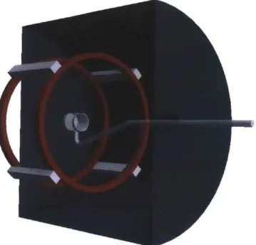
A pumping cell in which MEOP will be performed is to be located several meters away from EBIS. The magnetic field must be sufficiently uniform such that depolarization effects from transverse gradients are minimized. While it is possible to polarize in the strong kilogauss fringe fields with the aid of gradient correction coils, it is planned to instead construct cylindrical soft steel shielding inside which the pumping cell, Helmholtz coils, and polarization optics will be located. This provides maximum flexibility in the pumping cell location and allows polarization at low-field (~ 30 Gauss), where the MEOP process and our polarimetry methods are well-understood. A CAD drawing of the shielded Helmholtz pair is visible in Fig. (1-2), and a setup schematic in Fig. (1-3).
Figure 1-2: CAD drawing of shielded Helmholtz pair. (J. Maxwell) Helmholtz Pair 3 He Transfer B Shield Line
Pump Laser Elect
N 0ElectronRBeam
IonESource
U U U ---- ---- --- - Drift Tubes Ntjn Gun --- Ion Exrractor
i :00
N N N a 0 a
Chapter 2
Optical Pumping using the
Metastability Exchange Technique
near EBIS
A number of technical constraints have been taken into account in the design of the EBIS-based polarized ion source. Often, these constraints lend a choice of technique for how they may be addressed. In many of the sections that follow, the various options and their parameters will be discussed, and the rationale for the choice will be presented.
2.1
EBIS Overview
As an successor to the Tandem Van de Graaff ion sources at RHIC, a more efficient Electron Beam Ion Source (EBIS) has been developed and was commissioned in 2010 [1]. The EBIS is a 1.5 meter-long ion trap with a 15 keV electron beam enclosed in a 5T superconducting solenoid (Figures 2-1, 2-2). Over the past several years, it has been used with great success to prepare a vast variety of highly-ionized species.
In order to extract approximately 1012 3He++ per second, it is intended to flow in
2.1.1
EBIS Ion Trap Properties
In the following sections, the parameters for a number of properties of the EBIS ion trap are calculated. These include the operating pressure, ion trapping mechanisms,
average time spent in the trap, and energy deposition by the electron beam.
Ionization in EBIS
In [12], Seltzer and Berger apply Bethe theory and Sternheimer density-effect correc-tions in order to estimate the collision stopping power of electrons through various materials. They arrive at:
1 (dE 0.153536 ZB(T)
p dx 02 A
where 3 = v/c, Z is atomic number, A is atomic weight, and
B(T) = Bo(Tn) - 2In -j
where I is the mean excitation energy of the medium (41.8 eV for Helium), mc2 is
the rest energy of the electron (0.511 MeV), and 6 is the density effect correction. Furthermore,
Bo(T) = ln(T2
(T + 2)/2) + [1 + T2/8 - (2r + 1) In 2]/(r + 1)2
where T = T/mc2. T is the electron kinetic energy, which in EBIS is roughly 15 keV.
Evaluating this yields Bo(T) = -6.79.
The density-effect correction, which accounts for the material becoming polarized as a charged particle traverses it, is estimated by a numerical formula piecewise in the
parameter X =
j
logo (r(r + 2)) [12]. Below a certain a material-specific X0 value,J = 0. We find that this is the case for our parameters, where X = -0.6125 and for Helium, Xo = 2.191. Thus, 6 is indeed 0.
= 21.9 MeV p dx g cm- 2 With p = 1010 atoms/cm3 -+ 4.98 x 10⁻¹⁴ g/cm3, _I
()
= 1.09 x 10⁻¹² MeV/cm. p dx CmAs there are 6 x 10'" electrons passing over the length of EBIS (190 cm) each second, energy is deposited at a rate of 1.24 x 10¹⁰ MeV/sec. Since 79 eV are necessary to fully ionize a 3He atom, an extraction rate of 3 x
1012 ions/sec requires about 2.4 x 10⁸ MeV/sec. Thus, the energy provided by the electron beam is sufficient by more than a factor of ~ 102.
Pressure Inside EBIS
In order to design the vacuum systems for providing 3He flow to EBIS, the operating pressure must be estimated. Estimating that the EBIS turbopumps provide a vacuum outflow pumping speed of 100 1/s = 105 cm3
/s
at the trap, then the pressure P = Q/S[9] is
3 x 10¹⁴ s-1
P = 33 x 109 Cm-3 ~ 10-7 torr
105 cm3s-1
Ion Trapping
Once the 3He atoms become ionized in the electron beam, they are trapped by two main processes. For one, the positive ions are electrostatically attracted to the negatively-charged electron beam. Additionally, the positive ions are located in a strong 5T solenoidal magnetic field, and thus will undergo circular motion in the p, q5 plane. The radius of curvature of their motion is given by
Bqv = mV2
r
With B = 5T, q = 1.6 x 10⁻¹⁹ C, thermal velocity v = 103 m/s, m = 5 x 10-27 kg,
Magnetic ShIrns
SoleoiColectr
Electron Ion
Gun DrIft Tubes Extractor
+ +Anode A~eCTrapp"i Potential Extraction Potential Electron S'--- Colector - Cathode
I
ExtractorFigure 2-1: Top: a basic schematic of length of EBIS. (A. Pikin)
Transition region magnet coil
HV feedthroughs Gate valves
Electron gun Drift tubes bucking coil
Electron gun
EBIS. Below: the electric potential along the
Electron collector Gate valv bucking coil
CryopurnElectron collector Superconducting Exit lens solenoid
ryopump
Figure 2-2: A schematic of EBIS. (A. Pikin) m) Turbopump I ffR j ---- hill --- M-Turbopumn ryol Insulat-n
this means that the 3He++ ions are confined to the trap once ionized. Significantly, this implies that they will not later undergo wall bounces.
Time Spent in Trap
Another important figure is the approximate time spent in the trap, which is impor-tant as a figure of reference for polarization relaxation processes. First, the equilib-rium number of atoms in the trap is estimated. With a trap length of 1.5 m and an approximate diameter of 5 cm, the volume of gas in the trap is
257r333
- 150 cm3 = 2945 cm3 ~ 3, 000 cm3
4
Then this implies that there are
3 x 10⁹ m-3 .3 x 103 CM3
= 9 x 10¹² atoms inside EBIS at equilibrium. Then, comparing this with the extraction rate, the average time the atoms spend in the trap is
9 x 10¹²
s ~ 30 ins.
3 x 10¹⁴ s-1
So far, all discovered relaxation processes seem to occur over far longer time scales.
2.1.2
Flow from the Pumping Cell to EBIS
Polarized 3He atoms must be transferred from the pumping cell at Po ~ 1 torr to
EBIS at ~ 10-7 torr. It is calculated that the flow conductance between the pumping cell and EBIS must be approximately 8 x 10⁻⁶ l/s, based on principles outlined in [9]. A calibrated capillary leak at 10-5 atm-cc/sec at the pumping cell and a O(mm) diameter tube to EBIS will provide the necessary flow rate. The transfer line should be made of glass so that it can withstand high vacuum and so that it can be baked to minimize outgassing.
2.2
Possible Processes In EBIS Leading to
Depo-larization
There are a number of processes that take place inside EBIS that may lead to depo-larization. They include:
" Charge exchange. 3He+ + 3He++ 4 3He++ + 3He+
This has a cross section of - ~ 10-16 cm2, and is thus thought to occur at a
very low rate.
" Recombination. 3He+ + e
- 3He; 3He++ + e -+ 3He+
This is a 3-body process and thus thought to be unlikely. The cross section is
lowered by a factor of a2, thus -< 10-20 cm2
" Spin-exchange collisions. These are thought to occur at a low rate. The cross section for neutral Hydrogen spin-exchange collisions is approximately
1014 cm2; for 3He ions it is expected to be even lower. It is expected that
due to coulomb interactions, the ions will not even approach close enough for spin-exchange to occur.
" Wall depolarization of atoms inside EBIS. This is thought to be low because of the low probability of depolarization in a wall collision. Further study is underway to understand how many atoms will collide with the walls before being ionized.
2.2.1
A Note on Charge Exchange
It is important to investigate charge exchange because 3He+ has a free electron which can depolarize, and then cause the nucleus to depolarize via hyperfine coupling. If a 3He+ and a 3
He++ ion undergo charge exchange, depolarization in the 3He+
popula-tion can propagate to the 3He++ ions.
We approximate the rate of charge exchange as pip2VUV, where pi, P2 are the
densities of singly and doubly ionized 3He, V is the average velocity, o-
the charge exchange cross section, and V is the volume. We approximate the densities as both being 3 x 10 cm-3, which corresponds to 1% of the gas being ionized. Then
the rate is
(3 x 107 cm-3)(3 x 107 cm-3) 105 cm s-'
x10-16 cm-2 3 x 103 cm3 = 2.7 x 10 7 s-1
~ 3 x 10⁷ s-1
Since the rate of ion extraction is approximately 1012 S- 1, it is thought that even
if singly-charged ions depolarize and undergo charge exchange, the effects will be minimal.
2.2.2
Wall Bounces in Transfer Line
In [15], the relaxation time of 3He was measured on various surfaces. In an enclosed
space with surface area A, such as the pumping cell, the number of wall collisions per unit time is given by 1pA, where p is the density and V is the average velocity. With a measured relaxation time T, the probability to depolarize per collision is approximately
1
P ~~ 102
In a glass transfer line with an average number of collisions of 106, the expected depolarization is 1 part in 1014, which is negligibly small.
2.3
Magnetic Field Gradients
A strong transverse magnetic field gradient can cause depolarization if the field direc-tion in the particle's rest frame appears to oscillate near the Larmor frequency [10]. The polarization relaxation time of the 3He is related to the strength of the transverse magnetic field gradients by the equation [10]:
1 = - (V2)
(2.1)
T 3
JB
12 W 22 +1where for 3He, wo = 3.24 jB
11 kHz/Gauss and Tc ~ 2.2 x 10- 7p- 1, where p is in torr.
Such magnetic field gradient effects should be the dominant depolarization process in the transfer line. The polarization decays exponentially as exp(-t/), thus for a transfer line with T a function of position, this can be generalized to
P=Poexp -. (2.2)
for a particle spending time Ati at each location i. Assuming the transfer velocity is reasonably constant, we can factor out the Ati into the total transfer time F and number of locations N; thus
P = Po exp -. (2.3)
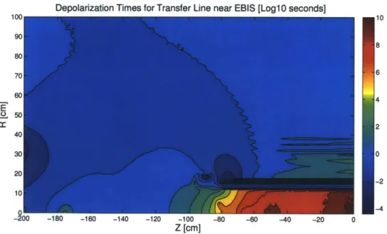
The relaxation time Tr is calculated at each point on the proposed path, and then Eq. (2.3) is computed. As an estimate, the transfer line is chosen to begin at (-110 cm, 70 cm), move to (-110 cm, 10 cm), then into EBIS at (-80 cm, 10 cm), the coordinate origin being the center of the trap, as in Fig. (2-3). With the short transfer times associated with molecular flow (10-3 seconds), the polarization retention along this sample path is calculated to be greater than 98%.
Since the gradient of the magnetic field is represented by a tensor, the transverse gradient (to the holding field) observed by the atom is dependent on its path. In
Depolarization Times for Transfer Line near EBIS [Loalo secondsl
.0.4
cc
Z [cm]
Figure 2-3: Simulated transfer line location in EBIS field
Depolarization Times for Transfer Line near EBIS [Log 10 secondsl
E
-100
Z [cm]
Figure 2-4: Heat-map showing polarization relaxation times near EBIS solenoid (cen-ter at origin). 110 -5 10 8 6 4 2 0 -2 -4 5 0
some cases, aligning the path of the atoms with a field line can produce a desirable reduction in the transverse gradient. However, the extent of the improvement depends greatly on the specific field configuration: in some instances, there is little benefit to following a field line, and in others, it is significant. As the atoms are bouncing along the interior of the transfer line, the direction of the velocity vector is changing rapidly, and thus in calculations, it is sometimes difficult to pin down the precise choice of "which" transverse gradient is applicable. Our interpretation of [10] leads us to believe that the chaotic motion of the atoms necessitates that the transverse gradients in all three dimensions should be added in quadrature in the squared term of Eq. (2.1).
Chapter 3
Experimental Realization
3.1
MIT Campus Lab
A lab on the MIT campus has been established for the development of the ion source. Tests have been performed on sealed 3He cells in order to understand and optimize
the polarization process. Two Keopsys 1OW "HePup" fiber lasers provide 1083 nm light for optical pumping of the C8 or C9 transitions. Light emerges from the fiber head in a linearly polarized state, which is then passed through a polarizing beam-splitting cube, a quarter-wave plate, and then beam expansion optics. After passing through the cell, a mirror reflects the transmitted laser light back through the cell for a second pass. The light continues back through the laser optics and upon reaching the quarter-wave plate becomes rotated in polarization relative to the original beam; thus, when passing through the polarizing beam-splitting cube (PBS), it is deflected into a Thorlabs DET36A silicon photodiode rather than the laser head. This allows a continuous measurement of absorption to be performed. Fig (3-1) shows a diagram of the setup. Data is acquired with a National Instruments PXIe-1078 DAQ crate, with a PXIe-6363 X Series Multifunction DAQ card connecting to two remote BNC connector pods (BNC-2110 and BNC-2111), and a PXIe-8370 MXI Express interface connecting to a PCIe slot on the lab computer. The DAQ system interfaces directly with LabView, in which routines are programmed. USB/serial connections interface with the controllers for Thorlabs SH1 shutters, the controllers for Thorlabs
LCC1112-667nm Polarimeter
MirrorPB
3He Concave A/4 Fiber Head
Cell Convex Lens Photodiode Lens
Helmholtz Coils
Figure 3-1: Diagram of the MIT lab setup.
B and LCC1111-C liquid crystal retarders, and a TTL control of the RF discharge, enabling nearly complete hardware automation.
3.1.1
Keopsys Lasers
The Keopsys 1083 nm ytterbium fiber lasers are manufactured for the purpose of 3He optical pumping, and can be operated at a power level of up to 10W. After a brief warm-up period (~ 10 minutes), the laser is tuned to either the Cs or C9 transition
by absorption spectroscopy on a secondary 3
He discharge cell. A secondary laser monitor fiber output is used for this so that the polarization setup is not disturbed and so that the calibration can be performed continuously. An infrared-optimized photomultiplier tube is positioned perpendicular to the monitor fiber output so that the re-radiated infrared light can be detected: at resonance, the light is reradiated primarily at ninety degrees. The transitions can be mapped this way by observing a strong signal when the laser is tuned to resonance.
The laser can be tuned either by a potentiometer on the device or by a ramp voltage input. The latter method has been used with a LabView-generated DC ramp in order to map the 3
He transitions at 1083 nm. The laser assigns a tune value based on the setting indicated by the potentiometer or input ramp voltage; however, this was seen to be unreliable day-to-day in predicting the precise output wavelength.
Combined with a slight drift in frequency during operation, this necessitated frequent and constant calibration. As the tuning cell absorption measurement can be made continuously and the wavelength can be controlled externally, a future revision of our data acquisition software will include a PID loop to follow the resonance and automatically keep the laser tuned.
3.1.2
Polarimetry
Several methods can be used to measure the 3He nuclear polarization. NMR is a
natural approach, but the coupling of nuclear and electronic spins can be harnessed to provide a simpler and more efficient polarization measurement. While the weak RF discharge is activated, various spectral lines can be analyzed in order to indi-rectly learn the nuclear polarization state. The two primary methods for polarization measurement involve infrared probe absorption, or optical polarimetry of the 667nm discharge line.
Performing the pump-probe polarimetry measurement is conceptually simple. A circularly polarized probe laser is directed transverse to the pumping cell. This cir-cularly polarized light consists effectively of two linearly polarized components, one that is transverse to the polarization axis, and one that is along it. The ratio of the transmitted power of transverse to longitudinally polarized light is calculated, giving the ratio of populations in the two 23S sublevels [14]. The population densities are known as a function of polarization, and thus the nuclear polarization can be inferred
[14].
A legacy polarimeter from previous MIT internal target tests has been explored as a first polarimetry option. This type of polarimeter indirectly measures the nuclear polarization by observing the circular polarization of the 667 nm discharge line. The calibration between optical and nuclear polarizations was performed in [6]. Light first passes through a rotating quarter-wave plate, which transforms the circularly polarized light into a rotating linearly polarized wave. This passes through a linear analyzer, which converts it to a wave of oscillating amplitude. The light passes through a 667 nm bandpass filter and is then detected by a photomultiplier tube. A
chopper on the rotating waveplate mount provides a reference signal by which a lock-in amplifier can measure the AC amplitude of the signal. A DC amplifier magnifies the constant level of the signal, and the ratio of the AC to DC components provides the optical polarization. There exists a correction factor of 1/ cos(6), where 6 is the angle between the polarimeter and the polarization axis, that must also be applied to compensate for observing the circularly polarized light from a nonzero angle of
incidence [11].
A new generation of 667 nm polarimetry is being developed using a liquid crystal retarder and photodiode to replace, respectively, the rotating quarter-wave plate and photomultiplier tube. The liquid crystal retarder is set to oscillate between the A/4 and the 3A/4 states, producing transmitted intensities I, and I2, lending an optical polarization of P = (I1 - 12)/(11 + 12). This method has higher statistical
uncer-tainty than the legacy method, as the signal being measured is at 1 Hz rather than at the high waveplate rotation frequency (~200 Hz), but the much greater simplicity lends to a strong reduction in potential systematic offsets. A Thorlabs PDF10A fem-towatt photoreceiver is sufficiently sensitive to provide the necessary precision in the polarization measurement (variation of less than 1%). When a circularly polarizing filter is placed before the polarimeter (to simulate measuring 100% polarization), the signal-to-noise as the liquid crystal retarder changes states is quite large. This can be seen in Fig. (3-2). When this optical polarimetry technique is used, the room lights are switched off and a sodium lamp is turned on, so that there is no ambient 667nm light that could interfere with the measurement.
3.2
Sealed Cell Production at Bates
Sealed cells for initial polarization tests have been produced at MIT-Bates. A sealed cell consists of two optically-clear windows (fused silica or #7740 Pyrex) of 2-inch di-ameter, connected by a 2-inch length #7740 Pyrex cylinder. The empty cells, custom manufactured by Finkenbeiner, Inc., of Waltham, MA, are initially produced with a glass to mini-conflat extension piece for attachment to filling equipment. Alternating
Photodiode Output for switched Liquid Crystal Retarder States
3IX.Dm 700Dm 900.0mL. 1.1 12 13 IA 1.5 1.6
Time [S]
Figure 3-2: Plot showing the PDF10A femtowatt photoreceiver output (volts) as the liquid crystal retarder changes states, with a circularly polarizing filter engaged.
cycles of baking and running of a strong RF discharge are used to clean the cell prior to filling. A residual gas analyzer is used to verify the removal of water vapor and other unwanted gases, and the spectral lines of the discharge are analyzed in order to verify purity. When finished, the cell is flamed off from the system. A schematic of the setup is visible in Fig. (3-4).
3.3
Preliminary Polarization
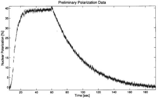
Polarization of a new sealed cell has been achieved and measured with the liquid crystal retarder technique. Fig. (4.4) shows a plot of the polarization versus time as the laser is first turned on and then off. This shows a preliminary achievement of approximately 40% nuclear polarization, with the quite satisfactory pump-up and discharge-on relaxation times of 4.9 ± 0.4 s and 38.3 ± 0.5 s, respectively. An increase of the polarization level is expected with further study and optimization.
11.0 10.0 9.0 8.0 7.0 6.0 5.0 4.0 3.0 2.0 1.0 0.0
V
00 1UO.m 300.m 1.7 1. 1.9 2.DPreliminary Polarization Data 40-35 - ' 30 - j 25 -N A 20-0 Q 15- 10-5 --0 20 40 60 80 100 120 140 160 180 Time [sec]
Figure 3-3: Plot of polarization versus time for new sealed cell, measured with liquid crystal retarder.
MKS Baratron
RGA Gauge
Glass Cell
Pump He3 Gas
Chapter 4
Test of Source Concept at RHIC
In order for the polarized ion source to be viable as planned, the polarized 3He atoms must retain their polarization in transit to EBIS. Theoretical predictions indicate the feasibility of this transfer. A test experiment, planned for the summer of 2013, is being designed to provide experimental verification.
3He atoms will be polarized in a shielded pumping cell, and then transferred to
a secondary cell inside the EBIS solenoid. In the secondary cell the polarization can be measured with a modified IR probe technique. Additionally, the polarization in the second cell can be inferred by measuring its effects on the polarization of the first cell. A schematic of the setup is visible in Fig. (1-3), and a CAD drawing in Fig. (4-2).
4.1
Polarization Measurement with a Two-Cell
Sys-tem
A two-cell system for measuring the polarization retention in transit to EBIS involves optically pumping 3He atoms in a pumping cell far from EBIS, allowing them to
diffuse into a second cell within the EBIS solenoid, and inferring the polarization in the second cell from that of the first. The polarization in the second cell can be inferred by killing the polarization in the pumping cell and measuring the inflow of
Stainless
Magnetic Shield Helmholtz Pair
Fiber
Teflon Valve
Figure 4-1: Test experiment setup schematic. (J. Maxwell)
polarized atoms. This method is explained in detail in [5].
4.1.1
Equilibrium Polarization Measurement
An alternative, simple, method for making a measurement of the polarization re-tention is to observe the change in equilibrium pumping cell polarization when the transfer line to the second cell is opened. The mechanics of such a measurement are described.
Based upon those in [5, 7], the rate equations for a two-cell system can be described as:
_(t)
P( - P(t) + + P() - P(t)) 7P T T PTtT
Tt T DP,(t) + T (4.1) (4.2)where PJ(t) is the polarization in the pumping cell, P(t) the test (EBIS) cell polar-ization, Tr and rt the relaxation times in the respective cells, T the "communication time" between cells, D the fraction of polarization retained in transit, and P the
Figure 4-2: Cutaway CAD drawing of test setup. (J. Maxwell)
EBIS Cell
Figure 4-3: Schematic of Two-Cell Setup
pump rate. The first term in each line represents relaxation within the respective cell, the second term reflects polarized 3He atoms flowing out of the cell, and the third term, polarized 3He flow into the cell. The final term in (4.1) reflects the
ongo-ing polarization by optical pumpongo-ing.
Assuming that there is negligible relaxation in EBIS at 5T, let Tt -+ 00. Then at
steady state, Eq. (4.2) indicates
and thus (4.1) becomes Pp Pp D2P P P+ "+P(
- PP)= 0.
T T T Solving, we find: P PP= 1 1-D2 *(4.4)To estimate the change in equilibrium polarization when the transfer line is opened, we first solve for P in the single-cell system. Examining only the pumping cell by dropping the transfer terms (i.e., letting D = 1), we find that if 50% polarza-tion is reached in the pumping cell (i.e., P, = 0.5), and T = 10 s, that P = 0.1. When
the transfer line is opened, using this value of P and a transfer time of T = 8.7 s, we examine the equilibrium polarization as D ranges from 0 to 1. With full polarization retention in the transfer, the equilibrium polarization remains 50%; with full loss, it is approximately 32%.
4.1.2
Sensitivity of EBIS Cell Polarization Measurement
Rearranging Eq. (4.4), it follows that:
D = 1 + - + PT 1 - -1 (4.5)
If the error in measuring D is dominated by the measurement error of Pp, then at D = 1, the error introduced is 6D ~ 1.746Pp. As D -+ 0, the error increases without
bound; at D = 0.4 it is roughly a factor of 10. Propagating error from Eq. (4.3), it follows that
6Pt = (D6Pp)2 + (Pp6D)2 (4.6)
Estimating 6Pp = 0.03 and using the above parameters, we find at D = 0.4, 6Pt e 0.1
and at D = 1, 6P _ 0.04. Thus, if the pumping cell polarization can be measured to ±3% accuracy, the EBIS cell polarization should be known to within +4-10% if
the polarization retention is above 40%. Estimating ballpark errors on the other
parameters to be 6f = 0.05, 6P = 0.005, 6r = 0.2 s, 6T = 1 s, the expected error
6Pt rises to ±4-12%.
4.1.3
Expected Polarization Retention in Transfer
Earlier, the polarization retention in a molecular flow transfer line was calculated to be greater than 98%. However, at a higher pressure in this configuration of 1 torr, the flow will not be molecular. While the "communication time" for polarization to equalize between the two cells is expected to be on the order of ~ 8.7 seconds (which would completely depolarize the atoms), the transfer time for any individual atom to flow through the transfer line at 1 torr is hoped to be small. This will be tested, and if necessary, a measurement method that avoids this complication will be developed.
4.2
Summary
Development is underway for providing a source of highly-polarized 3He ions to RHIC using metastability exchange optical pumping and the Electron Beam Ion Source. This will provide the world's first high-energy polarized neutron beam in a collider, a remarkable asset to studies of nuclear spin structure. Realization of the source at an MIT campus lab is ongoing, and a test of the concept is being prepared for summer 2013.
Bibliography
[1] Electron beam ion source. http://www.bnl.gov/cad/ebis/, August 2010. [2] W. E. Burcham, 0. Karban, S. Oh, and W. B. Powell. A source of polarized 3he
ions. Nuclear Instruments and Methods, 116(1):1-7, March 1974.
[3] Jacques Dupont-Roc, Michele Leduc, and Franck Laloe. New value for the metastability exchange cross section in helium. Phys Rev Lett, 27(8), 1971.
[4] D. 0. Findley, S. D. Baker, E. B. Carter, and N. D. Stockwell. A polarized 3he+ ion source. Nuclear Instruments and Methods, 71(2):125-132, June 1969.
[5] C. E. Jones et al. 3He(e, e') Quasielastic Asymmetry. Phys. Rev. C, 47, 1993.
[6] W. Lorenzon et al. Nmr calibration of optical measurement of nuclear polariza-tion in 3he. Phys. Rev. A, 47(468), 1993.
[7] J. D. Maxwell. Gas-transfer depolarization test for a polarized 'he ion source at rhic. 2012.
[8] RG Milner. "Development of a Polarized 3He Ion Source for RHIC," funded by the Office of Nuclear Physics Program for R&D for Next Generation Nuclear Physics Accelerator Facilities, 2010.
[9] Alexander Roth. Vacuum Technology. North Holland, 1982.
[10] L. D. Schearer and G. K. Walters. Nuclear spin-lattice relaxation in the presence of magnetic-field gradients. Phys. Rev., 139(5A):A1398-A1402, Aug 1965.
[11] Dirk De Schepper. Spin-dependent deep inelastic positron scattering from a
po-larized helium-3 internal gas target. PhD thesis, Massachusetts Institute of
Tech-nology, 1997.
[12] Stephen M. Seltzer and Martin J. Berger. Evaluation of the collision stopping power of elements and compounds for electrons and positrons. The International
Journal of Applied Radiation and Isotopes, 33(11):1189 - 1218, 1982.
[13] R. J. Slobodrian. New method for the production of polarized 3He ions based on the 23S1 state of 3He. Nuclear Instruments and Methods in Physics Research,
[14] C. Talbot, M. Batz, P. J. Nacher, and G. Tastevin. An accurate optical technique for measuring the nuclear polarisation of 3He gas. Journal of Physics: Conference
Series, 294, 2011.
[15] R. S. Timsit, J. M. Daniels, and A. D. May. Nuclear relaxation of 3He gas on various solid surfaces. Canadian Journal of Physics, 49(560), 1971.
![Table 1.1: An overview of the MEOP process, based on table by [3].](https://thumb-eu.123doks.com/thumbv2/123doknet/14732736.573408/14.918.194.697.235.353/table-overview-meop-process-based-table.webp)


![Figure 4-1: Test experiment setup schematic. (J. Maxwell) polarized atoms. This method is explained in detail in [5].](https://thumb-eu.123doks.com/thumbv2/123doknet/14732736.573408/34.918.120.768.106.437/figure-test-experiment-schematic-maxwell-polarized-method-explained.webp)
