HAL Id: hal-02359369
https://hal.archives-ouvertes.fr/hal-02359369
Submitted on 12 Nov 2019
HAL is a multi-disciplinary open access
archive for the deposit and dissemination of
sci-entific research documents, whether they are
pub-lished or not. The documents may come from
teaching and research institutions in France or
abroad, or from public or private research centers.
L’archive ouverte pluridisciplinaire HAL, est
destinée au dépôt et à la diffusion de documents
scientifiques de niveau recherche, publiés ou non,
émanant des établissements d’enseignement et de
recherche français ou étrangers, des laboratoires
publics ou privés.
Ultraspecific live imaging of the dynamics of zebrafish
neutrophil granules by a histopermeable fluorogenic
benzochalcone probe
Emma Colucci-Guyon, Ariane Batista, Suellen Oliveira, Magali Blaud, Ismael
Bellettini, Benoit Marteyn, Karine Leblanc, Philippe Herbomel, Romain
Duval
To cite this version:
Emma Colucci-Guyon, Ariane Batista, Suellen Oliveira, Magali Blaud, Ismael Bellettini, et al..
Ultra-specific live imaging of the dynamics of zebrafish neutrophil granules by a histopermeable fluorogenic
benzochalcone probe. Chemical Science , The Royal Society of Chemistry, 2019, 10 (12), pp.3654-3670.
�10.1039/c8sc05593a�. �hal-02359369�
1
ARTICLE
Ultraspecific live imaging of the dynamics of zebrafish neutrophil granules
by a histopermeable fluorogenic benzochalcone probe
Emma Colucci-Guyon1,2,*, Ariane S. Batista3,Ɨ, Suellen D. S. Oliveira4,Ɨ, Magali Blaud5, Ismael C. Bellettini6, Benoit S. Marteyn7,8, Karine Leblanc9, Philippe Herbomel1,2 & Romain Duval10,*
Neutrophil granules (NGs) are key components of the innate immune response and mark the development of the hematopoietic system in mammals. However, no specific fluorescent vital stain existed up to now to monitor their dynamics within a whole live organism. We rationally designed a benzochalcone fluorescent probe (HAB) featuring high tissue permeability and optimal photophysics such as elevated quantum yield, pronounced solvatochromism and target-induced fluorogenesis. Phenotypic screening identified HAB as the first cell- and organelle-specific small-molecule tracer of NGs in live zebrafish larvae, with no labeling of any other cell types or organelles. HAB staining was independent of the state of neutrophil activation, labeling NGs of both resting and phagocytically-active cells with equal specificity. By high-resolution live imaging, we documented the dynamics of HAB-stained NGs during phagocytosis. Upon zymosan injection, labeled NGs were rapidly recruited to the forming phagosome in live zebrafish phagocytosing neutrophils. Despite being a reversible ligand, HAB could not be displaced by high concentrations of pharmacologically-relevant competing chalcones, indicating that this specific labeling was the result of HAB precise physicochemical signature rather than a general feature of chalcones. However, one of the competitors was discovered as a promising interstitial fluorescent tracer illuminating zebrafish histology, similarly to BODIPY-ceramide. As a yellow-emitting histopermeable vital stain, HAB functionally and spectrally complements most genetically-incorporated fluorescent tags commonly used in live zebrafish biology, holding promise for the study of neutrophil-dependent responses relevant to human physiopathology such as developmental defects, inflammation and infection. Furthermore, HAB intensely labeled isolated live human neutrophils at the level of granulated subcellular structures consistent with human NGs, suggesting that the labeling of NGs by HAB is not restricted to the zebrafish model but also relevant to mammalian systems.
1Institut Pasteur, Unité Macrophages et Développement de l’Immunité, Paris, France. 2 CNRS UMR 3738, Paris, France. 3 Nanotechnology Engineering Program,
Instituto Alberto Luiz Coimbra de Pós-Graduação e Pesquisa de Engenharia - COPPE, Universidade Federal do Rio de Janeiro, Rio de Janeiro, 21941-972, Brazil. 4
Department of Anesthesiology, University of Illinois, Chicago, 60612, USA. Ɨ These co-authors contributed equally to this work. 5 LCRB, CNRS, Université Paris 5,
Sorbonne Paris Cité, Paris, 75006, France. 6 Departamento de Ciências Exatas e Educaçao, Universidade Federal de Santa Catarina, Blumenau, 89036-256,
Brazil. 7 Institut Pasteur, Unité de Pathogénie Microbienne Moléculaire, Paris, 75015, France. 8 INSERM UMR 786, Paris, France. 9
BioCIS, CNRS, Université Paris-Sud 11, Châtenay-Malabry, 92290, France. 10 MERIT, IRD, Université Paris 5, Sorbonne Paris Cité, Paris, 75006, France. Correspondence and requests for
materials should be adressed to E. C.-G. (email: emma.colucci@pasteur.fr) or to R. D. (email: romain.duval@ird.fr).
The zebrafish (Danio rerio), a precious vertebrate model in developmental biology, has recently emerged as a powerful non-mammalian model to study the development and the function of the immune system and to address host-pathogen interaction in the context of an entire live organism. The small size and the natural translucency of the zebrafish embryo and swimming larva make it possible to follow leukocyte deployment, behavior and functions in vivo at high resolution throughout the organism.1, 2 Maturation and deployment of myeloid cell lineages have been characterized for more than a decade. The zebrafish possesses a multi-lineage myeloid compartment with two types of granulocytes (heterophil/neutrophil and eosinophil granulocytes), and monocyte/macrophages, each with characteristic morphological and histochemical features. Macrophages appears during the first day of zebrafish development, followed by neutrophils that arise a day later, both leukocytes representing together a first, efficient immune system for the developing fish.3-5 Zebrafish neutrophils are morphologically and functionally similar to their mammalian counterpart. They are equipped with granules containing microbicidal compounds, they engulf and kill invading microorganisms, and have been extensively studied for their key roles in innate immunity and inflammation. The availability of transgenic fish lines expressing GFP or other fluorescent proteins under the control of specific neutrophil promoters allows the live imaging of neutrophil behavior in the context of the entire organism.6, 7 Although several small-molecule tracers of neutrophil granules have been described (e. g., quinacrine, LysoTrackerRed®, fluorescent tyramide conjugates or Sudan Black staining for endogenous peroxidase containing granules), they are either non-specific in terms of labeled organelles or limited to fixed samples and therefore unusable as vital stains for live animal imaging.5, 8, 9
In our development of fluorescent organic dyes for biological applications, we searched for small size, non-canonical fluorophores with novel structure-fluorescence relationships (SFRs)10, and identified the chalcone scaffold as such a motif.
Chalcones (1,3-diphenyl-2-propen-1-ones) have been investigated as fluorescent chemosensors for several analytical applications,11-13 and as pharmacological probes in a few cases.14-17 However, their development as fluotracers has remained hampered by suboptimal photophysics such as minute to modest quantum yields,11-13 by poor cytopermeabilities limiting biological labeling to cell surfaces14, 15 or extracellular deposits,17 by substantial non-specific labeling,16 and by
weakly documented SFRs.14-16, 18 These constitute severe drawbacks for potential fluorophores that are otherwise compact, easy to synthesize and also spectrally tractable by chemical modulation. They define the challenges to be addressed in the design of an optimal chalcone probe for biological studies.
2
Results
Probe synthesis and fluorescence structure-activity relationships. Chalcone itself is intrinsically non-fluorescent. Towards
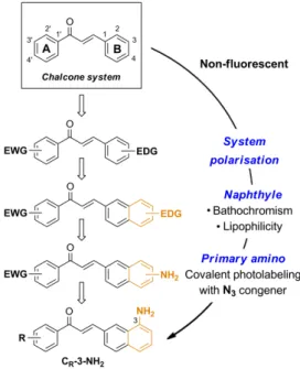
emissive chalcones possessing optimal photophysics and high cytopermeability for live-cell and live-animal imaging, we rationally delineated minimal structural modifications of the 1,3-diarylpropenone system (Scheme 1). Organic fluorophores are classically composed of push-pull moieties connected by means of an extended conjugation, allowing efficient excited state intramolecular charge transfers (ICTs).10 Accordingly, an electrodonating aromatic ring (B) would be conjugated to the enone system, with the arylcarbonyle (A) acting as electron-withdrawing moiety in the chalcone system (Fig. 1). The electrodonating aryle was chosen as a naphtyle group, known to increase fluorescence intensity while exerting bathochromic effects with respect to the phenyle group.10, 19 This shift is useful for microscopic imaging as it diminishes
light scattering, cellular auto-fluorescence and phototoxicity.20, 21 In addition, the more lipophilic naphthyle nucleus would
dramatically increase the ability of a given probe to penetrate cells and organelles. We finally settled on a pivotal primary amino group as the electrodonating substituent, having in mind its useful transformation to the azide for fluorogenic photolabeling strategies of cells or proteomes.22 Accordingly, we targeted the 3-aminobenzochalcone core as the optimal
fluorophore in this series, featuring the most extended captodative system, and expected to display maximal fluorescence intensity and bathochromism (Fig. 1).
Figure 1 Stepwise design of a minimal cytopermeable chalcone fluorophore (EWG, electro-withdrawing group; EDG,
electro-donating group; R, any substituent).
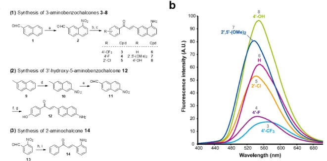
These 3-aminobenzochalcones were obtained by reduction of the corresponding nitro intermediates, accessed by Claisen-Schmidt condensation of various acetophenones with 8-nitro-2-naphthaldehyde 2. Starting from β-naphthaldehyde
1,23 a small library of benzochalcones 3-8 was thus obtained where the electron density of the acetophenyle part was tuned
from severely deficient (R = 4’-CF3) to strongly enriched (R = 4’-OH). Two close analogues were synthesized for the
physicochemical standardization of this new series: (i) 5-aminobenzochalcone 12 is a regioisomer of 3-aminobenzochalcone
8, possessing a less extended captodative system as a pseudo-para conjugation; (ii) 2-aminochalcone 14 is the exact
chalcone congener of 3-aminobenzochalcone 6, and should permit to quantify the influence of the naphthyle ring both in terms of fluorescence and lipophilicity. Compound 12 was obtained from β-methylnaphthalene 9 upon regioselective nitration24 then methyle oxidation. The obtained 6-nitro-2-naphthaldehyde 11 was turned to 5-aminobenzochalcone 12 by a “one-pot” condensation-reduction as for aminochalcones 3-8. aminochalcone 14 was obtained from 2-nitrobenzaldehyde 13 following the same strategy (Fig. 2a) (see Electronic Supplementary Information for all synthetic procedures).
3
Figure 2 Synthesis of representative compounds in the 3-aminobenzochalcone, 5-aminobenzochalcone and 2-aminochalcone series and SFRs in the 3-aminobenzochalcone series. (a) Reagents and conditions: a) HNO3 (40 equiv.),
H2SO4 (1.5 equiv.), AcOH, r. t., 13 % 2; b) substituted acetophenone (1 equiv.), NaOH (0.75-2 equiv.), EtOH, r. t.; c) SnCl2
(5-10 equiv.) or cat. Pd-C (10 %), H2, AcOH, r. t., 38 % 3, 31 % 4, 26 % 5, 37 % 6, 18 % 7, 69 % 8; d) HNO3 (1.25 equiv.), Ac2O, r.
t., 11 % 10; e) SeO2 (2 equiv.), neat, 150 °C, 28 % 11 (45 % based on conversion); f) 4-hydroxyacetophenone (1 equiv.),
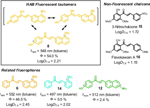
NaOH (2 equiv), EtOH, r. t.; g) Fe (10 equiv.), AcOH, reflux, 56 % 12; h) acetophenone (1 equiv.), NaOH (0.75 equiv), EtOH, r. t.; i) Pd-C (10 %), H2, AcOH, r. t., 12 % 14 (yields of chalcones indicated for two-steps, “one-pot” reactions). See Electronic Supporting Information for all synthetic procedures. (b) Semi-quantitative emission spectra of 3-aminobenzochalcones 3-8 (50 µM) in toluene at 20 °C. 3-Aminobenzochalcones were indeed fluorescent compounds, in agreement with our design of this novel fluorophore. However, while electro-withdrawing acetophenyle rings were expected to yield the most fluorescent compounds (Scheme 1), the strongest fluorescence was associated to the electrodonating 4’-hydroxy group (cpd 8), while a 4’-trifluoromethyl group (cpd 3) induced a much weaker fluorescence (Fig. 2b). The spectral properties of these fluorophores are given in Table S1 in the Electronic Supplementary Information and depicted in Fig. 3. 3-Aminobenzochalcones optimally display high Stokes shifts predicting an absence of internal quenching, with emission in the green-yellow region of the visible spectrum in toluene. As anticipated from Fig. 2, 4’-hydroxy-3-aminobenzochalcone 8 (HAB), with a 54.0 % quantum yield, was significantly more fluorescent than its non-substituted congener 6. This relation is validated by the further weaker quantum yield of 38.2 % of the p-trifluoromethyle derivative 3.Importantly, the favourable influence of a strongly electrodonating A ring called for revision of fluorophore representation in the chalcone series, with permanent enolization of the carbonyle occurring in an extended conjugation pattern similarly to the prototypical fluorescein or rhodamine fluorophores (Fig. 3). Further, HAB was far more fluorescent than its 5-amino regioisomer 12 with a ca. 20-fold higher quantum yield.
4
O NH2
8
HO
HAB Fluorescent tautomers
O NH2 λem= 548 nm (toluene) Φ= 54.0 % LogD7.4= 2.21 14 λem= 497 nm (toluene) Φ= 5.5 % LogD7.4= 2.02 O NH2 6 λem= 552 nm (toluene) Φ= 46.5 % LogD7.4= 2.45 OH NH2 O OH NH HO O HO 12 NH2 λem= 512 nm (toluene) Φ= 2.4 % Related f luorophores 3 4' 5 4' 3 2 O MeO 3-Nitrochalcone 15 LogD7.4= 1.72 OH OMe NO2 O MeO OH OMe OMe Flavokawain A 16 LogD7.4= 1.15
Non-f luorescent chalcones
Figure 3 Structure-fluorescence relationships and logD7.4 values at 20 °C in the 3-aminobenzochalcone,
5-aminobenzochalcone and 2-aminochalcone series (colours indicative of fluorescence emissions). This enormous difference in quantum yield between HAB and 12 cannot be explained solely by conjugation patterns: HAB features rotational hindrance of the 3-amino group (having peri relationships with H-2), a phenomenon absent in the 5-amino regioisomer 12.25, 26 As a result, HAB should be less susceptible to non-radiative desexcitation than 12, thereby favouring fluorescence transitions. Comparing the isoelectronic compounds 3-aminobenzochalcone 6 and 2-aminochalcone
14 showed as expected that the substitution of the (B) phenyle group for a naphthyle in 6 resulted in much stronger
fluorescent emission. Compound 6 thus possesses a quantum yield over eight-fold higher than 14, the latter also displaying cyan emission and a smaller Stokes shift. Noteworthy for future biological studies, the quantum yield of 54.0 % for the most fluorescent aminobenzochalcone HAB is higher than those of dansylamides (e. g., DPP)27 and indocyanines (e. g., ICG),28 prototypical fluorescent probes for live-cell and live-animal imaging, respectively, and is also, to our knowledge, the highest described to date in the chalcone series (see Table S1 in the Electronic Supplementary Information).
Solvatochromic behavior. HAB and its non-substituted analogue 6 exhibited pronounced solvatochromism (Δmaxλem ~ 140
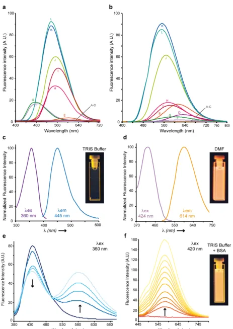
nm), being virtually non-fluorescent in water, biological buffers and alcohols, moderately emissive in polar aprotic solvents (e. g., ethyle acetate) and strongly fluorescent in non-polar solvents (e. g., toluene), with the exception of alcanes. While 6 was significantly fluorescent in n-hexane and n-octane, its 4’-hydroxy derivative HAB proved undetectable in these solvents, and showed only weak fluorescence in n-alkanols used to model biological lipids (e. g., n-heptanol) (Fig. 4a,b). Although the lack of fluorescence of HAB in biological buffers may be perceived as a limitation for imaging studies, it must be realized that the binding of HAB to amphipathic affinity sites of protein targets should result in a 4-500 fold fluorescence turn-on (FTO) according to Fig. 4b.10 As a proof-of-principle, bovine serum albumin (BSA) quenched HAB weak blue fluorescence (λex = 360 nm, λem
= 445 nm) in a dose-response manner while inducing the formation of a strongly yellow-emitting HAB-protein fluorescent complex (λex = 420 nm, λem = 578 nm) (Fig. 4e-f). A FTO of 21.5 (excitation at 420 nm, at BSA
concentration of 10 mg/ml) was observed upon binding of HAB to the protein (Fig. 4f), associated to an important bathochromic shift of its fluorescence spectrum (Δλex = 60 nm, Δλem = 133 nm) which was similar to that obtained in the
polar aprotic solvent DMF (Fig. 4d,f). This behaviour is opposite to that of chalcones binding the physiological sites of BSA and displaying slight ipsochromic shifts and quenching effects indicative of an apolar environment,18 suggesting that HAB binds non-conventional BSA sites. Overall, HAB should be featured with high signal-to-noise ratio and elevated pharmacological sensitivity, low or negligible predicted fluorescence in compartments such as the extracellular medium, the cytosol and the membranes under free form, and high fluorescence when target-bound (“in-target” fluorogenesis). Moreover, its absence of fluorescence in water or buffers has the potential to greatly simplify and time-optimize the staining protocols, with suppression of tedious washing steps and diffusion liabilities following dye administration.
5
Figure 4 HAB shows strong solvatochromism and protein-dependent fluorogenesis. (a,b) Semi-quantitative emission
spectra of 3-aminobenzochalcone 6 (Fig. 4a) and HAB (Fig. 4b) in various solvents (50 µM). A : H2O ; B : TRIS buffer 10 mM,
pH 7, NaCl 100 mM, MgCl2 5 mM; C : AcOH ; D : DMSO ; E : MeCN ; H: CHCl3; I: AcOEt; J : 1,4-dioxane ; K : benzene ; L :
toluene. Dissimilar solvent lettering between the figures are F: hexane; G: octane (Fig. 3a) and F: butanol; G: n-heptanol (Fig. 3b). (c,d) Normalized excitation spectra (purple lines) and fluorescence emission spectra (blue or orange lines) of HAB (50 µM) in TRIS buffer pH 7.0 (TRIS.HCl 10 mM, NaCl 100 mM, MgCl2 5 mM) (c) or DMF (d). (e) Fluorescence emission spectra of HAB (50 µM) titrated by BSA (0 to 1 mg/mL) in TRIS buffer with an excitation wavelength of 360 nm. (f) Fluorescence emission spectra of HAB (50 µM) titrated by BSA (1 to 10 mg/mL) in TRIS buffer with an excitation wavelength of 420 nm. Arrows indicate the decrease or increase in fluorescence following the addition of BSA. UVA-irradiated cuvettes in the corresponding conditions are shown as insets in (c), (d) and (f).
Cytopermeability evaluation. Lipophilicity is a key physicochemical parameter affecting cellular penetration and subcellular distribution in vitro, as well as pharmacokinetics in vivo.29 We measured the partition coefficients of key benzochalcones
compared to reference chalcones using n-octanol and pH 7.4 phosphate buffered saline (PBS), as to draw structure-cytopermeability relationships in this novel series (Fig. 3). As expected, the favorable influence of a naphthyle vs a phenyl ring regarding fluorescence was also exerted in terms of lipophilicity, as 3-aminobenzochalcone 6 and HAB possessed log
D7.4 values significantly higher than that of mere 2-aminochalcone 14. In particular, 6 was almost three times more
lipophilic than its exact 2-aminochalcone congener 14. On the other hand, the presence of a 4’-hydroxy group on HAB made it 2-fold more hydrophilic than its 4’-deshydroxy congener 6. Further, HAB was three to eleven times more lipophilic than 3-nitrochalcone 15 and natural flavokawain A 16 chosen as representative biologically-active chalcones30, 31 (Fig. 3). Overall, the neat amphipathicity (log D7.4 between 1.5 and 2.5), neutrality as well as small molecular weight (289.3 Da) of HAB32
6
HAB labels specific cells in live zebrafish embryos and swimming larvae. HAB, the most fluorescent, solvatochromic and
histopermeable derivative obtained, was tested in representative transparent animal models. We chose the nematode
Caenorhabditis elegans and the zebrafish Danio rerio to study the cell and organelle distribution of HAB in complete live
systems and assess its biological specificity. HAB could not be microscopically visualized in C. elegans due to excessive autofluorescence of the nematode (data not shown). On the other hand, HAB displayed a unique labeling pattern in zebrafish that deserved in-depth investigation. When HAB was incubated at 10 µM for 60 min on live zebrafish embryos and swimming larvae ranging from 24 hours post-fertilization (hpf) to 72 hpf, fluorescence emission was detected mainly in the 550-650 nm window following excitation at 488 nm, indicative of a non-aqueous environment (see Fig. 4b for the solvatochromic properties of HAB, and Fig. S2 in the Electronic Supplementary Information for the emission spectrum of
HAB in vivo). Since the fluo-labeling was maximal in the yellow-orange region of the visible spectrum, this suggested that HAB bound to specific proteins with strong target-induced bathochromic fluorogenesis and FTOs (Fig. 4e-h). This labeling
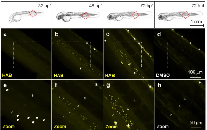
displayed a neat pattern of punctuated areas that first appeared by 32 hpf in the ventral tail of the embryo (Fig. 5a,e). The number and intensity of HAB fluorescent spots progressively increased at 48 hpf and 72 hpf following the known deployment of macrophages and neutrophils during zebrafish development,4, 5 suggesting that these cell types could be targeted by HAB. The shape and size of the fluorescent spots within the caudal region of the specimens suggested labeling of some subcellular compartment rather than whole cells.5 Noteworthy, the already autofluorescent pigment cells present in 72 hpf larva underwent a significant increase of fluorescence in presence of HAB (Fig. 4c,d) suggesting that the probe also accumulated within these cells due to their high content in aromatic pigments (i. e., pteridine and guanine derivatives).33, 34 Figure 5 HAB labels specific cells in live zebrafish from 32 hpf. Confocal fluorescence imaging of HAB labeling (10 µM) in live wild-type zebrafish embryos (32 and 48 hpf) and swimming larvae (72 hpf) following excitation at 488 nm and detection in the 550-650 nm range. The yellow-orange color is indicative of the fluorescence seen with the naked eye. Maximum intensity Z-projection images (2 µm serial optical sections) are shown. Arrows point to HAB label; asterisks mark pigment cells. A, artery; N, notochord; V, vein.
HAB labeling is reversible. HAB labeling was found to be fairly photostable in the conditions used so far, with ca. 50 %
fluorescence lost after 30 min of continuous illumination at 488 nm. However, since the embryos and larvae were systematically washed and mounted in HAB-free agarose for live imaging, the diffusion of HAB outside of its binding sites might be responsible for the loss of labeling overtime. To discriminate between free diffusion and photobleaching as the two possible causes for signal loss, we performed time-lapse imaging experiments in wich either of these two phenomena was kept to a minimum. To test the free diffusion hypothesis, 72 hpf zebrafish larvae were treated with 10 µM HAB for 1 hr, washed and mounted in tracer-free mounting medium, then imaged over time with short exposure-acquisition pulses every 10 min. Under these conditions of lower illumination hence limited photobleaching, the detection of HAB-labeled structures was no longer possible after ca. 1 hr, with again ca. 50 % of fluorescence lost after 30 min exposure (see Figure S3 in the Electronic Supplementary Information). To test the photobleaching hypothesis, we treated the 72 hpf larvae with
7
10 µM HAB for 1 hr and mounted them in agarose and tricaine containing 10 µM HAB. We then imaged the zebrafish larva embedded in HAB 10µM over time under continuous illumination (indicate time lapse here) by the 488 nm laser. Under these conditions of maximum photobleaching and abolished free diffusion, HAB labeling proved highly photostable, as it could be detected for up to 15 hrs of time-lapse imaging (see Video S4 in the Electronic Supplementary Information). This observation indicates that HAB bound in vivo continually exchanges with free non-fluorescent HAB, at a rate faster than the photobleaching of the bound HAB during continuous 488 nm laser illumination. This remarkable feature makes HAB an ideal live stain for both long-term and high temporal resolution confocal imaging of neutrophil granules in the zebrafish. These conditions of constant equilibrium by continuous contact, made possible by the absence of HAB fluorescence in aqueous media, constitute optimal settings for HAB labeling on the zebrafish model and are particularly suitable for long-term experiments.
HAB labels neutrophil granules in live zebrafish larvae. To identify the cell type(s) labeled by HAB, that according to their
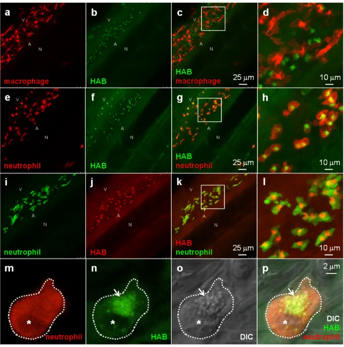
localization and developmental timing of detection could be the first leukocytes of the zebrafish embryo and larva (macrophages and/or neutrophils), we stained zebrafish transgenic 72 hpf larvae harbouring fluorescent macrophages or neutrophils35-37with HAB. Upon treatment with 10 µM HAB of Tg(Mfap4:mCherry-F) larvae possessing red-fluorescent macrophages expressing mCherry, we found that the probe did not co-localize with the red-fluorescent macrophages (Fig. 6a-d). Due to the wide emission range of the mCherry protein (560-760 nm), that overlapped with that of the yellow-green emitting HAB, mCherry was imaged following excitation at 552 nm and collecting only the “far red” portion of its fluorescence emission (660-750 nm), and HAB was excited at 448 nm and collecting the 550 - 650 nm range of its emission spectrum. Upon treatment with 10µM HAB of Tg(mpx:GAL4/UAS-E1b:nsfB-mCherry) larvae with neutrophils expressing mCherry in their cytoplasm and using the same HAB/mCherry acquisition parameters as above, we could show that HAB perfectly colocalized with all neutrophils of the imaged ventral tail region, labeling a specific compartment within them. (Fig. 6e-h). Using GFP-labeled neutrophils (Tg(mpx:GFP) strain), and sequentially detecting GFP emission in the 500-520 nm range and HAB using the “red” portion of its emission spectrum (> 600 nm) by exciting both fluorophores by the 448nm laser (Fig. 6i-l) we confirmed that HAB targeted a neutrophil specific compartment. Not only did HAB stained all neutrophils of the ventral tail region of 72 hpf larvae, where they are known to be particularly abundant2, 4, 37, but HAB-labeled neutrophils could also found in the eye, ear and yolk of 72 hpf larvae (data not shown), indicating that HAB labels the entire neutrophil population in vivo. Collectively, these observations demonstrated that zebrafish neutrophils are the cell type selectively targeted by HAB, where it specifically labels a compartment within these cells. To identify the compartment targeted by HAB within neutrophils, we performed high-resolution confocal imaging of HAB-labeled cells in 72 hpf larvae harbouring red neutrophils combining the fluorescence detection of HAB and mCherry with DIC microscopy. HAB was found to co-localize with neutrophil granules, which are easily recognizable in transmitted light / DIC imaging5, 38 for they are refractile and constantly moving in the cytoplasm (Fig. 6m-p). This colocalization was unambiguously demonstrated by the perfect overlap of fluorescence HAB signals and DIC images during time-lapse acquisitions of live patrolling neutrophils (see high-resolution Videos S5 and S6 in the Electronic Supplementary Information). Similar to their mammalian counterpart, the granules of zebrafish neutrophils contain myeloperoxidase.2, 38, 39 The diazo dye Sudan Black B (SB) is a classical lipid stain that specifically labels the myeloperoxidase-positive granules of zebrafish neutrophils,5, 40 as also
observed in human neutrophils.41, 42 The first SB-stained granules are detected by 33-35 hpf in zebrafish embryos, within the first, still immature embryonic neutrophils; this staining then increases in number and intensity, revealing the deployment of neutrophils during zebrafish development.5 To check that HAB was targeting the same granule population as SB in neutrophils, we tried to combine the two dyes. We first tested if the HAB label was compatible with the fixation protocol classically used to fix zebrafish embryos and larvae, but failed to detect HAB labeling in the larva post-fixation for two hours at room temperature with either 1 or 4 % formaldehyde, or when performing HAB labeling before the fixation. This could be due to the loss of HAB pharmacology in fixed samples, or to the reaction of HAB with formaldehyde, leading to fluorophore destruction: eventhough HAB is both a very weak organic base (predicted pKa value of 3.97 for its amino tautomers, see Figure S7 in the Electronic Supplementary Information) and a weak nucleophile, possessing the electronic features of a vinylogous amide, this possibility cannot be ruled out in absence of dedicated study. In any case, it was not possible to combine the SB staining with the HAB labeling.
8
Figure 6 HAB labels zebrafish neutrophil granules. (a-l) Confocal fluorescence imaging of HAB (10 µM) in live transgenic 72 hpf zebrafish larvae following excitation at 448 nm under equilibrium conditions. Detection parameters were as follow: for
HAB / mCherry: λEX 448 nm, λEM 550-650 nm, mCherry: λEX 552 nm, λEM 660-750 nm using sequential modes of acquisition.
For GFP / HAB: GFP λEX 448 nm, λEM 500-520 nm, HAB: λEX 448 nm, λEM 550-650 nm using sequential modes of acquisition.
Maximum intensity Z-projection images (2 µm serial optical sections) are shown. (m-p) High-resolution DIC and confocal fluorescence imaging of HAB (10 µM) in live 72 hpf zebrafish larva harbouring red neutrophils. Arrows point to HAB-labeled granules according to the DIC images in neutrophils. A single 0.4 µm optical section is shown. Boxes in Fig. 6c,g,k indicate the regions magnified in the insets (Fig. 6d,h,l respectively). Abbreviations used: A (aorta); N (notochord); V (vein); asterisk = nucleus. See Electronic Supplementary Information for Videos S5 and S6 related to Fig. 6m-p. HAB reveals the behavior of neutrophil granules during phagocytosis in live zebrafish larva. To circumvent this technical
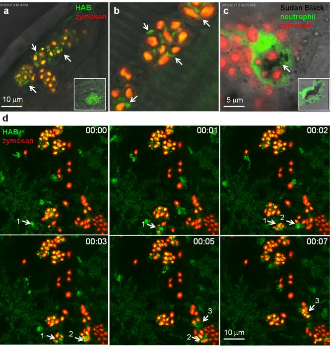
limitation, we sought indirect evidence for the identity of neutrophil granules as the targets of HAB. One of the most important functions of neutrophils is to eliminate invading microorganisms. Neutrophils are highly phagocytic cells, able to engulf and kill microbes with the arsenal of microbicidal compounds stored in their granules. Once they have engulfed microbes, neutrophils release the granule content (e.g., myeloperoxidase-produced hypochlorous acid and proteases) into the phagosome. We have shown that in vivo, zebrafish neutrophils degranulate into the phagosome following microbe phagocytosis and that the myeloperoxidase activity initially contained in the granules often relocalises to the phagosome, while the SB staining correlatively disappears.43 These steps have been advantageously reproduced with opsonized zymosan particles in cultured mammalian neutrophils, to show that neutrophil granules are recruited to the nascent phagosome in which they deliver their content by exocytosis.8, 44 To determine whether HAB would allow the visualization of neutrophil granule dynamics during phagocytosis in vivo, we vitally stained a zebrafish larva with HAB, then
9
subcutaneously injected red-fluorescent Cy5-zymosan, and immediately monitored the interaction between HAB-labeled neutrophil granules and zymosan particles by high resolution confocal imaging (Fig. 7). We found that upon zymosan phagocytosis, HAB-labeled granules are massively recruited to the particle-containing phagosomes (Fig. 7a,b,d and related Video S8 in ESI), similar to the myeloperoxidase containing, SB-labeled granules which relocalised around the zymosan-containing phagosome in the fixed larva (Fig. 7c). These observations strongly suggest that the myeloperoxidase-containing granules are the targets of HAB in vivo. They also represent, to our knowledge, the first documentation of the dynamics of neutrophil degranulation upon phagocytosis in an entire live organism. Figure 7 HAB reveals the dynamics of neutrophil granules upon phagocytosis of zymosan particles in live zebrafish. (a,b) Confocal live imaging of HAB-labeled neutrophil granules upon phagocytosis of subcutaneously-injected zymosan in a live 72 hpf zebrafish larva under equilibrium conditions. HAB is recruited to the forming phagosomes (arrows). Inset: HAB labeling of resting neutrophil. (c) Sudan Black (SB) staining of myeloperoxidase-containing neutrophil granules showing granule recruitment to the phagosome upon zymosan phagocytosis in fixed zebrafish larva. Inset: SB staining of a resting neutrophil; A single 1µm optical section is shown. (d) Frames extracted from an in vivo time-lapse confocal imaging sequence (time step = 1 min). Arrows point to HAB-labeled neutrophil granules that are recruited to the nascent zymosan containing phagosome. Three neutrophils (pointed with number 1 to 3) were tracked during the time lapse sequence. Maximum intensity Z-projection (1 µm serial optical sections). See Electronic Supplementary Information for Video S8 related to Fig. 7d.
10
HAB does not target neutrophil myeloperoxidase, and the staining of neutrophil granules by HAB is not a general feature of chalcones. To tentatively identify the specific protein target of HAB in the neutrophil granules, we performed HAB
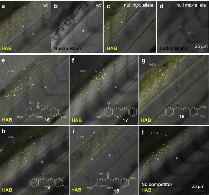
labeling in myeloperoxidase knock-out zebrafish larvae (“spotless” strain) under equilibrium conditions.45 While mutant
larvae showed the expected absence of SB staining, they nevertheless still exhibited a fluorescent labeling with HAB qualitatively and quantitatively indistinguishable from wild-type larvae (Fig. 8a-c). This result shows that the myeloperoxidase abundant in neutrophil granules is not the biochemical target of HAB. We also tested a selection of biologically-active chalcones as possible HAB competitors, based on their known or putative action on neutrophils such as inhibition of myeloperoxidase activity or repressing effects on pro-inflammatory mediators such as NF-κB, TNF-α, COX-2 and various interleukines.46-48 The natural chalcones flavokawain A 16 and cardamonin 17 are potent anti-inflammatory, proapoptotic and antitumour agents in mouse models, due to their ability to block NF-κB signaling.31, 49-51 Synthetic chalcone 18 is an inhibitor of chemokine CXCL12 that blocks its binding to several chemokine receptors, preventing eosinophil infiltration in an asthma mouse model.52 3-nitrochalcone 15 possesses pronounced antinociceptive properties in mice that are anti-inflammatory in origin.53 Last, 4-methylchalcone 19 was selected due to its homology with mere (unsubstituted) chalcone, reported to exert anti-myeloperoxidase and anti-migratory effects on zebrafish neutrophils.46 Larvae were first incubated with the competitors alone at 100 µM for 1 hr to assess their toxicity, as well as possible basal fluorescence in the zebrafish. Under these conditions, all specimens remain alive with no apparent toxicity from the chalcones. Moreover, using the same detection settings than for HAB at 10 µM (λEX 488 nm, λEM 550-650 nm), none of the
competitors showed fluorescence and their labeling was undistinguishable from that of the negative control DMSO (data not shown), except for chalcone 18 (see below and Fig. S9 in the Electronic Supplementary Information). When these various potential competitors were co-incubated at 100 µM with HAB at 10 µM in the same conditions, we could observe no extinction or diminution of HAB fluorescence in vivo with any of these competitors relative to the control (Fig. 8e-j). Moreover, fluorimetric association experiments in TRIS buffer or DMF (where HAB shows the same type of yellow-orange fluorescence as in neutrophil granules, see Fig. 4d) demonstrated that none of these chalcones, when incubated in presence of HAB at the same 10:1 stoichiometry, induced any significant qualitative or quantitative change in HAB fluorescence. The only exception was chalcone 18 which caused strong emission quenching in both media (data not shown), a feature not at play in zebrafish larvae in vivo (Fig. 8g). The binding of HAB to neutrophil granules thus appears to be the result of the probe’s precise physicochemical signature, rather than a general feature of chalcones.
11
Figure 8 HAB does not target zebrafish neutrophil myeloperoxidase, and its binding to neutrophil granules is not a general feature of chalcones. (a,c) Merged confocal fluorescence and bright-field imaging of HAB (10 µM) in live wild-type
(a) or “Spotless” (NL 144_01 mutant: null mpx allele) (c) 72 hpf zebrafish larvae following excitation at 488 nm and detection in the 550-650 nm range under equilibrium conditions. (b,d) SB staining of myeloperoxidase-containing neutrophil granules in bright-field imaging. (e-j) Merged confocal fluorescence and bright-field imaging of HAB (10 µM) in live wild-type 72 hpf zebrafish larvae co-treated with chalcones 15, 16 (flavokawain A), 17 (cardamonin), 18 or 19 (100 µM) following excitation at 488 nm and detection in the 550-650 nm range under diffusion conditions. The yellow-orange color is indicative of the fluorescence seen with the naked eye. Single 2 µm optical sections are shown. Abbreviations used: A (aorta); N (notochord); UGO (urogenital opening); V (vein). During the course of this competition study, we discovered that 4-hydroxy-3-methoxy-4‘-chlorochalcone (Cpd. 18) at 100 µM was responsible for unusual fluorescence in presence of HAB (Fig. 8g), and deserved further investigation. When incubated alone at the same concentration on 72 hpf larvae and excited at 488 nm, chalcone 18 exhibited a green-yellow fluorescence that illuminated the anatomy of the specimens. Indeed, confocal imaging showed that the fluorescence of 18 delineated the trunk neuromasts, somite muscle fibers and boundaries, blood vessels, notochord, spinal cord, spinal canal and caudal hematopoietic tissue (Fig. S9 in the Electronic Supplementary Information). Detailed examination revealed no detectable intracellular staining, but interstitial histological labeling. This behavior is reminiscent of that of BODIPY-ceramide, a fluorescent dye previously used at similarly high concentration as a histological counterstain for the confocal imaging of live zebrafish embryos.54, 55 The fact that chalcone 18 was not cytopermeable even at high concentration further validates the global design of HAB, a benzochalcone congener, as a histo- and cytopermeable tracer for in vivo studies. Interestingly, chalcone 18 was recently evaluated in the zebrafish model as an inhibitor of the chemokine CXCL12a, for its activity against the collective migration of cells of the posterior lateral line primordium. At 10 µM, 18 showed moderate biological effects and was not fluorescently detected by GFP filters.56 The complete study of chalcone 18 regarding
12
fluorescence detection threshold, photostability, phenotypic effects and long-term toxicity is currently underway, in order to validate this compound as a novel anatomical interstitial live stain in the zebrafish model.
HAB labels granules in live human primary neutrophils. In order to validate HAB as a specific vital stain of neutrophil
granules not only in fish but also in mammals, and to assess its relevance for human studies, we attempted the HAB labeling of live primary human neutrophils freshly isolated from total blood. Human neutrophils exist under two morphologically- and phenotypically-distinct types depending on their state of activation. Whereas non-activated neutrophils consist in round-shaped non-adhering circulating cells, activated neutrophils are polyhedral cells expressing a number of cytoadhesins and adhering to endothelium walls, glass or plastic surfaces.57, 58 When stained with 10 µM HAB, non-activated neutrophils exhibited intense but diffuse fluorescent labeling, presumably due to the difficulty of resolving subcellular structures when imaging non-adherent cells (Fig. 9a-c). An equally intense labeling of cells at the level of subcellular granular structures was detected in the same conditions in activated neutrophils, consistent with the morphology and distribution of human neutrophil granules,59, 60 in addition to a strong perinuclear staining possibly corresponding to the nuclear membrane (Fig. 9d-f and S10 in the Electronic Supplementary Information ). To address the human blood cell selectivity issue, we assessed HAB labeling on other freshly-isolated cell populations from whole human blood (peripheral blood mononuclear cells, PBMC) in the same conditions than those used to image purified neutrophils.
HAB was found to exhibit an impressive selectivity for neutrophils over lymphocytes, monocytes, erythrocytes and
thrombocytes (platelets), that were all weakly to faintly labeled (Fig 9g-i). This selectivity seems to be the result of the accumulation of HAB in neutrophil granules, consistent with the complete cell and organelle specificity observed in the zebrafish model in vivo. Although these results deserve further investigation regarding the subset of granules labeled in human neutrophils,59 they constitute a strong presumption that the specific labeling of neutrophil granules by HAB in the zebrafish model is also relevant in mammalian systems.
13
Figure 9 HAB selectively labels live human primary neutrophils over other blood cell types. (a-c) and (d-f): Confocal
fluorescence and DIC imaging of HAB (10 µM) in live human neutrophils following excitation at 448 nm and detection in the 550-650 nm range. Single 0.4 µm optical sections are shown. (a-c) show resting, non-activated, non-adherent neutrophils. (d-f) show activated, adherent neutrophils: note HAB staining of neutrophils granules; (g-i): Confocal fluorescence and DIC imaging of HAB (10 µM) in live human lymphocytes (lc), erythrocytes (ec), monocytes (mc) and thrombocytes (tc) using the same settings than in (a-c) and (d-f). Single 0.4 µm optical sections are shown. The yellow-orange color is indicative of the fluorescence seen with the naked eye. Asterisk = nucleus.
Discussion
A novel 3-aminobenzochalcone, HAB, was rationally designed, synthesized and validated as a histopermeable and fluorogenic vital stain for in vivo biological studies. Photophysically, HAB has elevated Stokes shift, the highest fluorescence quantum yield described to date in this series, elevated signal-to-noise ratio upon protein binding, and unprecedented photostability. This also chemically very stable tracer possesses the minimal architecture to be strongly fluorescent, and stains neutrophils in zebrafish embryo and larva with extraordinary specificity, with no labeling of other, even related, cell types (e. g., macrophages). Subcellularly, HAB proved specific of zebrafish neutrophil granules, with no staining of other organelles or compartments. This behavior was rationalized as target-induced bathochromic fluorogenesis based on various chemical and biochemical model systems. Neutrophil labeling was independent of the state of cell activation, since HAB stained the granules of both resting and phagocytically-active neutrophils with equal intensity and specificity. HAB labeling
in the zebrafish followed the deployment of neutrophil from 32 hpf embryo to 72 hpf swimming larva and was even visualised in neutrophil granules in 6 day-old larva with similar sensitivity despite the increase in tissue density and thickness (see Fig. S11 in the Electronic Supplementary Information). Due to its absence of toxicity up to 48 hrs incubation
14
and outstanding photostability, HAB is ideal for the long-term, high temporal resolution live imaging of developing zebrafish, with the potential of dynamic monitoring of the neutrophil granules in virtually any physiological or physiopathological context. To our knowledge, no other related tracer is endowed with such precious characteristics. Last,
HAB intensely labeled freshly purified neutrophils from human blood at the level of granular structures consistent with
neutrophil granules. Fluorescent dyes for neutrophil granules are limited, including quinacrine (QA),8 LysoTrackerRed® (LTR),9 dihydrorhodamine (DHR) 123,9 and rhodamine-thiolactones (RTLs),61 which are non-specific by essence. QA and LTR as weak bases constitute general acidotropic stains, sequestrated under their protonated form in various low pH organelles and vesicles (e. g., lysosomes,62-64 apoptotic vesicles,65 mast cell granules66, 67), to which mammalian basophil and neutrophil secreted granules also belong.68, 69 DHR 123 and RTLs are part of ROS-activated profluorophores used to detect hydrogen peroxide/peroxynitrite and hypochlorous acid, respectively, in activated neutrophils as well as in various other cell systems and organelles.61, 70-72 A comparative analysis of the various existing tracers of neutrophil granules is provided in Table 1. In marked contrast, the unique staining of live zebrafish neutrophil granules by HAB over other cell types and acidic compartments in the whole organism, together with HAB very weak basicity (about 105-fold lower than that of acidotropic amines63 with predicted pK
a value of 3.97 for the amino tautomers, see Figure S7 in the Electronic
Supplementary Information), supports a specific target-induced labeling of neutrophil granules by HAB. Although we showed that the myeloperoxidase present in the granules as is not involved in their specific staining by HAB, these granules also contain abundant neutral serine proteases (e. g., elastase, proteinase 3, cathepsin G),59 which constitute putative biochemical targets for HAB based on their reactivity towards reversible electrophilic inhibitors.73, 74 In this context, HAB has an immediate surrogate for proteomic interrogation under the form of a photoalkylative azido analogue (Fig. 1) easily accessible through Sandmeyer-type modification of the aromatic amino group.22 The covalent photolabeling of live
zebrafish larvae or isolated human neutrophils with a fluorogenic 3-azido-4‘-hydroxybenzochalcone, FACS purification of labeled cells followed by Western Blot identification of the covalently-stained molecular target(s) of HAB, therefore seems within reach. Overall, due to its optimal photophysical and physicochemical properties and unprecedented biological specificity, HAB constitutes a valuable small-molecule addition to the fluorescent proteins toolbox used in the zebrafish model, which it functionally and spectrally complements, and appears suitable for functional studies in live mammalian neutrophils. HAB therefore holds great promise as a vital stain to monitor the dynamics and behaviour of neutrophil granules in various aspects of the neutrophil function in the context of a live organism, with potential relevance to human physiology and physiopathology. Table 1 Functional comparison of HAB with existing tracers of neutrophil granules TRACER SPECIFICITY FOR NEUTROPHIL GRANULES HISTOPERMEABILITY FOR LIVE ANIMAL IMAGING
PHOTOSTABILITY FLUOROGENESIS REF.
Quinacrine No (acidotropic stain, DNA stain) Yes Yes No 65, 75-77 LysoTrackers No (acidotropic
stains) Yes No No
9, 65, 77-79 Acridine orange No (acidotropic stain, DNA/RNA stain) Yes Yes No 65, 80-83 Dihydrorhodamine 123 No (ROS/RNS-dependent - also labels mitochondria)
No (in vitro only) No Yes (ROS-activated) 9, 84-87
Rhodamine-thiolactones
No (ROS-dependent) Yes Yes Yes (ROS-reactive)
61, 88 Sudan Black B Yes (lipids/MPO-dependent) No (FA fixation in vivo) NA (colorimetric stain) NA (colorimetric stain) 5, 42, 89 Luminol No (ROS-dependent, MPO-amplified) No (in vitro only) NA (chemoluminescent stain) NA (chemoluminescent stain) 90 Tyramide-FITC / Cy3 Yes (MPO-reactive) No (FA fixation in vivo) No (FITC conjugate) No 5, 91 Yes (Cy3 conjugate)
15
MUB40 / RI-MUB40-Cy5 Yes (lactoferrin ligands)
No (peptides, FA fixation in vitro, in vivo) Yes No 60 NE680 Yes (elastase substrate) No (peptide, MeOH fixation ex vivo, intranasal instillation in mouse) Yes Yes (enzyme-activated) 92 Elastase, cathepsin G, proteinases 3 and 4 substrates Yes (enzyme substrates) No (peptides, FA fixation in vitro) Yes Yes (enzyme-activated) 93 HAB Yes (pH, ROS, MPO-independent fluorescence) Yes (amphipathic small-molecule, logD7.4 = 2.21) Yes (> 15 hrs under continuous 488 nm irradiation) Yes (target protein-induced FTO) Our study Abreviations: FA, formaldehyde; FITC, fluorescein isothiocyanate; FTO, Fluorescence Turn-On; MeOH, methanol; MPO, myeloperoxidase; NA, non-applicable; RNS, reactive nitrogen species; ROS, reactive oxygen species.
Methods
Compound synthesis, structural characterization and pKa prediction. Detailed synthetic procedures and analytical
identification for compounds 2-8, 10-12 and 14, as well as pKa prediction for HAB, can be found in the Electronic Supporting Information.
Zebrafish. Transgenic and mutant stocks of zebrafish were raised and staged according to Westerfield.94 AB wild-type fish and transgenic lines Tg(mpx:GFP)i114, Tg(mpx:GAL4.VP16)i22, Tg(Mfap4:mCherry-F) and Tg(UAS-E1b:nfsB.mCherry)c264
were used.35-37 Embryos were reared at 28 °C or 24 °C according to the desired speed of development. All timings in the text refer to the developmental stage at the reference temperature of 28.5 °C.94 Embryo and larvae were anesthetized with 200
μg/ml tricaine (Sigma-Aldrich) during live in vivo imaging.
HAB labeling of live zebrafish embryo and larva.
Equilibrium protocol (Fig. 6, 9, S10 and S11, Video S5, S6 and S7): 72 hpf or 6 dpf zebrafish larvae, either wild-type AB
specimens or transgenic specimens, were placed in individual wells (24-well culture plates; 10 embryos or larvae/well), incubated with HAB (10 µM in Volvic water from 10 mM stock solutions in DMSO) at room temperature for 1 hr in the dark, anaesthetized in buffered tricaine (Sigma) containing HAB (10 µM) then mounted in 35 mm glass-bottom dishes (Inagaki-Iwaki) in 1 % low-melting-point agarose containing HAB (10 µM) to ensure equilibrium conditions. The immobilized larvae were then covered with 2 ml Volvic water containing tricaine and HAB (10 µM) as described previously.43 Negative controls consisted of Volvic water containing 0.1 % DMSO. For wild-type AB specimens as well as transgenic specimens, HAB was incubated for 1 hr.
Diffusion protocol (Fig. 5, 7, 8, Fig. S4, S9, Video S8): Wild-type AB zebrafish embryos and swimming larvae (24 hpf to
72hpf) were placed in individual wells (24-well culture plates; 10 embryos or larvae/well), incubated with HAB (10 µM in Volvic water from 10 mM stock solutions in DMSO) at room temperature for 1 hr in the dark, washed, anaesthetized in
HAB-free buffered tricaine (Sigma) then mounted in 35 mm glass-bottom dishes (Inagaki-Iwaki) in HAB-free 1 %
low-melting-point agarose. The immobilized larvae were then covered with 2 ml Volvic water containing tricaine as described previously.43 Negative controls consisted of Volvic water containing 0.1 % DMSO. Practically, homogenous aqueous solutions of HAB were obtained by adding the drop of stock DMSO solutions on the inner wall of a plastic tube containing Volvic water at room temperature (20-23 °C), and vortexing without interruption for 20 s.
Sudan Black staining (Fig. 7). Embryos were fixed with 4% methanol-free formaldehyde (Poly-sciences, Warrington, PA) in
phosphate-buffered saline (PBS) for 2 hours at room temperature, rinsed in PBS, incubated in Sudan Black (SB; Sigma-Aldrich) for 20 minutes, washed extensively in 70% ethanol in water, then progressively rehydrated in 0.1% Tween 20 in PBS as previously decribed.5
Zymosan microinjection in zebrafish larvae (Fig. 7). Zebrafish larvae (72 hpf) were anaesthetized by immersion in buffered
tricaine immediately after the HAB labeling. They were injected with 1 nl of 0.4 x 10⁸/ml zymosan-Cy5 particles suspension using pulled borosilicate glass microcapillary (GC100F-15 Harvard apparatus) pipettes under a stereomicroscope (Stemi 2000, Carl Zeiss, Germany) with a mechanical micromanipulator (M-152; Narishige), and a Picospritzer III pneumatic microinjector (Parker Hannifin) set at a pressure of 20 psi and an injection time of 20 msec (subcutaneous injections) as previously described.43
Competition experiments (Fig. 8). 72 hpf wild-type AB zebrafish larvae were placed into individual wells as previously
described and co-incubated for 1 hr at room temperature with HAB (10 µM from a 10 mM stock solution in DMSO) and each competitor (100 µM from a 10 mM stock solution in DMSO) in Volvic water in the dark as previously described. Negative controls consisted of Volvic water containing 1.1 % DMSO. In this case, specimens were rapidly rinsed,
16
anaesthetized and mounted in HAB-free media for biological imaging. Practically, homogenous aqueous solutions of HAB +/- competitors were obtained by adding the drop of stock DMSO solutions on the inner wall of a plastic tube containg Volvic water at room temperature (20-23 °C), and vortexing without interruption for 20 s. HAB labeling of live human neutrophils (Fig. 9 and S10). Peripheral human blood was collected from healthy patients at the ICAReB service of the Pasteur Institute (authorization DC No. 2008-68). All donors gave written informed consent in accordance with the Declaration of Helsinki principles. Blood was collected from the antecubital vein into tubes containing sodium citrate (3.8 % final) as anticoagulant. Human polymorphonuclear neutrophils were purified as described previously.95 Briefly, plasma was removed by centrifugation (450 g, 15 min), and blood cells were resuspended in 0.9 % NaCl solution supplemented with 0.72 % Dextran. After red blood cells sedimentation, white blood cells were pelleted and further separated on a two layers Percoll (GE Healthcare) gradient (51-42 %) by centrifugation (240 g, 20 min). Peripheral Blood Mononuclear Cells (PBMC) (top layer) were isolated from polymorphonuclear neutrophils (bottom layer). Red blood cells were removed from the latter fraction using CD235a (glycophorin) microbeads (Miltenyi Biotec). Purified polymorphonuclear neutrophils were resuspended in the autologous plasma until experimental use. For HAB labeling, they were immediately put in RPMI 10 mM Hepes at 2 x 10⁶ cells/ml. 1 ml of neutrophil suspension was distributed in 35 mm glass-bottom dishes and incubated at room temperature for 20 min for neutrophils adhesion. The medium was then replaced by the HAB staining solution (at 1, 5 or 10 µM) in RPMI 10 mM Hepes; after 5-15 min at room temperature, HAB-labeled neutrophils were observed with a SP8 Leica confocal microscope, using a 63x water immersion objective. The 10 µM HAB concentration gave the best results.
Live imaging, image processing and analysis. Confocal microscopy to detect HAB single labeling in wild-type zebrafish
embryos and larvae was performed at 23-26 °C using a Leica SPE inverted microscope and a 16X oil immersion objective (PL FLUOTAR 16X/0.50 IMM). HAB was excited with a 488 nm laser and the fluorescence emission collected within the 550-650 nm range unless otherwise indicated. A Leica SP8 confocal microscope with a 20X oil immersion objective (HC PL APO CS2 20X/0.75 IMM) was used to live image transgenic larvae for colocalization studies and wt larvae for competition studies. Regarding HAB/mCherry acquisition, the following settings were applied: HAB (excitation 448 nm; emission 550-650 nm) and mCherry (excitation 552 nm; emission 660-750 nm) using a sequential acquisition mode. For GFP/HAB acquisition the following settings were applied: GFP (excitation 448 nm; emission 500-520 nm) and HAB (excitation 448 nm; emission > 600 nm). A 40X water immersion objective (HC PL APO CS2 40X / 1.1 water) was used also with the SP8 LEICA confocal microscope, to document at high resolution HAB labeling in neutrophils. For HAB-zymosan acquisition, the following settings were applied: HAB (excitation 488 nm; emission 491-601 nm) and Cy5-zymosan (excitation 638 nm; emission 683-795 nm). A 63x water immersion objective (HC PL APO CS2 63X / 1.2 water) was used to image HAB in live human neutrophils, following excitation at 448 nm and collecting the fluorescence emission in the 550-650 nm range. The 3D or 4D files generated by the confocal acquisitions were processed using the LAS-AF Leica software. Acquired Z-stacks (2 µm serial optical sections for images taken using 16X and 20X objectives) were projected using maximum intensity projection and exported as AVI or TIFF files. Z stacks acquired with the 40X or 63X objectives (0.4 µm to 1µm serial optical sections) were exported as AVI file. Frames were captured from the AVI files and handled with Powerpoint software to mount figures.
References
1. S. Masud, V. Torraca and A. H. Meijer, Curr Top Dev Biol, 2017, 124, 277-329. 2. G. J. Lieschke, A. C. Oates, M. O. Crowhurst, A. C. Ward and J. E. Layton, Blood, 2001, 98, 3087-3096. 3. D. Traver, P. Herbomel, E. E. Patton, R. D. Murphey, J. A. Yoder, G. W. Litman, A. Catic, C. T. Amemiya, L. I. Zon and N. S. Trede, Advances in Immunology, Vol 81, 2003, 81, 253-330. 4. P. Herbomel, B. Thisse and C. Thisse, Development, 1999, 126, 3735-3745. 5. D. Le Guyader, M. J. Redd, E. Colucci-Guyon, E. Murayama, K. Kissa, V. Briolat, E. Mordelet, A. Zapata, H. Shinomiya and P. Herbomel, Blood, 2008, 111, 132-141. 6. K. M. Henry, C. A. Loynes, M. K. B. Whyte and S. A. Renshaw, Journal of Leukocyte Biology, 2013, 94, 633-642. 7. E. A. Harvie and A. Huttenlocher, Journal of Leukocyte Biology, 2015, 98, 523-537. 8. E. Suzaki, H. Kobayashi, Y. Kodama, T. Masujima and S. Terakawa, Cell Motility and the Cytoskeleton, 1997, 38, 215-228. 9. C. F. Bassoe, N. Y. Li, K. Ragheb, G. Lawler, J. Sturgis and J. P. Robinson, Cytometry Part B-Clinical Cytometry, 2003, 51B, 21-29. 10. R. Duval and C. Duplais, Nat Prod Rep, 2017, 34, 161-193. 11. N. DiCesare and J. R. Lakowicz, Tetrahedron Letters, 2002, 43, 2615-2618. 12. J. Prabhu, K. Velmurugan and R. Nandhakumar, Spectrochim Acta A Mol Biomol Spectrosc, 2015, 144, 23-28. 13. C. G. Niu, A. L. Guan, G. M. Zeng, Y. G. Liu and Z. W. Li, Analytica Chimica Acta, 2006, 577, 264-270. 14. S. C. Lee, N. Y. Kang, S. J. Park, S. W. Yun, Y. Chandran and Y. T. Chang, Chem Commun (Camb), 2012, 48, 6681-6683. 15. M. Tomasch, J. S. Schwed, L. Weizel and H. Stark, Front Syst Neurosci, 2012, 6, 14.17
16. B. Zhou, P. Jiang, J. Lu and C. Xing, Arch Pharm (Weinheim), 2016, 349, 539-552. 17. T. Fuchigami, Y. Yamashita, M. Haratake, M. Ono, S. Yoshida and M. Nakayama, Bioorg Med Chem, 2014, 22, 2622-2628. 18. H. G. O. Alvim, E. L. Fagg, A. L. de Oliveira, H. C. B. de Oliveira, S. M. Freitas, M. A. E. Xavier, T. A. Soares, A. F. Gomes, F. C. Gozzo, W. A. Silva and B. A. D. Neto, Organic & Biomolecular Chemistry, 2013, 11, 4764-4777. 19. A. Bessette, T. Auvray, D. Desilets and G. S. Hanan, Dalton Transactions, 2016, 45, 7589-7604. 20. P. M. Alexander, H, Nature, 1962, 194, 2. 21. D. Kulms and T. Schwarz, Skin Pharmacology and Applied Skin Physiology, 2002, 15, 342-347.
22. S. J. Lord, H. L. Lee, R. Samuel, R. Weber, N. Liu, N. R. Conley, M. A. Thompson, R. J. Twieg and W. E. Moerner, J Phys Chem B, 2010, 114, 14157-14167. 23. S. D. Barker, K. Wilson and R. K. Norris, Australian Journal of Chemistry, 1995, 48, 1969-1979. 24. P. G. E. Alcorn, Wells, P. R., Australian Journal of Chemistry, 1965, 18, 1377-1389. 25. V. Balasubramaniyan, Chemical Reviews, 1966, 66, 567-+. 26. D. Skalamera, L. P. Cao, L. Isaacs, R. Glaser and K. Mlinaric-Majerski, Tetrahedron, 2016, 72, 1541-1546. 27. A. Niemann, J. Baltes and H. P. Elsasser, Journal of Histochemistry & Cytochemistry, 2001, 49, 177-185. 28. X. J. Yang, C. M. Shi, R. Tong, W. P. Qian, H. E. Zhau, R. X. Wang, G. D. Zhu, J. J. Cheng, V. W. Yang, T. M. Cheng, M. Henary, L. Strekowski and L. W. K. Chung, Clinical Cancer Research, 2010, 16, 2833-2844. 29. R. A. Duval, R. L. Allmon and J. R. Lever, J Med Chem, 2007, 50, 2144-2156.
30. P. Boeck, C. A. B. Falcao, P. C. Leal, R. A. Yunes, V. Cechinel, E. C. Torres-Santos and B. Rossi-Bergmann,
Bioorganic & Medicinal Chemistry, 2006, 14, 1538-1545. 31. X. L. Zi and A. R. Simoneau, Cancer Research, 2005, 65, 3479-3486. 32. M. J. Waring, Bioorg Med Chem Lett, 2009, 19, 2844-2851. 33. I. Ziegler, T. McDonald, C. Hesslinger, I. Pelletier and P. Boyle, J Biol Chem, 2000, 275, 18926-18932. 34. C. W. Higdon, R. D. Mitra and S. L. Johnson, PLoS One, 2013, 8, e67801. 35. Q. T. Phan, T. Sipka, C. Gonzalez, J. P. Levraud, G. Lutfalla and N. C. Mai, Plos Pathog, 2018, 14. 36. F. Ellett, L. Pase, J. W. Hayman, A. Andrianopoulos and G. J. Lieschke, Blood, 2011, 117, E49-E56. 37. S. A. Renshaw, C. A. Loynes, D. M. I. Trushell, S. Elworthy, P. W. Ingham and M. K. B. Whyte, Blood, 2006, 108, 3976-3978. 38. M. O. Crowhurst, J. E. Layton and G. J. Lieschke, Int J Dev Biol, 2002, 46, 483-492. 39. C. T. Pham, Nat Rev Immunol, 2006, 6, 541-550.
40. K. Wang, X. Fang, N. Ma, Q. Lin, Z. Huang, W. Liu, M. Xu, X. Chen, W. Zhang and Y. Zhang, Fish Shellfish
Immunol, 2015, 44, 109-116. 41. B. J. Bain, Am J Hematol, 2010, 85, 707. 42. W. van den Ancker, T. M. Westers, D. C. de Leeuw, Y. F. van der Veeken, A. Loonen, E. van Beckhoven, G. J. Ossenkoppele and A. A. van de Loosdrecht, Cytometry B Clin Cytom, 2013, 84, 114-118. 43. E. Colucci-Guyon, J. Y. Tinevez, S. A. Renshaw and P. Herbomel, J Cell Sci, 2011, 124, 3053-3059. 44. E. K. Macrae and K. B. Pryzwansky, Carlsberg Research Communications, 1984, 49, 315-322. 45. P. M. Elks, M. van der Vaart, V. van Hensbergen, E. Schutz, M. J. Redd, E. Murayama, H. P. Spaink and A. H. Meijer, PLoS One, 2014, 9, e100928. 46. Y. H. Chen, W. H. Wang, Y. H. Wang, Z. Y. Lin, C. C. Wen and C. Y. Chern, Molecules, 2013, 18, 2052-2060. 47. B. P. Bandgar, S. A. Patil, R. N. Gacche, B. L. Korbad, B. S. Hote, S. N. Kinkar and S. S. Jalde, Bioorganic & Medicinal Chemistry Letters, 2010, 20, 730-733. 48. J. Z. Wu, J. L. Li, Y. P. Cai, Y. Pan, F. Q. Ye, Y. L. Zhang, Y. J. Zhao, S. L. Yang, X. K. Li and G. Liang, Journal of Medicinal Chemistry, 2011, 54, 8110-8123. 49. D. J. Kwon, S. M. Ju, G. S. Youn, S. Y. Choi and J. Park, Food and Chemical Toxicology, 2013, 58, 479-486. 50. N. Wu, J. Liu, X. Z. Zhao, Z. Y. Yan, B. Jiang, L. J. Wang, S. S. Cao, D. Y. Shi and X. K. Lin, Tumor Biology, 2015, 36, 9667-9676. 51. S. Hatziieremia, A. I. Gray, V. A. Ferro, A. Paul and R. Plevin, British Journal of Pharmacology, 2006, 149, 188-198. 52. M. Hachet-Haas, K. Balabanian, F. Rohmer, F. Pons, C. Franchet, S. Lecat, K. Y. C. Chow, R. Dagher, P. Gizzi, B. Didier, B. Lagane, E. Kellenberger, D. Bonnet, F. Baleux, J. Haiech, M. Parmentier, N. Frossard, F. Arenzana-Seisdedos, M. Hibert and J. L. Galzi, Journal of Biological Chemistry, 2008, 283, 23189-23199. 53. F. de Campos-Buzzi, J. P. de Campos, P. P. Tonini, R. Correa, R. A. Yunes, P. Boeck and V. Cechinel-Filho,
Archiv Der Pharmazie, 2006, 339, 361-365.
