HAL Id: hal-00618801
https://hal.archives-ouvertes.fr/hal-00618801
Submitted on 3 Sep 2011
HAL is a multi-disciplinary open access
archive for the deposit and dissemination of sci-entific research documents, whether they are pub-lished or not. The documents may come from teaching and research institutions in France or abroad, or from public or private research centers.
L’archive ouverte pluridisciplinaire HAL, est destinée au dépôt et à la diffusion de documents scientifiques de niveau recherche, publiés ou non, émanant des établissements d’enseignement et de recherche français ou étrangers, des laboratoires publics ou privés.
Diagnostic accuracy of quantitative real-time PCR assay
versus clinical and Gram stain identification of bacterial
vaginosis
J.-P. Menard, C. Mazouni, F. Fenollar, Didier Raoult, L. Boubli, F. Bretelle
To cite this version:
J.-P. Menard, C. Mazouni, F. Fenollar, Didier Raoult, L. Boubli, et al.. Diagnostic accuracy of quantitative real-time PCR assay versus clinical and Gram stain identification of bacterial vaginosis. European Journal of Clinical Microbiology and Infectious Diseases, Springer Verlag, 2010, 29 (12), pp.1547-1552. �10.1007/s10096-010-1039-3�. �hal-00618801�
1 2 3 4 5 6 7 8 9 10 11 12 13 14 15 16 17 18 19 20 21 22 23 24 25 26 27 28 29 30 31 32 33 34 35 36 37 38 39 40 41 42 43 44 45 46 47 48 49 50 51 52 53 54 55 56 57 58 59 60
Diagnostic accuracy of quantitative real-time PCR assay versus clinical and Gram stain identification of bacterial vaginosis
Menard Jean-Pierre1,2, MD; Mazouni Chafika1, MD; Fenollar Florence2, MD PhD; Raoult
Didier2, MD PhD; Boubli Léon1, MD; Bretelle Florence1,2,3, MD PhD.
1
Service de Gynécologie Obstétrique, Hôpital Nord Marseille, Chemin des Bourrely 13915 Marseille, France
2
Unité des rickettsies, IFR 48, CNRS-IRD UMR 6236, Faculté de Médecine, Université de la Méditerranée, Marseille, France
3
Centre d’Investigation Clinique 9502, Marseille, France
Corresponding author:
Menard Jean-Pierre
Service de Gynécologie Obstétrique Hôpital Nord, Chemin des Bourrely 13915 Marseille, France
Tel + 33 4 91 96 46 72 Fax + 33 4 91 96 46 96
Email: jean-pierre.menard@mail.ap-hm.fr
Manuscript
1 2 3 4 5 6 7 8 9 10 11 12 13 14 15 16 17 18 19 20 21 22 23 24 25 26 27 28 29 30 31 32 33 34 35 36 37 38 39 40 41 42 43 44 45 46 47 48 49 50 51 52 53 54 55 56 57 58 59 60 Abstract
Purpose To determine the diagnostic accuracy of quantitative real-time PCR assay in
diagnosing bacterial vaginosis versus the standard methods: Amsel criteria and Nugent score.
Methods Amsel criteria, Nugent score, and results from the molecular tool were obtained
independently from vaginal samples of 163 pregnant women who reported abnormal vaginal symptoms before 20 weeks' gestation. To determine the performance of the molecular tool, we calculated the kappa value, sensitivity, specificity and positive and negative predictive values.
Results Either or both of the Amsel criteria ( 3 criteria) and the Nugent score (score 7)
indicated that 25 women (15%) had bacterial vaginosis; the remaining 138 women did not. DNA levels of Gardnerella vaginalis or of Atopobium vaginae exceeded 109 copies/mL or
108 copies/mL, respectively, in 34 (21%) of the 163 samples. Complete agreement between
both reference methods and high concentrations of G. vaginalis and A. vaginae was found in 94.5% (154/163 samples, kappa value = 0.81, 95% confidence interval0.70-0.81). The nine samples with discordant results were categorized as intermediate flora by the Nugent score. The molecular tool predicted bacterial vaginosis with a sensitivity of 100%, a specificity of 93%, a positive predictive value of 73%, and a negative predictive value of 100%.
Conclusions The quantitative real-time PCR assay shows excellent agreement with the results
of both reference methods for the diagnosis of bacterial vaginosis.
Keywords: Atopobium vaginae; Bacterial vaginosis; Diagnostic accuracy; Gardnerella
1 2 3 4 5 6 7 8 9 10 11 12 13 14 15 16 17 18 19 20 21 22 23 24 25 26 27 28 29 30 31 32 33 34 35 36 37 38 39 40 41 42 43 44 45 46 47 48 49 50 51 52 53 54 55 56 57 58 59 60 Introduction
Bacterial vaginosis (BV) is a common cause of vaginal symptoms in women of reproductive age 1. Mounting evidence associates BV, determined by clinical Amsel criteria or the Gram stain-based Nugent score, with susceptibility to sexually transmitted diseases, pelvic
inflammatory diseases, preterm labor and preterm delivery 2. Unfortunately, although BV is associated with adverse pregnancy outcomes, regardless of the presence of clinical symptoms, the benefits of screening for and treating BV in pregnant women remain unclear 3-5.The subjective clinical diagnosis is of limited value in assessing an asymptomatic population, and half the women with BV are asymptomatic 6. The artificial Nugent score category of intermediate flora has increased the complexity of the clinical approach to vaginal flora: a significant percentage of the women tested have an intermediate score, but this category remains remarkably uncharacterized 7,8,9. Consequently obstetricians and gynaecologists continue to seek a reliable method for an objective analysis of abnormal vaginal flora. Recent advances in molecular techniques have increased our knowledge of the microbial ecosystem of BV and associated some bacteria with BV for the first time 10,11. Of these, one of the most interesting may be Atopobium vaginae, an anaerobic and fastidious bacteria recently reported to be highly associated with BV 10,12,13. Specific proportions of different
Lactobacillus species are related to the overgrowth of particular BV-associated bacteria, with
the disappearance of Lactobacillus species related to the development of Gardnerella
vaginalis and A. vaginae, in particular 12,13. Although G. vaginalis and A. vaginae are both
commonly present in normal flora, high vaginal concentrations of both are highly specific for BV 12,13. We previously proposed a reliable and accurate molecular tool based on the combination of high vaginal quantification of G. vaginalis (DNA level 109
copies/mL) and
A. vaginae (DNA level 108 copies/mL). It resulted in sensitive and specific BV diagnosis when the Gram stain-based Nugent score was used as the gold standard 14. One of the
1 2 3 4 5 6 7 8 9 10 11 12 13 14 15 16 17 18 19 20 21 22 23 24 25 26 27 28 29 30 31 32 33 34 35 36 37 38 39 40 41 42 43 44 45 46 47 48 49 50 51 52 53 54 55 56 57 58 59 60
limitations of that study, however, was it did not apply a clinical approach to vaginal flora, by using the Amsel criteria, an alternative and commonly accepted diagnostic method for BV, as previous molecular studies have done 11,13,15.
The aim of the present study was to compare the diagnostic accuracy of the quantitative real-time PCR assay for G. vaginalis and A. vaginae for the diagnosis of BV with the two most commonly accepted methods, the Amsel criteria and the Nugent score.
1 2 3 4 5 6 7 8 9 10 11 12 13 14 15 16 17 18 19 20 21 22 23 24 25 26 27 28 29 30 31 32 33 34 35 36 37 38 39 40 41 42 43 44 45 46 47 48 49 50 51 52 53 54 55 56 57 58 59 60
Materials and Methods
A prospective study of pregnant women took place at the North University Hospital of Marseille (France). Pregnant women were enrolled from September 2008 to November 2009. Vaginal samples were used to screen for BV in women who reported abnormal vaginal symptoms before 20 weeks' gestation. We selected only women with singleton pregnancies and for whom vulvovaginal candidiasis, trichomoniasis and gonococcal infection were ruled out after clinical examination and laboratory tests. The University's institutional review board (Ethics Committee of the University of Aix-Marseille, France) approved the study (in a decision dated September 19, 2008), and all participants provided written informed consent.
Before vaginal sampling, the physician performed a speculum examination to note the appearance of the vaginal discharge, the spontaneous vaginal odour, and the odour after the addition of 10% potassium hydroxide. Vaginal pH was measured with pH strips and a colourimetric scale. After the clinical examination, the physicians took two vaginal samples from each woman at the same time, on two different sterile cytobrushes rotated against the vaginal wall (Scrinet, 5.5 mm, Laboratory CCD International, Paris, France). One sample from the first cytobrush was rolled onto a glass slide for Gram staining with an automated stainer (Model 7320 Aerospray Gram Slide Stainer; Wescor, Logan, UT) and Nugent scoring. The second cytobrush was transferred to a sterile tube containing 500 µl of BME Baral Medium (Invitrogen, Carlsbad, CA) and stored at –80°C until DNA extraction and molecular quantification.
The two methods used to diagnose BV were (1) clinical diagnosis, based on the combination of any three of the following four criteria 16: vaginal pH greater than 4.5, thin homogeneous
1 2 3 4 5 6 7 8 9 10 11 12 13 14 15 16 17 18 19 20 21 22 23 24 25 26 27 28 29 30 31 32 33 34 35 36 37 38 39 40 41 42 43 44 45 46 47 48 49 50 51 52 53 54 55 56 57 58 59 60
vaginal discharge, clue cells on microscopic examination of vaginal fluid, and "fishy" amine odour after the addition of 10% potassium hydroxide (positive “whiff” test);(2)
microbiological diagnosis, based on Gram staining graded according to the Nugent score 17 to determine the presence of normal (score of 0 to 3) or intermediate flora (score of 4 to 6), or BV (score of 7 to 10). Clinicians and laboratory staff made their diagnoses independently, unaware of each other’s results.
The quantitative molecular tool was based on a specific quantitative real-time polymerase chain reaction (qPCR) assay and serial dilutions of a plasmid suspension, as previously described 14.It targeted Lactobacillus species, A. vaginae, G. vaginalis, and a human albumin gene. Human albumin gene quantification was used as an internal control to
provide evidence for DNA presence and DNA quality.Briefly, after DNA extraction, a qPCR assay was performed with a Stratagene MX 3000P (Agilent, La Jolla, CA). The amplification program was run at 50°C for 2 min and at 95°C for 15 min, followed by 45 cycles at 95°C for 30 s and at 60°C for 1 min. Five µL of (1) a pure undiluted DNA sample, (2) a DNA sample diluted to 1/10 µL, (3) a DNA sample diluted to 1/100 µL, or (4) the serially diluted plasmid suspension was added to the 20-µL PCR mix that contained the Quantitect Probe PCR Kit mix (Qiagen, Courtaboeuf, France), the two pairs of primers, the two probes, and 100 U Uracil DNA glycosylase (Sigma-Aldrich, Saint Quentin Fallavier, France). All plasmid scale solutions were tested in duplicate. Negative controls were introduced in each reaction plate. To validate the quality and reproducibility of each PCR result, we verified for each reaction plate of each sample that (1) the standard curve remained linear and reproducible, (2) the cycle threshold values for all microorganisms tested in
undiluted and diluted samples were reproducible and linear, and (3) the range of values for the number of albumin copies in the vaginal samples used as an internal control was narrow. The
1 2 3 4 5 6 7 8 9 10 11 12 13 14 15 16 17 18 19 20 21 22 23 24 25 26 27 28 29 30 31 32 33 34 35 36 37 38 39 40 41 42 43 44 45 46 47 48 49 50 51 52 53 54 55 56 57 58 59 60
quality and reproducibility of each PCR result were validated, and none of the 163 samples used in this study had to be excluded. The quantitative molecular tool made it possible to reduce the detection limit for all microorganisms to 10 copies per 5 µL (103 copies per mL).
The final results were expressed as copies of microorganism DNA per mL of vaginal suspension.
To determine the accuracy of the molecular tool for the diagnosis of BV, the subjects were subsequently classified into two groups according to each method for diagnosing BV described above (Amsel criteria and Nugent score): those with BV and those without BV. Agreement between the two reference methods and high vaginal concentrations of A. vaginae (DNA level 108 copies/mL) and G. vaginalis (DNA level 109
copies/mL) was calculated with the kappa value, determined by the javastat online statistics package 18. A kappa of 1.0 indicates perfect agreement. Values greater than 0.75 represent excellent agreement and values 0.40-0.75 fair to good agreement 19. The sensitivity, specificity, and predictive values of A. vaginae DNA 108 copies/mL and G. vaginalis DNA 109
copies/mL to predict BV were compared with reference methods. The statistical analysis used the UBC Bayesian Calculator Type 2 20.
Results
The study included all 163 pregnant women. The mean age of the study population was 29 (±7) years. All women included had speculum examinations during which vaginal samples were taken. The Amsel criteria, Nugent score and molecular quantification of Lactobacillus species, G. vaginalis, A. vaginae, and human albumin gene were successfully obtained for each woman.
1 2 3 4 5 6 7 8 9 10 11 12 13 14 15 16 17 18 19 20 21 22 23 24 25 26 27 28 29 30 31 32 33 34 35 36 37 38 39 40 41 42 43 44 45 46 47 48 49 50 51 52 53 54 55 56 57 58 59 60
Table 1 reports the BV results according to both reference methods. Fifteen women had BV according to both the Amsel criteria and Nugent score, while 10 had discordant results
according to these two methods: (i) 5 women had normal flora according to Amsel criteria but a Nugent score 7; (ii) 5 women had BV according to Amsel criteria but a Nugent score < 7. In all, 25 (15%) of the 163 women had BV according to at least one of these methods. The remaining 138 women (85%) did not have BV, as shown by both methods.
The molecular characteristics of the vaginal flora are reported in Tables 2 and 3. No
Lactobacillus species was detected by the qPCR method in 32 (19.60%) of the 163 samples.
The DNA level of G. vaginalis equalled or exceeded 109 copies/mL for 22 women (13.5%),
and that of A. vaginae equalled or exceeded 108 copies/mL for 30 (18.40%). In all, 34 (21%)
of the 163 samples had high vaginal concentrations of G. vaginalis or A. vaginae or both. The molecular profile of the 25 samples of vaginal flora identified as BV by either the Amsel criteria or Nugent score is presented in Table 3: 18 had no Lactobacillus species detected by qPCR, and all had high vaginal concentrations of G. vaginalis or A. vaginae or both.
Complete agreement between both reference methods for diagnosing BV and the molecular tool (that is, high vaginal concentrations of G. vaginalis or A. vaginae) occurred in 94.5% (154/163 samples). There was disagreement for nine samples (5.5%). All nine had high vaginal concentrations of G. vaginalis ( 109 copies/mL) or A. vaginae ( 108
copies/mL), did not have BV according to the Amsel criteria, and were categorized as intermediate flora by the Nugent score (Table 2). There was substantial agreement (kappa=0.81; 95% confidence interval 0.70-0.81) between the reference methods and high vaginal concentrations of both microorganisms.Compared with the reference methods, the molecular tool had a sensitivity
1 2 3 4 5 6 7 8 9 10 11 12 13 14 15 16 17 18 19 20 21 22 23 24 25 26 27 28 29 30 31 32 33 34 35 36 37 38 39 40 41 42 43 44 45 46 47 48 49 50 51 52 53 54 55 56 57 58 59 60
of 100%, a specificity of 93%, a positive predictive value of 73% and a negative predictive value of 100% for predicting BV (Table 4).
Discussion
We examined the accuracy of a quantitative real-time PCR assay for G. vaginalis and A.
vaginae for the diagnosis of BV compared with both the Amsel criteria and the Nugent score,
both reference methods for this diagnosis. The originality of the present study is its
simultaneous consideration of both the clinical characteristics of vaginal flora, according to the Amsel criteria, and the categorization of vaginal flora, according to the Gram stain-based Nugent score, to determine the accuracy of the molecular tool for diagnosing BV.
Good agreement (kappa=0.81) and high sensitivity (100%) and specificity (93%) were reported for the molecular tool in relation to both reference methods. These findings
demonstrate the interest of molecular prediction of BV. The advantage of our PCR-based test is its ability to predict BV accurately when the standard methods result in false negatives. In this study, of the 25 flora samples with BV, 10 (40%) had discordant results for the Amsel criteria and the Nugent score (Table 3). Five samples categorized as BV by the Nugent score were normal according to the Amsel criteria. Those results are not surprising because the sensitivity of Amsel criteria for BV diagnosis is reported to be poor 21. For the Nugent score, 5 samples categorized as BV by the Amsel criteria were rated intermediate according to the Nugent score. The Nugent scoring system is excellent in diagnosing samples as either normal or BV, but the intermediate flora presents problems 7,8. Vaginal smears with intermediate flora may be considered as heterogeneous flora that most commonly include normal flora rather than BV, according to the definition of Amsel criteria 22. Furthermore,
1 2 3 4 5 6 7 8 9 10 11 12 13 14 15 16 17 18 19 20 21 22 23 24 25 26 27 28 29 30 31 32 33 34 35 36 37 38 39 40 41 42 43 44 45 46 47 48 49 50 51 52 53 54 55 56 57 58 59 60
recent PCR assays report the heterogeneous character of intermediate flora, with some of them suggesting a molecular profile more similar to that of BV than to normal samples 13,14.
The molecular criteria’s lower positive predictive value of 73% suggests that some positive molecular results are false positives (5.5%), i.e., although the molecular profiles of G.
vaginalis and A. vaginae are identical to those seen in BV patients, the women do not meet
the clinical ( 3 Amsel criteria) or microbiologic criteria (Nugent score 7)that define BV (Table 2). Alternatively, however, these results may represent true positives for the molecular condition of BV that were missed by traditional diagnostic tools. The false negatives of both standard methods reported above may support this alternative explanation. In any case, our molecular tool, based on PCR quantification of G. vaginalis and A. vaginae, clearly defines a reproducible and standardized molecularly defined BV, irrespective of the clinical and microscopic characteristics of vaginal flora. The clinical implications of this molecular condition should help to improve our understanding of BV, especially during pregnancy. Thus, we recently reported that high vaginal concentrations of G. vaginalis and A. vaginae are associated with a significant risk of preterm delivery in women with preterm labor 23.
Some points must be discussed as potential limitations of our study. First, we did no attempt to test for each bacterial species known to be found in the vagina, but the spectrum of targeted microorganisms is significant in BV. It is possible that quantification of additional vaginal microorganisms would modify the results. Second, repeat vaginal sampling to detect changes in the vaginal flora could provide valuable information about the pathogenesis and natural history of the molecular condition of BV. Third, quantitative PCR assays have recently
1 2 3 4 5 6 7 8 9 10 11 12 13 14 15 16 17 18 19 20 21 22 23 24 25 26 27 28 29 30 31 32 33 34 35 36 37 38 39 40 41 42 43 44 45 46 47 48 49 50 51 52 53 54 55 56 57 58 59 60
Consequently, the same threshold established in amenorrheic pregnant women may not be appropriate for non-pregnant women. In conclusion, comparison with the reference
methods of the Amsel criteria and the Nugent score shows that the quantitative real-time PCR assay, targeting G. vaginalis and A. vaginae, is a reliable tool for the diagnosis of BV. The pathogenesis of the molecular condition of BV should be investigated.
1 2 3 4 5 6 7 8 9 10 11 12 13 14 15 16 17 18 19 20 21 22 23 24 25 26 27 28 29 30 31 32 33 34 35 36 37 38 39 40 41 42 43 44 45 46 47 48 49 50 51 52 53 54 55 56 57 58 59 60 Acknowledgement.
The authors thank Dr Mireille Henry for her expertise in molecular biology, Stéphanie Junoy for her technical assistance, Jo Ann Cahn for editing the manuscript.
Disclosure of interests.
Université de la Méditerranée has applied for a patent on the quantitative molecular tool used in this study. European Patent Office N° 2087134.
Ethics approval
Institutional Review Board (Ethics committee of the University Aix-Marseille, France) approved the study (in a decision dated September 19, 2008).
1 2 3 4 5 6 7 8 9 10 11 12 13 14 15 16 17 18 19 20 21 22 23 24 25 26 27 28 29 30 31 32 33 34 35 36 37 38 39 40 41 42 43 44 45 46 47 48 49 50 51 52 53 54 55 56 57 58 59 60 References
1 Allsworth JE, Peipert JF. Prevalence of bacterial vaginosis: 2001-2004 National Health and Nutrition Examination Survey data. Obstet Gynecol 2007;109:114-20.
2 Larsson PG, Bergström M, Forsum U, Jacobsson B, Strand A, Wölner-Hanssen P. Bacterial vaginosis. Transmission, role in genital tract infection and pregnancy outcome: an enigma. APMIS 2005;113:233-45.
3 Leitich H, Kiss H. Asymptomatic bacterial vaginosis and intermediate flora as risk factors for adverse pregnancy outcome. Best Pract Res Clin Obstet Gynaecol 2007;21:375-90. 4 U.S. Preventive Services Task Force. Screening for bacterial vaginosis in pregnancy to prevent preterm delivery: U.S. Preventive Services Task Force recommendation statement. Ann Intern Med 2008;148:214-9.
5 Nygren P, Fu R, Freeman M, Bougatsos C, Klebanoff M, Guise JM, U.S. Preventive Services Task Force. Evidence on the benefits and harms of screening and treating pregnant women who are asymptomatic for bacterial vaginosis: an update review for the U.S.
Preventive Services Task Force. Ann Intern Med 2008;148:220-33.
6 Klebanoff MA, Schwebke JR, Zhang J, Nansel TR, Yu KF, Andrews WW. Vulvovaginal symptoms in women with bacterial vaginosis. Obstet Gynecol 2004;104: 267-72.
7 Larsson PG, Carlsson B, Fahraeus L, Jakobsson T, Forsum U. Diagnosis of bacterial vaginosis: need for validation of microscopic image area used for scoring bacterial morphotypes. Sex Transm Infect 2004;80:63-7.
8 Libman MD, Kramer M, Platt R, Montreal Prematurity Study Group. Comparison of Gram and Kopeloff stains in the diagnosis of bacterial vaginosis in pregnancy. Diagn Microbiol Infect Dis 2006;54:197-201.
9 McDonald HM, Brocklehurst P, Gordon A. Antibiotics for treating bacterial vaginosis in pregnancy. Cochrane Database Syst Rev 2007;CD000262.
1 2 3 4 5 6 7 8 9 10 11 12 13 14 15 16 17 18 19 20 21 22 23 24 25 26 27 28 29 30 31 32 33 34 35 36 37 38 39 40 41 42 43 44 45 46 47 48 49 50 51 52 53 54 55 56 57 58 59 60
10 Fredricks DN, Fiedler TL, Marrazzo JM. Molecular identification of bacteria associated with bacterial vaginosis. N Engl J Med 2005;353:1899-911.
11 Fredricks DN, Fiedler TL, Thomas KK, Oakley BB, Marrazzo JM. Targeted PCR for detection of vaginal bacteria associated with bacterial vaginosis. J Clin Microbiol
2007;45:3270-6.
12 De Backer E, Verhelst R, Verstraelen H, Alqumber MA, Burton JP, Tagg JR, Temmerman M, Vaneechoutte M. Quantitative determination by real-time PCR of four vaginal
Lactobacillus species, Gardnerella vaginalis and Atopobium vaginae indicates an inverse
relationship between L. gasseri and L. iners. BMC Microbiol 2007;7:115.
13 Bradshaw CS, Tabrizi SN, Fairley CK, Morton AN, Rudland E, Garland SM. The association of Atopobium vaginae and Gardnerella vaginalis with bacterial vaginosis and recurrence after oral metronidazole therapy. J Infect Dis 2006;194:828-36.
14 Menard JP, Fenollar F, Henry M, Bretelle F, Raoult D. Molecular quantification of
Gardnerella vaginalis and Atopobium vaginae loads to predict bacterial vaginosis. Clin Infect
Dis 2008; 47:33-43.
15 Fredricks DN, Fiedler TL, Thomas KK, Mitchell CM, Marrazzo JM. Changes in Vaginal Bacterial Concentrations with Intravaginal Metronidazole Therapy for Bacterial Vaginosis as Assessed by Quantitative PCR. J Clin Microbiol 2009;47:721-726.
16 Amsel R, Totten PA, Spiegel CA, Chen KC, Eschenbach D, Holmes KK. Nonspecific vaginitis. Diagnostic criteria and microbial and epidemiologic associations. Am J Med 1983;74:14-22.
17 Nugent RP, Krohn MA, Hillier SL. Reliability of diagnosing bacterial vaginosis is improved by a standardized method of gram stain interpretation. J Clin Microbiol 1991;29:297-301.
1 2 3 4 5 6 7 8 9 10 11 12 13 14 15 16 17 18 19 20 21 22 23 24 25 26 27 28 29 30 31 32 33 34 35 36 37 38 39 40 41 42 43 44 45 46 47 48 49 50 51 52 53 54 55 56 57 58 59 60 Accessed 5 December 2009.
19 Forsum U, Jakobsson T, Larsson PG, Schmidt H, Beverly A, Bjørnerem A, Carlsson B, Csango P, Donders G, Hay P, Ison C, Keane F, McDonald H, Moi H, Platz-Christensen JJ, Schwebke J. An international study of the interobserver variation between interpretations of vaginal smear criteria of bacterial vaginosis. APMIS 2002;110:811-8.
20 UBC Bayesian Calculator Type 2. Available at:
http://spph.ubc.ca/sites/healthcare/files/calc/bayes.html. Accessed 5 December 2009. 21 Sha BE, Chen HY, Wang QJ, Zariffard MR, Cohen MH, Spear GT. Utility of Amsel criteria, Nugent score, and quantitative PCR for Gardnerella vaginalis, Mycoplasma hominis, and Lactobacillus spp. for diagnosis of bacterial vaginosis in human immunodeficiency virus-infected women. J Clin Microbiol 2005;43:4607-12.
22 Ison CA, Hay PE. Validation of a simplified grading of Gram stained vaginal smears for use in genitourinary medicine clinics. Sex Transm Infect 2002;78:413-5.
23 Menard JP, Mazouni C, Salem-Cherif I, Fenollar F, Raoult D, Boubli L, Gamerre M, Bretelle F. High vaginal concentrations of Atopobium vaginae and Gardnerella vaginalis in women undergoing preterm labor. Obstet Gynecol 2010;115:134-40.
24 Srinivasan S, Liu C, Mitchell CM, Fiedler TL, Thomas KK, Agnew KJ, Marrazzo JM, Fredricks DN. Temporal variability of human vaginal bacteria and relationship with bacterial vaginosis. PLoS One 2010;5:e10197.
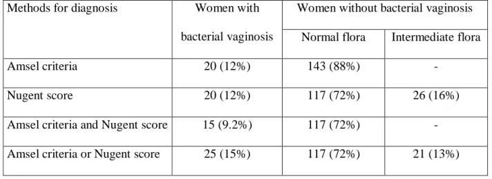
Table 1: Characteristics of vaginal flora according to the Amsel criteria and Nugent score.
Methods for diagnosis Women with bacterial vaginosis
Women without bacterial vaginosis Normal flora Intermediate flora
Amsel criteria 20 (12%) 143 (88%) -
Nugent score 20 (12%) 117 (72%) 26 (16%)
Amsel criteria and Nugent score 15 (9.2%) 117 (72%) - Amsel criteria or Nugent score 25 (15%) 117 (72%) 21 (13%)
Table 2: Molecular characteristics of vaginal flora for women with bacterial vaginosis (n=25) and without bacterial vaginosis (n=138), with the Amsel criteria and Nugent score as
reference methods. PCR quantification (bacteria/mL) Women with bacterial vaginosis n=25
Women without bacterial vaginosis n=138 Normal flora n=117 Intermediate flora n=21 No Lactobacillus species 18 (72%) 8 (6.8%) 6 (28%) G. vaginalis 109/mL 18 (72%) 0 4 (19%) A. vaginae 108/mL 24 (96%) 0 6 (28%) G. vaginalis 109/mL and/or A. vaginae 108/mL 25 (100%) 0 9 (43%)
Table 3: Molecular loads for Lactobacillus species, Gardnerella vaginalis and Atopobium
vaginae for women with bacterial vaginosis (n=25) according to Amsel criteria ( 3 criteria)
or Nugent score (score 7).
Method for diagnosis PCR quantification (bacteria/mL) N Amsel criteria Nugent score Lactobacillus G. vaginalis A. vaginae
1 BV BV - 1.15 x10⁹ 1.09 x10⁸ 2 BV BV 1.83 x10³ 2.69 x10¹⁰ 5.31 x10⁸ 3 BV BV - 1.19 x10⁹ 8.73 x10⁸ 4 BV BV - 4.47 x10⁸ 8.34 x10¹⁰ 5 BV BV - 4.73 x10⁹ 4.84 x10⁹ 6 BV BV 2.38 x10⁸ - 1.40 x10⁹ 7 BV BV - 6.32 x10⁸ 2.30 x10⁹ 8 BV BV - 1.88 x10⁹ 7.20 x10⁸ 9 BV BV 9.20 x10⁵ 3.10 x10⁹ 1.07 x10⁹ 10 BV BV - 1.80 x10⁹ 2.20 x10⁹ 11 BV BV - 2.60 x10⁸ 7.60 x10⁸ 12 BV BV - 7.75 x10⁹ 1.05 x10⁸ 13 BV BV - 1.40 x10⁹ 1.30 x10⁸ 14 BV BV - 3.40 x10⁹ 1.50 x10⁹ 15 BV BV 5.67 x10⁸ 3.05 x10⁹ 6.16 x10⁹ 16 BV IF 2.38 x10⁹ - 2.04 x10⁹ 17 BV IF - 1.02 x10⁹ 4.80 x10⁸ 18 BV IF - 5.67 x10⁹ 1.52 x10⁹ 19 BV IF - 3.42 x10⁹ 6.87 x10⁹ 20 BV IF - 1.80 x10⁹ 1.80 x10⁸ 21 NF BV - 5.30 x10⁹ 1.14 x10⁹ 22 NF BV - - 2.04 x10⁸ 23 NF BV - 1.56 x10¹⁰ 1.05 x10⁹ 24 NF BV 3.78 x10⁸ 5.41 x10⁹ 3.82 x10⁷ 25 NF BV 2.44 x10⁷ 1.50 x10⁸ 2.00 x10⁸
Table 4: Sensitivity, specificity and predictive values with 95% confidence intervals of the PCR quantification for the prediction of bacterial vaginosis among women with bacterial vaginosis (n=25) and without bacterial vaginosis (n=138), with the Amsel criteria and Nugent score as reference methods.
Microorganism threshold quantification to predict bacterial vaginosis % (95% confidence intervals) Sensitivity Specificity Predictive value Positive Negative G. vaginalis 109/mL 0.72 (0.54-0.90) 0.97 (0.94-0.99) 0.82 (0.66-0.98) 0.95 (0.91-0.99) A. vaginae 108/mL 0.96 (0.88-1) 0.96 (0.92-0.99) 0.80 (0.66-0.94) 0.99 (0.98-1) G. vaginalis 109/mL and/or A. vaginae 108/mL 1 0.93 (0.89-0.97) 0.73 (0.59-0.88) 1


![[PDF] Support de Cours Ajax gratuit | Cours informatique](data:image/gif;base64,R0lGODlhAQABAIAAAP///wAAACH5BAEAAAAALAAAAAABAAEAAAICRAEAOw==)