HAL Id: hal-00963711
https://hal.archives-ouvertes.fr/hal-00963711
Submitted on 21 Mar 2014HAL is a multi-disciplinary open access archive for the deposit and dissemination of sci-entific research documents, whether they are pub-lished or not. The documents may come from teaching and research institutions in France or abroad, or from public or private research centers.
L’archive ouverte pluridisciplinaire HAL, est destinée au dépôt et à la diffusion de documents scientifiques de niveau recherche, publiés ou non, émanant des établissements d’enseignement et de recherche français ou étrangers, des laboratoires publics ou privés.
Feature Extraction for Cortical Sulci Identification
Nicolas Royackkers, Houssam Fawal, Michel Desvignes, Marinette Revenu,
J.-M. Travère
To cite this version:
Nicolas Royackkers, Houssam Fawal, Michel Desvignes, Marinette Revenu, J.-M. Travère. Feature Extraction for Cortical Sulci Identification. 9th SCIA, 1995, Uppsala, Sweden. pp.1147-1154. �hal-00963711�
Feature Extraction for Cortical Sulci Identification
Nicolas Royackkers
1, Houssam Fawal
1, Michel Desvignes
1,
Marinette Revenu
1, Jean-Marcel Travere
21GREYC-ISMRA (CNRS, URA 1526) 6, Bd Maréchal Juin 14050 CAEN Cedex FRANCE 2CYCERON (CEA) Bd Becquerel BP 5229 14074 CAEN Cedex FRANCE
email: [email protected], [email protected]
Abstract
The use of PET in quantitative measurement of brain activity requires the superimposition of some anatomical data coming from other sources, like MRI. In the frame of structural and anatomical data matching for a great number of patients, we developed a method for the automatic identification of cortical sulci on 3D MR images. The knowledge used is automatically extracted from a database containing a few pictures where sulci have been previously recognized. Proportional correction mechanisms, based on Talairach’s grid, are proposed. They intend to adapt sulci statistical models to the particular features of any brain, in order to make the recognition easier. Our identification method is efficient and robust for the superficial part of six major sulci.
1. Introduction
Positron emission tomography (PET) is now widely used for the study of brain functional activity. PET is neither destructive nor invasive and is an in-vivo examination technique able to produce pictures of cerebral activity. Unfortunately, PET images do not reflect the anatomy of patients. In this way, when a particular activity area is identified in a PET examination, it cannot be localized in the corresponding brain. That is why a matching between PET and anatomical data from atlases or other images is necessary to make the most of PET information (multimodal data fusion) [12].
So as to allow the interpretation of a PET examination, a solution consists in the identification of the cortical sulci on a 3D magnetic resonance image (MRI) of the same patient. The resulting spatial network is sufficiently accurate to offer a good precision in the localization of functional areas. Cortical sulci identification is difficult because of the high interindividual variability of brain anatomy. In previous works [4,12], we have described a method for the recognition of the superficial part of six major sulci from MRI. This identification is based on metric and topologic features adapted from paper atlases, which generally do not contain suitable information for automatic recognition.
A more general approach has then been developed, where a set of features is automatically extracted from a database containing previously recognized instances of sulci. It allows to build a model of the cortical topography, describing sulci and their topologic relations. An individual correction method, based on Talairach's grid, is also proposed. It aims at making the data coming from various patients more homogeneous. The models of sulci we get should increase the efficiency of the identification process.
2. Anatomical and functional data fusion: different approaches
2.1. Computerized atlases
Computerized brain atlases are easy-to-use tools in the frame of the fusion of anatomical data with different modalities. They are built with information from paper atlases [11], from post-mortem examinations of frozen brains [7], from the two latter sources [8], or from MR images [5]. Typically, computerized atlases are composed of axial sections in which the major elements of brain anatomy can be found. In each section, anatomical elements are represented by a set of closed contours limiting “regions of interest” (ROI). 3D approaches have been proposed too; they use “volumes of interest” (VOI) [5,11].
All computerized atlases represent a single model of human cerebral anatomy. The underlying assumption is that all normal and adult brains, at least at a certain level of representation, have the same structure [2]. It can be verified for the general organization of the brain. However, the large intersubject local variability of cerebral anatomy must be underlined (e.g. shape, depth and localization of the sulci). Global size and pattern can also be very different from one brain to another. Therefore, the superimposition of a computerized atlas to an image needs the use of correction mechanisms.
2.2. Proportional atlases
Proportional atlases contain the representation of a generic anatomy in particular coordinate systems in which the anatomical structures are localized proportionally to brain dimensions. The superimposition of such an atlas to any examination is performed by applying scaling corrections. Among the numerous available methods, the proportional grid system of Talairach [14] produces the most stable results [9,13]. Yet, it cannot fully correct anatomical differences between subjects, since the localization variation interval represents more than 10% of the length of an average brain [13,14].
2.3. Deforming atlases
Deforming atlases, as well as proportional atlases, for the result of an individual matching between anatomical data (deformed atlas) and functional data (PET image), require the use of an intermediate image (usually MRI). This picture allows the identification of the anatomical structures of the studied patient, in order to guide the matching of the atlas. Global matching of the atlas onto the image is the first step in these methods. It consists in applying rigid transformations to the atlas so as to correct major differences between the two representations. Performing local deformations on the atlas is the second stage of the matching. Local rigid transformations on each region of interest can then be carried out [5]. But usually an elastic matching of the atlas is performed [2,7]. This technique consists in representing the atlas as a kind of rubber ball which is submitted to external forces, and in matching its surface with the surface of the brain contained in the image [2]. Matching can also be carried out with the contours of some anatomical elements or with the whole grey matter/white matter interface [7].
The elastic matching methods seem quite elegant, but they do not manage to reconcile simplicity with efficiency. Indeed, when the matching process only uses the surface of the brain, the atlas surface is successfully mapped on the image brain surface, but it is not the same for anatomical structures. So the method finally fails in taking into account intersubject local variability of cerebral anatomy. On the contrary, when matched elements are the anatomical landmarks themselves, the mapped atlas represents the studied brain anatomy in a satisfying way. But these elements must be interactively (i.e. manually) pointed at in the image, which represents a tedious task [1].
3. An identification method for six major sulci on 3D MR images
In collaboration with the CYCERON PET research center of Caen, we have developed a method for the automatic identification of the superficial part of six major sulci on 3D MR images. It consists in a direct recognition of anatomical elements for each subject. A full description of the identification procedure can be found in [4].
3.1. Method description
3.1.1. Cortical topography detection
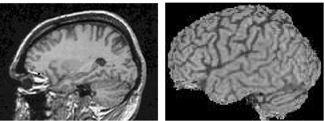
3D MR images are obtained from adult volunteers without any cerebral pathology. They are composed of 120 sections of 256x256 isotropic voxels. An automatic processing localizes the midsagittal plane and the anterior and posterior commissures (AC & PC) of Talairach’s grid. Next, the brain is isolated from the rest of the head [1] (Fig. 1).
FIG. 1. MR images (sagittal projections): top of a head and isolated brain
The brain contained in the pre-processed image is composed of three constituents: white matter, grey matter and cerebro-spinal fluid (CSF). The CSF allows to detect the sulci it fills, thanks to its particular grey level values. So the volume composed of all the sulci is isolated with a multi-level thresolding operation. A 3D skeletonization is then applied to this volume. The Tsao algorithm is used [15], after having added an artificial layer on the surface of the brain, in order to obtain the skeleton at the surface level. The resulting structure is thin in one direction, centered inside the initial volume and respectful of the original object topography. Nevertheless, the superficial layer blocks the skeletonization in a perpendicular direction to the surface. A curve thinning is applied on the surface. Superficial part of the sulci then appears as 3D digital curves (Fig. 2).
Sulci identification is performed mainly on superficial part of the sulci. All the same, we compute a depth for each superficial sulci point to take into account partially the structure present inside the brain [3].
3.1.2. Identification of a sulcus
Our method identifies the superficial part of each one of the recognized sulci separately (at the present time: lateral, precentral, central, postcentral, superior frontal and superior temporal sulci in the two hemispheres).
A rough localization of the sought sulcus in a small area makes up the first step of the identification process. Thus, a search area is defined. It is usually composed of the union of several parallelepipeds defined in Talairach’s grid. The linear structure that constitutes the superficial line of the whole set of sulci is then partitioned in “topological segments” defined as sets of voxels joining two terminal points or nodes of the structure. In order to prepare and to facilitate the sulcus identification, three kinds of processes can be applied: the deletion of segments whose features are not compatible with the sought curve ones, the fusion of connected segments after such suppressions, and the division of long and very curved segments (to keep the average direction meaningful).
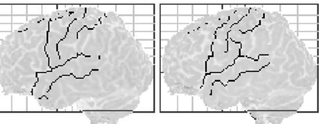
The identification by itself is initialized with the choice of a first segment. As a first step, long enough segments whose directions in axial and sagittal projections are compatible with expected values are selected. The best candidate, chosen for its maximum depth (lateral sulcus) or length (other sulci) is then recorded. Next, a sulcus following is performed from each one of the end of the segment, in order to find the missing parts of the sulcus. If existing, connected segments are examined and the one whose direction is closest to the expected slope is selected. On the contrary, the near enough segment which verifies best this criterion is chosen. Once selected, a segment is merged with the first one. The sulcus following continues until no segment can be found to extend the sulcus in the search area (Fig. 3).
FIG. 3. The six identified sulci on the surface of the left and right hemispheres 3.2. The need for a more general approach
Some major weaknesses of the developed identification process can be stressed. At first, there is no synthetic and explicit model of the sulci features. Next, most of the knowledge is determined or adapted from paper atlases. A heavy experimental work is then needed on each sulcus in order to obtain good results. Furthermore, as well as the information used, the identification processes developed are different for each recognized sulcus. Thus, they cannot be re-used to extend the identification to other sulci. At last, the treatment is static: a single solution is always produced, and no indication is given to the user about its validity (it is the best result according to the sulcus model, but can it be considered as satisfactory? And do equivalent solutions exist?).
4. Cortical topography description & automatic knowledge extraction
4.1. Purposes
From a database composed of some 3D MR images where sulci have already been recognized, the aim is to extract complete, homogeneous and ready-to-process statistical characteristics in order to establish a quantitative model of the cortical topography.
The defined processes and the extracted models should allow to validate and improve the choice of the information used for sulci recognition. The study of anatomical interindividual and interhemisphere variations (asymmetries) should be made easier. It should also enable the production and registration of data that are independent to algorithms, as well as the development of stages for the validation and comparison of potential solutions. Finally, the generalization of identification processes and their application to new sulci should be facilitated by the use of a unique sulci model.
4.2. A model of the cortical topography
4.2.1. Model features
Single sulcus features: each sulcus identified in the database is characterized by its principal direction, its gravity center, its length, some statistics on its global depth, its spatial and geodesic straightness, and its continuity.
Geometrical relations between sulci: for each couple of sulci appearing in the same database image, the connection points (if they exist), the relative localization of gravity centers, and the angle between the axes are computed.
4.2.2. Extraction and validation of the data
All the processes involved in the automatic extraction of the above-mentioned features have been implemented. At the present time, they are using already identified sulci contained in a database composed of 9 MR images.
When possible, results have been compared to sulci features given in anatomical books. The extracted values were then proved to be very close to those measured by neuroanatomists. For instance, the difference between the average brain volume computed and the measure obtained in [6] with a similar protocol is less than 2%. The average difference between the average sulci depths computed in our model and the values given by [10] is about 7%, though the atlas used post-mortem examinations.
Nevertheless, these results cannot be used directly for an individual identification.
4.3. Correction mechanisms
4.3.1. The need for an individual data correction
All the computed features are influenced by the shape and the size of the different brains: localization, direction, depth and other sulci characteristics depend on brain morphology (Fig. 4). Now, with only 9 MR images, some very important variations can be noticed about brain dimensions. For instance, a difference of 13% on length-to-width ratio, 32% on width and 75% on volume can be found. That is why computing features in different images to use them for sulci identification in another image is a nonsense: atlases show general statistics that are not adapted to each individual examination where the recognition has to be performed.
So as to solve this problem, we propose an original approach based on Talairach's proportional grid system, in order to adapt extracted features to any brain image.
Dist a n c e
O rie nt a tio n
Le n g th
R e l a tiv e orie nt a tio n
FIG. 4. Influence of the shape and the size of an object on its superficial curve features
4.3.2. Adapting mechanisms
Our approach consists in converting the coordinates of database sulci points towards the coordinate system of the picture where the recognition has to take place, by means of Talairach's grid [14]. Next, all the previously defined sulci features are computed on this image. Thus, all data are homogeneous because they are obtained in the same anatomical coordinate system. Moreover, they take into account the studied brain characteristics. Actually, our approach can be considered as the opposite of Talairach's one: the grid is not used to localize cortical structures in a standard brain, but the anatomical information contained in a database are adapted to the brain under study.
4.4. Statistical models of sulci
4.4.1. Sulci and types of sulci
There are as many computed individual models of a sulcus as available images. These models are used to build a unique model for each type (or class) of a sulcus (e.g. the lateral sulcus from left hemisphere model). It can then be used for identification tasks. To build the statistical model of a sulcus class, the variation interval, the average and the standard deviation of each individually extracted feature are determined.
The case of depth is particular: our depth is not corrected by the use of Talairach’s grid. Depth correction is made proportionally to the ratio of brain volumes. The statistics defined for other features are then computed on depth information.
4.4.2. Other knowledge
Some data used during the identification are not obtained from the previously defined models. They are the search area and the expected local directions and depths of a sulcus. This intrinsically statistical knowledge is automatically determined from the surface of the brain contained in the picture where the identification is performed.
Search area: By means of Talairach’s grid system, the database instances of a sulcus type are transferred in the analyzed image (proportional correction) and projected onto the surface of the brain it contains (non-linear correction). The resulting area shows the localization of the same sulcus in a learning set of images. It is then extended by an automatic multi-dilation process in order to reach a compact zone whose edges look as if they were manually drawn (Fig. 5).
FIG. 5. Building of a search area (lateral sulcus from the left hemisphere)
Local depth: Each point of the search area is associated to a local average depth. During the identification of a sulcus, the expected local depth can then be easily computed for each voxel on the brain surface with some of its closest neighbours belonging to the area.
Local direction: As well as local depths, local directions are very useful to get a precise representation of the sulci, especially the most curved ones. Two different approaches have been developed so as to build a shape model of a set of sulci. One is based on a morphologic treatment of the search area, the other on a parametric description of the set of sulci used to build this area. They both build a curve onto the surface of the brain under examination. Computing the local direction associated to a point of the search zone then amounts to take into account the closest part of the curve.
5. Results and conclusion
In order to perform the fusion of functional data from PET images and anatomical information from 3D MR images, a fully automatic method for the identification of the superficial part of six major sulci has been developed. Identified sulci have been compared to manual identifications done by two independent medical specialists teams. On the learning set of 9 brain images, 106 of 108 sulci have been correctly identified. On a test set of 4 other brain examinations, 45 on 48 sulci seem to be correctly recognized (this last result is not yet validated), which would give a success rate greater than 90%. Our method seems to be efficient and robust, but it cannot be easily applied to other sulci. Automatically extracting homogeneous knowledge is a first step towards the generalization of the process. The sulci identified in a database are first projected in the analyzed image, by means of Talairach’s grid system. A set of features is then extracted so as to represent these sulci and their geometrical relations. A statistical model of each type of sulcus is finally built. It is ready-to-process and respectful of the studied brain morphology. Search area and local information are determined in the same way.
Our first objective is now to use all the extracted data. Once a unique sulci model is used, common identification processes must be developed. The identification mechanisms should not change much as far as their results are good. However, several curves could be recorded first in each search area in order to select the best one later. As a matter of fact, an individual validation stage must be developed so as to find the best solution among a few sets of segments, using comparison with the extracted model of the sulcus under examination. Finally, a collective validation step must also be developed. It will choose the best set of sulci, compared with the whole cortical topography model described as a graph. The issue is then to determine the closest graph to the model.
Other improvements are also planned, such as the extension of the sulci model in order to take into account ramified structures, or the building of a more declarative representation of the processes so as to improve the identification flexibility.
Acknowledgements
The authors wish to thank J. Yelnik from INSERM U106 (La Salpétrière Hospital, Paris) and J.M. Constans from the Hospital of Caen for their contribution to this work.
References
[1] Allain P. - Imagerie par résonance magnétique du cerveau: analyse automatique
tridimensionnelle et segmentation; application au traitement de données en tomographie par émissions de positons. Ph. D. Thesis, University of Caen, 1993.
[2] Bajcsy R., Kovacic S. - Multiresolution elastic matching. In: Computer vision,
graphics and image processing, 1989, vol.46, pp 1-21.
[3] Desvignes M., Fawal H., Revenu M., Bloyet D., Allain P., Travere J.M., Baron J.C. - Calcul de la profondeur en un point des sillons du cortex sur des images RMN tridimensionnelles. In: 14ème colloque GRETSI, Juan-les-Pins, 1993, pp 1267-1271. [4] Desvignes M., Fawal H., Revenu M., Bloyet D., Allain P., Tavere J.M., Baron J.C. - Reconnaissance du sillon latéral sur des images RMN 3D. In: RFIA, Paris, 1994, pp
685-690.
[5] Evans A.C., Marret S., Torrescorzo J., Ku S., Collins L. - MRI-PET correlation in three dimensions using a volume-of-interest (VOI) atlas. In: Journal of cerebral blood
flow and metabolism, 1991, vol. 11, pp A69-A78.
[6] Filipek P.A., Kennedy D.N., Caviness V.S., Rossnick S.L., Spraggins T.A., Starewicz P.M. - Magnetic resonance imaging-based brain morphometry: development and application to normal subjects. In: Annals of neurology, 1989, vol.25 n°1, pp 61-67.
[7] Gee J.C., Reivich M., Bajcsy R. - Elastically deforming 3D atlas to match anatomical brain images. In: Journal of computer assisted tomography, 1993, vol.17
n°2, pp 225-236.
[8] Greitz T., Bohm C., Holte S., Eriksson L. - A computerized brain atlas: construction, anatomical content, and some applications. In: Journal of computer
assisted tomography, 1991, vol.15 n°1, pp 26-38.
[9] Joliot M. - Traitement du signal de résonance magnétique nucléaire in vivo. Ph. D. Thesis, University of Paris Orsay, 1992.
[10] Ono M., Kubik S., Abernathey C.D. - Atlas of the cerebral sulci. Stuttgart: Georg
Thieme Verlag, 1990.
[11] Rademacher J., Galaburda A.M., Kennedy D.N., Filipek P.A., Caviness V.S.
-Human cerebral cortex: localization, parcellation and morphometry with magnetic resonance imaging. In: Journal of cognitive science, 1992, vol.4 n°4, pp 352-374.
[12] Revenu M., Allain P., Bloyet D., Desvignes M., Travere J.M. - Fusion individuelle
de données cérébrales multimodales: informations issues d’images numériques et connaissance expertes. To appear in: TDSI, France, 1995.
[13] Steinmetz H., Furst G., Freund H.S. - Cerebral cortical localization: application
and validation of the proportional grid system in MR imaging. In: Journal of computer
assisted tomography, 1989, vol.13 n°1, pp 10-19.
[14] Talairach J., Tournoux P. - Co-planar stereotaxic atlas of the human brain.
Stuttgart: Georg Thieme Verlag, 1988.
[15] Tsao Y.F., Fu K.S. - A parallel thinning algorithm for 3D pictures. In: Computer