Received: March 29, 2019. Accepted: July 24, 2019. Pre-published: August 1, 2019.
©2020 Ferrata Storti Foundation
Material published in Haematologica is covered by copyright. All rights are reserved to the Ferrata Storti Foundation. Use of published material is allowed under the following terms and conditions:
https://creativecommons.org/licenses/by-nc/4.0/legalcode. Copies of published material are allowed for personal or inter-nal use. Sharing published material for non-commercial pur-poses is subject to the following conditions:
https://creativecommons.org/licenses/by-nc/4.0/legalcode, sect. 3. Reproducing and sharing published material for com-mercial purposes is not allowed without permission in writing from the publisher.
Correspondence:
DAVIDE ROSSI davide.rossi@eoc.ch GIANLUCA GAIDANO gianluca.gaidano@med.uniupo.itHaematologica
2020
Volume 105(2):448-456
doi:10.3324/haematol.2019.219550 Check the online version for the most updated information on this article, online supplements, and information on authorship & disclosures: www.haematologica.org/content/105/2/448Ferrata Storti Foundation
B
IRC3 is a recurrently mutated gene in chronic lymphocytic leukemia
(CLL) but the functional implications of BIRC3 mutations are largely
unexplored. Furthermore, little is known about the prognostic impact
of BIRC3 mutations in CLL cohorts homogeneously treated with first-line
fludarabine, cyclophosphamide, and rituximab (FCR). By immunoblotting
analysis, we showed that the non-canonical nuclear factor-
κB pathway is
active in BIRC3-mutated cell lines and in primary CLL samples, as
docu-mented by the stabilization of MAP3K14 and by the nuclear localization of
p52. In addition, BIRC3-mutated primary CLL cells are less sensitive to
flu-darabine. In order to confirm in patients that BIRC3 mutations confer
resist-ance to fludarabine-based chemoimmunotherapy, a retrospective
multicen-ter cohort of 287 untreated patients receiving first-line FCR was analyzed
Biological and clinical implications of
BIRC3
mutations in chronic lymphocytic leukemia
Fary Diop,1* Riccardo Moia,1* Chiara Favini,1Elisa Spaccarotella,1Lorenzo De Paoli,1Alessio Bruscaggin,2Valeria Spina,2Lodovico Terzi-di-Bergamo,2
Francesca Arruga,3Chiara Tarantelli,4Clara Deambrogi,1Silvia Rasi,1Ramesh
Adhinaveni,1Andrea Patriarca,1Simone Favini,1Sruthi Sagiraju,1Clive
Jabangwe,1Ahad A. Kodipad,1Denise Peroni,1Francesca R. Mauro,5Ilaria Del
Giudice,5Francesco Forconi,6,7Agostino Cortelezzi,8Francesco Zaja,9Riccardo
Bomben,10Francesca Maria Rossi,10Carlo Visco,11Annalisa Chiarenza,12Gian
Matteo Rigolin,13Roberto Marasca,14Marta Coscia,15Omar Perbellini,16
Alessandra Tedeschi,17Luca Laurenti,18Marina Motta,19David Donaldson,20
Phil Weir,20Ken Mills,21Patrick Thornton,22Sarah Lawless,20Francesco Bertoni,4Giovanni Del Poeta,23Antonio Cuneo,13Antonia Follenzi,24Valter
Gattei,10Renzo Luciano Boldorini,25Mark Catherwood,20Silvia Deaglio,3Robin
Foà,5Gianluca Gaidano1° and Davide Rossi2°
1Division of Hematology, Department of Translational Medicine, Amedeo Avogadro
University of Eastern Piedmont, Novara, Italy; 2Institute of Oncology Research and
Oncology Institute of Southern Switzerland, Bellinzona, Switzerland; 3Department of
Medical Sciences, University of Turin & Italian Institute for Genomic Medicine, Turin, Italy;
4Università della Svizzera Italiana, Institute of Oncology Research, Bellinzona, Switzerland; 5Hematology, Department of Cellular Biotechnologies and Hematology, Sapienza
University, Rome, Italy; 6Cancer Sciences Unit, Southampton Cancer Research UK and
National Institute for Health Research Experimental Cancer Medicine Centre, University of Southampton, Southampton, UK; 7Division of Hematology, University of Siena, Siena, Italy; 8Department of Hematology Oncology, Foundation IRCCS Ca’ Granda Ospedale Maggiore
Policlinico and University of Milan, Milan, Italy; 9Clinica Ematologica, DAME, University of
Udine, Udine, Italy; 10Clinical and Experimental Onco-Hematology Unit, Centro di
Riferimento Oncologico, Istituto di Ricovero e Cura a Carattere Scientifico (IRCCS), Aviano, Italy; 11Department of Cell Therapy and Hematology, Ospedale San Bortolo, Vicenza, Italy; 12Division of Hematology, Azienda Ospedaliera Universitaria Policlinico-OVE, Catania, Italy; 13Hematology Section, Azienda Ospedaliero Universitaria Arcispedale S. Anna, University of
Ferrara, Ferrara, Italy; 14Division of Hematology, Department of Oncology and Hematology,
University of Modena and Reggio Emilia, Modena, Italy; 15Division of Hematology, Azienda
Ospedaliero Universitaria Città della Salute e della Scienza and University of Turin, Turin, Italy; 16Section of Hematology, Department of Medicine, University of Verona, Verona, Italy; 17Department of Oncology/Haematology, Niguarda Cancer Center, Niguarda Ca Granda
Hospital, Milan, Italy; 18Fondazione Policlinico Universitario A. Gemelli, Rome, Italy; 19Department of Hematology, Spedali Civili, Brescia, Italy; 20Clinical Haematology, Belfast
City Hospital, Belfast Health and Social Care Trust, Belfast, Northern Ireland, UK; 21Centre
for Cancer Research and Cell Biology (CCRCB), Queen’s University Belfast, Belfast, Northern Ireland, UK; 22Department of Haematology, Beaumont Hospital, Dublin, Ireland; 23Department of Hematology, Tor Vergata University, Rome, Italy; 24Department of Health
Sciences, University of Eastern Piedmont Amedeo Avogadro, Novara, Italy; 25Department
of Pathology, University of Eastern Piedmont Amedeo Avogadro, Novara, Italy.
**FD and RM contributed equally to this work °GG and DR contributed equally to this work.
Introduction
Nuclear factor-κB (NF-κB) signaling is a key component of the development and evolution of chronic lymphocytic leukemia (CLL).1Two NF-κB pathways exist, namely the canonical and non-canonical pathways.2 The former is triggered by B-cell receptor signaling via Bruton tyrosine kinase (BTK), while the latter is activated by members of the tumor necrosis factor (TNF) cytokine family.3 Upon receptor binding, the TRAF3/MAP3K14-TRAF2/BIRC3 negative regulatory complex of non-canonical NF-κB sig-naling is disrupted, MAP3K14 (also known as NIK), the central activating kinase of the pathway, is released and activated to induce the phosphorylation and proteasomal processing of p100, thereby leading to the formation of p52-containing NF-κB dimers. The p52 protein dimerizes with RelB to translocate into the nucleus, where it regu-lates gene transcription. BIRC3 (Baculoviral IAP Repeat Containing 3) is a negative regulator of non-canonical NF-κB. Physiologically, BIRC3 (also known as cIAP2) cat-alyzes MAP3K14 protein ubiquitination in a manner that is dependent on the E3 ubiquitinine ligase activity of its C-terminal RING domain. MAP3K14 ubiquitination results in its proteasomal degradation.4
B-cell neoplasia often pirates signaling pathways by molecular lesions to promote survival and proliferation. Although according to bioinformatics criteria BIRC3 is one of the candidate driver genes of CLL, the functional impli-cations of BIRC3 mutations are partially unexplored.5-7 Furthermore, little is known about the prognostic impact of BIRC3 mutations in CLL cohorts homogeneously treat-ed first-line with fludarabine, cyclophosphamide, and rit-uximab (FCR).7
FCR is the most effective chemoimmunotherapy regi-men for the manageregi-ment of CLL in young and fit patients devoid of TP53 disruption.8Survival after FCR is, howev-er, variable, and is affected by the molecular characteris-tics of the CLL clone.9Deletion of 17p and TP53 mutations are present in most, but not all patients who are refractory to chemo-immunotherapy, which prompts the identifica-tion of addiidentifica-tional biomarkers associated with early failure of FCR.10-12
Methods
Functional studies
The human CLL cell line MEC1, the splenic marginal zone lym-phoma cell lines SSK41 and VL51, the mantle cell lymlym-phoma cell lines MAVER-1, Z-138 and JEKO-1, the human HEK-293T cell line, as well as primary CLL cells were used in functional experiments. The entire non-canonical NF-κB pathway was assessed by western blot analysis. Quantitative real-time polymerase chain reaction
(qRT-PCR) was utilized to analyze the non-canonical NF-κB signa-ture. Primary CLL were exposed to fludarabine and venetoclax for 24-48 h and apoptosis was measured using the eBioscience Annexin V Apoptosis Detection Kit APC (ThermoFisher). Details are supplied in the Online Supplementary Methods.
Cancer personalized profiling by deep sequencing
A retrospective multicenter cohort of 287 untreated CLL patients receiving first-line therapy with FCR was analyzed for mutations, including 173 patients from a previously published multicenter clinical series and 114 new patients not included in our previous report.10 The study was approved by the EthicalCommittee of the Ospedale Maggiore della Carità di Novara asso-ciated with the Amedeo Avogadro University of Eastern Piedmont (study number CE 67/14). Further information is provided in the
Online Supplementary Methods. A targeted resequencing gene panel
was designed to include: (i) coding exons plus splice site of 24 genes known to be implicated in CLL pathogenesis and/or prog-nosis; (ii) 3’UTR of NOTCH1; and (iii) enhancer and promoter region of PAX5 (size of the target region: 66627bp) (Table S1).6,7
The next-generation sequencing libraries for genomic DNA (gDNA) were constructed using the KAPA Library Preparation Kit (Kapa Biosystems) and those for RNA were constructed using the RNA Hyper Kit (Roche). Multiplexed libraries (n=10 per run) were sequenced using 300-bp paired-end runs on a MiSeq sequencer (Illumina) to obtain a coverage of at least 2000x in >90% of the tar-get region (66627 bp) in 80% of cases (Online Supplementary Table
S2). A robust and previously validated bioinformatics pipeline was
used for variant calling (Online Supplementary Appendix).
Statistical analysis
Progression-free survival (PFS) was the primary endpoint. Survival analysis was performed with the Kaplan-Meier method and compared between strata using the log-rank test. To account for multiple testing, adjusted P values were calculated using the Bonferroni correction. The adjusted association between exposure variables and PFS was estimated by Cox regression. Internal vali-dation of the multivariate analysis was performed using a boot-strap approach. Statistical significance was defined as a P value <0.05 (Online Supplementary Appendix).
Results
BIRC3 mutations associate with activation
of non-canonical nuclear factor-
κB signaling
In order to map unique BIRC3 mutations in CLL com-prehensively, we compiled somatically confirmed variants identified in the current CLL study cohort with those identified in previous studies13 or listed in public CLL mutation catalogues (Figure 1A). Virtually all BIRC3 muta-tions were frameshift mutamuta-tions or stop codons clustering in two hotspot regions comprised between amino acids
by targeted next-generation sequencing of 24 recurrently mutated genes in CLL. By univariate analysis
adjusted for multiple comparisons BIRC3 mutations identify a poor prognostic subgroup of patients in whom
FCR treatment fails (median progression-free survival: 2.2 years, P<0.001) similar to cases harboring TP53
mutations (median progression-free survival: 2.6 years, P<0.0001). BIRC3 mutations maintained an
inde-pendent association with an increased risk of progression with a hazard ratio of 2.8 (95% confidence interval
1.4-5.6, P=0.004) in multivariate analysis adjusted for TP53 mutation, 17p deletion and IGHV mutation
sta-tus. If validated, BIRC3 mutations may be used as a new molecular predictor to select high-risk patients for
novel frontline therapeutic approaches.
367-438 and amino acids 537-564. BIRC3 variants were predicted to generate aberrant truncated transcripts caus-ing the elimination or truncation of the C-terminal RING domain of the BIRC3 protein. The RING domain of
BIRC3 harbors the E3 ubiquitin ligase activity that is essential for proteasomal degradation of MAP3K14, the central activating kinase of non-canonical NF-κB signaling. This observation points to non-canonical NF-κB activation
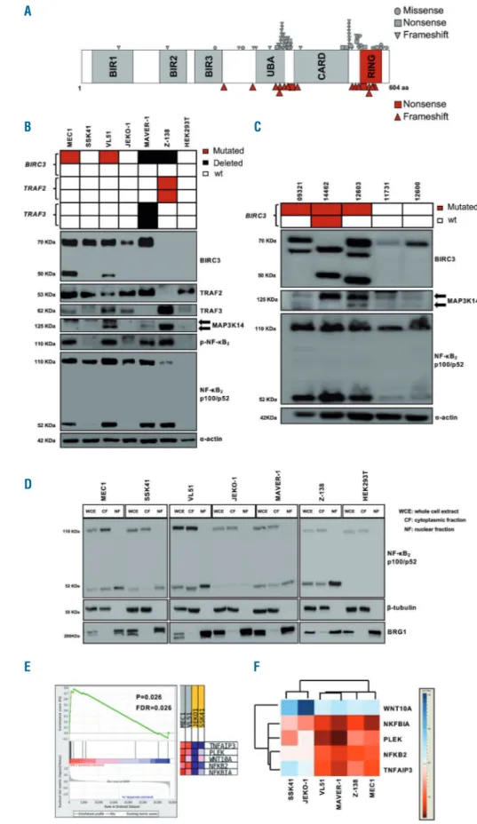
Figure 1. The non-canonical nuclear factor-κB pathway is active in BIRC3-mutated chronic lymphocytic leukemia cell lines and primary samples. (A) Disposition of BIRC3 mutations across the protein. The mutations identified by Landau et al.6, Puente et al.7and from a
public CLL mutation catalogue (COSMIC v85) are plotted in gray. Individual BIRC3 mutations identified in the cur-rent studied cohort and in our previous study13are plotted in red. (B) Western
blot analysis of BIRC3 protein expres-sion and NF-κB2activation and
process-ing in the splenic marginal zone lym-phoma (SMZL) cell lines SSK41, VL51 and in the chronic lymphocytic leukemia (CLL) cell line MEC1, carrying wildtype (wt) or disrupted BIRC3. The MAVER-1 and Z-138 cell lines were used as posi-tive controls of non-canonical NF-κB activation, harboring genetic activation of non-canonical NF-κB signaling. The JEKO-1 and HEK 293T cell lines were used as negative controls for non-canonical NF-κB signaling. α-actin was used as a loading control. Color codes indicate the gene status in each cell line. The aberrant BIRC3 band expressed in MEC1 and VL51 cell lines corresponds in size to the predicted BIRC3-truncated protein, encoded by the mutant allele. (C) Western blot analysis showing BIRC3 expression and NF-κB2 processing in purified primary
tumor cells from five CLL and SMZL patients carrying wildtype or disrupted BIRC3. Color codes indicate the gene status in each cell line. The aberrant BIRC3 bands in patients 09321, 14462 and 12603 correspond in size to the predicted BIRC3-truncated protein encoded by the mutant allele. α-actin was used as a loading control. (D) Western blot of whole cell extract, cyto-plasmic or nuclear fractions of the SMZL and CLL cell lines probed for the NF-κB2 subunits p100 and p52. The MAVER-1 and Z-138 cell lines served as positive controls while the JEKO-1 and HEK 293T cell lines were used as nega-tive controls. β-tubulin and BRG1 served as controls for the purity of the cytoplasmic and nuclear fractionations, respectively. (E) Gene set enrichment analysis score and distribution of non-canonical NF-κB target genes along the rank of transcripts differentially expressed in the SMZL cell lines SSK41, VL51 and in the CLL cell line MEC1. The JEKO-1 cell line was used as a negative control. (F) Validation of expression of non-canonical NF-κB target genes in the same SMZL and CLL cell lines as deter-mined by quantitative real-time poly-merase chain reaction analysis. Changes of gene expression were nor-malized to GAPDH expression; relative quantities were log2normalized to
con-trol samples (the mantle cell lymphoma cell line, JEKO-1).
A
B C
D
through MAP3K14 stabilization as the predicted function-al consequence of BIRC3 mutations in CLL.
The non-canonical NF-κB signaling was profiled by immunoblotting in B-cell tumor cell lines and primary CLL cells with different genetic make-up in the non-canonical NF-κB pathway to verify whether BIRC3 mutations lead to constitutive non-canonical NF-κB activation. Additional genetic features of the above-mentioned cell lines and pri-mary CLL cells are shown in Online Supplementary Table
S3. In the VL51 splenic marginal zone lymphoma cell line
and in the MEC1 CLL cell lines, both harboring endoge-nous truncating mutations of the BIRC3 gene, non-canon-ical NF-κB signaling was constitutively active, as docu-mented by the stabilization of MAP3K14, phosphoryla-tion of NF-κB2, its processing from p100 to p52, as well as the nuclear localization of p52 (Figure 1B-D). Consistent with the biochemical clues of non-canonical NF-κB activa-tion, the gene expression signature of the VL51 and MEC1 cell lines was significantly enriched in non-canonical NF-κB target genes (Figure 1E, F). Non-canonical NF-NF-κB signal-ing in BIRC3-mutated cells was consistent with that in mantle cell lymphoma cell lines known to harbor a dis-rupted TRAF3/MAP3K14-TRAF2/BIRC3 negative regula-tory complex by loss of TRAF3 or TRAF2.14Like BIRC3-mutated cell lines, primary CLL samples harboring inacti-vating mutations of BIRC3 also showed stabilization of MAP3K14 and NF-κB2processing from p100 to p52 (Figure 1C), thus confirming that non-canonical NF-κB activation is also a feature of primary cells harboring BIRC3 variants. MAP3K14 stabilization is largely associated with BIRC3 mutations. Indeed all seven cases harboring non-canonical NF-κB genetic lesions showed either a strong or a slight MAP3K14 band, while, conversely, only one out of five cases lacking a non-canonical NF-κB lesion had MAP3K14 expression (Fisher exact test, P=0.01).
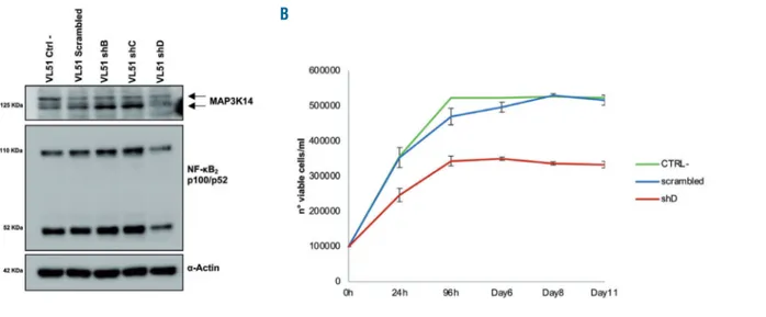
MAP3K14 was genetically targeted by shRNA to test whether BIRC3-mutated cells are addicted to its stabiliza-tion. Compared to non-targeting shRNA, the most effi-cient anti-MAP3K14 shRNA-D resulted in a partial silenc-ing of MAP3K14 and in decreased NF-κB2 processing from p100 to p52. This translated into a reduced cell viability of
the BIRC3-mutated VL51 cell line transduced with shRNA-D. This observation indicates that MAP3K14 sta-bilization is a vulnerability of BIRC3-mutated cells (Figure 2). In order to test the contribution of BTK to non-canon-ical NF-κB signaling when it is activated through BIRC3 mutations, BIRC3-mutated cell lines, as well as cell lines harboring a disrupted or competent TRAF3/MAP3K14-TRAF2/BIRC3 negative regulatory complex were treated with ibrutinib at different dosages and non-canonical NF-κB signaling activation was probed by immunoblotting of the NF-κB2processing from p100 to p52. Processing from p100 to p52 was unaffected by ibrutinib treatment in cell lines harboring BIRC3 mutations (Figure 3) or a disrupted TRAF3/MAP3K14-TRAF2/BIRC3 negative regulatory complex, consistent with the notion that BIRC3 mutations activate non-canonical NF-κB by bypassing BTK blockade by ibrutinib.14
BIRC3 mutations confer resistance to fludarabine
in primary chronic lymphocytic leukemia cells
We performed in vitro pharmacological studies on pri-mary CLL cells to verify the vulnerabilities of BIRC3-mutated cells. CLL cells purified from patients carrying
BIRC3 mutations were treated with increasing doses of
fludarabine. Drug-induced apoptosis was compared to that of samples harboring TP53 mutations, which repre-sent a control for fludarabine resistance. CLL cells devoid of genetic lesions in either BIRC3 or TP53 were used as a control for fludarabine sensitivity. The molecular charac-teristic of the ex-vivo CLL cells are listed in Online
Supplementary Table S4.
BIRC3-mutated cells showed delayed
fludarabine-induced cell death, as no response was observed after 24 h of treatment, at variance with TP53- and BIRC3-wildtype samples. At this time point, cell viability curves of BIRC3-mutated samples overlapped almost completely with those of TP53-disrupted samples, which are known to be fludarabine resistant (Figure 4A). At 48 h, the viability of
BIRC3-mutated cells was lower than that of
TP53-mutat-ed samples, but higher than that of TP53- and BIRC3-wild-type samples (Figure 4B).
Figure 2. Knockdown of MAP3K14 by RNA interference in VL51 cells.(A) Western blot analysis for MAP3K14 expression and for NF-κB2processing of p100 to p52.
(B) VL51 cell viability assessed by trypan blue after transduction with lentiviral vectors expressing the shRNA-D_MAP3K14 (shD: in red), a scrambled shRNA (scram-bled: in blue), and in non-transfected cells (CTRL: in green).
In order to assess whether BIRC3 mutations interfere with apoptosis, primary CLL cells were treated with vene-toclax. Venetoclax treatment resulted in a similar reduc-tion of cell viability in BIRC3-mutated cells, TP53-mutated cells and BIRC3/TP53-wildtype cells (Figure 4C, D). Such divergent sensitivity to fludarabine and venetoclax of
BIRC3-mutated CLL cells indirectly suggests that BIRC3
mutations likely affect the upstream DNA damage response pathway rather than downstream apoptosis as a mechanism of inducing cell death.
Patients harboring BIRC3 mutations are at risk
of FCR failure
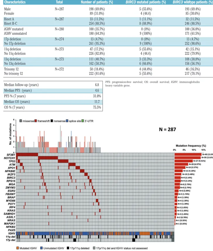
In order to confirm in vivo in patients that BIRC3 muta-tions confer resistance to fludarabine-based chemoim-munotherapy, we correlated the BIRC3 mutation status with PFS of CLL patients treated with FCR. Mutational profiling was performed in 287 patients who received first-line FCR. The baseline features of the study cohort were consistent with progressive, previously untreated CLL (Table 1). The median follow-up was 6.8 years, with
a median PFS and overall survival of 4.6 and 11.7 years, respectively (Table 1) consistent with observations in clinical trial cohorts.15As expected, SF3B1 and NOTCH1 were the most frequently mutated genes identified in 13.9% and in 13.6% of patients, respectively, followed by TP53 in 9.4% and ATM in 6.9% of patients. BIRC3 was mutated in 3.1% of patients, reflecting the data reported in previous studies.6,7,13 Overall, 154/287 (53.6%) cases harbored at least one non-synonymous somatic mutation in one of the 24 CLL genes included in our panel (range, 1-5 mutation per patient), which is con-sistent with the typical mutational spectrum of CLL requiring first-line treatment (Figure 5, Online
Supplementary Table S5).6,7,16
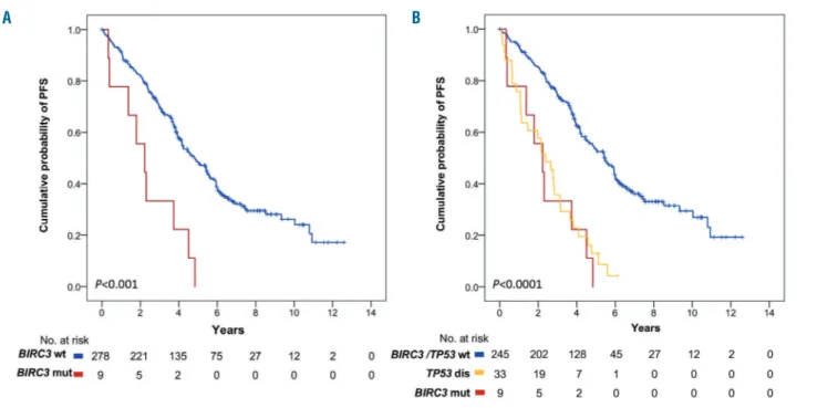
By univariate analysis adjusted for multiple compar-isons, among the genes analyzed in our panel, only TP53 mutations (median PFS of 2.6 years; P<0.0001) and
BIRC3 mutations (median PFS of 2.2 years; P<0.001)
(Figure 6A) were associated with significantly shorter PFS (Table 2). Following FCR treatment, the PFS of
BIRC3-mutated patients was similar to that of cases
har-Figure 3. The non-canonical nuclear factor-κB pathway is not switched off by ibrutinib in BIRC3-mutated cell lines.Western blot showing p100/p52 expression in (A) MEC1 and (B) VL51 cell lines that harbor BIRC3 mutations. (C) MAVER-1 and (D) Z-138 cell lines, known to be affected by non-canonical NF-κB pathway gene mutations and resistant to ibrutinib were used as positive controls. (E) The JEKO-1 cell line, known to be devoid of NF-κB pathway gene mutations and sensitive to ibrutinib, was used as a negative control. All cell lines were treated with different concentrations of ibrutinib for 72 and 96 h. DMSO: dimethylsulfoxide.
A B
boring TP53 disruption (Figure 6B). Consistently, BIRC3-mutated patients had a lower likelihood of achieving complete response (22.2%) at the end of FCR compared to BIRC3-wildtype cases (76.7%; P=0.001). Well-known molecular prognostic biomarkers of CLL, such as unmu-tated IGHV gene status and 17p deletion also associated with a significantly shorter PFS, supporting the represen-tativeness of the study cohort (Table 2). By multivariate analysis including variables showing a multiplicity-adjusted significant association with PFS, BIRC3 muta-tions maintained an independent association with PFS, with a hazard ratio of 2.8 (95% confidence interval: 1.4-5.6; P=0.004) (Table 2).
Discussion
The results of this study provide evidence that: (i) BIRC3 mutations are associated with activation of the non-canonical NF-κB pathway and with resistance to fludara-bine in vitro; and (ii) BIRC3-mutated patients, like cases harboring TP53 disruption, are subject to failure of FCR chemoimmunotherapy.
The mere presence of somatic mutations is insufficient
to implicate a gene in cancer. Cancer geneticists and bioin-formaticians differentiate “passenger” events, likely being randomly acquired, to distinguish them from mutations targeting candidate “cancer-driver” genes, likely implicat-ed in the tumor biology, according to a statistical defini-tion. Any given gene is labeled as a candidate “cancer driv-er” if it harbors somatic point mutations at a statistically significant rate or pattern in cancer samples. In CLL, more than 40 genes fulfill the statistical definition of a candidate “cancer driver”, including BIRC3, but few of them are bio-logically validated (i.e. SF3B1, NOTCH1, TP53, ATM,
FBXW7).6,7,17-20 The BIRC3 gene codes for a protein that ubiquitinates and negatively regulates the central activat-ing kinase of the non-canonical NF-κB pathway, namely MAP3K14 (also known as NIK).21,22In lymphoid malignan-cies, the NF-κB pathway is a pivotal and positive mediator of cell proliferation and survival.5,23,24With regards to CLL,
BIRC3 mutations are absent in patients with monoclonal
B-cell lymphocytosis, are rare at the time of diagnosis (3-4%), but are detectable in approximately 25% of fludara-bine-refractory patients.13 In this study, we verified the biological consequences of BIRC3 mutations, showing that they are associated with activation of the non-canon-ical NF-κB pathway, that BIRC3-mutated lymphoid cells
Figure 4. Responses of primary cells lines to fludarabine and venetoclax.(A-D) Viability of BIRC3-mutated (n=6 patients, red line), TP53-mutated (n=8 patients, black line) and wildtype (n=7 patients, blue line) primary CLL cells treated with different concentrations of fludarabine for 24 h (A) and 48 h (B) or venetoclax for 24 h (C) and 48 h (D). The pairwise P values are listed in the tables below the respective figures. M, mutated; WT, wildtype; NT, not treated.
A B
are addicted to the non-canonical NF-κB pathway, and that BIRC3-mutated CLL are resistant to fludarabine both
in vitro and in patients. It still remains to be clarified
whether NF-κB activation is the only molecular pathway that causes chemo-refractoriness in BIRC3-mutated CLL or whether other mechanisms are also involved.24-29
Table 1. Clinical data of FCR-treated chronic lymphocytic leukemia patients according to BIRC3 mutational status.
Characteristics Total Number of patients (%) BIRC3 mutated patients (%) BIRC3 wildtype patients (%) Male N=287 198 (69.0%) 5 (55.6%) 193 (69.4%) Female 89 (31.0%) 4 (44.4) 85 (30.6%) Binet A N=287 33 (11.5%) 1 (11.1%) 32 (11.5%) Binet B-C 254 (88.5%) 8 (88.9%) 246 (88.5%) IGHV mutated N=280 100 (35.7%) 0 (0%) 100 (36.0%) IGHV unmutated 180 (64.3%) 9 (100%) 171 (61.5%) 17p deletion N=274 13 (4.7%) 0 (0%) 13 (4.7%) No 17p deletion 261 (95.3%) 9 (100%) 252 (90.6%) 11q deletion N=273 47 (17.2%) 5 (55.6%) 42 (15.1%) No 11q deletion 226 (82.8%) 4 (44.4) 222 (79.9%) 13q deletion N=273 111 (40.7%) 3 (33.3%) 108 (38.8%) No 13q deletion 162 (50.3%) 6 (66.6%) 156 (56.1%) Trisomy 12 N=272 50 (18.4%) 4 (44.4%) 46 (16.5%) No trisomy 12 222 (81.6%) 5 (55.6%) 217 (78.1%) Median follow-up (years) 6.8
Median PFS (years) 4.6 PFS % (7 years) 31.0% Median OS (years) 11.7 OS % (7 years) 75.5%
Figure 5. Mutational profile of the FCR-treated cohort.Case-level mutational profiles of 287 patients treated with FCR (fludarabine, cyclophosphamide, rituximab). Each column represents one tumor sample, each row represents one gene. The fraction of tumors with mutations in each gene is plotted on the right. The number and type of mutations in each patient are plotted above the heatmap. Mutations are highlighted in red. IGHV mutational status, 17p deletion and 11q deletion are plotted at the bottom of the heatmap.
PFS: progression-free survival; OS: overall survival; IGHV: immunoglobulin heavy variable gene.
The introduction of FCR was a breakthrough in the man-agement of young, fit CLL patients, leading to improve-ments in both PFS and overall survival compared to those achieved with previous treatment regimens. In both clinical trials and real-life cohorts,10-12 IGHV mutation status and
TP53 disruption emerged as strong predictors of poor
response to FCR. However, these molecular biomarkers do
not fully capture all high-risk patients destined to relapse. We propose BIRC3 mutations as a new biomarker for the identification of patients at high risk of FCR failure, simi-larly to cases harboring TP53 disruption. If validated in independent series, BIRC3 mutations may turn out as a new molecular predictor of FCR resistance that could be used to select patients to be treated with novel targeted agents.
Table 2. Univariate and multivariate analyses of progression-free survival.
Univariate analysis Multivariate analysis Internal bootstrapping validation Characteristics 7-y PFS Median 95% CI P P* HR LCI UCI P HR LCI UCI Bootstrapping (%) PFS (y) selection (%) Binet A 40.3% 4.5 2.46.6 0.356 -Binet B-C 30.0% 4.6 3.8-5.4 - - - - - - IGHV mutated 49.3% 6.5 3.8-9.2 <0.001 0.003 - - - 0.001 - - - 98.8% IGHV unmutated 23.0% 3.9 3.5-4.4 1.8 1.3 2.6 1.9 1.3 2.7 No 11q deletion 33.4% 5.0 4.25.9 0.025 0.700 -11q deletion 13.9% 3.6 2.4-4.9 - - - - - - No 17p deletion 33.0% 4.8 4.1-5.6 <0.0001 <0.0001 - - - <0.0001 - - - 99.5% 17p deletion nr 1.1 0-2.6 4.0 2.2 7.5 4.9 2.5 9.8 TP53 wildtype 33.8% 5.4 4.3-5.8 <0.0001 <0.001 - - - 0.030 - - - 73.3% TP53 mutated nr 2.8 2.0-3.5 1.7 1.1 2.8 1.8 1.1 3 BIRC3 wildtype 32.2% 4.8 4.1-5.6 <0.001 0.005 - - - 0.004 - - - 91.1% BIRC3 mutated nr 2.2 0.9-3.5 2.8 1.4 5.6 3.4 1.6 7.3 EGR2 wildtype 31.5% 4.7 3.95.4 0.015 0.420 -EGR2 mutated nr 1.5 0-3.8 - - - - - - ATM wildtype 32.5% 4.8 4.15.6 0.029 0.812 -ATM mutated nr 3.2 2.4-4.1 - - - - - -
y: year; P: P-value; P*: Bonferroni correction; PFS: progression-free survival; CI: confidence interval; HR: hazard ratio; LCI: lower boundary of the confidence interval; UCI: upper boundary of the confidence interval; IGHV: immunoglobulin heavy variable gene; nr: not reached.
Figure 6. Kaplan-Meier estimates of progression-free survival in BIRC3-mutated patients. (A) Cases harboring BIRC3 mutations are represented by the red line. BIRC3-wildtype cases are represented by the blue line. (B) Cases harboring BIRC3 mutations are represented by the red line. Cases harboring TP53 disruption (including TP53 mutation and/or 17p deletion) are represented by the yellow line. Patients devoid of BIRC3 mutation and TP53 disruption are represented by the blue line.
Non-canonical NF-κB activation by BIRC3 mutations bypasses the block of BTK by ibrutinib. Consistently, NF-κB activation and cell survival are unaffected by ibrutinib in both CLL cells (our study) and mantle cell lymphoma cells.14If this preclinical evidence is validated in ibrutinib-treated patients, BIRC3 mutations may also translate into a biomarker for informing selection of novel agents.
Acknowledgments
This work was supported by: Molecular bases of disease dis-semination in lymphoid malignancies to optimize curative thera-peutic strategies, (5 x 1000 n. 21198), Associazione Italiana per la Ricerca sul Cancro Foundation, Milan, Italy (grants to GG and RF) and Progetti di Rilevante Interesse Nazionale (PRIN),
(2015ZMRFEA), Rome, Italy; partially funded by the AGING Project – Department of Excellence – DIMET, Università del Piemonte Orientale, Novara, Italy; partially funded by Novara AIL ONLUS, Novara, Italy; Associazione Italiana per la Ricerca sul Cancro (AIRC IG-17314); Swiss Cancer League, ID HSR-4660-11-2018, Bern, Switzerland; Research Advisory Board of the Ente Ospedaliero Cantonale, Bellinzona, Switzerland; European Research Council (ERC) Consolidator grant CLL-CLONE ID: 772051; grant n. 320030_169670/1 Swiss National Science Foundation, Berne, Switzerland; Fondazione Fidinam, Lugano, Switzerland; Nelia & Amadeo Barletta Foundation, Lausanne, Switzerland; Fond’Action, Lausanne, Switzerland; Translational Research Program, grant n. 6594-20, The Leukemia & Lymphoma Society, New York, USA.
References
1. Mansouri L, Papakonstantinou N, Ntoufa S, Stamatopoulos K, Rosenquist R. NF-κB acti-vation in chronic lymphocytic leukemia: a point of convergence of external triggers and intrinsic lesions. Semin Cancer Biol. 2016; 39:40-48
2. Bonizzi G, Karin M. The two NF-kappaB activation pathways and their role in innate and adaptive immunity. Trends Immunol. 2004;25(6):280-288.
3. Oeckinghaus A, Hayden MS, Ghosh S. Crosstalk in NF-κB signaling pathways. Nat Immunol. 2011;12(8):695-708.
4. Sun SC. The noncanonical NF-κB pathway. Immunol Rev. 2012;246(1):125-140. 5. Asslaber D, Wacht N, Leisch M, Qi Y, et al.
BIRC3 expression predicts CLL progression and defines treatment sensitivity via enhanced NF-κB nuclear translocation. Clin Cancer Res. 2019;25(6):1901-1912. 6. Puente XS, Beà S, Valdés-Mas R, et al.
Non-coding recurrent mutations in chronic lym-phocytic leukaemia. Nature. 2015;526 (7574):519-524.
7. Landau DA, Tausch E, Taylor-Weiner AN, et al. Mutations driving CLL and their evolu-tion in progression and relapse. Nature. 2015;526(7574):525-530.
8. Hallek M, Cheson BD, Catovsky D, et al. Guidelines for diagnosis, indications for treatment, response assessment and sup-portive management of chronic lymphocytic leukemia. Blood. 2018;131(25):2745-2760. 9. Gaidano G, Rossi D. The mutational
land-scape of chronic lymphocytic leukemia and its impact on prognosis and treatment. Hematology Am Soc Hematol Educ Program. 2017;2017(1):329-337.
10. Rossi D, Terzi-di-Bergamo L, De Paoli L, et al. Molecular prediction of durable remis-sion after first-line fludarabine-cyclophos-phamide-rituximab in chronic lymphocytic leukemia. Blood. 2015;126(16):1921-1924.
11. Thompson PA, Tam CS, O'Brien SM, et al. Fludarabine, cyclophosphamide, and ritux-imab treatment achieves long-term disease-free survival in IGHV-mutated chronic lym-phocytic leukemia. Blood. 2016;127(3):303-309.
12. Fischer K, Bahlo J, Fink AM, et al. Long-term remissions after FCR chemoimmunotherapy in previously untreated patients with CLL: updated results of the CLL8 trial. Blood. 2016;127(2):208-215.
13. Rossi D, Fangazio M, Rasi S, et al. Disruption of BIRC3 associates with fludara-bine chemorefractoriness in TP53 wild-type chronic lymphocytic leukemia. Blood. 2012;119(12):2854-2862.
14. Rahal R, Frick M, Romero R, et al. Pharmacological and genomic profiling iden-tifies NF-κB-targeted treatment strategies for mantle cell lymphoma. Nature Medicine. 2014;20(1):87-92.
15. Hallek M, Fischer K, Fingerle-Rowson G, et al. Addition of rituximab to fludarabine and cyclophosphamide in patients with chronic lymphocytic leukaemia: a randomised, open-label, phase 3 trial. Lancet. 2010;376(9747):1164-1174.
16. Stilgenbauer S, Schnaiter A, Paschka P, et al. Gene mutations and treatment outcome in chronic lymphocytic leukemia: results from the CLL8 trial. Blood. 2014;123(21):3247-3254.
17. Grossmann V, Kohlmann A, Schnittger A, et al. Recurrent ATM and BIRC3 mutations in patients with chronic lymphocytic leukemia (CLL) and deletion 11q22-q23. Blood. 2012;120(21):1771.
18. Rose-Zerilli MJ, Forster J, Parker H, et al. ATM mutation rather than BIRC3 deletion and/or mutation predicts reduced survival in 11q-deleted chronic lymphocytic leukemia: data from the UK LRF CLL4 trial. Haematologica. 2014;99(4):736-742. 19. Baliakas P, Hadzidimitriou A, Sutton LA, et
al. Recurrent mutations refine prognosis in
chronic lymphocytic leukemia. Leukemia. 2015;29(2):329-336.
20. Nadeu F, Delgado J, Royo C, et al. Clinical impact of clonal and subclonal TP53, SF3B1, BIRC3, NOTCH1, and ATM mutations in chronic lymphocytic leukemia. Blood. 2016;127(17):2122-2130.
21. Vince JE, Wong WW, Khan N, et al. IAP antagonists target cIAP1 to induce TNFalpha-dependent apoptosis. Cell. 2007;131(4):682-693.
22. Jost PJ, Ruland J. Aberrant NF-kappaB signal-ing in lymphoma: mechanisms, conse-quences, and therapeutic implications. Blood. 2007;109(7):2700-2707.
23. Raponi S, Del Giudice I, Ilari C, et al. Biallelic BIRC3 inactivation in chronic lymphocytic leukaemia patients with 11q deletion identi-fies a subgroup with very aggressive disease. Br J Haematol. 2019;185(1):156-159. 24. Hewamana S, Lin TT, Jenkins C, et al, The
novel nuclear factor-kappaB inhibitor LC-1 is equipotent in poor prognostic subsets of chronic lymphocytic leukemia and shows strong synergy with fludarabine. Clin Cancer Res. 2008;14(24):8102-8111. 25. Hewamana S, Alghazal S, Lin TT, et al. The
NF-κB subunit Rel A is associated with in vitro survival and clinical disease progres-sion in chronic lymphocytic leukemia and represents a promising therapeutic target. Blood. 2008;111:4681-4689.
26. Beg AA, Baltimore D. An essential role for NF-κB in preventing TNF-α-induced cell death. Science. 1996;274:782-784. 27. Wang CY, Mayo MW, Baldwin AS, Jr.
TNF-and cancer therapy-induced apoptosis: potentiation by inhibition of NF-κB. Science. 1996;274:784-787.
28. Webster GA, Perkins ND. Transcriptional cross talk between NF-κB and p53. Mol Cell Biol. 1999;19:3485-3495.
29. Nakanishi C, Toi M. Nuclear factor-κB inhibitors as sensitizers to anticancer drugs. Nat Rev. 2005;5:297-309.