Cellular/Molecular
Acid-Sensing Ion Channels in Postoperative Pain
Emmanuel Deval,
1Jacques Noël,
1Xavier Gasull,
1,2Anne Delaunay,
1Abdelkrim Alloui,
3,4Vale´rie Friend,
1Alain Eschalier,
3,4Michel Lazdunski,
1and Eric Lingueglia
11Institut de Pharmacologie Mole´culaire et Cellulaire, Unite´ Mixte de Recherche 6097 Centre National de la Recherche Scientifique/Universite´ de Nice-Sophia Antipolis, 06560 Valbonne, France,2Laboratory Neurophysiology, Facultat de Medicina, Universitat de Barcelona-Institut d’Investigacions Biome`diques August Pi i Sunyer, 08036 Barcelona, Spain,3Inserm, U 766, F-63001 Clermont-Ferrand, France, and4Clermont Universite´, Universite´ d’Auvergne, Pharmacologie fondamentale et clinique de la douleur, BP 10448, F-63000 Clermont-Ferrand, France
Iatrogenic pain consecutive to a large number of surgical procedures has become a growing health concern. The etiology and pathophys-iology of postoperative pain are still poorly understood, but hydrogen ions appear to be important in this process. We have investigated the role of peripheral acid-sensing ion channels (ASICs), which form depolarizing channels activated by extracellular protons, in a rat model of postoperative pain (i.e., hindpaw skin/muscle incision). We report high levels of ASIC-type currents (⬃77%)insensoryneurons innervating the hindpaw muscles, with a prevalence of ASIC3-like currents. The ASIC3 protein is largely expressed in lumbar DRG neurons innervating the plantar muscle, and its mRNA and protein levels are increased by plantar incision 24 h after surgery. Pharma-cological inhibition of ASIC3 channels with the specific toxin APETx2 orin vivo knockdown of ASIC3 subunit by small interfering RNA led
to a significant reduction of postoperative spontaneous, thermal, and postural pain behaviors (spontaneous flinching, heat hyperalgesia, and weight bearing). ASIC3 appears to have an important role in deep tissue but also affects prolonged pain evoked by skin incision alone. The specific homomeric ASIC1a blocker PcTx1 has no effect on spontaneous flinching, when applied peripherally. Together, these data demonstrate a significant role for peripheral ASIC3-containing channels in postoperative pain.
Introduction
Pain caused by surgical procedures is a significant clinical prob-lem. Persistent postoperative pain occurs in patients undergoing thoracotomy, coronary artery bypass surgery, inguinal hernia re-pair, cesarean section, limb amputations, and breast, gallbladder, or lung surgeries (for review, see Perkins and Kehlet, 2000; Bove et al., 2009). Patients report both resting and evoked pain, the latter being caused by coughing, movement, or pressure applica-tion on or near the wound (Moiniche et al., 1997). Intensity of acute postoperative pain is a predictor of chronic pain (Perkins and Kehlet, 2000) and often complicates the recovery process after surgery (Pavlin et al., 2002), prolonging hospitalization, predisposing patients to greater morbidity, and increasing stress (Wu et al., 2003; Duarte et al., 2005). Optimizing pain manage-ment after surgery is thus important to enhance patient comfort and expedite recovery.
The etiology of postoperative pain is still poorly understood, and the mechanisms involved are probably multiple, including tissue injury related to the incision itself, secondary inflamma-tion, and damage to nerves caused by tissue retraction during the
surgery (Flatters, 2008). The mechanisms of postincisional noci-ception are distinct from other pain conditions (Pogatzki-Zahn et al., 2007). Hydrogen ions appear as key mediators in this pro-cess, and local decrease of tissue pH is correlated with pain be-haviors in rat models of surgical pain stemming from skin and/or muscle incision (Woo et al., 2004). The transient receptor poten-tial vanilloid 1 (TRPV1) channel has been reported to contribute to heat hypersensitivity after plantar incision in mice (Banik and Brennan, 2009). On the other hand, nonselective or non-subtype-specific inhibitors of acid-sensing ion channels (ASICs), such as amiloride and A-317567, have also suggested a possible implication of these channels in postoperative pain (Dube´ et al., 2005). ASICs form a family of voltage-independent cation chan-nels activated by extracellular acidification and expressed by no-ciceptors (Waldmann et al., 1997a,b; Chen et al., 1998; Ba¨ssler et al., 2001).
In the present work, we have investigated the role of pe-ripheral ASICs in rat postoperative pain developed following hindpaw skin/muscle incision (Brennan et al., 1996). We re-port high levels of ASIC3-type currents and a large expression of the protein in lumbar DRG neurons innervating the rat hindpaw muscles. Plantar incision is associated with an in-crease in ASIC3 mRNA level and protein expression in lumbar sensory neurons. The pharmacological blockade of ASIC3 with the specific sea anemone toxin APETx2 (Diochot et al., 2004) and the in vivo knockdown of ASIC3 expression with a specific small interfering RNA (siRNA) (Deval et al., 2008) both led to a significant reduction of postoperative pain be-haviors, supporting the implication of peripheral ASIC3 in postoperative pain.
Received Oct. 4, 2010; revised Feb. 7, 2011; accepted Feb. 26, 2011.
Author contributions: E.D., J.N., and E.L. designed research; E.D., J.N., X.G., A.D., A.A., V.F., and E.L. performed research; E.D., X.G., A.D., and E.L. analyzed data; E.D., A.E., M.L., and E.L. wrote the paper.
We thank Drs. Anne Baron, Sylvie Diochot, and Miguel Salinas for helpful discussion, and C. Chevance for secre-tarial assistance. We thank the Association Franc¸aise contre les Myopathies, the Agence Nationale de la Recherche, the Institut UPSA de la Douleur, the Association pour la Recherche sur le Cancer, the Fondation pour la Recherche Médicale, and Grant FIS08/0014 (from Institut Salud Carlos III, Spain) for financial support.
The authors declare no conflict of interest.
Correspondence should be addressed to Emmanuel Deval at the above address. E-mail: [email protected]. DOI:10.1523/JNEUROSCI.5266-10.2011
Materials and Methods
Primary culture of labeled dorsal root ganglia neurons. Lumbar (L3-L6) DRGs were dissected bilaterally from adult male Wistar rats previously injected intramuscularly with the retrograde fluorescent dye DiI (5⫻ 1 l into hindpaw muscles). After an enzymatic dissociation with 0.1% collagenase, neurons were then plated on collagen-coated 35 mm Petri dishes (Biocoat) and maintained in culture at 37°C (95% air/5% CO2) in DMEM containing 5% fetal calf serum. Electrophysiological experi-ments were performed 1– 8 d after plating. Culture media were changed 1 d after plating and then every 3 d of culture by replacing half the media. ASIC currents were only considered when their amplitude was larger than 30 pA. The culture conditions did not dramatically change the properties of ASIC-like currents. A total of 33.3% of the cells (7 of 21 neurons) have been used 1–2 d after plating, and 66.7% (14 of 21 neu-rons) have been used⬎2 d after plating (between 3 and 8 d). A total of 71.4% of 1–2-d-old neurons (5 of 7 neurons) and 78.6% of older neurons (11 of 14 neurons) presented pH 6.6-evoked ASIC-like current, with 20 and 18.2% of type 1 current, and 80 and 72.7% of type 2 current, respec-tively. One neuron in the older population expressed an ASIC-type cur-rent partly inhibited by the PcTx1 toxin and with an intermediate inactivation rate that was not classified as type 1 or type 2.
Electrophysiology. We used the whole-cell configuration of the patch-clamp technique to measure membrane currents (voltage-patch-clamp) or membrane potentials (current-clamp). Recordings were made at room temperature using an RK-400 amplifier (Bio-Logic Science Instruments) with a 3 kHz low-pass filter (Krohn-Hite). Data were sampled at 10 kHz, digitized by a Digidata 1322A A-D/D-A converter (Molecular Devices), and recorded on a hard disk using pClamp software (version 9; Molecu-lar Devices). Patch pipettes (1– 4 M⍀) contained the following (in mM):
135 KCl, 2.5 Na2-ATP, 2 MgCl2, 2.1 CaCl2, 5 EGTA, 10 HEPES, pH 7.25 with KOH. Various pH-buffered solutions and drugs of interest were applied to individual patch-clamped cells using a homemade microper-fusion system driven by microsolenoid valves (Sirai) allowing rapid so-lution changes. The control bath soso-lution contained the following (in mM): 145 NaCl, 5 KCl, 2 MgCl2, 2 CaCl2, 10 HEPES, pH 7.4 with NaOH.
ASIC currents were induced by shifting one of eight outlets of the mi-croperfusion system from the pH 7.4 control solution to an acidic test solution. For the experiments performed on cultured DRG neurons, glucose (10 mM) was added to the control bath solution.
Quantitative RT-PCR experiments. Changes in rat ASIC3 expression in the postoperative pain model were evaluated in lumbar dorsal root gan-glia by reverse transcription quantitative PCR (RT-qPCR) 24 h after surgery. L4 –L6 ganglia from both sides of the animal were removed, RNA was extracted with the RNeasy micro kit (Qiagen), and cDNA was prepared with cloned AMV First-Strand cDNA synthesis kit (Invitrogen) and used for qPCR in a Light-Cycler480 (Roche Products). ASIC3 ex-pression was compared between ipsilateral and contralateral sides for the postoperative pain model and for naive animals that did not have any surgery. Primers used are as follows: 18S aagtccctgccctttgtacaca/gatc-cgagggcctcactaaac; and ASIC3 cacccaatgacttgcactgg/taggcagcatgttcag-cagg. For each animal, ASIC3 expression was normalized against 18S and converted to fold induction relative to the contralateral side.
Retrograde labeling of hindpaw muscle afferents and immunohistochem-istry. Dorsal root ganglion neurons innervating the plantar hindpaw muscle in male Wistar rats (8 –11 weeks) were labeled by intramuscular injections of 3⫻ 2l of the fluorescent dye fast blue (Polysciences) at a concentration of 2% in saline. Dye was injected 2 weeks before killing the rats, and lumbar (L5) DRGs were removed and processed for immuno-histochemistry. Freshly isolated DRGs were embedded in Tissue-Tek compound (Sakura Finetek, Bayer Diagnostic) and rapidly frozen in iso-pentane cooled by liquid nitrogen. Cryostat sections (12m thickness) were then prepared. Sections were fixed in 4% paraformaldehyde, per-meabilized with 0.1% Tween 20, and blocked with 2% horse serum be-fore overnight incubation at 4°C with an anti-ASIC3 rabbit polyclonal antibody (1:400; Alomone Labs) followed by a goat anti-rabbit Alexa 594 secondary antibody (1:200; Invitrogen). Specificity of the primary anti-body was previously confirmed on ASIC3 knock-out mice (Ikeuchi et al., 2009) and on the recombinant channel (data not shown). Staining was
visualized using an Axiovert 200M microscope (Carl Zeiss). Fast blue-labeled neurons were identified by their blue fluorescence following ex-posure to ultraviolet light at 350 nm. The images were analyzed with ImageJ software (National Institutes of Health). ASIC3 expression was quantified as the percentage of total fast blue-positive neurons. Consec-utive slices were not used to avoid double counting.
Animal handling and plantar incisional pain model. Adult male Wistar rats (Charles River) were housed with ad libitum access to food and water, in a temperature-controlled room with a 12 h light/dark cycle, at least 1 week before the surgical procedure. The plantar incisional pain model was then operated as initially described by Brennan et al. (1996). Rats were anesthetized with isoflurane 2.5%, and the skin of the op-erated foot was sterilized with povidone iodine 10% (Merck) and placed in the hole of a sterile drape. A 1 cm longitudinal incision was made through skin and fascia of the plantar face of a hindpaw, starting at 0.5 cm from the proximal edge of the heel. The plantar muscle was then elevated with forceps and incised longitudinally with the blade keeping attention to leave muscle origins intact. After hemostasis, the wound was apposed with two sutures of 5/0 polyamide monofilament (Filapeau, Peters Surgical). Vehicle (20l of NaCl 0.9%), APETx2 (20 l at 20 M), or PcTx1 (20l at 120 nM) was applied directly to the
wounds using a micropipette just before the second suture was made. The concentrations of APETx2 and PCTx1 used correspond to six times the concentrations that produce the maximal inhibitory effects on recombinant ASIC3 and ASIC1a currents, respectively. For recov-ery from anesthesia, animals were placed in new cages with special nonabrasive dry litter (Litaspen Premium, SPPS). Behavioral pain tests were performed 4 and 24 h after surgery.
Intrathecal injection of siRNA. Ten microliters of a siRNA (2g)/i-Fect (Neuromics) mix was injected intrathecally between the L4 and L5 ver-tebrae of rats using a Hamilton syringe and a 25 gauge needle. Animals received one injection per day for 4 d (Fig. 4 A, protocol). ASIC3 (CUA-CACGCUAUGCCAAGGAdtdt) and the corresponding scramble (GCUCACACUACGCAGAGAUdtdt) siRNAs have been previously de-scribed (Deval et al., 2008).
Behavioral pain tests. Spontaneous postoperative pain was assessed by counting the number of spontaneous flinches of the operated paw (flinching test). Rats were placed in transparent plastic boxes, and the number of flinches was counted over a period of 5 min.
Thermal hyperalgesia was measured with the radiant heat test (Bio-seb). Rats were placed in transparent plastic boxes on an elevated floor. A radiant heat source was then placed under one animal foot and main-tained until the rat lifted its foot.
A dynamic weight-bearing test (Bioseb) measures the postural equi-librium on freely moving rats. The rats were placed in a transparent chamber (30⫻ 30 cm) on a calibrated weight transducer pad. Animals were moving freely in the chamber, and the transducer measured the repartition of the rat’s average body weight on each paw over a 5 min time period. Naive rats equilibrate equally their body weight on each hindpaw, and a difference of weight distribution reflects pain experienced by rats. The weight-bearing difference was calculated as the absolute value of the difference between the weights on the two hindpaws.
Statistical analysis. Data are presented as the mean⫾ SEM. Statistical differences between different sets of data were assessed by perform-ing parametric or nonparametric tests where appropriate (see figure legends).
Results
ASIC-type currents activated by moderate acidifications in sensory neurons innervating the hindpaw muscles of the rat
ASIC channel functional properties were evaluated in rat DRG neurons innervating the hindpaw muscles identified by retro-grade labeling with the fluorescent dye DiI. Whole-cell voltage-clamp currents evoked by extracellular acidification from a resting pH 7.4 to a test pH 6.6 were recorded. The value for the test pH 6.6 was chosen because it corresponds to the tissue pH measured in vivo in rat muscle incision models of postoperative pain (Woo et al., 2004). We found that 77⫾ 13% of the sensory
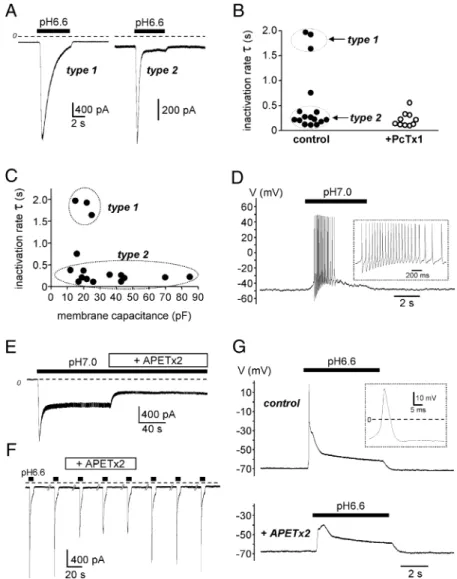
neurons innervating the hindpaw muscles presented transient current with ASIC-like kinetics when exposed to pH 6.6 (data from 2 different cultures, n⫽ 9 of 10 and 7 of 11 neurons, respec-tively). The ASIC-expressing neurons had an average membrane capacitance of 32.3⫾ 5.2 pF and an average resting potential of ⫺53.4 ⫾ 2.9 mV. Two different types of ASIC currents were observed and were readily distinguished based on their inactiva-tion kinetics (Fig. 1 A). Type 1 current exhibited a slow inactivat-ing rate ( ⫽ 1841 ⫾ 104 ms, present in 3 of 16 ASIC-expressing neurons) (Fig. 1 B) similar to the inactivation rate observed for the current elicited by recombinant ASIC1a ( ⫽ 1712 ⫾ 93 ms,
n⫽ 10, data not shown) (Baron et al., 2008). This current was
largely inhibited by the toxin PcTx1 (78⫾ 20% of inhibition of the current ampli-tude) (Fig. 1 B), a specific blocker of ASIC1a homomeric channels (Escoubas et al., 2000). These data indicate that, in DRG neurons innervating hindpaw mus-cles, the slow-inactivating type 1 current was mainly carried by homomeric ASIC1a channels. Conversely, type 2 current had rapid inactivation kinetics ( ⫽ 217 ⫾ 26 ms, present in 12 of 16 ASIC-expressing neurons) (Fig. 1 B) close to the inactiva-tion rates recorded for recombinant ASIC3 channels ( ⫽ 234 ⫾ 11 ms, n ⫽ 14, data not shown) (Waldmann et al., 1997b). PcTx1 had a small effect (32⫾ 5% inhibition) on 70% of type 2 current-expressing neurons (7 of 10 neurons tested), showing the presence of low levels of homomeric ASIC1a channels together with ASIC3-type current in these neu-rons. The type 2 current was inhibited by the toxin APETx2, which blocks both the peak and the window current of ASIC3-containing channels (Diochot et al., 2004) (Fig. 1 E). Indeed, application of APETx2 (5M) on a neuron displaying a mixture
of ASIC currents resulted in a 50% inhibi-tion of the peak amplitude evoked at pH 6.6 (Fig. 1 F). Importantly, APETx2 also decreased neuronal excitability in re-sponse to pH 6.6 (Fig. 1G).
Finally, in 1 of 16 neurons expressing ASIC-type currents, the current kinetic displayed an intermediate inactivation rate ( ⫽ 752 ms) (Fig. 1B). This current was partly inhibited by the PcTx1 toxin (46% of inhibition), and the remaining current had an intermediate inactivation rate ( ⫽ 550 ms), which probably reflects the activity of a complex mix of homo-meric ASIC1a and other heterohomo-meric ASIC channels.
A detailed analysis of the whole-cell membrane capacitance of ASIC-type current-expressing neurons (Fig. 1C) showed that the type 1 current, mainly supported by homomeric ASIC1a chan-nels, was expressed in small DRG neu-rons, with a cell capacitance of⬃20 pF, while the type 2 currents, mainly sup-ported by ASIC3 channels, were distributed in both small and large DRG neurons with cell capacitance ranging from 10 to 85 pF. Endogenous type 2 current (i.e., ASIC3-type) conferred to sensory neurons the capacity to electrically respond to very small acidifications near the physiological pH since activation of this current by a shift from resting pH 7.4 to pH 7.0 was sufficient to depolarize the membrane and promote action potential firing (Fig. 1 D). Together, these data indicate that in neurons inner-vating hindpaw muscles, ASIC3-containing channels, alone or together with homomeric ASIC1a channels, support most of the ASIC current elicited by moderate external acidosis in a range that is relevant to incisional pain (6.6⬍ pH ⬍ 7.4).
Figure 1. Electrophysiological characterization of ASIC channels in DRG neurons innervating the rat hindpaw muscles. A, Typical ASIC currents recorded in voltage-clamp (holding potential⫺80 mV) from retrogradely labeled sensory neurons innervating hindpaw muscles. Current was activated by a rapid switch of the external pH from 7.4 to 6.6, as indicated by the black bars above the current traces. The dashed line represents the zero current level. According to their inactivation rates (),twomaintypesofASIC current can be distinguished (type 1 and type 2). B, Analysis of the inactivation rate of native pH 6.6-induced ASIC currents in control condition and in the presence of the PcTx1 toxin. The slow-inactivating type 1 current is blocked by the toxin. C, Functional distribution of type 1 and type 2 ASIC currents as a function of the neuron membrane capacitance. D, Current-clamp experiment performed on a neuron exhibiting a type 2 ASIC current. A slight acidification of the external medium to pH 7.0 depolarizes the membrane sufficiently to trigger firing. Action potentials are magnified in the inset. E, Effect of APETx2 on recombinant sustained ASIC3 current recorded at⫺80mVfromF11-transfectedcells.CurrentwasactivatedbyexternalpHchangesfrompH7.4to7.0,as indicated by the black bar. The APETx2 toxin was applied externally at 1M, as indicated. F, Effect of APETx2 (5M) on native pH
6.6-evoked ASIC current recorded at⫺80 mV from retrogradely labeled sensory neurons innervating hindpaw muscles. The fast component of the current (type 2) is largely inhibited by the toxin. G, Current-clamp experiment performed on the same neuron as in F. The depolarization induced by the pH 6.6-evoked ASIC current was sufficient to trigger an action potential (magnified in the inset). The presence of APETx2 inhibits the pH 6.6-evoked depolarization and the action potential threshold is not reached.
ASIC3 is largely expressed in sensory neurons innervating the hindpaw plantar muscle
Functional data show a high level of ASIC3-type currents in rat DRG neurons innervating the hindpaw skeletal muscles, and we have analyzed the expression of the corresponding protein by immuno-histochemistry. This analysis was done on sensory neurons innervating the plantar hindpaw muscle [i.e., the muscle incised in the model of postoperative pain of Brennan et al., (1996) used in the present study]. We identified this population of sensory neurons in L5 DRG by retrograde labeling through injection of the fluores-cent dye fast blue in the plantar muscle followed by immunohistochemical stain-ing for ASIC3. In naive rats, a large num-ber of sensory neurons that innervate the plantar muscle expressed ASIC3 (75 ⫾ 2%; n⫽ 4 rats; 217 fast blue-labeled neu-rons analyzed) (Fig. 2 A, B). Moreover, ⬃62% of the ASIC3-labeled neurons have diameters in the range of those recorded with patch-clamp (i.e., from 10 to 45m), which is consistent with the percentage of sensory neurons that innervate the hind-paw muscles and exhibit ASIC3-type cur-rents (⬃57%, 12 of 21) (Fig. 1). Analysis of the soma size of the total population of ASIC3-expressing neurons showed a large expression in small-diameter neurons and
a significant expression in medium- to large-diameter neurons (Fig. 2C) (674 ASIC3-positive neurons analyzed), which is in good agreement with previous results (Voilley et al., 2001; Mol-liver et al., 2005). The distribution of ASIC3 in neurons innervat-ing the hindpaw plantar muscle (i.e., ASIC3 and fast blue-positive neurons) was slightly different from that of ASIC3 in the whole population of neurons, in which it was detected in a greater pro-portion of medium- to large-diameter neurons (Fig. 2C) (118 fast blue- and ASIC3-positive neurons analyzed). This distribution pattern is consistent with the large range of cell membrane capac-itance observed on ASIC3-type neurons with the patch-clamp technique (Fig. 1C).
ASIC3 contributes to the detection of postincisional pain
Because ASIC3 is largely expressed in sensory neurons innervat-ing the hindpaw muscles (includinnervat-ing the plantar muscle), where it constitutes the main ASIC-type current (Figs. 1, 2), we have an-alyzed its contribution to postoperative muscular pain in the plantar postincisional pain model in rat (Brennan et al., 1996). Postincisional pain behaviors were first analyzed 4 h after surgery (Fig. 3A) by measuring spontaneous flinching (Fig. 3B), heat hy-peralgesia (Hargreaves test) (Fig. 3C), and hindpaw weight bear-ing (dynamic weight-bearbear-ing test) (Fig. 3D). Plantar muscle incision caused significant hyperalgesia in all three behavioral tests. We then tested the effect of pharmacological inhibition of ASIC3 by the specific inhibitory peptide APETx2 (Diochot et al., 2004). APETx2 was known to block the peak of the acid-induced current flowing through homomeric and heteromeric ASIC3 channels (Diochot et al., 2004). We show here that APETx2 also inhibits the ASIC3 sustained window current
evoked by moderate external acidification to pH 7.0 (Fig. 1 E), an acidification well in the range of the tissue pH reached after sur-gical incision in the rat (Woo et al., 2004). This sustained current has been proposed to be important for pain perception (Yagi et al., 2006; Deval et al., 2008; Salinas et al., 2009). Local intraoper-ative application of APETx2 (20l, 20 M) significantly reduced
the spontaneous pain behavior of operated rats compared with vehicle-treated animals (Fig. 3B) (reduction of the numbers of flinches by 52%, p⬍ 0.001, n ⫽ 19 and 28, respectively). Con-versely, animals treated with PcTx1 (a potent and specific inhib-itory spider toxin for homomeric ASIC1a channels; 20l, 120 nM) did not show any significant reduction in the number of
flinches (Fig. 3B, left) (n⫽ 10). The role of ASIC3 was further confirmed by in vivo knockdown of ASIC3 expression through intrathecal injections of a specific siRNA targeting the ASIC3 mRNA (Fig. 3A). Uptake of this siRNA by lumbar DRG neu-rons innervating rat hindpaw and specific knockdown of ASIC3 mRNA versus other ASIC subunits and TRPV1 have been previously described (Deval et al., 2008). Rats treated with the ASIC3 siRNA displayed a significant decrease of the postoperative spontaneous pain behavior compared with an-imals injected with the control scramble siRNA (63% reduc-tion of the number of flinches, p⬍ 0.05, n ⫽ 10) (Fig. 3B, right). The flinching score of rats treated with the scramble siRNA (Fig. 3B, right) was not significantly different from the score of vehicle-treated animals (Fig. 3B, left) (p⫽ 0.39, un-paired t test).
Hindpaw skin and muscle incisions have been shown to evoke primary heat hyperalgesia (Brennan et al., 1996) (i.e., hyperalge-sia inside the area injured by the incision). Consistent with this
Figure 2. Analysis of ASIC3 expression in sensory neurons innervating the hindpaw plantar muscle. A, Immunolocalization of ASIC3 in lumbar DRG neurons. Fast blue-positive neurons retrogradely labeled from the plantar muscle (left, blue) and ASIC3-positive neurons (middle, red) in L5 DRG of naive rats. Merged image (right) shows fast blue-ASIC3-positive neurons expressing (arrows) or not expressing (arrowhead) ASIC3 (scale bar, 100m). B, Percentage of fast blue (FB)-positive neurons expressing the ASIC3 subunit (data from 4 rats, 217 FB-labeled neurons analyzed). C, Distribution of ASIC3-positive neurons in L5 DRG neurons as a function of soma diameter. Histograms represent the percentage of the total population of ASIC3-positive neurons or of the ASIC3⫹ FB-positive cells associated with different soma diameter ranges. The proportion of medium to large neurons expressing ASIC3 is larger in neurons that innervate the hindpaw muscle compared with the whole population of ASIC3-positive cells.
previous observation, animals treated with the vehicle displayed primary heat hyperalgesia measured by the Hargreaves test (ratio of ipsilateral to contralateral withdrawal latency significantly re-duced in comparison with control animals, p⬍ 0.001) (Fig. 3C). Intraoperative infusion of APETx2 induced a modest analgesia since the latency became not significantly different from the one of control animals (Fig. 3C). We also performed a Hargreaves test on animals where ASIC3 expression was knocked down by repet-itive intrathecal injections of siRNA (Fig. 3D). The animals that were knocked down still developed heat hyperalgesia after sur-gery (Fig. 3D) (p⬍ 0.01), but the hyperalgesia was significantly attenuated compared with scramble siRNA-treated animals (Fig. 3D) (p⬍ 0.001). These data suggest that ASIC3 significantly con-tributes to primary heat hyperalgesia induced by the surgical pro-cedure, but that inhibition of the channel is not sufficient to fully prevent hyperalgesia.
Surgical procedures provoke postural pain (guarding pain). We have used a weight-bearing test to evaluate postural equilibrium on freely moving rats follow-ing surgery (Fig. 3 E, F ). A difference in weight distribution between the operated and nonoperated paws reflected the in-tensity of postoperative pain presented by rats. The hindpaw weight-bearing differ-ence measured 4 h after surgery was abol-ished in animals treated with ASIC3 siRNA (Fig. 3F ), but was not significantly decreased in animals pretreated with local application of APETx2 (Fig. 3E). This could be explained by a slow onset of the pharmacological inhibition of ASIC3 by intraoperative application of the toxin on the weight-bearing behavior because of delayed toxin availability in deep muscu-lar tissue. Comparatively, the knockdown inhibition obtained with repetitive siRNA treatment was already maximal at the time of surgery. Anyway, the effect of the siRNA suggests a participation of ASIC3 in postoperative postural pain. Together, these data reveal that peripheral inactiva-tion of ASIC3 produces a significant anal-gesia 4 h after surgery in a rat model of incisional pain, and therefore support the involvement of ASIC3-containing chan-nels in postoperative pain.
ASIC3 expression is upregulated in the model of postincisional pain
Animals treated with either local applica-tion of vehicle or intrathecal injecapplica-tions of scramble siRNA still experienced pain 24 h after surgery, showing a hindpaw weight-bearing difference of⬃40 g (p ⬍ 0.001 compared with naive animals) (Fig. 4 A). Pain was largely attenuated by both local application of APETx2 during sur-gery and ASIC3 knockdown (Fig. 4 A). We then analyzed modifications in the ex-pression of ASIC3 mRNA and protein in sensory neurons innervating the injured plantar muscle after surgery. RT-qPCR experiments revealed that animals showed an increased level of ASIC3 transcripts in ipsilateral DRGs (L4 –L6) compared with their contralateral counterparts 24 h after surgery (fold expres-sion, ipsilateral vs contralateral DRGs: 1.50⫾ 0.12; n ⫽ 4) (Fig. 4 B), while naive nonoperated animals showed no difference (fold expression: 0.99⫾ 0.12; n ⫽ 5) (Fig. 4B). Consistent with the increase in ASIC3 transcripts, the number of ASIC3-positive neu-rons that innervate the incised muscle (i.e., fast blue retrogradely labeled neurons from plantar muscles) was also significantly in-creased 24 h after surgery in the ipsilateral L5 DRG (controlateral vs ipsilateral, 74⫾ 2% and 87 ⫾ 1% of fast blue-labeled neurons, respectively; n⫽ 4 rats; p ⬍ 0.05; at least 268 fast blue-labeled neurons analyzed in each group) (Fig. 4C). The distribution of ASIC3-expressing neurons innervating the hindpaw plantar muscle was modified in the ipsilateral DRGs 24 h after surgery with a large increase in the proportion of small-sized neurons
Figure 3. Effect of ASIC3 inhibition on postoperative pain 4 h after surgery. A, Protocol used to test the effect of the ASIC inhibitory toxins (local application during surgery) and of the siRNAs (repetitive intrathecal injections before surgery) on acute postoperative pain (4 h after surgery). B, Effect of APETx2 20Mand PcTx1 120 nM(left), and siRNAs (scramble and ASIC3si; right)
on spontaneous pain measured as the number of flinches of the ipsilateral hindpaw over a period of 5 min (**p⬍ 0.01, Kruskal– Wallis test followed by a Dunn’s post hoc test; *p⬍0.05,Mann–Whitneytest).C,D,Effectoflocalapplicationof20MAPETx2 (C)
and ASIC3 knockdown (D) on postoperative thermal hyperalgesia evaluated with the Hargreaves test. (C: ***p⬍0.001,compared with naive animals, one-way ANOVA followed by a Tukey’s post hoc test; D:⽤⽤⽤p⬍ 0.001,⽧⽧p⬍ 0.01, compared with scramble and ASIC3si controls (ctr), respectively, paired t tests, and ***p⬍ 0.001, unpaired t test). E, F, Effect of local application of 20MAPETx2 (E) and knockdown of ASIC3 (F ) on postural pain evaluated with the dynamic weight-bearing test (*p⬍0.05and
(Fig. 4 D) (118 and 164 neurons analyzed for contralateral and ipsilateral DRGs, re-spectively). The distribution pattern in contralateral DRGs from operated ani-mals was similar to the pattern in DRGs from naive rats (Fig. 2C). This suggests, together with the similar fraction of fast blue-positive neurons that expressed ASIC3 in naive animals and in contralat-eral DRG of operated rats (75⫾ 2% and 74⫾ 2%, respectively; n ⫽ 4 rats; at least 217 fast blue-labeled neurons analyzed in each group) (Figs. 2 B, 4C), that the sur-gery had a unilateral effect on ASIC3 ex-pression, with a selective increase in the DRGs innervating the operated paw. Our observations demonstrate that postopera-tive pain induced by plantar incision 24 h after surgery involves peripheral ASIC3 since (1) its expression is significantly in-creased in sensory neurons innervating the incised muscle, especially in small-diameter neurons, and since (2) the post-operative pain is abolished when ASIC3 is inactivated.
Discussion
Postincisional pain models have been very useful to investigate the pathophysiology of postoperative pain (Brennan et al., 1996; Pogatzki et al., 2002; Duarte et al., 2005). One of the best characterized mod-els is the one developed by Brennan et al. (1996), where hindpaw incision induces
evoked and nonevoked pain in agreement with clinical evidence (Pogatzki-Zahn et al., 2007). Peripheral and central sensitizations seem to be involved in the development and maintenance of surgical pain (Pogatzki-Zahn et al., 2007). Of particular interest is the local pH decrease at the site of incision (between pH 6.91 and 6.54, depending on the incision model) (Woo et al., 2004), sug-gesting that H⫹-gated depolarizing channels in primary afferent neurons such as ASICs and TRPV1 could contribute to postop-erative pain.
TRPV1 has been reported to mediate spontaneous activity and heat sensitization of cutaneous primary afferents after plan-tar incision (Banik and Brennan, 2009), and TRPV1-containing nociceptors have been implicated either in mechanical allodynia and hyperalgesia (Pospisilova and Palecek, 2006) or in heat hy-peralgesia and guarding pain (Hamalainen et al., 2009; Kang et al., 2010). The chemosensitivity of C-fibers innervating the rat hindpaw skin also supports a possible implication of ASICs in chemical sensitization (Kang and Brennan, 2009; Kang et al., 2010). This was further suggested by the effect, in the incision model, of amiloride (a poorly selective) and A-317567 (a nondis-criminant) blockers of ASICs (Dube´ et al., 2005). We show here that ASIC3 is highly expressed in skeletal muscle afferents of the rat hindpaw, where it is activated by moderate acidifications, and that selective pharmacological block of its activity or in vivo knockdown of its expression produces a significant analgesia af-ter surgery.
ASIC3-containing channels are particularly well suited to de-tect local changes in pH associated with postoperative pain and to contribute to nociceptor sensitization: (1) they exhibit the
high-est sensitivity to extracellular protons among ASIC channels (Yagi et al., 2006; Deval et al., 2008; Salinas et al., 2009); (2) they are activated in the range of pH recorded after skin and/or muscle incision (Woo et al., 2004); and (3) they display a sustained de-polarizing current in response to very moderate acidosis that is compatible with the detection of nonadaptating pain. This sus-tained window current, which is activated for pH values between 6.5 and 7.4 and is due to the overlap between the pH-dependent activation and inactivation of the channel (Yagi et al., 2006; Deval et al., 2010), has been proposed to contribute to pain during myocardial ischemia and cutaneous inflammation (Yagi et al., 2006; Deval et al., 2008). This window current is blocked by APETx2, as shown in the present study. We have recently shown that peripheral ASIC3 channels in rat skin nociceptors are sensors of acidic pain and are able to integrate several molecular signals produced during inflammation to contribute to primary hyper-algesia (Deval et al., 2008). Although surgical hypersensitivity seems to involve an inflammatory component, the pathophysio-logical mechanism associated with incision is clearly different from the one causing pain during inflammation (Pogatzki-Zahn et al., 2007). Indeed, inhibition of ASIC3 fully eliminates primary heat hyperalgesia caused by inflammation (Deval et al., 2008), but we report here that it does not completely reverse primary heat hyperalgesia caused by incision. It has also been shown that deep tissue (i.e., muscle) rather than skin incision is critical for the development of guarding pain, which is similar to pain at rest in postoperative patients, and for spontaneous activity in both primary sensory neurons (Xu and Brennan, 2010) and dorsal horn neurons (Xu and Brennan, 2009). Interestingly, ASIC3 has
Figure 4. Postoperative pain and ASIC3 expression 24 h after surgery. A, Dynamic weight-bearing test performed in rats treated with scramble or ASIC3 (ASIC3si) siRNAs (intrathecal injections, see experimental protocol in Fig. 3A), vehicle or local application of APETx2 (**p⬍0.01,***p⬍0.001,one-wayANOVAfollowedbyaTukey’sposthoctest).B,RT-qPCRanalysisofthelevelofASIC3 mRNA in lumbar DRGs (L4 –L6) from naive and operated rats. Data are represented as the ratio of ipsilateral over contralateral DRG (*p ⬍0.05,unpairedttest,n⫽4rats).C,Percentageoffastblue(FB)-positiveneuronsexpressingtheASIC3proteinincontralat-eral and ipsilat⬍0.05,unpairedttest,n⫽4rats).C,Percentageoffastblue(FB)-positiveneuronsexpressingtheASIC3proteinincontralat-eral DRG 24 h after surgery (*p⬍ 0.05, paired t test, n ⫽ 4 rats, 268 and 290 FB-labeled neurons analyzed, respectively). D, Distribution of ASIC3-positive neurons innervating the plantar muscle (i.e., FB-positive) as a function of soma diameter in contralateral and ipsilateral L5 DRG 24 h after surgery (n⫽ 4 rats, 268 and 290 FB-labeled neurons analyzed, respectively). Data represent the number of ASIC3-positive neurons expressed as the percentage of total fast blue-positive neurons (i.e., the sum of the bars is 74% for ASIC3⫹FB-contra and 87% for ASIC3⫹FB-ipsi). contra, Contralateral; ipsi, ipsilateral.
been involved in muscular pain caused by repeated acid injec-tions and inflammation in mouse (Sluka et al., 2003, 2007). The more robust effect seen here after knockdown of ASIC3 on weight bearing compared with primary thermal hyperalgesia may thus reflect an important role of ASIC3 in deep tissue, including mus-cle (Sutherland et al., 2001; Sluka et al., 2003; Ikeuchi et al., 2008), after surgery. However, postural pain that appears 24 h postsur-gery in animals subjected to skin incision alone is also blocked by APETx2 (supplemental Fig. 1, available at www.jneurosci.org as supplemental material), also suggesting a contribution of ASIC3 to prolonged skin pain. ASIC3 thus appears to play an important role in the primary afferent fibers innervating skin and deep tis-sues such as muscle, sensing pain caused by acidosis and increas-ing the nociceptive input to the spinal cord, which results in central sensitization. In good agreement with this, experiments performed in rats have reported that activation of nociceptors by acid stimuli induces central sensitization at the level of the dorsal spinal cord (Carpenter et al., 2001), and that deep tissue incision (i.e., plantar muscle) increases both the proportion of dorsal horn neurons with spontaneous activity and the rate of this spon-taneous activity (Xu and Brennan, 2009).
Tissue-induced release of mediators, ischemia, and local aci-dosis are important for primary hypersensitivity and sensitiza-tion of nociceptors in the incisional pain models (Woo et al., 2004). A greater response to pH 6.0 and lactic acid of the afferent fibers innervating the rat hindpaw skin has been described after incision (Kang and Brennan, 2009). Lactate enhances the activity of ASIC3 (Immke and McCleskey, 2001), and the increase in tissue lactate concentration (and possibly other mediators) that parallels the decrease in tissue pH after incision may potentiate the acid-induced activity of ASIC3 (Woo et al., 2004; Kim et al., 2007). This effect can be reinforced by the increased expression of ASIC3 in nociceptors 24 h after incision that was observed in the present work. Interestingly, nerve growth factor, which is an im-portant regulator of ASIC3 gene expression (Mamet et al., 2002, 2003), has been involved in nociceptor sensitization and heat hyperalgesia after incision (Zahn et al., 2004).
In conclusion, this study involves peripheral ASIC3 channels in postincisional pain. ASIC3 sustained current activated in the pH range reached after tissue incision is likely to be an important contributor to sensory neuron depolarization and hyperexcit-ability. Interestingly, a single intraoperative application of an ASIC3 inhibitor evokes a potent analgesic effect 24 h after sur-gery. ASIC3 may thus represent a new pharmacological target for the management of postoperative pain in addition to wound in-filtration with local anesthetics (Liu et al., 2006; Scott, 2010) and other currently available approaches.
References
Banik RK, Brennan TJ (2009) Trpv1 mediates spontaneous firing and heat sensitization of cutaneous primary afferents after plantar incision. Pain 141:41–51.
Baron A, Voilley N, Lazdunski M, Lingueglia E (2008) Acid sensing ion channels in dorsal spinal cord neurons. J Neurosci 28:1498 –1508. Ba¨ssler EL, Ngo-Anh TJ, Geisler HS, Ruppersberg JP, Gru¨nder S (2001)
Mo-lecular and functional characterization of acid-sensing ion channel (ASIC) 1b. J Biol Chem 276:33782–33787.
Bove SE, Flatters SJ, Inglis JJ, Mantyh PW (2009) New advances in muscu-loskeletal pain. Brain Res Rev 60:187–201.
Brennan TJ, Vandermeulen EP, Gebhart GF (1996) Characterization of a rat model of incisional pain. Pain 64:493–501.
Carpenter KJ, Nandi M, Dickenson AH (2001) Peripheral administration of low pH solutions causes activation and sensitisation of convergent dorsal horn neurones in the anaesthetised rat. Neurosci Lett 298:179 –182. Chen CC, England S, Akopian AN, Wood JN (1998) A sensory
neuron-specific, proton-gated ion channel. Proc Natl Acad Sci U S A 95: 10240 –10245.
Deval E, Noe¨l J, Lay N, Alloui A, Diochot S, Friend V, Jodar M, Lazdunski M, Lingueglia E (2008) ASIC3, a sensor of acidic and primary inflammatory pain. EMBO J 27:3047–3055.
Deval E, Gasull X, Noe¨l J, Salinas M, Baron A, Diochot S, Lingueglia E (2010) Acid-sensing ion channels (ASICs): pharmacology and implication in pain. Pharmacol Ther 128:549 –558.
Diochot S, Baron A, Rash LD, Deval E, Escoubas P, Scarzello S, Salinas M, Lazdunski M (2004) A new sea anemone peptide, APETx2, inhibits ASIC3, a major acid-sensitive channel in sensory neurons. EMBO J 23:1516 –1525.
Duarte AM, Pospisilova E, Reilly E, Mujenda F, Hamaya Y, Strichartz GR (2005) Reduction of postincisional allodynia by subcutaneous bupiva-caine: findings with a new model in the hairy skin of the rat. Anesthesiol-ogy 103:113–125.
Dube´ GR, Lehto SG, Breese NM, Baker SJ, Wang X, Matulenko MA, Honore´ P, Stewart AO, Moreland RB, Brioni JD (2005) Electrophysiological and in vivo characterization of A-317567, a novel blocker of acid sensing ion channels. Pain 117:88 –96.
Escoubas P, De Weille JR, Lecoq A, Diochot S, Waldmann R, Champigny G, Moinier D, Me´nez A, Lazdunski M (2000) Isolation of a tarantula toxin specific for a class of proton-gated Na⫹ channels. J Biol Chem 275:25116 –25121.
Flatters SJ (2008) Characterization of a model of persistent postoperative pain evoked by skin/muscle incision and retraction (SMIR). Pain 135:119 –130.
Hamalainen MM, Subieta A, Arpey C, Brennan TJ (2009) Differential effect of capsaicin treatment on pain-related behaviors after plantar incision. J Pain 10:637– 645.
Ikeuchi M, Kolker SJ, Burnes LA, Walder RY, Sluka KA (2008) Role of ASIC3 in the primary and secondary hyperalgesia produced by joint in-flammation in mice. Pain 137:662– 669.
Ikeuchi M, Kolker SJ, Sluka KA (2009) Acid-sensing ion channel 3 expres-sion in mouse knee joint afferents and effects of carrageenan-induced arthritis. J Pain 10:336 –342.
Immke DC, McCleskey EW (2001) Lactate enhances the acid-sensing Na⫹ channel on ischemia-sensing neurons. Nat Neurosci 4:869 – 870. Kang S, Brennan TJ (2009) Chemosensitivity and mechanosensitivity of
no-ciceptors from incised rat hindpaw skin. Anesthesiology 111:155–164. Kang S, Wu C, Banik RK, Brennan TJ (2010) Effect of capsaicin treatment
on nociceptors in rat glabrous skin one day after plantar incision. Pain 148:128 –140.
Kim TJ, Freml L, Park SS, Brennan TJ (2007) Lactate concentrations in incisions indicate ischemic-like conditions may contribute to postopera-tive pain. J Pain 8:59 – 66.
Liu SS, Richman JM, Thirlby RC, Wu CL (2006) Efficacy of continuous wound catheters delivering local anesthetic for postoperative analgesia: a quantitative and qualitative systematic review of randomized controlled trials. J Am Coll Surg 203:914 –932.
Mamet J, Baron A, Lazdunski M, Voilley N (2002) Proinflammatory medi-ators, stimulators of sensory neuron excitability via the expression of acid-sensing ion channels. J Neurosci 22:10662–10670.
Mamet J, Lazdunski M, Voilley N (2003) How nerve growth factor drives physiological and inflammatory expressions of acid-sensing ion channel 3 in sensory neurons. J Biol Chem 278:48907– 48913.
Moiniche S, Dahl JB, Erichsen CJ, Jensen LM, Kehlet H (1997) Time course of subjective pain ratings, and wound and leg tenderness after hysterec-tomy. Acta Anaesthesiol Scand 41:785–789.
Molliver DC, Immke DC, Fierro L, Pare´ M, Rice FL, McCleskey EW (2005) ASIC3, an acid-sensing ion channel, is expressed in metaboreceptive sen-sory neurons. Mol Pain 1:35.
Pavlin DJ, Chen C, Penaloza DA, Polissar NL, Buckley FP (2002) Pain as a factor complicating recovery and discharge after ambulatory surgery. Anesth Analg 95:627– 634.
Perkins FM, Kehlet H (2000) Chronic pain as an outcome of surgery. A review of predictive factors. Anesthesiology 93:1123–1133.
Pogatzki EM, Niemeier JS, Brennan TJ (2002) Persistent secondary hyper-algesia after gastrocnemius incision in the rat. Eur J Pain 6:295–305. Pogatzki-Zahn EM, Zahn PK, Brennan TJ (2007) Postoperative pain–
clinical implications of basic research. Best Pract Res Clin Anaesthesiol 21:3–13.
Pospisilova E, Palecek J (2006) Post-operative pain behavior in rats is reduced after single high-concentration capsaicin application. Pain 125:233–243.
Salinas M, Lazdunski M, Lingueglia E (2009) Structural elements for the generation of sustained currents by the acid pain sensor ASIC3. J Biol Chem 284:31851–31859.
Scott NB (2010) Wound infiltration for surgery. Anaesthesia 65 [Suppl 1]:67–75.
Sluka KA, Price MP, Breese NM, Stucky CL, Wemmie JA, Welsh MJ (2003) Chronic hyperalgesia induced by repeated acid injections in muscle is abolished by the loss of ASIC3, but not ASIC1. Pain 106:229 –239. Sluka KA, Radhakrishnan R, Benson CJ, Eshcol JO, Price MP, Babinski K,
Audette KM, Yeomans DC, Wilson SP (2007) ASIC3 in muscle mediates mechanical, but not heat, hyperalgesia associated with muscle inflamma-tion. Pain 129:102–112.
Sutherland SP, Benson CJ, Adelman JP, McCleskey EW (2001) Acid-sensing ion channel 3 matches the acid-gated current in cardiac ischemia-sensing neurons. Proc Natl Acad Sci U S A 98:711–716.
Voilley N, de Weille J, Mamet J, Lazdunski M (2001) Nonsteroid anti-inflammatory drugs inhibit both the activity and the inflammation-induced expression of acid-sensing ion channels in nociceptors. J Neurosci 21:8026 – 8033.
Waldmann R, Champigny G, Bassilana F, Heurteaux C, Lazdunski M
(1997a) A proton-gated cation channel involved in acid-sensing. Nature 386:173–177.
Waldmann R, Bassilana F, de Weille J, Champigny G, Heurteaux C, Lazdun-ski M (1997b) Molecular cloning of a non-inactivating proton-gated Na⫹ channel specific for sensory neurons. J Biol Chem 272:20975–20978. Woo YC, Park SS, Subieta AR, Brennan TJ (2004) Changes in tissue pH and temperature after incision indicate acidosis may contribute to postoper-ative pain. Anesthesiology 101:468 – 475.
Wu CL, Naqibuddin M, Rowlingson AJ, Lietman SA, Jermyn RM, Fleisher LA (2003) The effect of pain on health-related quality of life in the immedi-ate postoperative period. Anesth Analg 97:1078 –1085.
Xu J, Brennan TJ (2009) Comparison of skin incision vs. skin plus deep tissue incision on ongoing pain and spontaneous activity in dorsal horn neurons. Pain 144:329 –339.
Xu J, Brennan TJ (2010) Guarding pain and spontaneous activity of nocice-ptors after skin versus skin plus deep tissue incision. Anesthesiology 112:153–164.
Yagi J, Wenk HN, Naves LA, McCleskey EW (2006) Sustained currents through ASIC3 ion channels at the modest pH changes that occur during myocardial ischemia. Circ Res 99:501–509.
Zahn PK, Subieta A, Park SS, Brennan TJ (2004) Effect of blockade of nerve growth factor and tumor necrosis factor on pain behaviors after plantar incision. J Pain 5:157–163.