CARDIAC
Performance of turbo high-pitch dual-source CT for coronary CT
angiography: first ex vivo and patient experience
Fabian Morsbach&Sonja Gordic&Lotus Desbiolles&Daniela Husarik&
Thomas Frauenfelder&Bernhard Schmidt&Thomas Allmendinger Simon Wildermuth&Hatem Alkadhi&Sebastian Leschka
Received: 3 February 2014 / Revised: 15 April 2014 / Accepted: 28 April 2014 / Published online: 17 May 2014 # European Society of Radiology 2014
Abstract
Objectives To evaluate image quality, maximal heart rate allowing for diagnostic imaging, and radiation dose of turbo high-pitch dual-source coronary computed tomographic angi-ography (CCTA).
Methods First, a cardiac motion phantom simulating heart rates (HRs) from 60-90 bpm in 5-bpm steps was examined on a third-generation dual-source 192-slice CT (prospective ECG-triggering, pitch 3.2; rotation time, 250 ms). Subjective image quality regarding the presence of motion artefacts was interpreted by two readers on a four-point scale (1, excellent; 4, non-diagnostic). Objective image quality was assessed by calculating distortion vectors. Thereafter, 20 consecutive pa-tients (median, 50 years) undergoing clinically indicated CCTA were included.
Results In the phantom study, image quality was rated diag-nostic up to the HR75 bpm, with object distortion being 1 mm or less. Distortion increased above 1 mm at HR of 80-90 bpm. Patients had a mean HR of 66 bpm (47-78 bpm). Coronary segments were of diagnostic image quality for all patients with HR up to 73 bpm. Average effective radiation dose in patients was 0.6±0.3 mSv.
Conclusions Our combined phantom and patient study indi-cates that CCTA with turbo high-pitch third-generation dual-source 192-slice CT can be performed at HR up to 75 bpm
while maintaining diagnostic image quality, being associated with an average radiation dose of 0.6 mSv.
Key points
• CCTA is feasible with the turbo high-pitch mode.
• Turbo high-pitch CCTA provides diagnostic image quality up to 73 bpm.
• The radiation dose of high-pitch CCTA is 0.6 mSv on average.
Keywords High-pitch CT . Heart rate . Radiation dose . Image quality . Coronary CT angiography
Introduction
Technical advances in computed tomography (CT) have given radiologists powerful tools to reduce patient dose [1,2]. This is particularly true for coronary CT angiography (CCTA), in which patient dose has been lowered considerably in the past decade [1]. One of the most recent techniques for reducing radiation dose was prospective electrocardiography (ECG)-triggering at a high pitch, which became available with second-generation dual-source CT [3–5]. With this technique, the entire heart volume can be imaged within the diastolic phase of one heartbeat with an ECG-triggered high-pitch acquisition (table feed 458 mm/s). Hereby, radiation dose could be reduced to range around 1 mSv [3–5]. However, this mode is limited to patients with regular and relatively low heart rates <60 beats per minute (bpm) [6–8].
The use of automated attenuation-based tube voltage selec-tion and tube current in CCTA has shown addiselec-tional potential for reducing radiation dose while maintaining image quality [9]. In addition, iterative reconstructions can be used in CTCA for another reduction in radiation dose [10]. For example, Schuhbaeck et al. [11] showed that radiation dose can be
F. Morsbach
:
S. Gordic:
L. Desbiolles:
D. Husarik:
T. Frauenfelder
:
H. Alkadhi (*):
S. LeschkaInstitute of Diagnostic and Interventional Radiology, University Hospital Zurich, Raemistrasse 100, 8091 Zurich, Switzerland e-mail: [email protected]
L. Desbiolles
:
S. Wildermuth:
S. LeschkaDivison of Radiology and Nuclear Medicine, Kantonsspital St. Gallen, St. Gallen, Switzerland
B. Schmidt
:
T. Allmendingerlowered to below 0.1 mSv when using a protocol at 80 kVp and when the patient exhibits a heart rate of 60 bpm or less.
Recently, third-generation dual-source 192-slice CT was introduced, which is characterised by another acceleration of table feet to 737 mm/s (pitch 3.2 at a broader detector) and a gantry rotation time of 250 ms (compared with 280 ms of second-generation dual-source CT), allowing for ECG-gated CT at a temporal resolution of 66 ms. Based on these technical features, it is expected that CCTA with third-generation dual-source 192-slice CT may be feasible at higher heart rates as compared to second-generation dual-source CT at a diagnostic image quality.
The purpose of this feasibility study was to evaluate the image quality, maximal heart rate allowing for diagnostic imaging and radiation dose of turbo high-pitch dual-source CCTA with prospective ECG-gating in a cardiac motion phantom study, and to confirm these results in vivo.
Material and methods
Ex-vivo study
Phantom preparation
The ex vivo part of our study included images with a cardiac motion phantom [12] coupled to a three-dimensional (3D) motion simulator (QRM-Sim4D-Cardio; Quality Assurance in Radiology and Medicine, Moehrendorf, Germany). The motion simulator permits a range of motion of 80 × 40 × 80 mm (x-, y- and z-axis) and a maximum frequency of 3 Hz, thereby creating motion sequences simulating 3D heart motion with corresponding simulated ECG (Fig.1), which can be fed out to the CT machine’s ECG control panel. This allows for an ECG-synchronised CT image resembling phys-iological coronary artery movement. Attached to the 3D mo-tion simulator is the momo-tion arm with a probe holder fitted for multiple tubes. We used three parallel arranged tubes mimick-ing coronary arteries filled with contrast medium (iopromidum, Ultravist 370; Bayer Schering Pharma, Berlin, Germany) and saline in order to achieve an attenuation of 350 HU at 100 kVp. The inner diameter of the tubes was 3 mm. The tubes were submerged in a water tank and were inserted in the mediastinal aperture of a fitted chest phantom consisting of materials made from epoxy resin and additives, such as calci-um carbonate, magnesicalci-um oxide, hydroxyapatite and micro-spheres to obtain soft tissue, lung and bone equivalent struc-tures [12].
The phantom was set to simulate heart rates starting from 60 bpm and increasing in steps of 5 bpm each consecutive image. The maximum heart rate was 90 bpm.
CT data acquisition—phantom
All examinations were performed on a third-generation dual-source CT system (SOMATOM Force, Siemens Healthcare, Forchheim, Germany) equipped with integrated circuit detec-tors (Stellar Infinity; Siemens Healthcare) [13]. Examinations were performed using the turbo high-pitch mode at a pitch of 3.2 (table feed 737 mm/s), and using the following imaging parameters: tube voltage of 100 kVp with automated tube-current modulation and a reference tube tube-current-time product of 270 mAs/rotation; collimation of 96×0.6 mm; slice acqui-sition of 192×0.6 mm by means of a z-flying focal spot; gantry rotation time of 250 ms. The examinations were per-formed with prospective ECG-gating and acquisition start was synchronised to 60 % of the R-R interval of the simulated ECG.
Images were reconstructed using advanced model-based iterative reconstruction (ADMIRE, strength level 3; Siemens Healthcare) with a medium soft tissue kernel (Bv40) and a slice thickness of 0.6 mm at an increment of 0.4 mm (field of view, 200 mm; pixel matrix, 512 × 512). Images were anonymised and transferred to an external workstation (Multi-Modality Workplace; Siemens Healthcare) for further analysis.
Subjective image analysis—phantom
Two independent readers (F.M. and S.G. with 3 years of experience in cardiac imaging each) rated the image quality of the tubes in three different cross-sections on multiplanar
Fig. 1 Movement pattern output in x-, y- and z-axis of the 3D motion simulator for a simulated heart rate of 60 bpm
reformations visualising the upper, middle and lower part of each tube. The rating was performed using a four-point Likert scale as previously shown [14]: 1=excellent image quality without visible object distortion or image blurring; 2=good image quality with minor object distortion or blurring; 3=fair image quality, intermediate blurring; 4=non-diagnostic image quality with severe object distortion or blurring. Scores of 1- 3 were considered as having a diagnostic image quality.
Objective image quality analysis—phantom
Objective image quality was assessed by calculating the object distortion vector as previously described [14]. Using multiplanar reconstructions, one reader (L.D. with 4 years of experience in cardiac imaging) measured the change of the outer diameter of the tube (x) and the change of the tube length (z) for all datasets and calculated the object distortion vector (d) using Eq.1.
d¼pffiffiffiffiffiffiffiffiffiffiffiffiffiffix2þ z2 ð1Þ
Patient study
Patient population
Between November 2013 and January 2014, 33 consecutive patients undergoing clinically indicated CCTA were included. All patients were referred to CCTA for evaluation of suspected coronary artery disease, had an intermediate risk of coronary artery disease and suffered from atypical chest pain. The indications were in accordance with current guidelines and recommendations [15]. Patients were included if the patients’
ECG after nitrate application (see below) indicated a heart rate ≤75 bpm and when no arrhythmias were detected. Exclusion criteria were impaired renal function (estimated glomerular filtration rate <30 ml/min) (n=0), known hypersensitivity to iodinated contrast material (n=0), pregnancy (n=0), and high and/or irregular heart rate (n=13). CCTA in these 13 patients was performed in the step-and-shoot (n=5) or in the spiral mode with retrospective ECG-gating (n=8). Thus, the final study population consisted of 20 patients (median age, 50 years; age range, 43-82 years; 17 men, 3 women). Full patient demographics are shown in Table1.
IRB approval was obtained; written informed consent was waived because of the retrospective design of the study.
CT data acquisition—patients
The images ranged from the level of the tracheal bifurcation to the diaphragm. All patients received a single oral dose of 2.5 mg isosorbiddinitrate sublingually (Isoket, Schwarz Pharma, Monheim, Germany) 3 min prior to the scan. Sixty
millilitres contrast media (iopromidum, Ultravist 370, 370 mg iodine/ml; Bayer Schering Pharma, Berlin, Germany) were injected in an antecubital vein, followed by a chaser of 40 ml diluted contrast media (20 % vol) with a dual-head power injector (Stellant; Medrad, Inianola, USA) at a flow rate of 6.0 ml/s. Image initiation was controlled by bolus tracking with a region of interest (ROI) in the ascending aorta, using a signal attenuation threshold of 120 HU. Examinations were performed with prospective ECG-gating during one heart cycle starting at 60 % of the RR-Interval. Automated tube voltage (CAREkV; Siemens) and tube current modulation (CAREDose; Siemens) was used with the following imaging parameters: 100 reference kVp, 270 reference mAs, field-of-view (FoV) 200 mm, pixel matrix 512×512. Data were re-constructed with advanced iterative reconstruction (ADMIRE, strength level 3) with a medium soft tissue kernel (Bv40), a slice thickness of 0.6 mm and an increment of 0.4 mm.
Image analysis
Signal-to-noise ratio (SNR), contrast-to-noise ratio (CNR) and image noise were assessed as objective determinants of image quality by the two readers who also performed the objective image quality readout. Calculations of the SNR and CNR in the aorta were independently performed by readers as
Table 1 Patient characteristics, image quality, imaging parameters, and radiation dose parameters
No. of patients 20
Age (range) [years] 50 (43-82)
Female sex 15 % (3/20)
Heart rate (range) [bpm] 67±9 (47-78)
Body mass index (range) [kg/m2] 25.5±2.1 (22.6-28.5)
Total no. of coronary artery segments 270
Excellent image quality (score 1) 82.1 % (222/270)
Good image quality (score 2) 10.4 % (28/270)
Moderate image quality (score 3) 7.1 % (19/270)
Non-diagnostic image quality (score 4) 0.4 % (1/270)
Attenuation (range) [HU] 556±127 (393-755)
Image noise (range) [HU] 32±10 (26- 47)
SNR (range) 17±14 (11- 48)
CNR (range) 28±10 (19- 48)
Tube voltage (range) [kVp] 86±10 (70- 100)
Tube current-time product (range) [mAs] 447±144 (358-616)
Scan length (range) [mm] 125±6 (110- 138)
CTDIvol(range) [mGy·cm] 2.9±0.9 (1.5- 3.4)
DLP (range) [mGy·cm-1] 46±20 (23-84)
Radiation dose estimate (range) [mSv] 0.6±0.3 (0.3- 1.1)
SNR singal-to-noise ratio, CNR contrast-to-noise ratio, CTDIVolCT
previously shown [16]. First, the vessel contrast was calculat-ed as the difference in the mean attenuation (in HU) between the aorta at the level of the left main artery (LM) and the mean attenuation in the epicardial fat tissue at the same slice posi-tion. Attenuation in the ascending aorta was measured with a ROI of predefined size (155 mm2) avoiding calcifications and plaques. Second, image noise was determined as the standard deviation (SD) of attenuation in the ROI in the ascending aorta. Third, the SNR was calculated by dividing the attenu-ation in the aorta by the SD, while CNR was calculated using Eq.2.
CNR¼ ðattenuationaorta−attenuationfat tissueÞ SDaorta
ð2Þ All images were independently evaluated and classified by the two independent radiologists who previously performed the image analysis of the phantom study. The readers were blinded to any text information in the images and any clinical information. For analysis of CCTA data, coronary arteries were segmented according to the 15-segment model of the American Heart Association [17]. The intermediary artery was designated as segment 15, if present. All segments with a diameter of at least 1.5 mm at their origin were included.
The image datasets were presented the observers in random order. For the classification of subjective image quality each coronary artery segment was classified using the same mod-ified Likert scale used above in the phantom study [14]. A score of 1-3 was considered acceptable in terms of image quality for routine clinical diagnostics.
Estimation of the CT radiation dose
For an estimation of the CT radiation dose, the CT volume dose index (CTDIvol), the dose-length-product (DLP) and the
scan length were recorded, as previously shown [18]. The effective dose of CTCA was derived from the product of the dose-length product and a conversion coefficient for the chest according to a method proposed by the European Working Group for Guidelines on Quality Criteria in CT [19]. A conversion coefficient of k=0.014 mSv·mGy-1·cm-1was ap-plied [19,20].
Statistical analysis
Quantitative variables were expressed as mean±standard de-viation and categorical variables as frequencies or percent-ages. The inter-reader agreement regarding qualitative evalu-ation was analysed by using the intra-class correlevalu-ation coeffi-cient (ICC, two-way random). According to Landis and Koch [19], a value between 0 and 0.20 was considered as slight agreement; 0.21-0.40 as fair; 0.41-0.60 as moderate; 0.61-0.80 as substantial; 0.81-1 as almost perfect agreement. Wilcoxon signed-rank test was used to test for significant difference in image quality. Pearson’s correlation coefficients were used to test for the inter-observer agreement of the noise and contrast attenuation measurements. A two-tailed P value of <0.05 was considered to indicate statistically significant differences. All statistical analysis was conducted using IBM SPSS Statistics (release 21; Chicago, IL, USA).
Results
Ex vivo study
The inter-observer agreement for subjective image quality grading was almost perfect (ICC=0.894, P<0.001). Image quality was diagnostic for all images at a simulated heart rate up to 75 bpm, while at heart rates of 80-90 bpm 63 % of tubes (17/27) were considered of non-diagnostic image quality (Table 2). The image quality at heart rates ≤75 bpm was significantly superior to that at heart rates of≥80 bpm (P= 0.005). Figure 2 illustrates the image quality of the middle tube at the various heart rates.
The distortion vector remained low at heart rates up to 75 bpm (0 mm) and increased to 1 mm at 80 bpm and continued to increase to more than 4 mm at 90 bpm (Table2
and Fig.3).
Patient study
Patients had an average heart rate of 66±9 bpm (range, 47-78 bpm). In one patient, heart rate prior to examination was
Table 2 Subjective and objective image quality in the phantom study in relation to the simulated heart rate Simulated heart rate
60 bpm 65 bpm 70 bpm 75 bpm 80 bpm 85 bpm 90 bpm
Excellent image quality (score 1) 100 % (9/9) 100 % (9/9) 100 % (9/9) – – – –
Good image quality (score 2)
– – – 89 % (8/9) 11 % (1/9) – –
Moderate image quality (score 3) – – – 11 % (1/9) 67 % (6/9) 33 % (3/9) –
Non-diagnostic image quality (score 4) – – – – 22 % (2/9) 67 % (6/9) 100 % (9/9)
below 75 bpm but slightly increased during data acquisition to an average of 78 bpm. The examination duration for CCTA was 160±11 ms (141-176 ms). The automatic attenuation-based tube voltage selection algorithm choose 70 kVp in one patient, 80 kVp in four patients, 90 kVp in three patients and 100 kVp in two patients, with corresponding increases in effective tube currents (range, 358-616 mAs/rotation).
The inter-observer agreements were almost perfect for measurements of the attenuation (mean difference, 30±13 HU; r=0.84, P<0.01) and noise (mean difference, 8±4 HU; r=0.81, P<0.01). Thus, the average of measurements from both readers was used for further analysis.
The inter-observer agreement was almost perfect for sub-jective image quality per coronary segment (ICC=0.905; P<0.001).
Both readers graded in heart rates of up to 73 bpm all coronary segments of diagnostic image quality (i.e. scores of 1-3) (Table2and Fig.4). In the one patient with a heart rate during image acquisition of 78 bpm, the middle segment of the right coronary artery (RCA) was of non-diagnostic image quality due to severe blurring, while the other coronary seg-ments in this patient were of diagnostic image quality (Fig.5).
The average radiation dose of all CCTA studies in patients was 0.6±0.3 mSv (Table2).
Discussion
The high-pitch acquisition mode of dual-source systems re-cently introduced into CCTA permits imaging of the entire heart within one heart beat by continuous and fast movement of the table during CT data acquisition. This mode constitutes an important step forward towards high quality imaging at low radiation doses [3,5,21]. With second-generation dual-source CT, the pitch can be increased to a maximum of 3.4 which corresponds to a table speed of 458 mm/s at the given detector width, thus resulting in an acquisition time of the heart of around 0.25 s. Using the high-pitch data acquisition mode with prospective ECG-gating with second-generation dual-source CT, radiation exposure for CCTA can be reduced to around 1 mSv.
As a drawback of the high-pitch acquisition, the projection data of the entire heart needs to be acquired in a single diastasis of the cardiac cycle necessitating a relatively low heart rate (and hence, a relatively long diastole). Thus, the application of high-pitch CCTA has been restricted to patients with a heart rate of 60 bpm or below when using second-generation dual-source CT [5,6,14,22,23]. In addition, the heart rhythm needs to be stable as the optimal starting time-point for image acquisition has to be foreseen several heart-beats earlier.
The results of our phantom and in vivo feasibility study indicate that the heart rate threshold for high-pitch acquisition with third-generation dual-source CCTA is elevated to 75 bpm. The relevant technical advances of third-generation compared with second-generation dual-source CT in regard to CCTA is the higher acquisition speed (737 mm/s compared with 458 mm/s) and, in addition, the further improvement in temporal resolution (66 ms owing to a gantry rotation time of 0.25 s, compared with 75 ms at a gantry rotation time of 0.28 s). Therefore, compensation of object movement is su-perior in third-generation dual-source CT due to the faster z-axis coverage at higher heart rates and the higher temporal resolution of each acquired slice decreasing the geometric object distortion and vessel blurring. The results of the phan-tom study are supported by our first clinical observations in
Fig. 2 Image quality examples of the contrast medium filled tubes at the different simulated heart rates. Image quality is excellent to good (score 1 and 2) for heart
rates of≤75 bpm and fair to
non-diagnostic (score 3 and 4) at heart
rates≥80 bpm
Fig. 3 Plot of the distortion vector [mm] at the different simulated heart rates [bpm]. Note the increasing distortion of the tubes at 80 bpm and higher heart rates
clinically indicated CCTA. Turbo high-pitch CCTA could be successfully performed in all patients with diagnostic image quality at heart rates up to 73 bpm, while image quality of one segment in a patient with a heart rate of 78 bpm was of non-diagnostic image quality. The average radiation dose in the patient part of our study was 0.6 mSv, representing a further reduction as compared to previous reports with second-generation dual-source CT.
We used automated attenuation-based tube potential selec-tion as another means for radiaselec-tion dose reducselec-tion as previ-ously shown [9]. This technique has automatically selected 80 kVp in most of the patients, yielding a high and diagnostic image quality at a high CNR of the vessels. This is in accor-dance with results from a recent study by Cao et al. [24], who investigated image quality in 120 patients randomly assigned with an 80-kVp protocol. All data were reconstructed with an iterative reconstruction algorithm, which has repetitively shown to be beneficial in terms of image quality in low-dose CCTA [11].
Some limitations of our study have to be addressed. The transfer of the phantom study results into clinical practice is limited. We used a cardiac motion phantom in order to simu-late the natural movement of coronary arteries at different heart rates. However, the cardiac phantom simulates a stable heart rhythm, while heart rate variability during CCTA acqui-sition has been reported to be an important contributor to image quality impairment [25, 26]. Moreover, the tubes in our phantom study were of uniform size and orientation. In order to compensate for these limitations, however, we also included in this study the results of 20 patients referred for clinically indicated CCTA who underwent a similar CT pro-tocol. Still, the small patient population used in our study and the lack of a reference standard for the assessment of coronary artery stenosis necessitates future studies in larger population. Moreover, this study did not exploit all options of the imaging system for lowering the radiation dose to the lowest possible dose, but was aimed at an evaluation of the heart rate depen-dency of the turbo-flash option for CCTA. For example, a
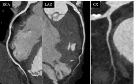
Fig. 4 CCTA in a 50-year-old patient with a heart rate of 73 bpm during data acquisition. Image quality was excellent to good (score 1 and 2) in all segments of the RCA, LAD and LCX. Radiation dose of this CCTA study was 0.5 mSv
Fig. 5 CCTA in a 50-year-old patient with a heart rate of 78 bpm. Image quality was excellent to good (score 1 and 2) in all segments of the LAD and LCX, while image quality was non-diagnostic (score 4) for segment 2 of the RCA due to blurring artefacts (arrow). Radiation dose of this CCTA study was 0.6 mSv
higher strength level of the iterative reconstruction used is associated with a lower image noise, which could be translat-ed to a lower radiation dose (through a rtranslat-eduction of the effective tube current). Finally, we did not test the accuracy of turbo flash CCTA compared with catheter coronary angi-ography. However, the purpose of this study was to determine the image quality, required heart rate, and radiation dose of the turbo high-pitch mode for CCTA. Finally, the results of this study are only valid for the investigated third-generation dual-source CT system in the turbo-flash mode. Modern CT sys-tems from different vendors provide other acquisition tech-niques which are beyond the scope of this study.
In conclusion, our combined phantom and in vivo study indicates that CCTA with turbo high-pitch third-generation dual-source 192-slice CT can be performed at heart rates up to 75 bpm while maintaining diagnostic image quality, being associated with an average radiation dose of 0.6 mSv.
Acknowledgments The scientific guarantor of this publication is
Sebastian Leschka. The authors of this manuscript declare relationships with the following companies: B. Schmidt and T. Allmendinger are employees of Siemens Healthcare. H. Alkadhi and S. Leschka previously served as speakers for Siemens Healthcare. The authors state that this work has not received any funding. No complex statistical methods were necessary for this paper. Institutional Review Board approval was not required because of use of a phantom and also the retrospective nature of the study of clinically indicated CCTA. Written informed consent was waived by the Institutional Review Board. Methodology: retrospective experimental study performed at one institution
References
1. Alkadhi H, Leschka S (2011) Radiation dose of cardiac computed tomography - what has been achieved and what needs to be done. Eur Radiol 21:505–509
2. Tamm EP et al (2011) Quality initiatives: CT radiation dose reduc-tion: how to implement change without sacrificing diagnostic quality.
Radiographics 31:1823–1832
3. Achenbach S et al (2009) Coronary computed tomography angiog-raphy with a consistent dose below 1 mSv using prospectively electrocardiogram-triggered high-pitch spiral acquisition. Eur Heart
J 31:340–346
4. Achenbach S et al (2009) High-pitch spiral acquisition: a new scan mode for coronary CT angiography. J Cardiovasc Comput Tomogr 3:
117–121
5. Leschka S et al (2009) Diagnostic accuracy of high-pitch dual-source CT for the assessment of coronary stenoses: first experience. Eur
Radiol 19:2896–2903
6. Alkadhi H et al (2010) Low-dose, 128-slice, dual-source CT coronary angiography: accuracy and radiation dose of the high-pitch and the
step-and-shoot mode. Heart 96:933–938
7. Goetti R et al (2010) High-pitch dual-source CT coronary
angiogra-phy: systolic data acquisition at high heart rates. Eur Radiol 20:2565–
2571
8. Stolzmann P et al (2011) Predictors of image quality in high-pitch
coronary CT angiography. AJR Am J Roentgenol 197:851–858
9. Layritz C et al (2013) Automated attenuation-based selection of tube voltage and tube current for coronary CT angiography: reduction of
radiation exposure versus a BMI-based strategy with an expert
in-vestigator. J Cardiovasc Comput Tomogr 7:303–310
10. Moscariello A et al (2011) Coronary CT angiography: image quality, diagnostic accuracy, and potential for radiation dose reduction using a novel iterative image reconstruction technique-comparison with
tra-ditional filtered back projection. Eur Radiol 21:2130–2138
11. Schuhbaeck A et al (2013) Image quality of ultra-low radiation exposure coronary CT angiography with an effective dose <0.1 mSv using high-pitch spiral acquisition and raw data-based
iterative reconstruction. Eur Radiol 23:597–606
12. Ulzheimer S, Kalender WA (2003) Assessment of calcium scoring
performance in cardiac computed tomography. Eur Radiol 13:484–
497
13. Morsbach F et al (2013) Stenosis quantification in coronary CT angiography: impact of an integrated circuit detector with iterative
reconstruction. Invest Radiol 48:32–40
14. Farshad-Amacker NA et al (2013) Effect of high-pitch dual-source CT to compensate motion artifacts: a phantom study. Acad Radiol 20:
1234–1239
15. Taylor AJ et al (2010) ACCF/SCCT/ACR/AHA/ASE/ASNC/ NASCI/SCAI/SCMR 2010 Appropriate Use Criteria for Cardiac Computed Tomography. A Report of the American College of Cardiology Foundation Appropriate Use Criteria Task Force, the Society of Cardiovascular Computed Tomography, the American College of Radiology, the American Heart Association, the American Society of Echocardiography, the American Society of N u c l e a r C a r d i o l o g y, t h e N o r t h A m e r i c a n S oc i e t y f o r Cardiovascular Imaging, the Society for Cardiovascular Angiography and Interventions, and the Society for Cardiovascular Magnetic Resonance. Circulation 122:e525-555
16. Husmann L et al (2006) Influence of cardiac hemodynamic parame-ters on coronary artery opacification with 64-slice computed tomog-raphy. Eur Radiol 16:1111–1116
17. Austen WG et al (1975) A reporting system on patients evaluated for coronary artery disease. Report of the Ad Hoc Committee for Grading of Coronary Artery Disease, Council on Cardiovascular Surgery, American Heart Association. Circulation 51:5–40 18. Stolzmann P et al (2007) Radiation dose estimates in dual-source
computed tomography coronary angiography. Eur Radiol 18:592– 599
19. Menzel HG, Schibilla H, Teunen D (eds) (2000) European guidelines on quality criteria for computed tomography. Publication No. EUR 16262 EN. European Commission, Luxembourg
20. Einstein AJ et al (2010) Radiation dose from single-heartbeat coro-nary CT angiography performed with a 320-detector row volume
scanner. Radiology 254:698–706
21. Lell M et al (2009) High-pitch electrocardiogram-triggered computed tomography of the chest: initial results. Invest
Radiol 44:728–733
22. Baumuller S et al (2009) Dual-source versus 64-section CT coronary angiography at lower heart rates: comparison of accuracy and
radia-tion dose. Radiology 253:56–64
23. Sun G et al (2012) 320-detector row CT coronary angiography: effects of heart rate and heart rate variability on image quality,
diagnostic accuracy and radiation exposure. Br J Radiol 85:e388–
e394
24. Cao JX et al (2013) Radiation and contrast agent doses reductions by using 80-kV tube voltage in coronary computed tomographic
angi-ography: a comparative study. Eur J Radiol 83:309–314
25. Leschka S et al (2008) Effect of decrease in heart rate variability on the diagnostic accuracy of 64-MDCT coronary angiography. AJR
Am J Roentgenol 190:1583–1590
26. Matt D et al (2007) Dual-source CT coronary angiography: image quality, mean heart rate, and heart rate variability. AJR Am J
![Fig. 3 Plot of the distortion vector [mm] at the different simulated heart rates [bpm]](https://thumb-eu.123doks.com/thumbv2/123doknet/14811717.611583/5.892.347.804.78.297/fig-plot-distortion-vector-different-simulated-heart-rates.webp)