Sebastian Leschka Paul Stolzmann Lotus Desbiolles Stephan Baumueller Robert Goetti Thomas Schertler Hans Scheffel Andre Plass Volkmar Falk Gudrun Feuchtner Borut Marincek Hatem Alkadhi Received: 30 June 2009 Revised: 30 July 2009 Accepted: 6 August 2009
Published online: 16 September 2009
# European Society of Radiology 2009
Diagnostic accuracy of high-pitch dual-source
CT for the assessment of coronary stenoses:
first experience
Abstract Objectives: The objective was to prospectively investigate the diagnostic accuracy of high-pitch (HP) dual-source computed tomogra-phy coronary angiogratomogra-phy (CTCA) compared with catheter coronary an-giography (CCA) for the diagnosis of significant coronary stenoses.
Methods: Thirty-five patients (seven women; mean age 62±8 years) underwent both CTCA and CCA. CTCA was performed with a second-generation dual-source CT system permitting data acquisition at an HP of 3.4. Patients with heart rates >60 bpm were excluded from study enrolment. All coronary segments were evaluated by two blinded and independent observers with regard to image quality on a four-point scale (1: excellent to 4: non-diagnostic) and for the presence of significant coro-nary stenoses (defined as diameter narrowing exceeding 50%). CCA served as the standard of reference. Radiation dose values were calculated using the dose-length product.
Results: Diagnostic image quality was found in 99% of all segments
(455/459). Non-diagnostic image quality occurred in a single patient with a sudden increase in heart rate imme-diately before and during CTCA. Tak-ing segments with non-evaluative image quality as positive for disease, the sensitivity, specificity and positive and negative predictive values were 94, 96, 80 and 99% per segment and 100, 91, 88 and 100% per patient. The effective radiation dose was on average 0.9±0.1 mSv. Conclusion: In patients with heart rates≤60 bpm, CTCA using the HP mode of the dual-source CT system is associated with high diag-nostic accuracy for the assessment of coronary artery stenoses at sub-milli-Sievert doses.
Keywords Dual-source CT . Coronary angiography . High pitch . Catheter coronary angiography . Coronary artery disease . Diagnostic accuracy
Introduction
Computed tomography (CT) coronary angiography (CA) represents a non-invasive imaging technique for the assessment of coronary artery disease with high diagnostic accuracy [1–8]. Reported sensitivities range from 91 [9] to 100% [10,11] and specificities range from 82 [4] to 97% [3, 9]; negative predictive values were invariably high, ranging from 95 to 100% [3,10,11].
This high diagnostic accuracy has been obtained through rapid developments in the spatial and temporal resolution of CTCA. However, advances in CT technology developments were paralleled by an increased patient radiation dose [12], increasing from about 4–6 mSv with 4-section CTCA [13] up to 12 mSv on average [14] with 64-section CT. This was mainly caused by the thin detector widths and the low helical pitch values, the latter being required for data acquisition in the retrospective electrocardiography (ECG)-gating mode. S. Leschka . P. Stolzmann .
L. Desbiolles . S. Baumueller . R. Goetti . T. Schertler . H. Scheffel . G. Feuchtner . B. Marincek . H. Alkadhi (*)
Institute of Diagnostic Radiology, University Hospital Zurich, Raemistrasse 100, 8091 Zurich, Switzerland e-mail: [email protected] Tel.: +41-1-2553662 Fax: +41-1-2554443 A. Plass . V. Falk
Clinic for Cardiovascular Surgery, University Hospital Zurich, Raemistrasse 100,
8091 Zurich, Switzerland H. Alkadhi
Cardiac MR PET CT Program, Massachusetts General Hospital and Harvard Medical School,
Heightened awareness of increasing radiation exposure from CT imaging among radiologists and referring physicians resulted in the introduction of several dose-reduction techniques including ECG-based tube current modulation [15], anatomy-based tube current modulation, lowering of the tube voltage [16] and prospective ECG-gating [17,18]. With these techniques, effective radiation doses of CTCA could be lowered down to∼1.5–2.5 mSv with the first generation of dual-source CT.
Recently, the second generation of dual-source CT systems was introduced, providing high detector coverage with the use of two 128-section detectors. This dual-source CT system also allows CTCA examinations to be performed at high pitch (HP) values of up to 3.4 [19]. In the latest generation 64-slice CT, the maximally applicable pitch is limited to values around 0.2 to 0.3 to ensure gapless volume coverage of the heart in 5–10 s. In the HP acquisition mode that is unique to dual-source CT, the second detector system can be used to fill these gaps [19]. By combining HP and large detector coverage, the CTCA acquisition time is reduced to a quarter of a second, allowing depiction of the entire heart within a single heart beat.
However, while the diagnostic accuracy of the first dual-source CT system was as high as described for 64-section CT [6,16,17], the feasibility, image quality, and accuracy of second-generation dual-source CT for the detection of coronary artery stenosis have not been investigated so far. The purpose of this study was to prospectively investigate the diagnostic accuracy of HP dual-source CTCA for the assessment of coronary artery stenoses compared with the reference standard catheter coronary angiography (CCA).
Materials and methods
Study population
The study protocol had IRB approval, and all patients gave informed consent to participate in the study.
Thirty-five patients (7 women, 28 men; mean age 62± 8 years; age range 48–74 years) were included in the study and underwent CTCA prior to CCA. Both examinations were performed within a time interval of 30 days. All CTCA studies were clinically indicated and were per-formed for preoperative evaluation of cardiac and thoracic morphology before surgery or percutaneous interventions of the aortic (n=21) and mitral valves (n=14). Patients with unstable heart rhythm, a heart rate>60 bpm (assumed to be too high for use of the HP mode), patients with previous bypass surgery or coronary interventions, or those with contraindications for the administration of either contrast media or beta blockers (if heart rate >60 bpm) were not eligible for study enrolment. Patients were also excluded from this study if a target heart rate≤60 bpm (n=5) could not be achieved after the administration of IV beta-blockers.
Patients who were excluded from study participation were either investigated using retrospectively ECG-gated acqui-sition protocols if not eligible for high-pitch acquiacqui-sitions or using other modalities if general contraindications for contrast-enhanced CT were present.
According to the criteria published by Diamond and Forrester [20], each patient in this population had a low (n=11) to intermediate (n=24) pre-test probability of CAD. Premedication
Fifteen patients (43%) continued taking their baseline beta-receptor antagonist medication at the time of CTCA. In 10 patients (29%), 5–10 mg intravenous metoprolol (Beloc i.v., AstraZeneca, Zug, Switzerland) was administered for reducing the heart rate to ≤60 bpm. The heart rate was lowered to achieve diagnostic image quality in all coronary segments in single diastolic reconstructions [15].
Dual-source CT protocol and image reconstruction
All patients underwent dual-source CT (Somatom Defini-tion Flash, Siemens Healthcare, Forchheim, Germany). All patients received a single dose of 2.5 mg isosorbiddinitrate s. l. (Isoket, Schwarz Pharma, Monheim, Germany). Then, 60 mL of iopromide (Ultravist 370, 370 mg/mL, Bayer Schering Pharma, Berlin, Germany) was injected at a flow rate of 6 mL/s followed by 60 mL of saline solution. Contrast-agent application was controlled by bolus-track-ing in the ascendbolus-track-ing aorta (signal attenuation threshold 100 HU). Data acquisition was initiated with a mean delay of 9 s after reaching the threshold. CT parameters were as follows: detector collimation 2×64×0.6 mm, slice acqui-sition 2×128×0.6 mm by means of a z-flying focal spot, gantry rotation time 280 ms, pitch 3.4, tube current time product 320 mAs per rotation, and tube voltage 100 kV. CTCA was performed from 2 cm below the level of the tracheal bifurcation to the diaphragm. A craniocaudal imaging direction was chosen, and the start phase for image acquisition of the most cranial slice was selected at 60% of the R-R interval in all patients.
Images were reconstructed with a slice thickness of 0.6 mm, a reconstruction increment of 0.4 mm, and using a soft-tissue convolution kernel (B26f). In the case of vessel wall calcifications, additional images were reconstructed using a sharp-tissue convolution kernel (B46) to compen-sate for blooming artefacts.
CTCA data analysis
Coronary segments were defined according to a reporting system of the American Heart Association (AHA) [21]. The right coronary artery (RCA) was defined to include
segments 1–4, the left main artery (LM) to consist of segment 5, the left anterior descending artery (LAD) to include segments 6–10, and the left circumflex artery (LCX) to include segments 11–15. The intermediary artery was designated as segment 16, if present, and considered to belong to the LAD. All diameter measurements were performed with an electronic calliper tool provided with the workstation (Syngo MultiModality Workplace; Siemens Healthcare, Forchheim, Germany).
CTCA data analysis was performed by two blinded and independent observers with 8 and 3 years of experience in cardiac radiology. Both were unaware of the clinical history and the results of the CCA. First, both readers independently rated the image quality of each coronary segment on a four-point scale as follows: 1, excellent (no artefacts, unrestricted evaluation); 2, good (minor artefacts, good diagnostic quality); 3, adequate (moderate artefacts, still acceptable and diagnostic), and 4, non-diagnostic (severe artefacts impairing accurate evaluation). Second, both observers assessed all coronary artery segments for the presence of haemodynamically significant stenoses. Significant stenosis was defined as luminal diameter narrowing exceeding 50%. Vessel diameters were measured on reconstructions perpen-dicularly oriented to the vessel centreline using electronic callipers. For any disagreement in data analysis, consensus agreement was appended.
Catheter coronary angiography
CCA was performed according to standard techniques and at least two views in different planes were obtained for each coronary artery. One experienced observer who was aware of the patients’ clinical history but blinded to the results from CTCA evaluated all angiograms with regard to the presence (diameter reduction>50%) or absence of signif-icant stenoses. Coronary artery segments were defined according to the AHA reporting system described above [21], similar to CTCA.
Estimation of radiation dose
For the estimation of CT radiation dose, the CT volume dose index (CTDIvol) and the dose-length product (DLP) were recorded and used for estimations. The effective dose of CTCA was derived from the product of the DLP and a conversion coefficient (k = 0.017 mSv·mGy−1·cm−1) for the chest according to a method proposed by the European Working Group for Guidelines on Quality Criteria in CT [22].
Statistical analysis
All statistical analyses were performed using commercially available software (SPSS, release 17.0, SPSS, USA).
Quantitative variables are expressed as mean ± standard deviation and categorical variables as frequencies and percentages. Kappa statistics were calculated for inter-observer agreements for image quality read-out and assessment of significant coronary artery stenosis with CTCA. Correlation between image quality of each coro-nary segment and BMI was assessed using Spearman’s correlation analysis.
Sensitivity, specificity, positive predictive value and negative predictive value were calculated from chi-squared tests of contingency. CCA was considered the standard of reference. Non-evaluative segments were censored as positive findings [6]. Statistics for diagnostic accuracy of CTCA were calculated on a per-segment, per-vessel (i.e. at least one significant stenosis or absence of any significant stenosis in one coronary artery), and on a per-patient basis (i.e. at least one significant stenosis or absence of any significant stenosis per patient). A P value of <0.05 was considered statistically significant.
Results
CTCA and CCA were successfully performed in all patients without side effects. The average heart rate was 58±3 bpm, and the body mass index of the study patients was 26.2±3.1 kg/m2 on average, ranging from 19.1 to 31.4 kg/m2. The CTCA acquisitions had an average image length of 117±11 mm and an average acquisition time of 0.27±0.05 s. The average start of the CTCA acquisition was at 59±8% of the R-R interval, and the average end of acquisition was at 78±7% of the R-R interval.
Prevalence of coronary artery stenosis
A total of 459 segments were evaluated in 35 patients. CCA identified 67 coronary artery stenoses with a luminal diameter narrowing of more than 50% in 14 patients (40%). Single-vessel disease was present in 26% (9/35) and multi-vessel disease in 14% (5/35). Significant coronary artery stenosis was absent in 60% of patients (21/35). Coronary artery stenosis was most commonly present in the LAD (11/35; 31%) and less often in the CX (5/35; 14%), the RCA (4/35; 11%) and LM (3/35; 9%).
Image quality
Inter-observer agreement for image quality rating was good (kappa=0.69). Diagnostic image quality was found in 99% of all segments (455/459) (Fig.1). Excellent image quality without any motion artefacts (i.e. score 1) was present in 76% of the coronary segments (352/459), minor artefacts (i.e. score 2) occurred in 19% (90/459), and moderate artefacts (i.e. score 3) in 4% (21/459). Image quality was
rated as non-diagnostic (i.e. score 4) in 1% of segments (4/459). A total of 97% of patients (34/35) had a diagnostic evaluation of the entire coronary artery tree. In one patient, the heart rate increased immediately before the CTCA acquisition started, and CTCA was performed in a single heart beat at 80 bpm, resulting in a CTCA phase acquisition of 80–11% (Fig.2). No deterioration of image quality was observed with increasing BMIs (P=0.78). Consequently, the image quality of the mid and distal RCA, the distal RCX, and the posterior descending artery were non-diagnostic. In this patient no significant coronary artery stenosis was identified by CCA. Acquisition was therefore repeated using retrospective ECG gating.
Diagnostic accuracy of HP dual-source CTCA
The kappa value for coronary artery stenosis detection with CTCA was 0.78 indicating a good inter-observer agreement between both readers. CTCA correctly recognised 63 of the 67 significant stenoses detected with CCA (94%) (Fig.3). In 12 segments, lesions were incorrectly graded as being
stenotic on CTCA. Including the 4 non-diagnostic segments in the analysis and considering them to be positive for disease, a total of 16 false-positive ratings occurred. In four segments, CTCA underestimated the severity of stenosis.
In the vessel-based analysis, CTCA correctly identified 22 of the 23 vessels as having at least one significant stenosis at CCA (96%). Five vessels were falsely classified as being stenotic, and one vessel was falsely classified as being non-stenotic with CTCA. In 2% of vessels (2/105), non-diagnostic segments resulted in false-positives on a vessel-based analysis as no stenosis was present in the other segments of the vessel that could be evaluated.
In the per-patient analysis, CTCA correctly identified at least one significant coronary artery stenosis in all patients with significant stenoses at CCA. In one patient without coronary artery disease at CCA, CTCA suspected signif-icant stenosis, and one patient with non-diagnostic image quality had no significant coronary artery stenosis at CCA. Thus, unnecessary CCA would have been performed in clinical practice, and these patients were considered as false-positives in the per-patient analysis. The diagnostic Fig. 1 HP dual-source CTCA
in a 67-year-old woman before aortic valve replacement (heart rate 55 bpm). Curved reforma-tions of the right coronary artery (a), left anterior descending ar-tery (b) and left circumflex artery (c) demonstrate normal coronary arteries without sub-stantial stenoses. Image quality was classified as excellent (i.e. score 1) in all coronary seg-ments. Catheter coronary angi-ography of the right (d) and left coronary arteries (e) confirms the absence of substantial coro-nary artery disease
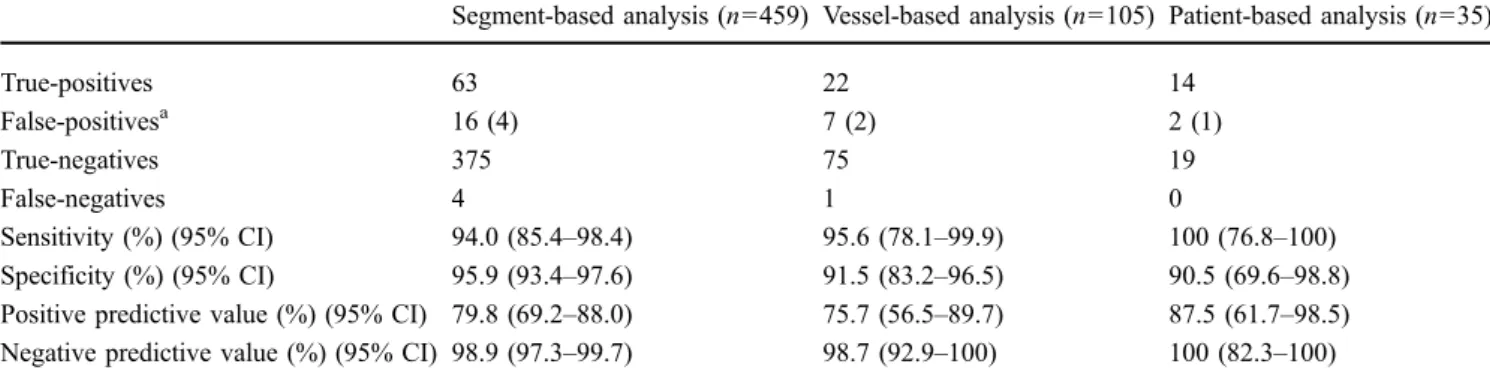
accuracy of CTCA in segment, vessel and per-patient analyses is summarised in Table1.
Radiation doses
The average DLP of the CTCA acquisitions was 54± 5 mGy·cm (range 46–63 mGy·cm). The estimated radiation dose was 0.9±0.1 mSv (range 0.8–1.1 mSv). In 80% of patients (28/35), HP dual-source CTCA resulted in a radiation dose of below 1 mSv.
Discussion
This study introduces an HP data acquisition technique for spiral CTCA. The HP mode of dual-source CT permits imaging of the entire heart within one heart beat by continuous and fast movement of the table during CT acquisition. In single-source CT, the pitch is restricted to values below 1.5 for gapless projection data acquisition in
non-cardiac examination. However, commonly, lower helical pitch values in the range 0.2–0.3 were mandatory in single-source CTCA to provide sufficient projection data for image reconstruction. With dual-source CT, however, a pitch of 3.4 can be used corresponding to a table speed of 480 mm/s, thus resulting in an acquisition time of the heart of 0.25 s. The second tube-detector combination is then used to fill in occurring z-axis gaps. In this way, the heart could be imaged with a field of view of 30 cm covered by both detectors using a quarter of rotation for each detector system [19]. Thus each individual slice has the high temporal resolution of a quarter of a rotation corresponding to 75 ms for the CT system used in our study.
Based on the results of our study, the following conclusions can be drawn: First, HP dual-source CTCA allows imaging of the coronary arteries with diagnostic image quality at regular heart rates of below 60 bpm. Second, HP dual-source CTCA provides a high diagnostic accuracy for the evaluation of coronary artery stenosis comparable to that of 64-section and first-generation dual-source CT systems. Third, HP dual-dual-source CTCA allows Fig. 2 HP dual-source CTCA
in a 55-year-old man with a body mass index of 29.6 kg/m2 before mitral valve replacement. The ECG signal (a) demon-strates that the initial heart rate of 60 bpm increased to 80 bpm immediately before the CTCA acquisition, and phase acquisi-tion of CTCA was at 80–11% of the R-R interval. Curved refor-mation of the right coronary artery (b) demonstrates the non-diagnostic image quality of the mid and distal segments (arrows)
imaging of the entire heart in a quarter of a second at an average radiation dose estimate below 1 mSv.
High diagnostic accuracy for the assessment of coronary artery stenosis has been repeatedly reported with CTCA. Although reported sensitivities range from 91 [9] to 100% [10, 11] and specificities from 82 [4] to 97% [3, 9], negative predictive values were invariably high, ranging from 95 to 100% [3, 10, 11]. This led to the widely accepted conclusion that a normal CTCA reliably rules out significant coronary artery stenosis and further invasive workup with CCA can be omitted. This concept presumes that the entire coronary artery tree can be imaged with a diagnostic image quality, and no segment must be excluded
from analysis. In our study using an HP mode for CTCA acquisition provided by the latest generation of dual-source CT, sensitivity was 100%, specificity was 91% and negative predictive value was 100% on a patient-based analysis. While these values somewhat resemble those reported with the first-generation dual-source CT [3,4,9, 10,23], we did not exclude non-diagnostic segments from analysis but rather considered these as positive ratings on an intent-to-diagnose basis. Moreover, by using the HP mode, the CTCA acquisition time was reduced to approximately one quarter of a second, allowing imaging of the entire coronary artery tree within a single heart beat. Finally, because of the short CTCA acquisition time and HP, the radiation dose was on average 0.9 mSv.
One general limitation of CTCA is the problem of non-diagnostic image quality. The rate of non-non-diagnostic segments has been reported to be as high as 12% with 64-section CTCA [4] and to range from 1 to 6% with first-generation dual-source CTCA [6,7, 18, 24], and usually affects around 10% of patients. In our study, the rate of coronary segments with non-diagnostic image quality was below 1%. Furthermore, all non-diagnostic segments were found in a single patient with an acceleration of the heart rhythm immediately before the CTCA acquisition. This change in heart rate interfered with the planned diastolic CTCA acquisition, but was taken between 80 and 11% of the R-R interval. Hence, rapid cardiac motion during systole hindered artefact-free coronary imaging.
Both high and irregular heart rhythms can be problematic especially when applying the HP mode. The acquisition time window for HP CTCA of about 0.27 s is sufficient for motion-free imaging of the coronary arteries in patients with a low heart rate and subsequently a long resting period of the coronary arteries in mid-diastole [25]. Thus, application of the HP mode for CTCA may be limited in patients with higher heart rates and subsequent shortening of the diastolic resting period. In order to still image these patients with CTCA, the latest generation of dual-source CT provides— similarly to the first generation but at an increased temporal resolution—protocols using either prospective or retro-spective ECG-gating, the latter with low helical pitch values. Fig. 3 HP dual-source CT coronary angiography in a 53-year-old
man before mitral valve replacement and with suspected coronary artery disease (heart rate 48 bpm). Curved reformation of the right coronary artery (a) demonstrates stenosis in the proximal segment (arrow). CCA of the right (b) confirms the stenosis in the proximal right coronary artery (arrowhead)
Table 1 Diagnostic accuracy of HP dual-source CTCA compared with CCA
Segment-based analysis (n=459) Vessel-based analysis (n=105) Patient-based analysis (n=35)
True-positives 63 22 14 False-positivesa 16 (4) 7 (2) 2 (1) True-negatives 375 75 19 False-negatives 4 1 0 Sensitivity (%) (95% CI) 94.0 (85.4–98.4) 95.6 (78.1–99.9) 100 (76.8–100) Specificity (%) (95% CI) 95.9 (93.4–97.6) 91.5 (83.2–96.5) 90.5 (69.6–98.8)
Positive predictive value (%) (95% CI) 79.8 (69.2–88.0) 75.7 (56.5–89.7) 87.5 (61.7–98.5)
Negative predictive value (%) (95% CI) 98.9 (97.3–99.7) 98.7 (92.9–100) 100 (82.3–100)
a
The data acquisition technique of HP dual-source CTCA requires foreseeing the optimal phase of CTCA acquisition several seconds before imaging, as time is needed for acceleration of the table. Therefore, irregularities in the heart rhythm are problematic because the phase of CTCA acquisition would move to earlier or later R-R intervals if the heart rate were to suddenly decrease or increase.
We acknowledge the following study limitations. First, we included a relatively small group of only 35 patients. Certainly, future studies with larger patient populations are needed to confirm our preliminary experience as described in this study. Second, all patients were imaged with the use of 100-kV protocols. Although we believe that there will be a limit in obese patients, we did not observe a deterioration of image quality due to increased noise. Therefore, the current study fails to define upper BMI limits for 100-kV CTCA acquisitions. Third, we only performed HP dual-source CTCA in patients with a stable heart rate below 60 bpm. This study exclusion criterion was based on the theoretical consideration that the acquisition time of a quarter of a second would permit motion artefact-free
imaging only in the long resting periods of the coronary arteries in mid-diastole when the heart rate is low. Further feasibility studies are required to investigate which heart rate conditions are feasible for applying the HP mode with dual-source CTCA and whether the HP mode can be applied also in patients with higher heart rates while maintaining high diagnostic accuracy.
Conclusions
Our study results indicate that HP dual-source CTCA provides a high diagnostic accuracy for the assessment of coronary stenoses combined with a 1% rate of non-diagnostic coronary segments and a radiation dose below 1 mSv as evidenced in patients with heart rates≤60 bpm. Further studies including larger patient populations are warranted to confirm our initial results and to further evaluate the use of the HP and other CT parameters in second-generation dual-source CTCA in patients with higher heart rates.
References
1. Brodoefel H, Burgstahler C, Tsiflikas I et al (2008) Dual-source CT: effect of heart rate, heart rate variability, and calcification on image quality and diagnostic accuracy. Radiology 247:346–355
2. Johnson TR, Nikolaou K, Busch S et al (2007) Diagnostic accuracy of dual-source computed tomography in the diagnosis of coronary artery disease. Invest Radiol 42:684–691
3. Leschka S, Alkadhi H, Plass A et al (2005) Accuracy of MSCT coronary angiography with 64-slice technology: first experience. Eur Heart J 26:1482– 1487
4. Raff GL, Gallagher MJ, O’Neill WW et al (2005) Diagnostic accuracy of non-invasive coronary angiography using 64-slice spiral computed tomography. J Am Coll Cardiol 46:552–557
5. Ropers U, Ropers D, Pflederer T et al (2007) Influence of heart rate on the diagnostic accuracy of dual-source computed tomography coronary angi-ography. J Am Coll Cardiol 50:2393– 2398
6. Scheffel H, Alkadhi H, Plass A et al (2006) Accuracy of dual-source CT coronary angiography: first experience in a high pre-test probability population without heart rate control. Eur Radiol 16:2739–2747
7. Weustink AC, Meijboom WB, Mollet NR et al (2007) Reliable high-speed coronary computed tomography in symptomatic patients. J Am Coll Car-diol 50:786–794
8. Miller JM, Dewey M, Vavere AL et al (2009) Coronary CT angiography using 64 detector rows: methods and design of the multi-centre trial CORE-64. Eur Radiol 19:816–828
9. Leber AW, Knez A, von Ziegler F et al (2005) Quantification of obstructive and nonobstructive coronary lesions by 64-slice computed tomography: a comparative study with quantitative coronary angiography and intravascular ultrasound. J Am Coll Cardiol 46:147– 154
10. Mollet NR, Cademartiri F, van Mieghem CA et al (2005)
High-resolution spiral computed tomo-graphy coronary angiotomo-graphy in patients referred for diagnostic con-ventional coronary angiography. Circulation 112:2318–2323
11. Pugliese F, Mollet NR, Runza G et al (2006) Diagnostic accuracy of non-invasive 64-slice CT coronary angiog-raphy in patients with stable angina pectoris. Eur Radiol 16:575–582
12. Alkadhi H (2009) Radiation dose of cardiac CT–what is the evidence? Eur Radiol 19:1311–1315
13. Achenbach S, Giesler T, Ropers D et al (2001) Detection of coronary artery stenoses by contrast-enhanced, retro-spectively electrocardiographically-gated, multislice spiral computed tomography. Circulation 103:2535– 2538
14. Hausleiter J, Meyer T, Hermann F et al (2009) Estimated radiation dose asso-ciated with cardiac CT angiography. JAMA 301:500–507
15. Leschka S, Scheffel H, Desbiolles L et al (2007) Image quality and recon-struction intervals of dual-source CT coronary angiography: recommenda-tions for ECG pulsing windowing. Invest Radiol 42:543–549
16. Alkadhi H, Scheffel H, Desbiolles L et al (2008) Dual-source computed tomo-graphy coronary angiotomo-graphy: influ-ence of obesity, calcium load, and heart rate on diagnostic accuracy. Eur Heart J 29:766–776
17. Scheffel H, Alkadhi H, Leschka S et al (2008) Low-dose CT coronary angiog-raphy in the step-and-shoot mode: diagnostic performance. Heart 94:1132–1137
18. Stolzmann P, Leschka S, Scheffel H et al (2008) Dual-source CT in step-and-shoot mode: noninvasive coronary an-giography with low radiation dose. Radiology 249:71–80
19. Achenbach S, Marwan M, Schepis T et al (2009) High-pitch spiral acquisition: a new scan mode for coronary CT angiography. J Cardiovasc Comput Tomogr 3:117–121
20. Diamond GA, Forrester JS (1979) Analysis of probability as an aid in the clinical diagnosis of coronary-artery disease. N Engl J Med 300:1350–1358 21. Austen WG, Edwards JE, Frye RL et al (1975) A reporting system on patients evaluated for coronary artery disease. Report of the Ad Hoc Committee for Grading of Coronary Artery Disease, Council on Cardiovascular Surgery, American Heart Association. Circula-tion 51:5–40
22. Menzel HG, Schibilla H, Teunen D (eds) (2000) European guidelines on quality criteria for computed tomogra-phy. Publication no. EUR 16262 EN. European Commission, Luxembourg
23. Ong TK, Chin SP, Liew CK et al (2006) Accuracy of 64-row multidetector com-puted tomography in detecting coronary artery disease in 134 symptomatic pa-tients: influence of calcification. Am Heart J 151(1323):e1321–e1326 24. Rixe J, Rolf A, Conradi G et al (2008)
Image quality on dual-source com-puted-tomographic coronary angiogra-phy. Eur Radiol 18:1857–1862 25. Husmann L, Leschka S, Desbiolles L et
al (2007) Coronary artery motion and cardiac phases: dependency on heart rate– implications for CT image reconstruction. Radiology 245:567–576