Publisher’s version / Version de l'éditeur:
Environmental science and technology, 44, 17, pp. 6775-6781, 2010
READ THESE TERMS AND CONDITIONS CAREFULLY BEFORE USING THIS WEBSITE. https://nrc-publications.canada.ca/eng/copyright
Vous avez des questions? Nous pouvons vous aider. Pour communiquer directement avec un auteur, consultez la première page de la revue dans laquelle son article a été publié afin de trouver ses coordonnées. Si vous n’arrivez pas à les repérer, communiquez avec nous à PublicationsArchive-ArchivesPublications@nrc-cnrc.gc.ca.
Questions? Contact the NRC Publications Archive team at
PublicationsArchive-ArchivesPublications@nrc-cnrc.gc.ca. If you wish to email the authors directly, please see the first page of the publication for their contact information.
NRC Publications Archive
Archives des publications du CNRC
This publication could be one of several versions: author’s original, accepted manuscript or the publisher’s version. / La version de cette publication peut être l’une des suivantes : la version prépublication de l’auteur, la version acceptée du manuscrit ou la version de l’éditeur.
For the publisher’s version, please access the DOI link below./ Pour consulter la version de l’éditeur, utilisez le lien DOI ci-dessous.
https://doi.org/10.1021/es101206t
Access and use of this website and the material on it are subject to the Terms and Conditions set forth at
Noninvasive probing of inhibitory effects of cylindrospermopsin and
microcystin-LR using cell-based impedance spectroscopy
Male, Keith B.; Tom, Roseanne; Durocher, Yves; Greer, Charles; Luong,
John H. T.
https://publications-cnrc.canada.ca/fra/droits
L’accès à ce site Web et l’utilisation de son contenu sont assujettis aux conditions présentées dans le site LISEZ CES CONDITIONS ATTENTIVEMENT AVANT D’UTILISER CE SITE WEB.
NRC Publications Record / Notice d'Archives des publications de CNRC:
https://nrc-publications.canada.ca/eng/view/object/?id=f4668468-8b3b-43c4-babf-832fe09773f2
https://publications-cnrc.canada.ca/fra/voir/objet/?id=f4668468-8b3b-43c4-babf-832fe09773f2
Noninvasive Probing of Inhibitory
Effects of Cylindrospermopsin and
Microcystin-LR Using Cell-Based
Impedance Spectroscopy
K E I T H B . M A L E , R O S E A N N E T O M ,
Y V E S D U R O C H E R , C H A R L E S G R E E R , A N D J O H N H . T . L U O N G *
Biotechnology Research Institute, National Research Council Canada, Montreal, Quebec, Canada H4P 2R2
Received April 16, 2010. Revised manuscript received July 26, 2010. Accepted August 2, 2010.
In an effort to develop a noninvasive method for assessment
of cyanobacterial toxins in drinking water, plausible cytotoxicity/
inhibition of microcystin-LR and cylindrospermopsin was
evaluated by cell-substrate impedance sensing (ECIS) using
three different cell lines. Sf9 insect cells were attached to
concanavalin A coated gold electrodes, whereas Chinese hamster
ovary (CHO) and human embryo kidney (HEK) cells were
attached to a fibronectin or laminin coated gold surface.
Cytotoxic or inhibitory effects were dependent upon the cell
line and the extracellular matrix (ECM) coating. Neither toxin
exhibited any appreciable effect on the insect cells. In contrast,
cytotoxicity of cylindrospermopsin on CHO cells was attested
by both ECIS and viability tests. The half-inhibition concentration
(ECIS
50) of cylindrospermopsin for CHO cells was ∼2 µg/mL
(ppm) after 20 h of exposure and 4 µg/mL (ppm) after 30 h of
exposure for a laminin or fibronectin coated surface. ECIS
confirmed no significant effect of cylindrospermopsin on HEK
cells. Microcystin-LR was also tested with CHO cells, resulting
in an ECIS
50value of ∼12 µg/mL (ppm) after 25 h of exposure
for a laminin coated gold surface. The effect of
microcystin-LR on CHO cells probed by ECIS was inhibitory rather than cytotoxic,
as confirmed by cell viability assays.
Introduction
There are an increasing number of communities, especially in Canada with recreational and drinking water contaminated by cyanobacterial blooms as high as 250,000 cells/mL water or ∼30 mg/L biomass (1). Cyanobacteria (blue-green algae) are widely distributed and can dominate eutrophic (phos-phorus and nitrogen rich) aquatic environments such as lakes and reservoirs. Some cyanobacteria can produce toxins that attack different organs, tissues, and metabolic pathways: hepatotoxins, neurotoxins, cytotoxins, dermatotoxins and genotoxins, protease inhibitors, and other toxins of poorly known activity (2). Hepatotoxins, cyclic heptapeptides, are potent inhibitors of eukaryotic protein phosphatases 1 and 2A and potent liver tumor promoters (3). They are produced by members of the cyanobacterial genera Microcystis,
Ana-baena, Aphanizomenon, Oscillatoria, Nostoc, and Cylin-drospermopsis (4, 5). The major bloom forming cyanobacterial
species, Microcystis aeruginosa, produces potent hepatotoxins called microcystins which are a series of more than 75 cyclic
peptides sharing a common structure first isolated in 1986 (6). The health risk assessment elements of microcystin-LR have prompted the World Health Organization (WHO) (7) to establish a guideline value for drinking water. As established by Health Canada, the drinking water should not exceed 1.5 µg/L (ppb) microcystin-LR. Unfortunately, a number of communities in Quebec, Canada have regularly exceeded this guideline level (8). The microcystin-LR level found in the four eutrophic lakes tested varied over the growing season from May to October with a peak in toxicity generally sometime in August or September. In Missisquoi Bay, Lake Champlain, Quebec public health advisories have been issued for eight consecutive years from 2000 to 2008. Similar drinking water issues caused by cyanobacterial blooms have also been reported in hypereutrophic lakes and farm dugouts in Alberta, Canada (9, 10).
Upon ingestion, typical symptoms of cyanobacteria toxin poisonings (CTP) include nausea, vomiting, diarrhea, and fever. Long-term exposure can cause liver damage and even death as in the tragedy involving 52 human fatalities in Caruara, Brazil (11, 12). Most CTP occur when water blooms accumulate as thick surface scums which humans would generally avoid, and drinking water is usually treated to prevent high concentrations of cyanotoxin being present, thus sparing severe poisoning of humans by the oral route. However, in the Brazilian case the lethality occurred through the intravenous route at a dialysis clinic, where the levels of microcystin-LR in the water used for treatment were deter-mined to be 19.5 µg/L (ppb).
Cylindrospermopsin, produced by a variety of cyano-bacterial genera including Cylindrospermopsis,
Aphanizome-non, Umezakia, and Anabaena, is a cyclic guanidine alkaloid
which has cytotoxic, hepatotoxic, neurotoxic, and genotoxic effects. Besides acting as a hepatotoxin, it affects the kidneys, spleen, thymus, and heart (13). Cylindrospermopsis raciborskii was retrospectively implicated (14) as a causative agent in a human poisoning incident in 1979 in the tropical and subtropical waters of Australia. The toxin responsible was later identified and given the name cylindrospermopsin (15). This toxin has since also been reported in New Zealand (16), Japan (17), Thailand (18), China (19), Israel (20), and Europe (21, 22). Similarly to microcystin-LR, risk assessment cal-culations show that the guideline values for cylindrosper-mopsin in drinking water should be in the 1 µg/mL (ppb) range (23).
The embodiment of Electric Cell-Substrate Impedance Sensing (ECIS) is a small gold electrode (250 µm diameter) deposited on the bottom of culture wells and immersed in a culture medium inoculated with a specific cell line (24, 25). Using culture medium as the electrolyte, a constant current source applies a small ac current of ∼1 mA at 4000-5000 Hz between the small detecting electrode and a large counter electrode. The arrival of cells to the electrode and their attachment act as insulating particles because their plasma membrane will interfere with the free space immediately above the electrode for current flow. The changing impedance can be continuously monitored and interpreted to reveal information about cell spreading. Detailed information of the principle of ECIS can be found elsewhere (26, 27). The attachment and spreading of cells attached to the surface of a gold electrode precoated with an extracellular matrix (ECM) will be affected by the presence of toxic/inhibitory com-pounds, resulting in a disruption of the current flow at the substratum level. This technique has been employed for monitoring the cytotoxic effects of explosives at levels of ppm (28, 29), heavy metals (30), and nanomaterials including
* Corresponding author e-mail: john.luong@cnrc-nrc.gc.ca.
Environ. Sci. Technol.2010, 44, 6775–6781
10.1021/es101206t 2010 American Chemical Society VOL. 44, NO. 17, 2010 / ENVIRONMENTAL SCIENCE & TECHNOLOGY96775 Published on Web 08/11/2010
quantum dots (31). In addition the technique was capable of probing the inhibitory effects of closely related compounds isolated from Antrodia camphorata (32) at micromolar levels and destruxins from Metarhizium anisopliae (33). In these instances the inhibition occurred at the level of cell adhesion and spreading with little effect on the overall growth of the cells.
This paper describes an online technique based on ECIS for the continuous assessment of the behavior of insect and mammalian cells exposed to cylindrospermopsin and mi-crocystin-LR. The attachment and spreading of the cells will result in a significant change in the measured impedance. Exposure of the cells to cylindrospermopsin and microcystin-LR, if cytotoxic or inhibitory at the substratum level, will lead to alterations in cell behavior and the resulting chemical effect can be screened by measuring the impedance change. To our knowledge, this is the first demonstration for the use of ECIS to probe the effect of cylindrospermopsin and microcystin-LR on three different cell lines.
Materials and Methods
Cell Lines and Culture Conditions. Insect Cell Line.
Spodoptera frugiperda Sf 9 cells were maintained in 125-mL
disposable Erlenmeyer flasks with a working volume of 20 mL in serum-free SF-900 II medium (Gibco BRL, Canadian Life Technologies, Burlington, ON, Canada). Cells were cultured weekly at 0.4 × 10⁶cells/mL at 27 °C, pH 6.2, with
agitation at 110 rpm. The monitoring of the cell count and viability by the trypan blue exclusion assay during the growth was performed with a CEDEX Innovatis cell counter (Bielefeld, Germany). Sf9 cells, inoculated at an initial cell density of 0.4 × 10⁶cells/mL, were grown to the midexponential phase 2.5-3.0 × 10⁶cells/mL, and the resulting cells were aseptically
centrifuged at 1500 rpm for 4 min. Pellets were thereafter suspended at a cell concentration of 3 × 10⁶cells/mL in a
fresh medium.
Chinese Hamster Ovary (CHO) Cell Line. CHO cells were
maintained in 125-mL disposable Erlenmeyer flasks with a working volume of 20 mL in serum-free Freestyle CHO Expression medium (Invitrogen, Burlington, ON, Canada). Cells were subcultured every 2-3 days at 0.2 × 10⁶or 0.05
× 106 cells/mL, respectively, in a humidified incubator controlled at 5% CO2and 37 °C, on an orbital shaker platform
set at 120 rpm. Cell counts and viability were monitored, using trypan blue dye exclusion, with a CEDEX Innovatis cell counter (Bielefeld, Germany). CHO cells were grown to the exponential phase 1.0-2.5 × 10⁶cells/mL. The cell
concen-tration was adjusted to 2 × 10⁶ cells/mL for standard
experiments.
Human Embryo Kidney (HEK) Cell Line. HEK cells were
maintained in 125-mL disposable Erlenmeyer flasks with a working volume of 20 mL in serum-free F17 medium (Invitrogen, ON, Canada). Cells were subcultured every 2-3 days at 0.3 × 10⁶
or 0.2 × 10⁶cells/mL, respectively, in a
humidified incubator controlled at 5% CO2at 37 °C, on an
orbital shaker platform set at 120 rpm. Cell counts and viability were monitored, using trypan blue dye exclusion, with a CEDEX Innovatis cell counter (Bielefeld, Germany). HEK cells were grown to the midexponential phase 1.0-2.0 × 10⁶cells/mL. The cell population was adjusted to 2 × 10⁶ cells/mL for standard experiments.
Electrode Coating and Cell Addition. Concanavalin A
(Con A, 0.40 mL, 0.5 mg/mL, prepared fresh daily in 50 mM PBS, pH 7.4 with sonication for 1 h) was added into each of the 8 wells of a sensing chip (8W1E, Applied Biophysics, Troy, NY) to coat the detecting gold electrodes for analysis with insect cells. Similarly, fibronectin (0.1 mg/mL) or laminin (0.1 mg/mL) was added to the sensing chips for analysis with HEK and CHO cell lines. Con A, fibronectin, or laminin binds quickly (90% of the change occurs in the first 10 min) to the
electrode surface. Sensing chips needed to be pretreated with
L-cysteine (10 mM) to remove the build-up of films which otherwise interfere with the absolute resistance readings. After protein adsorption (∼30-60 min), the wells were washed 3 times with 0.85% NaCl, and 0.4 mL of culture medium was placed in each well. The impedance baseline was monitored for 1-2 h at 27 °C in a humidified chamber with the ECIS impedance system. The wells were emptied, and 0.4 mL of insect cell suspension (∼2-3 × 10⁶cells/mL, depending on
the cell line) was added into each well. Microcystin-LR (Abraxis, Warminster, PA) was dissolved in ethanol (∼0.5 mL) with sonication to a concentration of 1 mg/mL, while cylindrospermopsin (Abraxis, Warminster, PA) was dissolved in water to 1 mg/mL. The toxins were added to insect cell suspensions (1.5 mL at 3 × 10⁶ cells/mL) at various
concentrations before adding 0.4 mL to 2 or 3 wells to test for possible inhibitory effects. Different concentrations including a control with 30 µL of ethanol (for microcystin-LR experiments) were tested simultaneously. Similarly mam-malian cells (2 × 10⁶cells/mL) were added (0.4 mL) to wells
and incubated in a humidified incubator at 37 °C and 5% CO2.
Impedance Measurement with ECIS. In brief, the system
(model 100, Applied Biophysics, Troy, NY, USA) is equipped with 16 sample wells (2 chips of 8 wells, each containing a singly addressable detecting electrode). A common counter gold electrode (0.2 cm2) is shared by the 8 detecting electrodes
(0.5 × 10⁻³cm2- 250 µm diameter), and the two electrodes
(detecting gold electrode and counter gold electrode) of the well (10-mm square wells -volume ∼0.4 mL) are connected to a lock-in amplifier of the ECIS system. Cell behavior should not be affected if the applied potential is 1 V AC or less (noninvasive). The impedance of each well was measured every 2 min at 4 kHz, and the system acquires resistance, impedance, and capacitance data. However, as larger changes occurred in the resistance, we have focused on these changes in this study. Both data acquisition and processing were performed using software supplied by Applied Biophysics. The ECIS50value derived from the time response function,
f(C, t), was calculated as described by Xiao et al (29). For
simplification of plots and calculations, data points at 30 min intervals were selected from the raw resistance data. The sensing chip could be temporarily removed (pause function from the software) from the ECIS system incubator and placed on a Wilovert AFL 30 inverted microscope (Hund, Germany) equipped with a digital video camera (KP-D50U, Hitachi, Tokyo, Japan) to observe the cells during experimentation.
Results and Discussion
ECIS Models Using HEK and CHO Cells. As shown in Figure
1, Sf 9 insect cells were not affected by cylindrospermopsin up to 50 µg/mL (50 ppm) and microcystin-LR up to 20 µg/ mL (20 ppm). Such results were somewhat surprising since this cell line has been used to probe cytotoxicity and inhibition of various compounds (28, 32, 33). Therefore, the subsequent experiments focused on the mammalian cells HEK and CHO, which have also commonly been used in ECIS toxicity studies. CHO cells (2 × 10⁶cells/mL) were tested using gold surfaces
precoated with laminin or fibronectin and compared with the control. Adhesion was most rapid using fibronectin, although a latent response was also observed for laminin (Figure 2). A confluent layer of cells resulted after a few hours with fibronectin, whereas with laminin the confluence was only achieved after 10-15 h, although the resistance changes were similar. The protein concentration (0.1 mg/mL) used for precoating was optimal as similar results were obtained at 0.2 mg/mL. Both proteins could be used as models for toxin analysis as there could be differences in response to the toxins due to the difference in cell adhesion. HEK cells (2 × 10⁶cells/mL) adhered within 1 h with either a fibronectin
or laminin coating (figure not shown). The response was also more stable with laminin as the resistance signal drifted downward with time for fibronectin coated gold surfaces. Another important parameter was the cell concentration which was added to the wells of the sensing chips. Compared to insect cells with an optimal level at 3 × 10⁶ cells/mL
(28, 32, 33), and V79 mammalian cells from Chinese hamster lung fibroblast with an optimal level of 1 × 10⁶cells/mL (31),
the concentration required for forming a confluent layer with reproducible resistance results for HEK and CHO cells was found to be 2 × 10⁶cells/mL with fibronectin coated gold
surfaces. Lower cell concentrations resulted in lower resis-tance change, and the results were less reproducible. Microscopic observations also confirmed that fewer cells attached and spread on the gold surface.
Effect of Cylindrospermopsin on CHO Cells with Lami-nin-Coated Gold Surfaces. There was no significant effect
at 330 ng/mL (ppb) when cylindrospermopsin was added to CHO cells anchored on a laminin coated gold surface (Figure 3). However, at 660 ng/mL (ppb) the inhibitory effect was observed and became pronounced as the concentration increased to 3.3 µg/mL (3.3 ppm). At very high toxin concentrations (>6.6 µg/mL or 6.6 ppm), the CHO cells were severely inhibited after 15 h as the resistance change approached zero after a slight initial increase. During the first few hours, the profiles were similar regardless of toxin concentrations indicating an initial cell attachment. During the cell spreading phase, the toxin likely interfered with the process, and at high concentrations resulted in cell delami-nation from the laminin coated gold surface. At high
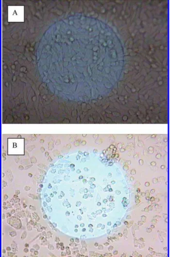
concentrations of cylindrospermopsin (6.6 µg/mL), cell morphology had become spherical compared to the control (Figure 4).
Half-Inhibition Concentration (ECIS50) for
Cylindrosper-mopsin. For the effector cells, the resistance change (∆Rs)
of the well is dependent on the number (No) of initial cells
attached on the detecting electrode, the toxicant concentra-tion (C), and the exposure time (t) as reported by Xiao et al (29). The resistance change normalized by Nois defined as FIGURE 1. (A) Resistance response (Ω) of Spodoptera
frugi-perda Sf9 cells on concanavalin A coated gold electrode
surfaces to various concentrations (µg/mL) of cylindrosper-mopsin: (a) 0, (b) 5, (c) 6.7, (d) 13.5, (e) 20, (f) 26.7, and (g) 50.
FIGURE 2. Resistance response (Ω) of CHO cells on (a) no protein coating, (b) fibronectin coated gold electrode surfaces, and (c) laminin coated gold electrode surfaces.
FIGURE 3. (A) Resistance response (Ω) of CHO cells on laminin coated gold electrode surfaces to various concentrations (µg/ mL) of cylindrospermopsin: (a) 0, (b) 0.33, (c) 0.66, (d) 1.3, (e) 3.3, and (f) 6.6.
FIGURE 4. Microscopic photos of CHO cells on the laminin coated gold electrode surface after 24 h: (a) without cylindro-spermopsin and (b) 6.6 µg/mL cylindrocylindro-spermopsin. Note: The diameter of the electrode shown in the figures is 250 µM.
the cell response to the toxicant measured by ECIS, f(C, t) ) ∆Rs/No. For a control with no toxicant, C is equal to zero,
and f(0, t) increases as the cells spread on the electrode and reach a plateau. In the presence of toxicant, f(C, t) after an initial increase the value decreases and even approaches zero, indicating total cell death at high toxicant concentrations. The inhibition concentration required to achieve 50% inhibition of the cytotoxicity response is defined as the half-inhibition concentration (ECIS50) or f(ECIS50, t)/f(0, t) ) 50%.
The ECIS50for cylindrospermopsin was calculated from
the data obtained in Figure 3. The time response function
f(C, t) was used to construct a series of inhibition curves at
any given time t0(>7.5 h) for the series of cylindrospermopsin
concentrations (0.33-6.6 µg/mL) used in Figure 3. Nofor
each well was assumed to be equivalent; therefore, ∆Rsdid
not have to be adjusted due to different Novalues. The time
response function, f(C, t), was then normalized by simply taking the ∆Rs, i.e., Rt-R0 at different cylindrospermopsin
concentrations and dividing the values by the ∆Rsat f(0, t).
The normalized time response function decreased as the concentration of cylindrospermopsin (>330 ng/mL) increased for all exposure times considered (Figure 5A). The ECIS50for
cylindrospermopsin was determined for each exposure time by extrapolating the value on the cylindrospermopsin concentration axis when the normalized time response function was 0.5. Figure 5B shows the relationship between the half-inhibition concentration and exposure time with the ECIS50for cylindrospermopsin estimated as ∼2 µg/mL
after 20 h of exposure. The average and standard error on the mean (SEM) were given for four runs. The ppm (µM) detection range observed with cylindrospermopsin was similar to that obtained for other toxins also using the ECIS technique (28-33). It should be noted that the detection range is similar (ppm) for cyanobacteria toxins such as microcystins using standard HPLC techniques (34, 35). Water samples generally contain very low levels of cyanobacteria toxins (ppb range); therefore, large samples (6 L) should be concentrated utilizing C18solid phase extraction designs to obtain concentrations
suitable for HPLC analysis or techniques such as ECIS (36). In principle, a preconcentration factor of a 1000-fold is feasible provided the availability of large sample volumes. Alternatively, some enzyme-linked immunosorbent assays (ELISA) or fluorescent immunochromatography techniques have been developed to measure the water samples directly in the ppb range (36-38). In addition changing the cell line used for ECIS testing may also be a possibility for improving sensitivity. Since cylindrospermopsin is a potent hepatotoxin, a hepatic cell line such as rat hepatic stellate cells could be an alternative choice.
Effect of Cylindrospermopsin on CHO Cells with Fi-bronectin-Coated Gold Surfaces. Cylindrospermopsin added
with CHO cells to fibronectin coated gold surfaces did not provoke any significant inhibitory effect at low concentrations (below 1.32 µg/mL). However, inhibition was observed at 3.30 µg/mL (3.3 ppm) and became pronounced as the concentration increased to 13.2 µg/mL (figure not shown). At very high toxin concentrations (>13.2 µg/mL or 13.2 ppm) and after 15 h of exposure, the resistance change approached zero after a slight initial increase. Such behavior indicated the CHO cells were severely inhibited. It should be noted that for laminin coated gold surfaces, during the first few hours the profiles were similar regardless of toxin concen-trations. As observed microscopically, at high cylindrosper-mopsin concentrations (13.2 µg/mL), the cell shape had become spherical compared to the control.
Half-Inhibition Concentration (ECIS50) for
Cylindrosper-mopsin. Similar to the case of laminin coated gold surfaces,
the ECIS50for cylindrospermopsin was calculated from the
data obtained using fibronectin coated gold surfaces. The time response function f(C, t) was used to construct a series of inhibition curves at any given time t0(>7.5 h) for the series
of toxin concentrations (3.3-20 µg/mL). The normalized time response function decreased as expected as the toxin concentration (>3.3 µg/mL) increased for all exposure times considered. The relationship between the half-inhibition concentration and exposure time, indicating that the ECIS50
for cylindrospermopsin was ∼4 µg/mL after 30 h of exposure. The ECIS50value for cylindrospermopsin was higher (6 µg/
mL at 20 h) using fibronectin coated gold surfaces, compared to laminin (2 µg/mL at 20 h) coated surfaces. It was reasoned that the toxin was likely able to penetrate the cells easier when they were not well spread on the electrode surface, since the spreading phenomenon seems slower when laminin was used as ECM for cell adhesion and spreading. Therefore the ECIS technique was more sensitive to cytotoxic effects of cylindrospermopsin using a laminin coated gold electrode compared to a fibronectin coated electrode although the fibronectin coated surface provided a more rapid cell adherence and increase in resistance.
Viability studies confirmed the cytotoxic effect of cylin-drospermopsin on CHO cells. CHO cells (1.5 × 10⁶cells/mL)
were grown in the presence of 2 µg/mL and 20 µg/mL of
FIGURE 5. (A) Cylindrospermopsin inhibition curves were obtained for each cylindrospermopsin concentration (Figure 3, curves a-f) at various exposure times (h): (a) 7.5, (b) 10, (c) 12.5, (d) 15, (e) 17.5, and (f) 20. The normalized time response function (y axis), f (C, t), was determined by taking the ∆Rs
(Figure 3, curves b-f), i.e., Rt-R0at different cylindrospermopsin
concentrations and dividing the values by the ∆Rs (∼5000 Ω,
Figure 3, curve a) at f (0, t). (B) Relationship between the half-inhibition concentration (ECIS50) and exposure time during
cell culture for cylindrospermopsin. The ECIS50 value obtained
for cylindrospermopsin was determined for each exposure time (Figure 5A, curves a-f) by extrapolating the value for the x-axis from the y-axis (0.5). (Data expressed as SEM, n ) 4).
cylindrospermopsin. After 6.5 h of exposure, there was little effect at 2 µg/mL; however, at 20 µg/mL the viability declined noticeably. Consistent with the ECIS results of Figure 3, only a modest effect at toxin concentrations below 3.3 µg/mL was observed after 5-7 h of exposure. Higher concentrations (6.6 µg/mL) trigged the cytotoxic effect, and after 24 h of exposure to 20 µg/mL cylindrospermopsin, the viability of the cells dropped to 83%. Cell growth was severely inhibited as the cell density declined substantially to 42% (1.55 × 10⁶cells/
mL) and 29% (1.06 × 10⁶cells/mL) at 2 µg/mL and 20 µg/mL
of cylindrospermopsin, respectively, when compared to the control (3.66 × 10⁶cells/mL).
Effect of Cylindrospermopsin on HEK Cells with Lami-nin-Coated Gold Surfaces. Cylindrospermopsin exhibited
no significant inhibitory effect on HEK cells adhered to laminin coated gold surfaces (figure not shown). Similar to the results with Sf 9 insect cells (Figure 1), at concentrations as high as 20 µg/mL (20 ppm) there was no significant difference compared to the control wells. The results obtained using HEK cells was not as reproducible as with CHO cells due to the drift of the signal response. Viability assays indicated a toxic effect of cylindrospermopsin on HEK cells. HEK cells (1.73 × 10⁶cells/mL) were grown in the presence
of 2 µg/mL and 20 µg/mL of cylindrospermopsin. After 6.5 h there was little effect at either concentration (viability >94% and similar cell densities); however, after 24 of exposure an effect was obvious, as the cell density decreased to 86% (2.88 × 10⁶cells/mL) at 2 µg/mL and 63% (2.11 × 10⁶cells/mL) at 20 µg/mL compared to the control (3.35 × 10⁶cells/mL).
The inhibitory effect of cylindrospermopsin was more significant with CHO cells compared to HEK cells. Therefore, ECIS with CHO cells adhered to laminin coating would be most suitable for testing the effect of cylindrospermopsin.
Effect of Microcystin-LR on CHO Cells with Laminin-Coated Gold Surfaces. The effect of ethanol was first tested
for CHO cells since hydrophobic microcystin-LR was dis-solved in ethanol. Ethanol exhibited a detrimental impact on the response signal at a concentration of 2%; however, at 0.5% the effect was not significant. Other solvents including dimethylsulfoxide also exhibited similar inhibition patterns. Microcystin-LR was dissolved in ethanol as a stock concen-tration of 5 mg/mL and diluted in the culture buffer for testing. Up to 25 µg/mL, the ethanol level was very low and should not have any effect on CHO cells. Microcystin-LR added with CHO cells to laminin coated gold surfaces resulted in an inhibitory effect as shown in Figure 6. Below 5 µg/mL there was no significant effect; however, by 8.3 µg/mL (8.3 ppm) inhibition was observed and became pronounced as the concentration increased to 25 µg/mL (25 ppm). During the first few hours, the profiles were similar regardless of microcystin-LR concentration confirming an initial cell
attachment to the electrode. During the cell spreading phase, the microcystin-LR likely interfered with the process, and at very high concentrations the cells might be actually delami-nated from the laminin coated gold surface.
Half-Inhibition Concentration (ECIS50) for
Microcystin-LR. The ECIS50for microcystin-LR was calculated from the
data obtained in Figure 6 using laminin coated gold surfaces. The time response function f(C, t) was used to construct a series of inhibition curves at any given time t0(>10 h) for the
series of microcystin-LR concentrations (8.3-25 µg/mL) used in Figure 6. The normalized time response function decreased as the cylindrospermopsin concentration (>8.3 µg/mL) increased for all exposure times considered (Figure 7A). Figure 7B shows the relationship between the half-inhibition concentration and exposure time, and the ECIS50for
mi-crocystin-LR was estimated as ∼12 µg/mL after 25 h of exposure. The average and standard error on the mean (SEM) were given for four runs. The ECIS50value for
microcystin-LR was higher (12 µg/mL at 25 h) for laminin coated gold surfaces compared to the cylindrospermopsin using laminin (2 µg/mL at 20 h-Figure 5B) and fibronectin (4 µg/mL at 30 h) coated surfaces. Such behavior indicated that this toxin
FIGURE 6. (A) Resistance response (Ω) of CHO cells on laminin coated gold electrode surfaces to various concentrations (µg/ mL) of microcystin-LR: (a) 0, (b) 8.3, (c) 13.3, (d) 20, and (e) 25.
FIGURE 7. (A) Microcystin-LR inhibition curves were obtained for each microcystin-LR concentration (Figure 6, curves a-e) at various exposure times (h): (a) 10, (b) 12.5, (c) 15, (d) 17.5, (e) 20, (f) 22.5, and (g) 25. The normalized time response function (y axis), f(C, t), was determined by taking the ∆Rs (Figure 6,
curves b-e), i.e., Rt-R0at different microcystin-LR concentrations
and dividing the values by the ∆Rs(∼4000 Ω, Figure 6, curve a)
at f(0, t). (B) Relationship between the half-inhibition con-centration (ECIS50) and exposure time during cell culture for
microcystin-LR. The ECIS50 value obtained for
cylindrosper-mopsin was determined for each exposure time (Figure 7A, curves a-g) by extrapolating the value for the x-axis from the y-axis (0.5). (Data expressed as SEM, n ) 4).
was not able to penetrate the cells as easily as cylindrosper-mopsin. This result was not surprising as reports have indicated that cultured cell lines do not actively take up microcystins (39). However, CHO cells genetically engineered to express the organic anion transporting polypeptide (OATP1B3) have been used in conjunction with a cell electronic sensing system (RT-CES) to measure a cytotoxic response to microcystins (40). In brief, cells are seeded into the plastic wells of the sensor device with 9 mm well-to-well spacing with the electronic sensors covering about 80% of the bottom surface area in each well. This impedance system is more complex and requires cells to be adhered first for 24 h before a potentially cytotoxic compound is added. Therefore, it is time-consuming and tedious for cell counting and cell viability assays since the number of adhered cells could be up to 18000 (41). In our system, small detection electrodes, about <0.063% of the well bottom surface area, only monitor the small percentage of the cells that adhere to the electrode (32). Thus, potential inhibitors/cytotoxins are added simultaneously with the cells, and the effects are noted immediately within a few hours. The detection sensitivity of our system is below microohm (42); therefore, the system can easily follow the behavior of one single cell (20-30 ohm cell).
Viability studies confirmed that there was little cytotoxic effect of microcystin-LR on CHO cells grown in the presence of 5 µg/mL and 25 µg/mL of microcystin-LR. After 33 h of exposure, the viability cell count at 2 µg/mL and 25 µg/mL was 99.7 and 99.2%, respectively. However, there was an effect on the CHO cell growth (total cell density) as at 2 µg/ mL and 25 µg/mL the cell concentrations were 95 and 74% of the controls (containing just ethanol), respectively. This was consistent with the ECIS results of Figure 6, i.e., very modest effect at microcystin-LR concentrations below 8.3 µg/mL after 25 h, whereas at higher concentrations (25 µg/ mL) the inhibition effect was observed. These inhibitory effects of microcystin-LR were similar to those reported for
Antrodia camphorata isolates and destruxins from Metar-hizium anisophliae (32, 33).
In summary, ECIS was capable of probing and quantifying the effect of two popular cyanobacterial toxins using three different cell lines. The tolerance of Spodoptera frugiperda Sf 9 cells to these toxins was surprisingly high; therefore, the insect cell might not be the best model for testing the cytotoxic/inhibitory effect of cyanobacterial toxins. Cylin-drospermopsin exhibited significant cytotoxicity on CHO cells but not on HEK cells. The effect of microcystin-LR on CHO cells probed by ECIS was inhibitory rather than cytotoxic, as confirmed by cell viability assays. Such results attested that CHO cells could be the best model for testing cytotoxicity or inhibition of cyanobacterial toxins.
Acknowledgments
The authors thank Johnny Montes of the Biotechnology Research Institute (BRI), National Research Council Canada (NRC), Montreal, Quebec, Canada for proving the Sf 9 insect cells.
Literature Cited
(1) Lahti, K.; Rapala, J.; Fardig, M.; Niemela, M.; Sivonen, K. Persistence of cyanobacterial hepatotoxin, microcystin-LR in particulate material and dissolved in lake water. Water. Res. 1997, 31, 1005–1012.
(2) Svrcek, C.; Smith, D. W. Cyanobacteria toxins and the current state of knowledge on water treatment options: a review. J.
Environ. Eng. Sci. 2004, 3, 155–185.
(3) MacKintosh, C.; Beattie, K. A.; Klumpp, S.; Cohen, P.; Codd, G. A. Cyanobacteria microcystin-LR is a potent and specific inhibitor of protein phosphatases 1 and 2A from both mammals and higher plants. FEBS Lett. 1990, 264, 187–192.
(4) Botes, D. P.; Tuinman, A. A.; Wessels, P. L.; Vilgoen, C. C.; Kruger, H.; Williams, D. H.; Santikarn, S.; Smith, R. J.; Hammond, S. J. The structure of cyanoginosin-LA, a cyclic heptapeptide toxin from cyanobacterium. Microcystis aeruginosa. J. Chem. Soc.,
Perkin Trans. 1984, 1, 2311–2318.
(5) Carmichael, W. W. Cyanobacteria secondary metabolites-the cyanotoxins. A review. J. Appl. Bact. 1992, 72, 445–459. (6) Krishnamurthy, T.; Carmichael, W. W.; Sarver, E. W. Toxic
peptides from freshwater cyanobacteria (blue-green algae). I. Isolation, purification and characterization of peptides from Microcystis auruginosa and Anabaena flos-aquae. Toxicon 1986,
24, 865–873.
(7) WHO. Report of the working group on chemical substances in
drinking water, Geneva, 22-26 April 1997; Section 5.2,
Micro-cystin-LR, World Health Organization: Geneva.
(8) Rolland, A.; Bird, D. F.; Giani, A. Seasonal changes in composition of the cyanobacterial community and the occurrence of hepatotoxic blooms in the eastern townships, Quebec, Canada.
J. Plank. Res. 2005, 27, 683–694.
(9) Prepas, E. E.; Kotak, B. G.; Campbell, L. M.; Evans, J. C.; Hrudey, S. E.; Rohrlack, T.; Christoffersen, K.; Kaebernick, M.; Neilan, B. A. Cyanobacterial protease inhibitor Microviridin J causes a lethal molting disruption in Daphnia pulicaria. Appl. Environ.
Microbiol. 2004, 70, 5047–5050.
(10) Kotak, B. G.; Zurawell, R. W.; Prepas, E. E.; Holmes, C. F. B. Microcystin-LR concentration in aquatic food web compart-ments from lakes of varying trophic status. Can. J. Fish. Aquat.
Sci. 1996, 53, 1974–1985.
(11) Jochimsen, E. M.; Carmichael, W. W.; An, J.; Denise, M. C.; Cookson, S. T.; Holmes, C. E. M.; Antunes, M. B. C.; Melo, F. D. A.; Lyra, T. M.; Barreto, V. S. T. Liver failure and death after exposure to microcystins at a hemodialysis center in Brazil. N. Engl. J. Med. 1998, 338, 873–878.
(12) Carmichael, W. W.; Azevedo, S. M. F. O.; Renato, J. S. A.; Molica, J. R.; Jochimsen, E. M.; Lau, S.; Rinehart, K. L.; Shaw, G. R.; Eaglesham, G. K. Human fatalities from cyanobacteria: chemical and biological evidence for cyanotoxins. Environ. Health
Perspect. 2001, 109, 663–668.
(13) Sivonen, K.; Jones, G. Cyanobacterial toxins. In Toxic
cyano-bacteria in water: a guide to their public health consequences, monitoring, and management; Chorus, I., Bartram, J., Eds.; E &
FN Spon: New York, 1999; pp 41-111.
(14) Hawkins, P. R.; Runnegar, M. T. C.; Jackson, A. R. B.; Falconer, E. R. Severe hepatotoxicity caused by the tropical cyanobac- terium(bluegreenalga)Cylindrospermopsisraciborskii(Woloszyn-ska) Seenaya and Subba Raju isolated from a domestic water supply reservoir. Appl. Environ. Microbiol. 1985, 50, 1292–1295. (15) Chiswell, R. K.; Shaw, G. R.; Eaglesham, G.; Smith, M. J.; Norris, R. L.; Seawright, A. A.; Moore, M. R. Stability of cylindrosper-mopsin, the toxin from cyanobacterium, Cylindropsermopsis
raciborskii: effect of pH, temperature, and sunlight on
decom-position. Environ. Toxicol. 1999, 14, 155–161.
(16) Stirling, D. J.; Quilliams, M. A. First report of the cyanobacterial toxin cylindrospermopsin in New Zealand. Toxicon 2001, 39, 1219–1222.
(17) Harada, K. I.; Ohtani, I.; Iwamoto, K.; Suzuki, M.; Watanabe, M. F.; Watanabe, M.; Terao, K. Isolation of cylindrospermopsin from a cyanobacterium Umezakia natans and its screening method. Toxicon 1994, 32, 73–84.
(18) Li, R.; Carmichael, W. W.; Brittain, S.; Eaglesham, G. K.; Shaw, G. S.; Mahakhant, A.; Noparatnaraporn, W.; Yongmanitchai, K.; Kaya, K.; Watanabe, W. W. Isolation and identification of the cyanotoxin cylindrospermopsin and deoxy- cylindrospermopsin from a Thailand strain of Cylindrospermopsis raciborskii (Cy-anobacteria). Toxicon 2001, 39, 973–980.
(19) Li, R.; Carmichael, W. W.; Brittain, S.; Eaglesham, G. K.; Shaw, G. R.; Liu, Y.; Watanabe, M. M. First report of the cyanotoxins cylindrospermopsin and deoxycylindrospermopsin from
Raphid-iopsis curvata (Cyanobacteria). J. Phycol. 2001, 37, 1121–1126.
(20) Banker, P. D.; Carmeli, S.; Hadas, O.; Teltsch, B.; Porat, R.; Subenik, A. Identification of cylindrospermopsin in
Aphani-zomenon ovalisporum (Cyanophycea) isolated from lake
Kin-neret, Israel. J. Phycol. 1997, 33, 613–616.
(21) Fastner, J.; Heinze, R.; Humpage, A. R.; Mischke, U.; Eaglesham, G. K.; Chorus, I. Cylindrospermopsin occurrence in two German Lakes and preliminary assessment of toxicity and toxin produc-tion of Cylindrospermopsis raciborskii (cyanobacteria) isolates.
Toxicon 2003, 42, 313–321.
(22) Preuβel, K.; Stru¨ ken, A.; Wiedner, C.; Chorus, I.; Fastner, J. First report on cylindrospermopsin producing Aphanizomenon
flos-aquae (Cyanobacteria) isolated from two German lakes. Toxicon 2006, 47, 156–162.
(23) Shaw, G. R.; Seawright, A. A.; Moore, M. R.; Lam, P. K. S. Cylindrospermopsin, a cyanobacterial alkaloid: evaluation of its toxicologic activity. Ther. Drug Monit. 2000, 22, 89–92. (24) Mitra, P.; Keese, C. R.; Giaever, I. Electric measurement can be
used to monitor the attachment and spreading of cells in tissue culture. Biotechniques 1991, 11, 504–509.
(25) Giaever, I.; Keese, C. R. A morphological biosensor for mam-malian cells. Nature 1993, 366, 591–592.
(26) Kowolenko, M.; Keese, C. R.; Lawrence, D. A.; Giaever, I. Measurement of macrophage adherence and spreading with weak electric fields. J. Immunol. Methods 1990, 127, 71–77. (27) Keese, C. R.; Karra, N.; Dillon, B.; Goldberg, A. M.; Giaever, I.
Electrical wound-healing assay for cells in vitro. In Vitro Mol.
Toxicol. 1998, 11, 183–192.
(28) Luong, J. H. T.; Habibi-Razaei, M.; Meghrous, J.; Xiao, C.; Male, K. B.; Kamen, A. Monitoring motility, spreading, and mortality of adherent insect cells using an impedance sensor. Anal. Chem. 2001, 73, 1844–1848.
(29) Xiao, C.; Lachance, B.; Sunahara, G.; Luong, J. H. T. Assessment of cytotoxicity using electric cell-substrate impedance sensing: concentration and time response function approach. Anal.
Chem. 2002, 74, 5748–5753.
(30) Xiao, C.; Luong, J. H. T. On-Line monitoring of cell growth and cytotoxicity using electric cell-substrate impedance sensing (ECIS). Biotechnol. Prog. 2003, 19, 1000–1005.
(31) Male, K. B.; Lachance, B.; Hrapovic, S.; Sunahara, G.; Luong, J. H. T. Assessment of cytotoxicity of quantum dots and gold nanoparticles using cell-based impedance spectroscopy. Anal.
Chem. 2008, 80, 5487–5493.
(32) Male, K. B.; Rao, Y. K.; Tzeng, Y.-M.; Montes, J.; Kamen, A.; Luong, J. H. T. Probing inhibitory effects of Antrodia camphorata isolates using insect cell-based impedance spectroscopy: toxicity vs chemical structure. Chem. Res. Toxicol. 2008, 21, 2127–2133. (33) Male, K. B.; Tzeng, Y.-M.; Montes, J.; Liu, B.-L.; Liao, W.-C.; Kamen, A.; Luong, J. H. T. Probing inhibitory effects of destruxins from Metarhizium anisopliae using insect cell based impedance
spectroscopy: inhibition vs chemical structure. Analyst 2009,
134, 1447–1452.
(34) Watanabe, M. F.; Oishi, S.; Harada, K.-I.; Matsuura, K.; Kawai, H.; Suzuki, M. Toxins contained in Microcystis species of cyanobacteria (blue-green algae). Toxicon 1988, 26, 1017–1025. (35) Meriluoto, J. A. O.; Eriksson, J. E.; Harada, K.-I.; Dahlem, A. M.; Sivonen, K.; Carmichael, W. W. Internal surface reversed-phase high-performance liquid chromatography separation of the cyanobacterial peptide toxins microcystin-LA, -LR, -YR, -RR and nodularin. J. Chromatogr. 1990, 509, 390–395.
(36) Pyo, D.; Park, G.; Choi, J.; Oh, C. Microcystin detection characteristics of fluorescence immunochromatography and high performance liquid chromatography. Bull. Korean Chem.
Soc. 2005, 26, 268–272.
(37) McDermott, C. M.; Feola, R.; Plude, J. Detection of cyanobacterial toxins (microcystins) in waters of northeastern Wisconsin by a new immunoassay technique. Toxicon 1995, 33, 1433–1442. (38) Pyo, D.; Choi, E.; Cha, G. S.; Lee, J.; Jung, S.; Kim, M. S. Production
and characterization of monoclonal antibodies against micro-cystin LR. Bull. Korean Chem. Soc. 2003, 24, 126–128. (39) Chong, M. W. K.; Gu, K. D.; Lam, P. K. S.; Yang, M.; Fong, W. F.
Study on the cytotoxicity of microcystin-LR on cultured cells.
Chemosphere 2000, 41, 143–147.
(40) Huang, D. H.; Mock, M.; Hagenbuch, B.; Chan, S.; Dmitrovic, J.; Gabos, S.; Kinniburgh, D. Dyanmic cytotoxic response to microcystins using microelectronic sensor arrays. Environ. Sci.
Technol. 2009, 43, 7803–7809.
(41) Xing, J. Z.; Zhu, L.; Jackson, J.-A.; Gabos, S.; Sun, X.-J.; Wang, X.-B.; Xu, X. Dynamic monitoring of cytotoxicity on micro-electronic sensors. Chem. Res. Toxicol. 2005, 18, 154–161. (42) Xiao, C. D.; Lachance, B.; Sunahara, G.; Luong, J. H. T. An
in-depth analysis of electric cell-substrate impedance sensing to study the attachment and spreading of mammalian cells. Anal.
Chem. 2002, 74, 1333–1339.