https://doi.org/10.1117/2.1200610.0467
Vous avez des questions? Nous pouvons vous aider. Pour communiquer directement avec un auteur, consultez la première page de la revue dans laquelle son article a été publié afin de trouver ses coordonnées. Si vous n’arrivez pas à les repérer, communiquez avec nous à PublicationsArchive-ArchivesPublications@nrc-cnrc.gc.ca.
Questions? Contact the NRC Publications Archive team at
PublicationsArchive-ArchivesPublications@nrc-cnrc.gc.ca. If you wish to email the authors directly, please see the first page of the publication for their contact information.
https://publications-cnrc.canada.ca/fra/droits
L’accès à ce site Web et l’utilisation de son contenu sont assujettis aux conditions présentées dans le site LISEZ CES CONDITIONS ATTENTIVEMENT AVANT D’UTILISER CE SITE WEB.
READ THESE TERMS AND CONDITIONS CAREFULLY BEFORE USING THIS WEBSITE.
https://nrc-publications.canada.ca/eng/copyright
NRC Publications Archive Record / Notice des Archives des publications du CNRC :
https://nrc-publications.canada.ca/eng/view/object/?id=23cfe0db-7543-4fb8-bdd1-403f5b36efa9
https://publications-cnrc.canada.ca/fra/voir/objet/?id=23cfe0db-7543-4fb8-bdd1-403f5b36efa9
NRC Publications Archive
Archives des publications du CNRC
This publication could be one of several versions: author’s original, accepted manuscript or the publisher’s version. / La version de cette publication peut être l’une des suivantes : la version prépublication de l’auteur, la version acceptée du manuscrit ou la version de l’éditeur.
For the publisher’s version, please access the DOI link below./ Pour consulter la version de l’éditeur, utilisez le lien DOI ci-dessous.
Access and use of this website and the material on it are subject to the Terms and Conditions set forth at
Inspection of hard-to-reach industrial parts using small-diameter
probes: optical tools originally developed for in-vivo coronary artery
inspection prove adept at profiling the internal surface of a worn
plasma torch electrode
SPIE Newsroom
10.1117/2.1200610.0467
Inspection of hard-to-reach
industrial parts using
small-diameter probes
Marc Dufour, Guy Lamouche, Bruno Gauthier, Christian Padioleau, and Jean-Pierre Monchalin
Optical tools originally developed for in-vivo coronary artery inspec-tion prove adept at profiling the internal surface of a worn plasma torch electrode.
Inspection of industrial surfaces sometimes requires threading an optical probe inside the part through small apertures only a few millimeters or even smaller in diameter. The surfaces can be imaged with specially bundled fibers. But accurate 3D mapping in the micron range is a more challenging problem.
Measuring the insertion depth of a connector pin in an elec-tronic panel is an easy task for instruments that are based on low-coherence interferometry (LCI). From the rear of the panel, the light can reach the pin extremity even if it is deep (5mm) in a narrow access hole (1mm), as illustrated in Figure 1. However, mapping the narrow, cylindrical shape of the internal wall sur-face of the hole itself requires getting the probe inside.
LCI-based optical inspection instrumentation has been devel-oping rapidly over the last 10 years, especially for cardiovascu-lar arterial wall mapping in vivo using small catheter probes.1
Advances in fiber-optic technology and micro-optical compo-nents make it possible to produce probes less than 1mm in di-ameter. But optical fibers are sensitive to temperature and me-chanical perturbations, which compromise absolute accuracy measurements.
Our solution to the problem has long been to use LCI in a common-path configuration. This approach provides a means to define a reference at the probe location and thus to compensate for perturbations. The probe can be installed hundreds of me-ters away from the control unit, yet still guarantee accuracy in the micron range.
The principles of common-path interferometry for industrial inspection (for a film thickness gauge) were first disclosed in a patent in 1967.2Progress made since in optical
telecommunica-Figure 1.This scheme shows measurement of the insertion depth of a pin in an electronic panel. LCI: low-coherence interferometry.
tions technologies now enables assembly of miniaturized and rugged interferometers based on the initial concept. The idea consists in comparing reflections of light from the sensing probe onto two surfaces: the sample surface and a reference surface. In Figure 2, the reference is the fiber end, which reflects a small fraction of the incoming light. The reflections are sent to an an-alyzing interferometer through a unique single-mode fiber. Ow-ing to the short coherence length of the source (less than 20µm), the interferometer is able to compare the distance traveled by the reflections and to render a precise measurement.
Our configuration integrates an efficient scanning delay line (patent pending) developed in collaboration with Novacam Technologies Inc..3, 4Thousands of measurements per second are
made over a depth range as long as 8mm at stand-off distances from 0 to 100cm. The probes are designed to scan a surface a single point at a time. Mapping the entire surface necessitates
10.1117/2.1200610.0467 Page 2/2
SPIE Newsroom
Figure 2.The optical probe is attached at the end of an optical fiber.
Figure 3.(a) The internal surface of the plasma torch electrode has a cylindrical shape, 8mm in diameter and 4cm long. A portion of the internal surface is visible in (b).
a separate mechanical system. A cylindrical map requires one translation drive and one rotation.
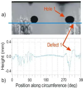
The internal surface of a plasma torch electrode, illustrated in Figure 3, was damaged by high-current electrical arcs. A raised point, for example, ‘Defect1’ in Figure 4, is an arc initiator and in-duces instabilities. To better understand the physics of this phe-nomenon, we mapped the internal surface using a 50mm-long, 3mm-diameter probe. The map is shown in Figure 4.
In summary, LCI using a common-path configuration en-ables highly-accurate, micron-scale measurements in indus-trial plants. The availability of micro-optical components
facil-Figure 4.(a) Shown is an unfolded 3D image of the internal surface of the electrode. Depth is encoded as black (deeper) to bright values. The two holes visible in the image are aligned 180 from each other. (b) Height variations of the surface along one full circle are indicated.
itates the assembly of submillimeter-sized probes. Integrating these technologies makes it easier to geometrically character-ize small industrial parts and surfaces that would otherwise be inaccessible.
Author Information
Marc Dufour, Guy Lamouche, Bruno Gauthier, Christian Padioleau and Jean-Pierre Monchalin Industrial Material Institute
National Research Council Canada Quebec, Canada
References
1. J. G. Fujimoto et al., High resolution in vivo intra-arterial imaging with optical
coher-ence tomography, Heart 82 (2), pp. 128–33, 1999.
2. P. A. Flournoy, Interferometric optical phase discrimination apparatus, US Patent 3319515, 1967.
3. M. L. Dufour et al., Surface inspection of hard to reach industrial parts using
low-coherence interferometry, Proc. SPIE 6343, p. 63431Z, 2006.
4. http://www.novacam.com
c