Acute Respiratory Distress Syndrome and Risk of
AKI among Critically Ill Patients
Michael Darmon, Christophe Clec’h, Christophe Adrie, Laurent Argaud, Bernard Allaouchiche, Elie Azoulay,
Lila Bouadma, Maı¨te´ Garrouste-Orgeas, Hakim Haouache, Carole Schwebel, Dany Goldgran-Toledano, Hatem Khallel, Anne-Sylvie Dumenil, Samir Jamali, Bertrand Souweine, Fabrice Zeni, Yves Cohen, and Jean-Franc¸ois Timsit
Abstract
Background and objectives Increasing experimental evidence suggests that acute respiratory distress syndrome (ARDS) may promote AKI. The primary objective of this study was to assess ARDS as a risk factor for AKI in critically ill patients.
Design, setting, participants, & measurements This was an observational study on a prospective database fed by 18 intensive care units (ICUs). Patients with ICU stays.24 hours were enrolled over a 14-year period. ARDS was defined using the Berlin criteria and AKI was defined using the Risk, Injury, Failure, Loss of kidney function, and End-stage kidney disease criteria. Patients with AKI before ARDS onset were excluded.
Results This study enrolled 8029 patients, including 1879 patients with ARDS. AKI occurred in 31.3% of patients and was more common in patients with ARDS (44.3% versus 27.4% in patients without ARDS; P,0.001). After adjustment for confounders, both mechanical ventilation without ARDS (odds ratio [OR], 4.34; 95% confidence interval [95% CI], 3.71 to 5.10) and ARDS (OR, 11.01; 95% CI, 6.83 to 17.73) were in-dependently associated with AKI. Hospital mortality was 14.2% (n=1140) and was higher in patients with ARDS (27.9% versus 10.0% in patients without ARDS; P,0.001) and in patients with AKI (27.6% versus 8.1% in those without AKI; P,0.001). AKI was associated with higher mortality in patients with ARDS (42.3% versus 20.2%; P,0.001).
Conclusions ARDS was independently associated with AKI. This study suggests that ARDS should be considered as a risk factor for AKI in critically ill patients.
Clin J Am Soc Nephrol 9: 1347–1353, 2014. doi: 10.2215/CJN.08300813
Introduction
AKI is common in critically ill patients and remains associated with poor outcomes (1–3). The main known risk factors for AKI in critically ill patients are absolute or relative hypovolemia, nephrotoxic drug exposure, sepsis, and comorbidities (4–8).
An increasing body of evidence points to deleteri-ous interactions between kidney and lung dysfunc-tions (9). Experimental studies suggest that AKI may increase the risk of lung injury, chiefly via the activa-tion of proinflammatory and proapoptotic pathways because of renal ischemia/reperfusion (9–13). Several lines of evidence suggest that mechanical ventilation and acute respiratory distress syndrome (ARDS) may have adverse effects on kidney function via three main mechanisms. First, positive-pressure ventilation may reduce cardiac output and increase central ve-nous pressure, thereby diminishing renal bloodflow, free water clearance, or the GFR (14–17). In addition, changes in arterial blood O2 or CO2 may influence renal vascular resistance, renal perfusion, or diuresis (18–22). Finally, emerging data suggest that ventila-tor-induced lung injury may not only affect the lung,
but may also lead to further systemic inflammation via the release of inflammatory cytokines (23–26).
Few studies have specifically addressed the associ-ation between respiratory failure and AKI (27–32). In addition, most of these studies were performed in specific patient populations and failed to adequately address the effect of ARDS on renal function (27,29– 32).
The primary objective of this study was to assess the influence of ARDS on subsequent AKI in unselected patients in the intensive care unit (ICU).
Materials and Methods
Study Design and Data Source
We conducted an observational study on a pro-spective multicenter database (OutcomeRea; http:// www.outcomerea.org) to assess influence of refrac-tory hypoxemia on subsequent AKI. The database, fed by 18 French ICUs, collects prospective data on daily disease severity, iatrogenic events, and nosoco-mial infections. Each year, each ICU includes a ran-dom sample of at least 50 patients who have ICU
Due to the number of contributing authors, the affiliations are provided in the Supplemental Material. Correspondence: Dr. Michael Darmon, Medical-Surgical Intensive Care Unit, Saint Etienne University Hospital, Avenue Albert Raimond, 42270 Saint Priest in Jarez, France. Email: michael. darmon@chu-st-etienne.fr
stays.24 hours. Each ICU can choose to obtain patients’ samples by taking either consecutive admissions to se-lected ICU beds throughout the year or consecutive admis-sions to all ICU beds for 1 month.
Study Population and Definitions
This study was approved by the institutional review board of the Clermont Ferrand University Hospital, which waived the need for informed consent in compliance with French law on database studies. This study was conducted in accordance with the Declaration of Helsinki.
We included consecutive patients aged.18 years who were entered into the database between January 1997 and April 2011. Patients with preexisting chronic kidney failure (defined as an eGFR,60 ml/min per 1.73 m2), with pre-renal dysfunction (transient AKI) as the main mechanism of AKI, with AKI predating ARDS, treatment-limitation decisions, left ventricular dysfunction, or ICU stays ,24 hours (and were thus unlikely to develop AKI after ARDS onset) were excluded.
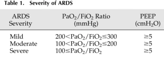
AKI was defined according to the Risk, Injury, Failure, Loss of kidney function, and End-stage kidney disease (RIFLE) criteria (33) and ARDS was defined as a PaO2/ FiO2ratio,300 mmHg in the absence of cardiogenic pul-monary edema (Table 1) (34). Because the 6- and 12-hour urine outputs were not recorded in the database, AKI def-inition and maximum renal severity were based upon changes in serum creatinine.
Baseline creatinine values were assessed using the four-variable Modification of Diet in Renal Disease (MDRD) equation. As recommended by the Acute Dialysis Quality Initiative Group, a normal eGFR of 75 ml/min per 1.73 m2 before ICU admission was assumed (35).
CKD was either defined according to the Acute Physi-ology and Chronic Health Evaluation II definition or was identified through a specific code according to data ex-tracted from the medical record. Transient AKI and left ventricular dysfunction were extracted from medical re-cords and identified through a specific code in the database. Data Collection
Data were collected daily by senior physicians and/or specifically trained study monitors in the participating ICUs and all of the collected data were extracted from medical records. For each patient, the investigators entered the data into a computer case-report form using data-capture software (RHEA; OutcomeRea) and imported all records into the OutcomeRea database. All codes and
definitions were established before study initiation. The data quality checking procedure was described elsewhere (36). The following information was recorded: age, sex, admission category (medical, scheduled surgery, or un-scheduled surgery), and origin (home, ward, or emergency department). Severity of illness was evaluated on thefirst ICU day using the Simplified Acute Physiology Score II (SAPS II), the Sequential Organ Failure Assessment (SOFA) score, and the Logistic Organ Dysfunction score (37–39). Knaus scale definitions were used to record pre-existing chronic organ failures including respiratory, car-diac, hepatic, renal, and immune system failures (40). Finally, the McCabe scoring system was assessed to obtain comparisons regarding the importance of host factors on the basis of the severity of the underlying disease, ranging from 1 (no fatal underlying disease) to 3 (rapidly fatal un-derlying disease) (41).
Quality of the Database
For most of the study variables, the data-capture soft-ware immediately ran an automatic check for internal consistency, generating queries that were sent to the ICUs for resolution before incorporation of the new data into the database. In each participating ICU, data quality was checked by having a senior physician from another par-ticipating ICU review a 2% random sample of the study data every other year. A 1-day data-capture training course held once a year was open to all OutcomeRea investigators and study monitors. All qualitative variables used in the analyses hadk coefficients.0.8 and all variables had inter-rater coefficients in the 0.67–1 range, indicating good to excellent reproducibility.
Statistical Analyses
Categorical variables are presented as n (%) and contin-uous variables are medians (interquartile ranges). Com-parisons of patients with and without AKI relied on chi-squared tests for categorical data and on the t test or Wilcoxon’s test, as appropriate, for continuous data. Risk factors associated with AKI were assessed using a mul-tivariate logistic regression model. The potential link be-tween ARDS and the subsequent development of AKI was assessed after adjusting for clinically pertinent con-founding factors and for factors significant in the univar-iate analysis. These factors were baseline comorbidities (diabetes mellitus, immunodeficiency, chronic cardiac and pulmonary dysfunction, and myeloma), sepsis, ad-ministration of nephrotoxic drugs (aminoglycosides, gly-copeptides, and/or iodinated contrast media), nonrenal organ failures (defined as the relevant specific SOFA com-ponent score.2), and age. Each of these variables was included in a stepwise logistic regression conditional model in which variables were selected according to their P value. Variables with a P value ,0.05 were maintained in thefinal model. Goodness of fit and discrimination of the model were determined using the Hosmer–Lemeshow statistic and the C statistic (area under the curve), respec-tively. Results are reported as adjusted odds ratios (ORs) with their 95% confidence intervals (95% CIs).
All P values are two tailed, and P values,0.05 are con-sidered significant. Statistical analyses were performed using the SAS 9.1 software package (SAS Institute, Cary, NC).
Table 1. Severity of ARDS
ARDS Severity
PaO2/FiO2Ratio (mmHg)
PEEP (cmH2O)
Mild 200,PaO2/FiO2#300 $5
Moderate 100,PaO2/FiO2#200 $5
Severe 100#PaO2/FiO2 $5
Severity is according to the Berlin definition (34). ARDS, acute respiratory distress syndrome; PEEP, positive end-expiratory pressure.
Results
Study Population
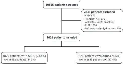
Of the 10,865 patients entered into the database during the study period, 2836 were excluded for the following reasons: history of CKD (n=672), transient AKI (n=130), AKI before ARDS onset (n=46), treatment-limitation deci-sions (n=1378), and evidence of left ventricular dysfunc-tion (n=610) (Figure 1). The remaining 8029 patients fulfilled the inclusion criteria and were enrolled in the study (Figure 1).
Table 2 reports the main patient characteristics at ICU admission. Overall, patients with AKI were older and were more often men; their acute illnesses were more se-vere as assessed on the basis of the SAPS II score or organ failures at ICU admission, and they had larger numbers of comorbidities.
ARDS and AKI
Overall, ARDS developed in 1879 patients (23.4%). AKI occurred after study inclusion in 832 patients with ARDS (44.3%) and in 1683 patients without ARDS (27.4%) (P,0.001). The median delay between ARDS and AKI was 2 days (interquartile range, 1–9).
We assessed AKI severity among patients with and without ARDS using the RIFLE criteria. Among patients without ARDS, AKI severity was R in 736 patients (43.7% of patients with AKI), I in 455 patients (27.0% of patients with AKI), and F in 492 patients (29.3% of patients with AKI). Among patients with ARDS, AKI severity was R in 171 patients (20.5% of patients with AKI), I in 276 patients (33.2% of patients with AKI), and F in 385 patients (46.3% of patients with AKI) (P,0.001).
Of the patients with ARDS, the severity of ARDS according to the Berlin classification was mild in 975 patients (51.9%), moderate in 379 patients (20.2%), and severe in 525 patients (27.9%).
Overall hospital mortality was 14.2% (n=1140) and was higher in patients with ARDS (27.9% versus 10.0% in pa-tients without ARDS; P,0.001) and in papa-tients with AKI (27.6% versus 8.1% in patients without AKI; P,0.001).
Among the patients with ARDS, those who subsequently developed AKI had a higher hospital mortality rate than those without AKI (42.3% versus 20.2%; P,0.001). Changes in AKI and ARDS rates during the study period are reported in the Supplemental Appendix.
Risk Factors for AKI
After adjustment for confounders, both mechanical ventilation without ARDS (OR, 4.34; 95% CI, 3.71 to 5.10) and ARDS (OR, 11.01; 95% CI, 6.83 to 17.73) were independently associated with AKI (Table 3). Other factors independently associated with AKI were shock, myeloma, age, diabetes mellitus, immunodeficiency, chronic liver or cardiac disease, and multiple myeloma. The model was well calibrated and the C statistic was 0.77. Figure 2 re-ports the cumulative incidence of AKI in patients with and without ARDS. Finally, a sensitivity analysis was per-formed to assess the influence of ARDS duration on AKI incidence before and after adjustment (Supplemental Ap-pendix).
The incidences of AKI before adjustment in patients with mild, moderate, and severe ARDS were 40%, 75.7%, and 29.5%, respectively. After adjustment for confounders, mild ARDS (OR, 2.42; 95% CI, 1.52 to 3.83), moderate ARDS (OR, 2.22; 95% CI, 1.38 to 3.55), and severe ARDS (OR, 2.58; 95% CI, 1.57 to 4.25) were significantly associated with AKI compared with patients without ARDS (Hosmer– Lemeshow goodness offit P=0.30; C statistic, 0.74).
Discussion
We found a significant independent association between ARDS and subsequent AKI. Although several studies point to physiologic mechanisms responsible for a deleterious effect of ARDS on other organs, little information was available on the clinical effect of ARDS on kidney function. Our results support the addition of ARDS to the list of risk factors for AKI in critically ill patients.
The growing evidence pointing to deleterious inter-actions between kidney and lung dysfunctions suggests a
Figure 1. | Flow chart of patients admitted during the study period. ARDS, acute respiratory distress syndrome; FLST, decision to forgo life-sustaining therapies.
partial explanation for the natural history of multiple organ dysfunctions in critically ill patients (9). In experimental studies in animals or healthy volunteers, acute lung injury adversely affected kidney function. The three main under-lying mechanisms were positive-pressure ventilation, hyp-oxemia, and systemic inflammation. Positive-pressure ventilation may modify the cardiac preload and has been
associated with systemic hemodynamic changes leading to decreases in the GFR, renal blood flow, and free water clearance (16,17,42). Moreover, activation of the sympa-thetic and renin-angiotensin systems, together with suppres-sion of atrial natriuretic peptide release, observed during positive pressure ventilation further decreases glomerular perfusion, GFR values, urine output, and sodium excretion
Table 2. Patient characteristics
Variable AKI (n=2515) No AKI (n=5514) P Value
Age, yr 65.9616 55.1618.4 ,0.001
Men 1472 (58.5) 3431 (62.2) 0.002
SAPS II score 50.1619.8 33.6616.5 ,0.001
Transfer from ward 1214 (48.3) 2390 (43.3) ,0.001
McCabe score 1 1507 (59.9) 3928 (71.2) ,0.001 2 826 (32.9) 1314 (23.8) 3 182 (7.2) 272 (4.9) Admission category Medical 1747 (69.5) 3885 (70.5) ,0.001 Scheduled surgery 471 (18.7) 769 (13.9) Unscheduled surgery 297 (11.8) 860 (15.6) Chronic comorbidities Heart disease 364 (14.5) 393 (7.16) ,0.001 Respiratory disease 313 (12.5) 822 (14.9) 0.003 Liver disease 170 (6.8) 283 (5.1) 0.003 Immunodeficiency 416 (16.5) 669 (12.1) ,0.001 Diabetes mellitus 401 (15.9) 519 (9.4) ,0.001 Myeloma 30 (1.2) 20 (0.4) ,0.001 ARDS 832 (33.1) 1047 (19) ,0.001 Hemodynamic failure 1127 (44.8) 1576 (28.6) ,0.001 Hepatic failure 848 (33.7) 1102 (20) ,0.001 Sepsis 1265 (50.3) 2841 (51.5) 0.3 Nephrotoxic drugs 1316 (52.3) 3108 (56.4) 0.001 ICU mortality 543 (21.6) 291 (5.3) ,0.001 Hospital mortality 693 (27.6) 448 (8.1) ,0.001
Data are reported as the mean6SD or n (%) unless otherwise specified. SAPS II, Simplified Acute Physiology Score version II (which can range from 0 to 155); ICU, intensive care unit.
Table 3. Factors independently associated with AKI after adjustment for confounders (stepwise logistic regression)
Variable OR (95% CI) P Value
Respiratory statusa No MV (reference)
MV, no ARDS 4.34 (3.71 to 5.10) ,0.001
ARDS 11.01 (6.83 to 17.73) ,0.001
Age (per yr) 1.04 (1.03 to 1.04) ,0.001
Chronic cardiac dysfunctionb 1.54 (1.30 to 1.82) ,0.001
Chronic liver diseaseb 1.41 (1.14 to 1.76) ,0.001
Immunodeficiencyb
1.68 (1.45 to 1.76) 0.002
Shock 4.23 (3.38 to 5.29) ,0.001
Diabetes mellitus 1.62 (1.38 to 1.90) ,0.001
Multiple myeloma 1.91 (1.01 to 3.61) ,0.001
Hosmer–Lemeshow statistics were as follows: k2=11.8; P=0.16; C statistic: 0.77. OR, odds ratio; 95% CI, 95% confidence interval; MV, mechanical ventilation.
aRespiratory status was inserted as the dummy variable.
bUnderlying comorbidities (chronic liver disease, chronic cardiac dysfunction, or immunodeficiency) were defined according to the Knaus definition (40).
(43). Moreover, hypoxemia and hypercapnia were previously shown to modify renal vascular resistances (18,19,21) and to increase diuresis (20,44,45). Interestingly, similar effects were reported in patients with chronic obstructive pulmonary dis-ease (19,21), as well as in renal transplant recipients (18) and critically ill patients with refractory hypoxemia (22). None of these studies, however, evaluated the long-term consequences of these physiologic alterations. Finally, systemic inflammation and biotrauma have been implicated in systemic organ dys-function during ARDS. Thus, several lines of evidence indicate that biotrauma induced by mechanical ventilation not only affects the lung, but also leads to further systemic inflamma-tion and organ dysfuncinflamma-tion via the release of inflammatory cytokines (23–26). Thus, in studies evaluating protective ven-tilation, higher tidal volumes were associated with not only higher levels of TNF-a, IL-1b, IL-6, and IL-8, but also with higher rates of AKI or higher numbers of days with AKI (23–26). Despite this large body of evidence suggesting an in-teraction between respiratory failure and AKI, few studies have evaluated the effect of respiratory failure on renal function by clearly assessing the time of mechanical ventilation initiation relative to the onset of AKI (27–32). A recent meta-analysis of these studies suggested that both ARDS and mechanical ventilation were associated with a 3-fold increase in the risk of AKI (46). Most of the studies included in this analysis were observational studies, how-ever, and focused on specific populations such as trauma patients (27), lung transplant recipients (29), cancer pa-tients (30), or papa-tients with hepatic failure (32). Thus, the general applicability of their findings is unclear. Further-more, these studies were not specifically designed to as-sess the influence of respiratory failure on AKI, and they did not separate the effect of mechanical ventilation from
that of ARDS (46). In this study, both ARDS and mechan-ical ventilation were found to be independently associated with usual and well described risk factors for AKI, namely, shock, diabetes mellitus, age, underlying cardiac or he-patic dysfunction, and myeloma. Our study provides valid data supporting the addition of both ARDS and mechan-ical ventilation to the list of risk factors for AKI in unse-lected ICU patients.
Our study has several limitations. First, the observational design and lack of information on ventilator settings preclude conclusions about factors that may have pro-moted the development of AKI. Additional studies are needed to further assess the influence of ventilator settings, blood O2and CO2alterations, and systemic inflammation on the occurrence of AKI. In addition, data regarding the mechanism of AKI, fluid balance management across groups, or diuretics use in patients with ARDS were un-available. Whether some differences regardingfluid man-agement or type of fluid used in each of the studied groups might have accounted for the observed difference regarding AKI rate remains to be evaluated. Furthermore, although we adjusted for the main nephrotoxic agents (e.g., contrast media and antimicrobial agents), we must acknowledge the lack of information regarding other nephrotoxic agents, such as angiotensin-converting en-zyme inhibitors or angiotensin receptor antagonists. In this study, and in accordance with current guidelines, baseline creatinine was back calculated from the MDRD assuming a 75 ml/kg per 1.73 m2GFR (35,47). Although this imputation was validated in previous works (48), it may overestimate the incidence of AKI by nearly 10% and may misclassify severity of AKI in up to 30% (49). Finally, the observational study design only allows us to conclude
that ARDS was independently associated with AKI. No conclusions can be drawn about the causal nature of this association, despite the numerous experimental studies suggesting causality. Further studies are needed to ad-dress this issue more specifically and to evaluate the in-fluence of ventilator settings on renal function.
In conclusion, the independent association demonstrated in our study between ARDS and subsequent AKI indicates that ARDS should be added to the list of risk factors for AKI in critically ill patients. Although no conclusions about causality can be drawn from our data, the strong exper-imental evidence suggesting causality supports the need for further studies in this field aimed at confirming our findings and evaluating the mechanisms underlying the ARDS-AKI association, most notably ventilator settings, in unselected ICU patients.
Acknowledgments
The authors thank A. Wolfe for help with this article.
This study was performed on the behalf of the OutcomeRea study group.
Disclosures None.
References
1. Chertow GM, Burdick E, Honour M, Bonventre JV, Bates DW: Acute kidney injury, mortality, length of stay, and costs in hospitalized patients. J Am Soc Nephrol 16: 3365–3370, 2005
2. Metnitz PGH, Krenn CG, Steltzer H, Lang T, Ploder J, Lenz K, Le Gall J-R, Druml W: Effect of acute renal failure requiring renal replacement therapy on outcome in critically ill patients. Crit Care Med 30: 2051–2058, 2002
3. Bagshaw SM: Short- and long-term survival after acute kidney injury. Nephrol Dial Transplant 23: 2126–2128, 2008 4. Bagshaw SM, Laupland KB, Doig CJ, Mortis G, Fick GH,
Mucenski M, Godinez-Luna T, Svenson LW, Rosenal T: Prognosis for long-term survival and renal recovery in critically ill patients with severe acute renal failure: A population-based study. Crit Care 9: R700–R709, 2005
5. Uchino S, Kellum JA, Bellomo R, Doig GS, Morimatsu H, Morgera S, Schetz M, Tan I, Bouman C, Macedo E, Gibney N, Tolwani A, Ronco C; Beginning and Ending Supportive Therapy for the Kidney (BEST Kidney) Investigators: Acute renal failure in critically ill patients: A multinational, multicenter study. JAMA 294: 813–818, 2005
6. Nisula S, Kaukonen K-M, Vaara ST, Korhonen A-M, Poukkanen M, Karlsson S, Haapio M, Inkinen O, Parviainen I, Suojaranta-Ylinen R, Laurila JJ, Tenhunen J, Reinikainen M, Ala-Kokko T, Ruokonen E, Kuitunen A, Pettila¨ V; FINNAKI Study Group: In-cidence, risk factors and 90-day mortality of patients with acute kidney injury in Finnish intensive care units: The FINNAKI study. Intensive Care Med 39: 420–428, 2013
7. Hoste EAJ, Doom S, De Waele J, Delrue LJ, Defreyne L, Benoit DD, Decruyenaere J: Epidemiology of contrast-associated acute kidney injury in ICU patients: A retrospective cohort analysis. Intensive Care Med 37: 1921–1931, 2011
8. Perner A, Haase N, Guttormsen AB, Tenhunen J, Klemenzson G, A˚neman A, Madsen KR, Møller MH, Elkjær JM, Poulsen LM, Bendtsen A, Winding R, Steensen M, Berezowicz P, Søe-Jensen P, Bestle M, Strand K, Wiis J, White JO, Thornberg KJ, Quist L, Nielsen J, Andersen LH, Holst LB, Thormar K, Kjældgaard A-L, Fabritius ML, Mondrup F, Pott FC, Møller TP, Winkel P, Wetterslev J; 6S Trial Group; Scandinavian Critical Care Trials Group: Hy-droxyethyl starch 130/0.42 versus Ringer’s acetate in severe sepsis. N Engl J Med 367: 124–134, 2012
9. Li X, Hassoun HT, Santora R, Rabb H: Organ crosstalk: The role of the kidney. Curr Opin Crit Care 15: 481–487, 2009
10. Kramer AA, Postler G, Salhab KF, Mendez C, Carey LC, Rabb H: Renal ischemia/reperfusion leads to macrophage-mediated in-crease in pulmonary vascular permeability. Kidney Int 55: 2362– 2367, 1999
11. Deng J, Hu X, Yuen PST, Star RA: Alpha-melanocyte-stimulating hormone inhibits lung injury after renal ischemia/reperfusion. Am J Respir Crit Care Med 169: 749–756, 2004
12. Hassoun HT, Lie ML, Grigoryev DN, Liu M, Tuder RM, Rabb H: Kidney ischemia-reperfusion injury induces caspase-dependent pulmonary apoptosis. Am J Physiol Renal Physiol 297: F125– F137, 2009
13. Hassoun HT, Grigoryev DN, Lie ML, Liu M, Cheadle C, Tuder RM, Rabb H: Ischemic acute kidney injury induces a distant or-gan functional and genomic response distinguishable from bi-lateral nephrectomy. Am J Physiol Renal Physiol 293: F30–F40, 2007
14. Farge D, De la Coussaye JE, Beloucif S, Fratacci MD, Payen DM: Interactions between hemodynamic and hormonal mod-ifications during PEEP-induced antidiuresis and antinatriuresis. Chest 107: 1095–1100, 1995
15. Jacob LP, Chazalet JJ, Payen DM, Villiers SM, Boudaoud S, Teillac P, Pruna AS, Idatte JM, Eurin BG: Renal hemodynamic and functional effect of PEEP ventilation in human renal trans-plantations. Am J Respir Crit Care Med 152: 103–107, 1995 16. Murdaugh HV Jr, Sieker HO, Manfredi F: Effect of altered
in-trathoracic pressure on renal hemodynamics, electrolyte excre-tion and water clearance. J Clin Invest 38: 834–842, 1959 17. Hall SV, Johnson EE, Hedley-Whyte J: Renal hemodynamics and
function with continuous positive-pressure ventilation in dogs. Anesthesiology 41: 452–461, 1974
18. Sharkey RA, Mulloy EM, Long M, O’Neill SJ: The effect of con-tinuous positive airway pressure (CPAP) on renal vascular re-sistance: The influence of renal denervation. Crit Care 3: 33–37, 1999
19. Sharkey RA, Mulloy EM, O’Neill SJ: The acute effects of oxygen and carbon dioxide on renal vascular resistance in patients with an acute exacerbation of COPD. Chest 115: 1588–1592, 1999 20. Hildebrandt W, Ottenbacher A, Schuster M, Swenson ER, Ba¨rtsch
P: Diuretic effect of hypoxia, hypocapnia, and hyperpnea in humans: Relation to hormones and O(2) chemosensitivity. J Appl Physiol (1985) 88: 599–610, 2000
21. Baudouin SV, Bott J, Ward A, Deane C, Moxham J: Short term effect of oxygen on renal haemodynamics in patients with hy-poxaemic chronic obstructive airways disease. Thorax 47: 550– 554, 1992
22. Darmon M, Schortgen F, Leon R, Moutereau S, Mayaux J, Di Marco F, Devaquet J, Brun-Buisson C, Brochard L: Impact of mild hypoxemia on renal function and renal resistive index during mechanical ventilation. Intensive Care Med 35: 1031–1038, 2009
23. Ranieri VM, Giunta F, Suter PM, Slutsky AS: Mechanical venti-lation as a mediator of multisystem organ failure in acute re-spiratory distress syndrome. JAMA 284: 43–44, 2000 24. Ranieri VM, Suter PM, Tortorella C, De Tullio R, Dayer JM,
Brienza A, Bruno F, Slutsky AS: Effect of mechanical ventilation on inflammatory mediators in patients with acute respiratory distress syndrome: A randomized controlled trial. JAMA 282: 54– 61, 1999
25. Liu KD, Glidden DV, Eisner MD, Parsons PE, Ware LB, Wheeler A, Korpak A, Thompson BT, Chertow GM, Matthay MA; National Heart, Lung, and Blood Institute ARDS Network Clinical Trials Group: Predictive and pathogenetic value of plasma biomarkers for acute kidney injury in patients with acute lung injury. Crit Care Med 35: 2755–2761, 2007
26. The Acute Respiratory Distress Syndrome Network: Ventilation with lower tidal volumes as compared with traditional tidal volumes for acute lung injury and the acute respiratory distress syndrome. N Engl J Med 342: 1301–1308, 2000
27. Vivino G, Antonelli M, Moro ML, Cottini F, Conti G, Bufi M, Cannata F, Gasparetto A: Risk factors for acute renal failure in trauma patients. Intensive Care Med 24: 808–814, 1998 28. Schortgen F, Lacherade JC, Bruneel F, Cattaneo I, Hemery F,
Lemaire F, Brochard L: Effects of hydroxyethylstarch and gelatin on renal function in severe sepsis: A multicentre randomised study. Lancet 357: 911–916, 2001
29. Rocha PN, Rocha AT, Palmer SM, Davis RD, Smith SR: Acute renal failure after lung transplantation: Incidence, predictors and impact on perioperative morbidity and mortality. Am J Transplant 5: 1469–1476, 2005
30. Lahoti A, Kantarjian H, Salahudeen AK, Ravandi F, Cortes JE, Faderl S, O’Brien S, Wierda W, Mattiuzzi GN: Predictors and outcome of acute kidney injury in patients with acute myelogenous leukemia or high-risk myelodysplastic syndrome. Cancer 116: 4063–4068, 2010 31. Jung JY, Park BH, Hong S-B, Koh Y, Suh GY, Jeon K, Koh SO, Kim JY, Cho JH, Choi HS, Park YB, Kim HC, Kim Y-S, Lim CY, Park MS: Acute kidney injury in critically ill patients with pandemic in-fluenza A pneumonia 2009 in Korea: A multicenter study. J Crit Care 26: 577–585, 2011
32. O’Riordan A, Brummell Z, Sizer E, Auzinger G, Heaton N, O’Grady JG, Bernal W, Hendry BM, Wendon JA: Acute kidney injury in pa-tients admitted to a liver intensive therapy unit with paracetamol-induced hepatotoxicity. Nephrol Dial Transplant 26: 3501–3508, 2011 33. Bellomo R, Ronco C, Kellum JA, Mehta RL, Palevsky P; Acute
Dialysis Quality Initiative workgroup: Acute renal failure -definition, outcome measures, animal models, fluid therapy and information technology needs: The Second International Con-sensus Conference of the Acute Dialysis Quality Initiative (ADQI) Group. Crit Care 8: R204–R212, 2004
34. Ranieri VM, Rubenfeld GD, Thompson BT, Ferguson ND, Caldwell E, Fan E, Camporota L, Slutsky AS; ARDS Definition Task Force: Acute respiratory distress syndrome: The Berlin Definition. JAMA 307: 2526–2533, 2012
35. Khwaja A: KDIGO Clinical Practice Guidelines for Acute Kidney Injury. Nephron Clin Pract 120: 179–184, 2012
36. Laupland KB, Zahar J-R, Adrie C, Minet C, Ve´sin A, Goldgran-Toledano D, Azoulay E, Garrouste-Orgeas M, Cohen Y, Schwebel C, Jamali S, Darmon M, Dumenil A-S, Kallel H, Souweine B, Timsit J-F: Severe hypothermia increases the risk for intensive care unit-acquired infection. Clin Infect Dis 54: 1064–1070, 2012 37. Timsit J-F, Fosse J-P, Troche´ G, De Lassence A, Alberti C,
Garrouste-Orgeas M, Bornstain C, Adrie C, Cheval C, Chevret S; OUTCOMEREA Study Group, France: Calibration and discrimi-nation by daily Logistic Organ Dysfunction scoring compara-tively with daily Sequential Organ Failure Assessment scoring for predicting hospital mortality in critically ill patients. Crit Care Med 30: 2003–2013, 2002
38. Le Gall JR, Lemeshow S, Saulnier F: A new Simplified Acute Physiology Score (SAPS II) based on a European/North American multicenter study. JAMA 270: 2957–2963, 1993
39. Vincent JL, Moreno R, Takala J, Willatts S, De Mendonc¸a A, Bruining H, Reinhart CK, Suter PM, Thijs LG; Working Group on Sepsis-Related Problems of the European Society of Intensive Care Medicine: The SOFA (Sepsis-related Organ Failure Assess-ment) score to describe organ dysfunction/failure. Intensive Care Med 22: 707–710, 1996
40. Knaus WA, Draper EA, Wagner DP, Zimmerman JE: APACHE II: A severity of disease classification system. Crit Care Med 13: 818– 829, 1985
41. McCabe WR, Jackson GG: Gram-negative bacteremia. I. Etiol-ogy and ecolEtiol-ogy. Arch Intern Med 110: 847–855, 1962 42. Kuiper JW, Groeneveld ABJ, Slutsky AS, Plo¨tz FB: Mechanical
ventilation and acute renal failure. Crit Care Med 33: 1408– 1415, 2005
43. Annat G, Viale JP, Bui Xuan B, Hadj Aissa O, Benzoni D, Vincent M, Gharib C, Motin J: Effect of PEEP ventilation on renal function, plasma renin, aldosterone, neurophysins and urinary ADH, and prostaglandins. Anesthesiology 58: 136–141, 1983
44. Ho¨hne C, Boemke W, Schleyer N, Francis RC, Krebs MO, Kaczmarczyk G: Low sodium intake does not impair renal compensation of hypoxia-induced respiratory alkalosis. J Appl Physiol (1985) 92: 2097–2104, 2002
45. Ho¨hne C, Krebs MO, Boemke W, Arntz E, Kaczmarczyk G: Evi-dence that the renin decrease during hypoxia is adenosine me-diated in conscious dogs. J Appl Physiol (1985) 90: 1842–1848, 2001
46. van den Akker JP, Egal M, Groeneveld JA: Invasive mechanical ventilation as a risk factor for acute kidney injury in the critically ill: A systematic review and meta-analysis. Crit Care 17: R98, 2013
47. Fliser D, Laville M, Covic A, Fouque D, Vanholder R, Juillard L, Van Biesen W; Ad-hoc working group of ERBP: A European Renal Best Practice (ERBP) position statement on the Kidney Disease Improving Global Outcomes (KDIGO) clinical practice guide-lines on acute kidney injury: Part 1: Definitions, conservative management and contrast-induced nephropathy. Nephrol Dial Transplant 27: 4263–4272, 2012
48. Za´vada J, Hoste E, Cartin-Ceba R, Calzavacca P, Gajic O, Clermont G, Bellomo R, Kellum JA; AKI6 investigators: A com-parison of three methods to estimate baseline creatinine for RIFLE classification. Nephrol Dial Transplant 25: 3911–3918, 2010
49. Siew ED, Matheny ME, Ikizler TA, Lewis JB, Miller RA, Waitman LR, Go AS, Parikh CR, Peterson JF: Commonly used surrogates for baseline renal function affect the classification and prognosis of acute kidney injury. Kidney Int 77: 536–542, 2010
Received:August 7, 2013 Accepted: April 2, 2014
Published online ahead of print. Publication date available at www. cjasn.org.
This article contains supplemental material online at http://cjasn. asnjournals.org/lookup/suppl/doi:10.2215/CJN.08300813/-/ DCSupplemental.