Plasticity in Unimodal and Multimodal Brain Areas Re
flects Multisensory Changes
in Self-Face Identi
fication
Matthew A. J. Apps
1,2, Ana Tajadura-Jiménez
3, Marty Sereno
4, Olaf Blanke
5and Manos Tsakiris
21
Nuf
field Department of Clinical Neuroscience, John Radcliffe Hospital, University of Oxford, Oxford OX3 9DU, UK,
2Lab of
Action and Body, Department of Psychology, Royal Holloway, University of London, Egham TW20 0EX, UK,
3UCL Interaction
Center (UCLIC), University College London, London WC1E 6BT, UK,
4Department of Psychological Science, Birkbeck, University
of London, WC1H 0DS, UK and
5Center for Neuroprosthetics and Brain-Mind Institute, Ecole Polytechnique Fédérale de Lausanne,
SV 2805, Switzerland
M.A.J. Apps and A. Tajadura-Jiménez declare equal contribution.
Address correspondence to Matthew A. J. Apps. Email: [email protected]
Nothing provides as strong a sense of self as seeing one
’s face.
Nevertheless, it remains unknown how the brain processes the
sense of self during the multisensory experience of looking at one
’s
face in a mirror. Synchronized visuo-tactile stimulation on one
’s own
and another
’s face, an experience that is akin to looking in the
mirror but seeing another
’s face, causes the illusory experience of
ownership over the other person
’s face and changes in
self-recog-nition. Here, we investigate the neural correlates of this enfacement
illusion using fMRI. We examine activity in the human brain as
par-ticipants experience tactile stimulation delivered to their face, while
observing either temporally synchronous or asynchronous tactile
stimulation delivered to another
’s face on either a specularly
congru-ent or incongrucongru-ent location. Activity in the multisensory right
temporo-parietal junction, intraparietal sulcus, and the unimodal
inferior occipital gyrus showed an interaction between the
synchro-nicity and the congruency of the stimulation and varied with the
self-reported strength of the illusory experience, which was recorded
after each stimulation block. Our results highlight the important
inter-play between unimodal and multimodal information processing for
self-face recognition, and elucidate the neurobiological basis for the
plasticity required for identifying with our continuously changing
visual appearance.
Keywords: enfacement, face recognition, fMRI, multisensory,
self-recognition
Introduction
The ability to represent the visual properties of one
’s face as
distinct from others is a fundamental aspect of human
self-awareness. Recognizing one
’s face in a mirror is a key
behav-ioral marker of self-awareness (
Gallup 1970
), an ability
ex-pressed by a small selection of species, including humans
(
Gallup 1970
;
Anderson and Gallup Jr. 2011
). Neuroimaging
studies of self-face recognition suggest that representations of
one
’s own facial appearance may be stored in a specialized
network of areas, which is engaged when viewing images of
one
’s own face (
Kircher et al. 2001
;
Uddin et al. 2005
,
2006
,
2008
;
Platek et al. 2006
,
2008
;
Devue et al. 2007
;
Kaplan et al.
2008
;
Sugiura et al. 2008
;
Platek and Kemp 2009
;
Devue and
Bredart 2011
;
Ramasubbu et al. 2011
;
Apps et al. 2012
;
Ma and
Han 2012
). Of these areas, the inferior occipital gyrus (IOG),
the inferior temporal gyrus (ITG), and the temporo-parietal
junction (TPJ) respond only to images of one
’s current, but not
one
’s past face, suggesting they continuously update a visual
representation of one
’s facial appearance (
Apps et al. 2012
).
These studies have used static face stimuli to investigate
self-recognition. However, the way in which infants and
nonhu-man primates succeed in recognizing their specular image
suggests that processes other than mere visual perception are
engaged in self-identi
fication. The normal experience of
looking into one
’s face in the mirror is accompanied by a
con-tinuous integration of tactile and proprioceptive events
per-ceived on one
’s face and visual events perceived on the
mirror-re
flection. Such processes putatively underlie responses
on the classic
“rouge” task of mirror self-recognition. In this
task, the placement of a red spot on the face of infants and
some primates while they are looking into a mirror, results in
behaviors that indicate a detection of the spot on that location
of the body, which are evaluated by a goal-directed movement
towards the red spot (
Gallup 1970
;
Bertenthal and Fischer
1978
;
Suarez and Gallup 1981
;
Suddendorf et al. 2007
;
Ander-son and Gallup Jr. 2011
). Such updating of the representation
of one
’s visual appearance during multisensory stimulation
may also underlie the assimilation of changes, and provide a
sense of continuity of one
’s self over time (
Tsakiris 2010
;
Apps
et al. 2012
). Therefore, the static stimuli used in the vast
majority of self-face recognition studies seem to violate the
dynamic, multisensory conditions present when looking in a
mirror and thus lack sensitivity for identifying activity related
to mirror self-recognition during multisensory stimulation.
While research investigating self-face recognition has used
static stimuli, studies of body-ownership and bodily illusions
have used multisensory stimulation to investigate where and
how visuo-tactile stimulation is integrated in the brain.
Neuroi-maging studies highlight that the premotor cortex (PMC),
intra-parietal sulcus (IPS), cerebellum, and TPJ are activated when
visuo-tactile stimulation causes an illusory sense of ownership
over the whole, or parts, of a body (
Ehrsson et al. 2004
;
Ionta
et al. 2011
;
Petkova et al. 2011
). More recently it has been
suggested that representations of one
’s self change through
the integration of visual and tactile information when looking
in a mirror (
Tajadura-Jiménez et al. 2012b
). The matching
between felt and observed sensorimotor signals purportedly
leads to the formation, and the updating, of a mental
represen-tation of one
’s visual appearance. Seeing an unfamiliar face
being touched at the same time as one
’s own face, a situation
akin to looking in a mirror but seeing another person
’s face,
changes one
’s ability to self-recognize and creates the illusory
experience of looking at one
’s self in the mirror (
Tsakiris 2008
;
Sforza et al. 2010
;
Tajadura-Jimenez et al. 2012a
). This
“enface-ment
” illusion arises due to the congruency between felt and
© The Author 2013. Published by Oxford University Press. All rights reserved.Cerebral Cortex January 2015;25:46–55 doi:10.1093/cercor/bht199
seen sensory events and does not arise from asynchronous
visuo-tactile stimulation. However, little is understood about
the neuronal processes that underpin this type of visuo-tactile
integration and create a sense that I am looking at
“me.”
Plasticity in self-recognition may therefore occur through the
integration of visuo-tactile information in multimodal areas
which leads to a sense of ownership over one
’s face.
Under-standing the neurobiological mechanisms that underpin the
multisensory experience of looking at one
’s face in a mirror
may therefore be crucial for understanding the plasticity of
self-recognition. However, no previous study has investigated
activity in the brain during the multisensory driven process of
experiencing a face as
“me.”
Does the multisensory experience of mirror-self recognition
engage areas previously known for their role in integrating
multisensory information and creating a sense of ownership
over the body, or alternatively, does it recruit a distinct network
which is activated when recognizing static images of the
self-face? Here, we used block-design fMRI to examine brain activity
during the enfacement illusion, as a corollary of mirror
self-recognition. Participants observed movies of an unfamiliar face
receiving tactile stimulation to the face while receiving tactile
stimulation themselves from an air puff system. The visuo-tactile
stimulation could be synchronized or not and on either a
specu-larly congruent or incongruent location on the faces. After each
block, participants rated the extent to which they experienced
the illusion on a 7-point Likert scale. This design enabled us to
examine where in the brain activity varies with the extent to
which participants experience the face that they see being
touched in synchrony with their own face as
“self.” We predict
that activity in brain areas that have previously been implicated
in self-face recognition and multisensory bodily illusions will
fluctuate with the experience of enfacement.
Methods
Participants
Fifteen female right-handed paid-volunteers (mean age = 25.8 years, SD = 3.88) gave their informed consent to participate. Only partici-pants who experienced the enfacement illusion in a preliminary behav-ioral session were invited to take part in this experiment, as explained below. The study was approved by the Royal Holloway, Psychology Departmental Ethics Committees, and conformed to regulations set out in the Birkbeck-UCL Neuroimaging Centre (BUCNI) MRI Rules of Operation (http://bucni.psychol.ucl.ac.uk/index.shtml).
Apparatus and Materials
Two different female“models” (∼20 years old), who were unfamiliar to the participants, were recorded being touched with a tap of a cotton bud on the right cheek or on the right hand side of the chin, at a random frequency ranging from 0.33 to 0.76 Hz, while they main-tained a neutral facial expression. This allowed 4 40 s “induction” movies to be produced, which differed in the unfamiliar face displayed and the part of the face being touched. In a pilot experiment, the 2 models were rated on scales of trustworthiness and physical attractive-ness, along with 8 other faces, by 11 participants, who did not take part in the subsequent parts of the study. Previous research has found a bidirectional link between the physical attractiveness that participants attribute to another person’s face and the strength of the enfacement illu-sion felt for that face (Paladino et al. 2010; Sforza et al. 2010). In addition, trustworthy faces are more likely to be identified as looking like the self (Verosky and Todorov 2010). We therefore ensured that the faces were equally evaluated in terms of trustworthiness and physical at-tractiveness, to avoid potential influences of the seen face in the pattern of the enfacement illusion. The 2 faces were not significantly different
on either measure (trustworthiness: t10= 0.65, P > 0.53; physical attrac-tiveness: t10= 1.26, P > 0.23). In addition, the models viewed in the synchronous and asynchronous conditions were counterbalanced across participants. Tactile stimulation was delivered to the left cheek of participants through puffs of air. To deliver the stimulation we used the arrangement ofHuang and Sereno (2007). The system consisted of an air compressor in the scanner control room, which provided input to a solenoid valve (Numatics) that was controlled by TTL pulses from a data acquisition and control card (National Instruments USB-6800). Plastic air tubes from the valve were connected to a blockfixed in the magnet behind the head coil. From this base, a tube with aflexible nozzle (Loc-Line) that could be freely positioned was used to direct air puffs to the participant’s cheek. The input air pressure (30–40 psi) was adjusted so that a 100-ms air puff was delivered, which felt akin to the level of tactile sensation experienced by a touch delivered from the cotton bud, with a similar duration. The system delivers pure unconta-minated air with a temperature comfortable for the participant. In this study, we used air puffs to deliver tactile stimulation to participants while inside the MRI scanner. To ensure that participants experienced the illusory experience when using different methods of stimulation to the participant and the model in the video, we performed a pilot study on participants who previously had reported experiencing the illusion. The pilot study highlighted that we could evoke the enfacement illu-sion using this paradigm. Thus, as in a previous study (Mazzurega et al. 2011), we show that the enfacement illusion can be induced even when the stimulation applied to the 2 faces is not identical. The tactile stimulation the participants received was therefore akin to the touch being delivered on the faces in the movies described above.
Visual stimuli were projected onto a screen which participants viewed via a mirror positioned above their face. Participants made responses (see below) using their right hand on a 4-button MRI-compatible response box. Brain images were acquired with a Siemens 1.5 Tesla Avanto MRI scanner at Birkbeck-University College London Centre for Neuroimaging (BUCNI). Presentation software (Neurobeha-vioral Systems, Inc., USA) was used to deliver stimuli and record responses. NI Measurement and Automation Explorer (Version 5.0.0f1) provided access to the data acquisition and control card (National Instru-ments USB-6800). Behavioral and fMRI Data were analyzed in Matlab 2006a, SPSS 19, SPM8, and MRICRON.
Procedure
Pre-MRI Screening Session
Thirty-six female right-handed paid-volunteers were tested on the en-facement illusion paradigm in a separate behavioral session, which took place a few weeks before the MRI session. Participants were exposed to 2 repetitions of 3 different visuo-tactile stimulation con-ditions, each lasting for 40 s and with their order randomized: synchro-nous congruent, synchronous incongruent, and asynchronous congruent, which are described in the subsequent section. The subjec-tive experience of participants during each visuo-tactile stimulation condition was assessed with a statement (I felt I was looking at my face), for which participants rated their level of agreement using a 7-item Likert scale, ranging from“strongly agree” (+3) to “strongly dis-agree” (−3). This statement was adopted from previous studies on the enfacement illusion (Tajadura-Jimenez et al. 2012a). A keypad was used for this purpose. The 15 participants who experienced the illusion most strongly (i.e., they agreed more strongly with the statement) in the syn-chronous congruent condition in comparison to the other 2 conditions; mean synchronous congruent = 1.73, SD = 1.13; mean synchronous incongruent =−0.68, SD = 1.57; mean asynchronous congruent = −1.89, SD = 1.16) were invited back to participate in the MRI session.
At the end of this MRI screening session, participants were pre-sented with 2 additional synchronous congruent conditions and were required to indicate the onset of the enfacement illusion by pressing a key in the keypad. The IMS lasted for 40 s, independently of the partici-pant key press. The average onset of the illusion across participartici-pants was 13.31 ± 8.56 s (M ± SD). This task revealed both within and across participant variability in the onset of the illusion. The responses across participants were highly variable (M = 13.3 s, SD = ±8.56, range = 5.56– 32.65 s). The responses within participants were in comparison
relatively consistent, although there was still variability, with the average difference in the keypresses from trial 1 to trial 2 being 5.32 s (SD = ±3.40, range = 0.20–10.28 s).
Scanning Session
Fifteen participants lay supine in an MRI scanner with thefingers of the right hand positioned on the response box. The nozzle connected to the solenoid valve was placed over the left cheek of the participant’s face, through which the air puffs were delivered at a frequency ranging from 0.33 to 0.76 Hz during 40 s periods while they watched the movies of tactile stimulation being delivered to the other’s face.
Experimental Design
We used a 2 × 2 factorial block design. Thefirst factor was the specular congruency of the visuo-tactile stimulation. While receiving tactile
stimulation on the left check, participants viewed tactile stimulation being delivered to the other’s face to either a specularly congruent location (i.e., the right cheek of the other person) as if the participant was looking at a mirror, or at an incongruent location (i.e., the chin). This incognruent condition controlled for the possibility that simply the synchrony of a seen touch on another’s face and a felt touch on one’s own face, even when the seen touch and the felt touch are on different locations of the face, could drive activity in multisensory areas. In the congruent conditions, the touch stimulated the same portion of the cheek of the face in the movie and of the participant’s face. The second factor was the temporal synchronicity of the visuo-tactile stimulation. This could either be synchronous, where touch on the participant’s face and the face in the movie was temporally synchro-nized, or asynchronous, with a lag of 1 s separating the stimulation on the participants face and that on the other’s face in the movie (see Fig.1a). This created a design with 4 conditions, synchronous
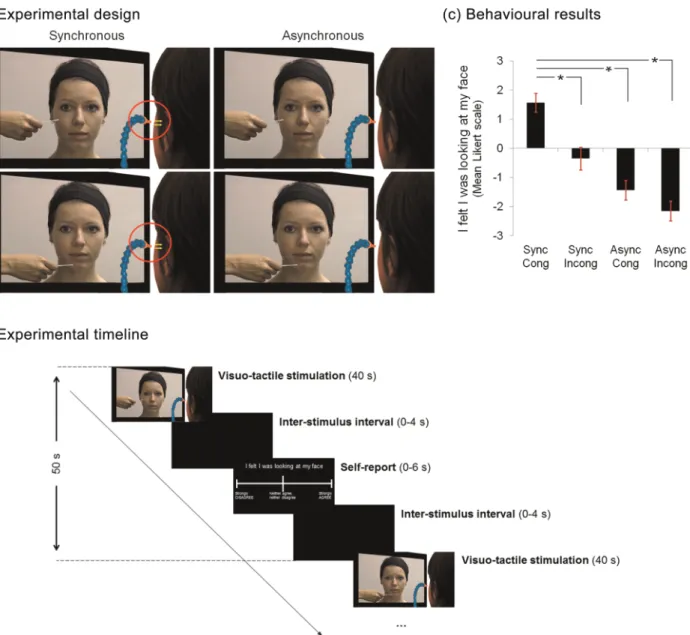
Figure 1. Experimental design, timeline, and behavioral results. (a) While in the scanner and in 40 s blocks, participants received tactile stimulation to their left cheek from puffs of air. The stimulation was akin to the cotton bud that was seen touching the face of another unfamiliar person in a movie which was played to the participant at the same time. The tactile stimulation on the 2 faces could be either synchronous or asynchronous and on either specularly congruent or incongruent locations. For the incongruent stimulation, participants observed the other person being touched on the chin. After each block of stimulation, participants rated the strength of the illusory experience on a 7-point Likert scale, ranging from“strongly agree” (+3) to “strongly disagree” (−3). (b) The block interval was 50 s, during which there was, a movie (40 s), followed by a blank screen presented for a variable interstimulus interval (0–4 s), followed by the question and Likert scale (maximum 6 s), followed by a blank screen presented for the remaining time to complete 50 s. (c) The mean Likert scale responses across participants for the 4 conditions are shown. As it can be seen, participants showed a stronger illusory experience in the synchronous, congruent condition, as predicted. Error bars depict standard error of the mean.
congruent (Sync-Cong), synchronous incongruent (Sync-Incong), asyn-chronous congruent (Async-Cong), and asynasyn-chronous incongruent (Async-Incong). The incongruent and the asynchronous conditions served as control conditions in which no enfacement illusion was ex-pected (Tsakiris 2008;Sforza et al. 2010;Tajadura-Jimenez et al. 2012a).
As in the behavioral session, after each block, participants were asked to report their level of agreement with the statement“I felt I was looking at my face,” using a 7-item Likert scale displayed on the screen. The participants used the response box for this task. A maximum of 6 s was allowed to answer the statement, which was suf fi-cient for all participants. The onset of the question was jittered ran-domly and uniformly over the 4 s period after the offset of the movies. Thus, the block interval was 50 s, during which there was, a movie (40 s), followed by a blank screen presented for a variable duration (0–4 s), followed by the question and Likert scale (maximum 6 s), and followed by a blank screen presented for the remaining time to complete 50 s (see Fig.1b). Participants used one button to move up the scale, one to move down, and a third to indicate that they had chosen their response. This question also afforded us the opportunity to analyze the data parametrically, with the responses to this question being used a predictor of activity during the corresponding movie. This was ben-eficial as it allowed us to examine the extent to which participants were experiencing the illusion in each block, rather than following the ap-proach used in previous designs (Ehrsson et al. 2004;Ionta et al. 2011;
Petkova et al. 2011), where“off-line” reports of the strength of the experience after the experiment are regressed against the BOLD response in different conditions. Thus, our design afforded us the op-portunity to examine activity that related to the experience of the illu-sion on-line. To analyze the behavioral responses, we performed pairwise comparisons between conditions and corrected for multiple comparisons using a Bonferroni correction (P < 0.05).
There were 3 experimental runs, each lasting∼10 min. In each run, the 4 conditions were repeated 3 times each, their order randomized, resulting in 12 trials completed in each run and a total of 36 trials. Image Acquisition
For each participant, T2* weighted echo planar images (EPI) were ac-quired. Thirty-five slices were acquired in an interleaved manner, at an oblique angle to the AC-PC line. A voxel size of 3 × 3 × 3 mm was used; TR = 3 s, TE = 50 ms, flip angle = 90°. Prior to the functional scans, high-resolution T1-weighted structural images were acquired at a resol-ution of 1 × 1 × 1 mm using an MPRAGE sequence.
Image Analysis
All preprocessing and statistical analyses were conducted using SPM8 (www.fil.ion.ucl.ac.uk/spm). The EPI images werefirst realigned and co-registered to the subject’s own anatomical image. The structural image was processed using a unified segmentation procedure combin-ing segmentation, bias correction, and spatial normalization to the MNI template (Ashburner and Friston 2005); the same normalization parameters were then used to normalize the EPI images. Lastly, a Gaus-sian kernel of 8 mm FWHM was applied to spatially smooth the images in order to conform to the assumptions of the GLM implemented in SPM8 (see below).
Statistical Analysis
Event Definition and Modeling
The data were analyzed using 2 different approaches. First, we ana-lyzed the data within the factorial design outlined above, with 2 factors Synchronicity (synchronous or asynchronous) and specular Con-gruency (congruent or incongruent). For each subject, we created a GLM in which there were 5 regressors for each of the 3 scanning ses-sions. Four events in each session corresponded to each of the 4 con-ditions (Sync-Cong, Sync-Incong, Async-Cong, Asyn-Incong). These were modeled as 40 s events, which were convolved with the canonical hemodynamic response function (HRF). Thefifth event in each session corresponded to the question periods after every block, which were modeled as 6 s blocks and convolved with the canonical HRF.
The second analysis we performed was a parametric analysis, which looked for activity that was scaled with the subjective experience of the
illusion, regardless of the condition in the factorial design. For this analysis, the regressors used were similar to those outlined for the fac-torial analysis. However, the regressors for the 4 experimental con-ditions in each session were collapsed into one regressor which corresponded to all conditions in that session. Afirst-order parametric modulator of that regressor was then created, using the responses on the question at the end of each block of IMS, to scale the canonical HRF. As such, this parametric modulator acted as a predictor of the level of activity based on the extent to which the participants self-reported the experience of the illusion“on-line.” That is, we used the responses to the question“I felt like I was looking at my face,” which were collected after the offset of stimulation in every block, regardless of the condition in the factorial design to which the block belonged. This approach allowed us to look block by block at the strength of the illusory experience, and not the presumed strength based on“off-line” questions before or after the scanning session, as in previous studies (Ehrsson et al. 2004;Ionta et al. 2011;Petkova et al. 2011). In both the factorial and parametric analyses, the residual effects of head motion were modeled in the analysis by in-cluding the 6 parameters of head motion acquired from the realignment stage of the preprocessing as covariates of no interest. Prior to the study, a set of planned experimental timings were carefully checked so that they resulted in an estimable GLM in which the statistical independence of the different event types was preserved.
First-Level Analysis
For the 2 analyses, SPM{t} contrast images were computed for each re-gressor at thefirst level.
Second-Level Analysis
SPM{t} contrast images from thefirst level were input into a second-level full factorial random effects ANOVA with pooled variance. An F-contrast was performed in the factorial analysis to look for voxels in which activity showed an interaction between synchronicity and con-gruency (we defined the contrast as [1, −1, −1, 1] with the Sync-Cong and Async-Incong conditions corresponding to the 2 positive contrast weights) with a linear combination of the betas across the 3 sessions. In the parametric analysis, F-contrasts were applied at the second level to look for areas in which activity varied statistically with a linear com-bination of the betas corresponding to the parametric modulator across the sessions.
To correct for multiple comparisons we used 2 approaches. First, we corrected using Familywise error rate (FWE, P < 0.05) correction across the whole brain. Second, to avoid false-negative results from the deployment of this conservative statistical threshold in areas previously implicated in static self-face recognition and in areas involved in bodily illusions, we applied small volume corrections of an 8 mm sphere around the MNI coordinates in the IPS, occipital face area (OFA), and the ITG fromApps et al. (2012),the premotor and cerebellar coordi-nates fromEhrsson et al. (2004), and the TPJ coordinate ofIonta et al. (2011)(see Table1). To ensure that activity in areas that others have previously implicated in self-face recognition with static images were
Table 1
Coordinates used for small volume corrections
Anatomical region MNI coordinate
Occipital
Right inferior occipital gyrus (BA 19)a
48,−62, −8 Temporal
Right posterior superior temporal gyrus (in tde temporo-parietal
junction region)b 54,−52, 26
Inferior temporal gyrus (BA 21)a
62,−12, −16 Parietal
Right intraparietal sulcus (BA 7)a
28,−62, 48 Frontal
Precentral gyrus (BA 6)c
51, 0, 48 Cerebellum
Lobule VIc
48,−57, −27 Coordinates taken from the areas responding to the current self-face in (a)Apps et al. (2012); to the mislocation of the body in space (b) inIonta et al. (2011); and when experiencing the rubber hand illusion (c) inEhrsson et al. (2004).
not engaged during the synchronous, congruent touch, we performed additional small volume corrections in 3 areas. We performed small volume corrections in the right insula (38, 22, 16 (Taliarach coordi-nates)), and the right Inferior Frontal Gyrus (48, 32, 14) fromDevue et al. (2007), as well as in the left Fusiform Gyrus (−42, −56, 16) from Sugiura et al. (2005). These coordinates were reported in the original papers in Taliarach coordinates and were converted into MNI coordi-nates (Calder et al. 2001) for the small volume corrections. However, we would not predict an effect in these areas, as we previously did notfind these areas to be involved in current self-face recognition (Apps et al. 2012). We used the coordinates of these other studies to avoid any effects of circularity that occur when using the results of one analysis to inform an additional nonorthogonal analysis applied to the same dataset (Kriegeskorte et al. 2009) (i.e., we did not use coordinates from the fac-torial analysis as corrections for the parametric analysis or vice versa). In addition, this enabled us to make important inferences about whether areas previously implicated in face recognition, or areas involved in mul-tisensory processing, are engaged during mirror self-recognition.
Results
Behavioral Results
A repeated-measures ANOVA on the responses to the statement
“I felt like I was looking at my face,” presented after each
stimu-lation block, revealed an interaction effect between
Synchro-nicity and Congruency (F
1,14= 16.84, P < 0.001; see Fig.
1
c).
Planned pairwise comparisons between the Sync-Cong
condi-tion and the other 3 control condicondi-tions (Async-Cong, t
14= 10.65,
P < 0.001; Async-Incong, t
14= 11.41, P < 0.001; and Sync-Incong,
t
14= 6.72, P < 0.001) showed signi
ficantly higher responses on
the Likert scale for the Sync-Cong than each of the other
con-ditions. Thus, participants experienced the illusory effect more
strongly in the Sync-Cong condition than in any other condition,
as predicted.
fMRI Results
To analyze the fMRI data, we employed 2 approaches. First, we
performed a factorial analysis to look for voxels in which
activity showed an interaction effect between Synchronicity
and Congruency. Second, we performed a parametric analysis
that looked for voxels in which activity in each block,
regard-less of the condition, was scaled with the responses on the
Likert scale to the enfacement question at the end of each
block of stimulation. The factorial analysis revealed activity in
several areas (see Table
2
) that survived whole-brain correction
for multiple comparisons. Small volume corrections around
the coordinates of previous studies (
Ehrsson HH et al. 2004
;
Ionta et al. 2011
;
Apps et al. 2012
) revealed interaction effects
in the right IPS (MNI coordinates: 28,
−58, 52; Z = 5.21,
P < 0.05 svc), the right IOG ( putatively in the OFA; 50,
−68,
−4; Z = 5.45, P < 0.05 svc) and the posterior portions of the
superior temporal gyrus in the right TPJ (54,
−48, 20; Z = 5.45,
P < 0.05 svc) and these regions were also identi
fied as parts of
clusters within the whole-brain analysis (P < 0.05 FWE).
Similar corrections around the ITG, premotor cortex, and
cer-ebellar coordinates did not reveal any interaction effects
(P > 0.05 uncorrected). We found no voxels that showed a
main effect of congruency or synchronicity even at a reduced
threshold (P < 0.005 uncorrected).
The parametric analysis did not
find any voxels in which
activity co-varied with the Likert scale responses, when
cor-recting for multiple comparisons across the whole brain.
Nevertheless, when applying small volume corrections around
the coordinates from previous studies, we found a negative
correlation between the illusory experience and the magnitude
of BOLD activity in the right TPJ (50,
−52, 26; Z = 3.11,
P < 0.05 svc). Examination of the beta coef
ficients seen in
Figure
2
shows that this effect may be driven by a decrease in
the negative BOLD response (a higher absolute response)
found in all conditions in the experiment. This is in line with
previous studies that have shown experimentally induced
negative BOLD responses in the TPJ (
Corbetta et al. 2008
;
Geng and Mangun 2011
), particularly in tasks that require
sub-jects to attend to differences between self and other, or during
perspective taking tasks (
Lombardo et al. 2011
;
Schnell et al.
2011
). In addition, the right IPS (28,
−56, 50; Z = 2.97, P < 0.05
svc) and the right IOG ( putatively in the OFA; 42,
−62, −10;
Z = 3.07, P < 0.05 svc) activity was found to positively co-vary
with the experience of the illusion on the enfacement question.
Small volume corrections around the ITG, premotor, and
cer-ebellar coordinates did not reveal any voxels that showed a
parametric effect. Thus, we
find that activity in the same
locations as reported in previous studies of self-face
recog-nition (
Apps et al. 2012
) and bodily illusions (
Ionta et al. 2011
)
varies parametrically with the extent to which the illusion was
experienced. In addition, they were in the same locations as
the activations identi
fied by the factorial analysis.
Activity in the Insula, the right inferior frontal gyrus and left
fusiform gyrus was not found to vary parametrically with the
strength of the illusion and did not show an interaction
between synchronicity and congruency even at a lowered
threshold (P < 0.005).
Our results show that activity in a network of areas is
modu-lated by the synchronicity and specular congruency of visuo-tactile
stimulation. Activity in 3 areas that show such an interaction
effect, the rTPJ, rIOG, and the rIPS,
fluctuates parametrically with
the extent to which multisensory stimulation leads to the illusory
experience of another
’s face being one’s own.
Discussion
We used fMRI to examine brain activity during the illusory
experience of identi
fication with another’s face that occurs
Table 2
Full table of results for the congruency × synchronicity interaction
Anatomical region MNI Coordinate
in mm (x,y,z) z-Value Occipital
Left lingual gyrus (BA 19) −14, −64, −2 8.90
Right inferior occipital gyrus (BA 19) 30,−96, −8 7.90 Left inferior occipital gyrus (BA 19) −22, −96, 0 7.83 Insula
Right short insula gyrus 36, 24, 14 6.44
Left short insula gyrus −36, 16, 8 5.36
Right long insula gyrus 38,−12, 10 5.35
Temporal
Right posterior superior temporal gyrus (in the TPJ region; BA 39/7)
58,−44, 18 6.08 Parietal
Left intraparietal sulcus (BA 7) −24, −62, 54 5.91 Right parietal operculum (secondary Somatosensory
cortex)
66,−10, 18 5.80 Right intraparietal sulcus (BA 7) 28,−58, 52 5.21 Frontal
Middle frontal gyrus (BA 46) 48, 8, 30 5.44
All results are whole-brain corrected (P < 0.05 FWE). The atlas of Duvernoy (Duvernoy 1999) was used for anatomical localization.
following synchronized visuo-tactile stimulation (
Tsakiris
2008
;
Sforza et al. 2010
;
Tajadura-Jimenez et al. 2012a
).
Activity in the right TPJ, IOG, and the IPS was modulated by
synchronous, congruent visuo-tactile stimulation between
one
’s own and another person’s face, and activity in these
areas varied parametrically with the extent to which
partici-pants were experiencing the illusion. We suggest that the
inter-play between the unimodal IOG and the multimodal TPJ and
IPS drives the dynamic process of self-identi
fication.
Our results support the notion that dynamic changes in
self-recognition involve plasticity in unimodal self-face
represen-tations. Lateral portions of the IOG contain patches which
respond selectively to particular categories of stimuli,
includ-ing the face selective OFA. Theories of face recognition,
sup-ported by neuroimaging studies, suggest that the OFA
processes individual facial features but does not process
con-figural information that leads to the representation of an
iden-tity (
Barton 2008
;
Kanwisher and Barton 2011
). This would
suggest that synchronous congruent visuo-tactile stimulation
to self and other leads to changes in the unimodal
represen-tations of the low-level visual features of the seen unfamiliar
face stimulus. Such plasticity in the face perception system may
account for the changes in the perceptual experience of the
face during the enfacement illusion, such as the assimilation of
features of the other
’s face in the mental representation of
one
’s own face, as has been documented in behavioral tasks
(
Tajadura-Jiménez et al. 2012a
). This is also similar to the
find-ings of imaging studies investigating multisensory stimulation
to the body, which reported plasticity in the extrastriate body
area during synchronous stimulation (
Ionta et al. 2011
).
Importantly, our
findings suggest that the experience of
self-identi
fication involves integration in multisensory brain areas.
The ventral IPS receives projections from portions of the
inferior and superior temporal sulci and the IOG (
Seltzer and
Pandya 1978
,
1980
,
1986
,
1994
;
Petrides and Pandya 2009
),
which contain face selective patches (
Allison et al. 2000
;
Haxby et al. 2000
;
Barraclough and Perrett 2011
;
Kanwisher
and Barton 2011
). The IPS also receives somatosensory and
vestibular input, suggesting involvement in integrating
body-related multisensory information (
Seltzer and Pandya 1980
,
1986
;
Lopez and Blanke 2011
). Neurophysiological studies in
monkeys and neuroimaging investigations in humans have
identi
fied bimodal neurons with topographically aligned
so-matosensory and visual receptive
fields in the IPS (
Duhamel
et al. 1998
;
Avillac et al. 2005
;
Sereno and Huang 2006
;
Huang
et al. 2012
). In addition, the IPS is activated during illusions
where body ownership is modulated such as during the
rubber-hand illusion and whole-body illusions (
Ehrsson et al.
2004
,
2005
;
Petkova et al. 2011
), and also when seeing touch
on another
’s face in a (
Cardini et al. 2011
). These
findings
suggest that the IPS integrates visual and somatosensory
infor-mation to create a coherent representation of one
’s body and
its peripersonal space, which results in predictions being
formed about the likelihood of upcoming somatosensory
input (
Blanke 2012
).
It is suggested that synchronized visuo-tactile stimulation
leads to an updating of the near-space representation of one
’s
face and hand that is processed in the IPS (
Brozzoli et al. 2011
,
2012a
,
2012b
;
Cardini et al. 2013
). Interestingly, it has also
been shown that a rapid, plastic re-mapping of the visuo-tactile
Figure 2. fMRI results. Activity in voxels that showed a significant interaction between Synchronicity and Congruency, and also in which activity varied parametrically with the illusory experience. Voxels that showed this response were found in the right TPJ (a), the right IOG (b) and the right IPS (c) and are displayed in the upper panels (P < 0.001 uncorrected is used for display purposes). Plots of the beta coefficients from the peak voxels from the factorial analysis are displayed in the lower panels.
peripersonal space around one
’s body occurs when the body is
seen in the mirror (
Maravita et al. 2002
). The process of
identi-fying with a body seen in the extrapersonal space, that is, in
the space behind the mirror, alters the processing of the visual
stimuli applied to the re
flected body, which even though are
seen to be in extrapersonal space they are now being
re-mapped as peripersonal stimuli through the mirror re
flection
(
Holmes et al. 2004
). Predictions about the body are therefore
rapidly
updated
during
multisensory
experience
while
exposed to a mirror re
flection. Our finding is consistent with
this view. In this study, as the face was not experienced as
“me” during the control conditions, the approaching cotton
bud was not predictive of an impending tactile stimulation to
the same location on the subject
’s face. Thus, only during the
illusory condition would an experience be akin to looking in a
mirror and would plastic updating occur to the perispersonal
space. This
finding suggests that the conditions that elicit the
enfacement illusion result in multisensory driven predictions
about upcoming somatosensory input, which are processed in
the IPS. Such an effect may be central to the experience of
ownership of one
’s face when looking in a mirror.
The TPJ is known for its role in integrating multisensory
information and in the processing of the
first-person
perspec-tive. The portion of the TPJ in the upper bank of the posterior
superior temporal sulcus (STS) and the adjacent portion of the
angular gyrus are connected to multisensory areas including
the ventral IPS, the anterior insula (AI) and the premotor
cortex, but also to visual areas including the lateral occipital
areas, inferior temporal cortex and additionally to the primary
and secondary somatosensory areas (
Seltzer and Pandya 1978
,
1989
,
1994
;
Barnes and Pandya 1992
;
Augustine 1996
;
Cipollo-ni and Pandya 1999
;
Petrides and Pandya 2009
;
Mars et al.
2012
). Notably, the cluster we identi
fied is distinct from the
portion of the TPJ often referred to as being part of the
default-mode network (
Mars et al. 2012
).
A recent study by
Ionta et al. (2011)
showed that stimulation
to the trunk, which causes the illusory experience of one
’s
body being located above its actual position, modulates activity
in the same portion of the TPJ that was activated in our study.
Lesions and transcranial magnetic stimulation (TMS)-induced
disruptions to this region elicit out of body experiences
(
Blanke and Mohr 2005
;
Ehrsson 2007
). Neuroimaging studies
also show that the same portion of the TPJ is engaged during
self-face recognition (
Uddin et al. 2005
;
Kaplan et al. 2008
;
Apps et al. 2012
), and some have suggested the same portion
is activated when processing others
’ mental states (
Saxe and
Kanwisher 2003
;
Frith and Frith 2006
;
Hampton et al. 2008
;
Aichhorn et al. 2009
). Interestingly, this area is also engaged
when processing the level of trust that one should have with
another and the level of similarity of another
’s face based on
how trustworthy it is (
Behrens et al. 2008
;
Hampton et al.
2008
). Increasing trust with another also increases the level of
perceived similarity between one
’s own and another’s face
(
Farmer, Mckay and Tsakiris, in press
). This seems to indicate
that the magnitude of the TPJ response is a function of the
extent to which perspectives, self or other, are being
pro-cessed. We found a reduction in the magnitude of the BOLD
response in the TPJ that was scaled with the experience of
en-facement. This result suggests that during synchronous
con-gruent stimulation participants represented and experienced
the seen face as self, while in the control conditions, they
rep-resented 2 individuals, the self and the other seen face. This
effect may be an important neural marker of visual
self-recognition, as seeing one
’s face in a mirror reflects a rare
in-stance in which a face is seen but is experienced as mine. In
addition, given the important role that this region has in
pro-cessing social information (
Behrens et al. 2008
;
Zaitchik et al.
2010
;
Carter et al. 2012
;
Mars et al. 2012
;
Santiesteban et al. in
press
), it is possible that plasticity in the representation of the
self-face in the TPJ may underpin changes in sociocognitive
processing that occur following the experience of enfacement
(
Maister et al. 2013
).
Neuroimaging studies have identi
fied regions that are
engaged during self-face recognition, when viewing static
visual stimuli (
Platek et al. 2008
;
Devue and Bredart 2011
).
Such studies have reported activity in many regions including:
the right TPJ, right IOG, right inferior/middle frontal gyrus
(IFG/MFG), the bilateral IPS, the right ITG, the posterior
cingu-late gyrus, the precuneus, the AI, the fusiform gyrus, and the
temporal poles. Many have argued that these areas therefore
re
flect the neural basis of mirror self-recognition (
Kircher et al.
2000
;
Uddin et al. 2005
;
Platek et al. 2006
;
Devue et al. 2007
;
Kaplan et al. 2008
;
Sugiura et al. 2008
;
Platek and Kemp 2009
;
Heinisch et al. 2011
;
Apps et al. 2012
;
Ma and Han 2012
).
Our study, by using multisensory stimulation shows that a
small subset of these regions, the TPJ, the IPS, and the IOG,
are involved in the process of experiencing a visually observed
face as
“me” and the multisensory process of mirror
self-identi
fication. This result therefore suggests that not all of the
regions previously implicated in self-face recognition, may
ac-tually be engaged when identifying one
’s self with an image
during online multisensory input.
While the question of maintenance of a self-face
represen-tation has been addressed in several studies with adults (see
Devue and Brédart (2011)
for a review), the neurocognitive
mechanisms that allow us to acquire and update, as opposed to
simply maintain a representation of our own face, remain
poorly understood. To frame this problem, consider how we
first come to form a mental representation of how we look like
at the ontogenetic level. Infants cannot have a priori
knowl-edge of their appearance. Thus, the initial acquisition of a
mental self-face representation cannot be explained by this
process of comparing an external stimulus to a mental
rep-resentation because a mental reprep-resentation of what we look
like does not exist a priori. An infant encountering a mirror for
the
first time must succeed in matching their sensorimotor
experience with the observed sensorimotor behavior of the
object seen inside the mirror (
Apps and Tsakiris in press
). This
matching between felt and observed sensorimotor signals over
time will lead to the formation of a mental representation of
visual appearance (i.e.,
“that is my body reflected in the
mirror; therefore, that is what I look like
”). This process of
self-identi
fication allows successful performance in the classic
rouge task of mirror self-recognition (
Gallup 1970
).
Further-more, as our physical appearance changes over time, the
mental representation of what we look like should possess
suf-ficient plasticity to ensure both the assimilation of changes and
a sense of continuity over time (
Apps et al. 2012
). Instead, it is
the infants
’ ability to integrate online sensorimotor signals
with visual feedback during mirror exposure that allows them
to realize that the face with the rouge spot that they see in the
mirror is their own. A similar process of asimilitating dynamic
multisensory input seems to underpin the updating of self-face
representations. It is therefore important to distinguish
between 3 key processes: 1) self-identi
fication, which allows
for the construction and acquisition of a mental representation
of appearance; 2) self-recognition, which allows for the
main-tenance of a stored mental representation; and 3)
self-updating, which allows for assimilation of physical changes
that will eventually be re
flected in the mental representation.
While most studies have focused on the second process for
which mnemonic representations seem to be crucial, recent
studies on the enfacement illusion have successfully
demon-strated how multisensory integration can be used to
under-stand the processes of self-identi
fication and self-updating.
We here expand this view by highlighting a set of unimodal
and multimodal brain areas that underpin the process of
self-identi
fication in response to current multisensory input. We
argue that the processes self-identi
fication, self-recognition
and self-updating may conform to a core component of the
principles of predictive coding within the free-energy principle
(
Friston 2005
,
2009
,
2010
;
Hesselmann et al. 2010
;
Apps and
Tsakiris in press
). This principle, a unifying theory of cortical
function, states that the brain generates a model of the world
through its sensory systems, which leads to predictions about
upcoming sensory input. Sensory input which is not predicted
causes surprise (or
“entropy”) in sensory systems. The brain
tries to reduce the average level of surprise across all sensory
systems. This reduction can occur in 2 ways. First, actions can
be performed with predictable outcomes to remove and avoid
surprise. Second, representations of the causes of sensory
events can be updated, to optimize predictions about future
sensory input. Our results and the effect of enfacement can be
explained within this framework. Before synchronous,
congru-ent stimulation, the other
’s face is not processed as “me.”
During stimulation, there is surprise induced by the
con-gruency of the seen and felt events. Participants are instructed
to remain motionless during stimulation and therefore they
cannot avoid surprise by performing actions. Thus, the only
way for the brain to minimize the surprise is by updating
rep-resentations of the self-face, with multimodal areas explaining
away surprise in unimodal sensory areas (
Apps and Tsakiris in
press
). Interestingly, this account argues that when stimuli
become predictable, the BOLD response in areas involved in
processing contextually relevant information is attenuated. In
our study, when the illusion is experienced and subjects are
processing the face as if it was their own in a mirror, the tactile
stimulation becomes more predictable (i.e., as the cotton bud
approaches the face, a tactile stimulation can be predicted on
the same location of one
’s own face). The free-energy principle
would therefore predict an attenuated response in areas that
process both visual and tactile information about one
’s own
face, during the illusory experience, as opposed to conditions
where separate visual and tactile information need to be
pro-cessed about one
’s own and another’s face (
Apps and Tsakiris
in press
).
Tentatively, our results support this claim. We showed the
involvement of both the unimodal IOG and the multimodal
TPJ and IPS in processing the multisensory driven changes in
the representation of a face, supporting the free-energy claim
that interactions between unimodal and multimodal areas
explain incoming sensory input. Also, we found evidence of
an attenuation of the BOLD response in the TPJ during the
illu-sion condition. Thus, our data support 2 key tenets of a
predic-tive coding account of self-recognition, which may offer an
improvement on past theoretical perspective (
Legrand and
Ruby 2009
), which have focused on the role of motor efference
for self-awareness. Here, we show that non-motor
multisen-sory information can also update representations and
predic-tions about the self. Future studies should therefore examine
whether predictive coding and the free-energy principle may
be fruitful explicators of the neural basis of self-recognition.
In conclusion, our study shows that plasticity in both
unimo-dal and multisensory areas during visuo-tactile stimulation
leads to another
’s face being perceived as one’s own. We argue
that such processes underpin mirror self-recognition and the
ontogeny of representations of one
’s visual appearance. These
findings may be crucial for understanding the neurobiological
processes that underpin our maintenance of a continuous
sense of self as we age, and also the accommodation of the
ex-tensive changes that may occur as a result of ageing,
recon-structive surgery, or traumatic events.
Funding
ESRC First Grant (RES-061-25-0233) and a European Research
Council (ERC-2010-StG-262853) grant to M.T., Bial Foundation
Bursary for Scienti
fic Research 2010/2011 to A.T.-J. and M.T.
Notes
Con
flict of Interest: None declared.
References
Aichhorn M, Perner J, Weiss B, Kronbichler M, Staffen W, Ladurner G. 2009. Temporo-parietal junction activity in theory-of-mind tasks: falseness, beliefs, or attention. J Cogn Neurosci. 21:1179–1192. Allison T, Puce A, McCarthy G. 2000. Social perception from visual
cues: role of the STS region. Trends Cogn Sci. 4:267–278.
Anderson JR, Gallup GG Jr. 2011. Which Primates Recognize Them-selves in Mirrors? Plos Biol. 9.
Apps MAJ, Tajadura-Jimenez A, Turley G, Tsakiris M. 2012. The differ-ent faces of one’s self: an fMRI study into the recognition of current and past self-facial appearances. Neuroimage. 63:1720–1729. Apps MAJ, Tsakiris M. The free-energy self: a predictive coding
account of self-recognition. Neurosci Biobehav Rev. In press. Ashburner J, Friston KJ. 2005. Unified segmentation. Neuroimage.
26:839–851.
Augustine JR. 1996. Circuitry and functional aspects of the insular lobe in primates including humans. Brain Res Rev. 22:229–244. Avillac M, Deneve S, Olivier E, Pouget A, Duhamel JR. 2005. Reference
frames for representing visual and tactile locations in parietal cortex. Nat Neurosci. 8:941–949.
Barnes CL, Pandya DN. 1992. Efferent cortical connections of multimo-dal cortex of the superior temporal sulcus in the rhesus-monkey. J Comp Neurol. 318:222–244.
Barraclough NE, Perrett DI. 2011. From single cells to social percep-tion. Philos Trans R Soc B Biol Sci. 366:1739–1752.
Barton JJS. 2008. Structure and function in acquired prosopagnosia: lessons from a series of 10 patients with brain damage. J Neuropsy-chol. 2:197–225.
Behrens TEJ, Hunt LT, Woolrich MW, Rushworth MFS. 2008. Associat-ive learning of social value. Nature. 456:245–U245.
Bertenthal BI, Fischer KW. 1978. Development of self-recognition in infant. Dev Psychol. 14:44–50.
Blanke O. 2012. Multisensory brain mechanisms of bodily self-consciousness. Nat Rev Neurosci. 13:556–571.
Blanke O, Mohr C. 2005. Out-of-body experience, heautoscopy, hallu-cination of neurological and autoscopic origin implications for neu-rocognitive mechanisms of corporeal awareness and self consciousness. Brain Res Rev. 50:184–199.
Brozzoli C, Gentile G, Ehrsson HH. 2012a. Neural bases of periperso-nal space in humans revealed by fMRI-adaptation. Cogn Process. 13:S23–S24.
Brozzoli C, Gentile G, Ehrsson HH. 2012b. That’s near my hand! Parietal and premotor coding of hand-centered space contributes to localization and self-attribution of the hand. J Neurosci. 32: 14573–14582.
Brozzoli C, Gentile G, Petkova VI, Ehrsson HH. 2011. fMRI Adaptation Reveals a Cortical Mechanism for the Coding of Space Near the Hand. J Neurosci. 31:9023–9031.
Calder AJ, Lawrence AD, Young AW. 2001. Neuropsychology of fear and loathing. Nat Rev Neurosci. 2(5):352–363.
Cardini F, Costantini M, Galati G, Romani GL, Ladavas E, Serino A. 2011. Viewing One’s Own Face Being Touched Modulates Tactile Perception: an fMRI Study. J Cogn Neurosci. 23:503–513.
Cardini F, Tajadura-Jiménez A, Serino A, Tsakiris. 2013. It feels like it’s me: interpersonal multisensory stimulation enhances visual remap-ping of touch from other to self. J Exp Psychol Hum Percpect Perform. 23(3):630–637.
Carter RM, Bowling DL, Reeck C, Huettel SA. 2012. A distinct role of the temporal-parietal junction in predicting socially guided decisions. Science. 337:109–111.
Cipolloni PB, Pandya DN. 1999. Cortical connections of the frontoparietal opercular areas in the rhesus monkey. J Comp Neurol. 403:431–458. Corbetta M, Patel G, Shulman GL. 2008. The reorienting system of
the human brain: from environment to theory of mind. Neuron. 58:306–324.
Devue C, Bredart S. 2011. The neural correlates of visual self-recognition. Conscious Cogn. 20:40–51.
Devue C, Collette F, Balteau E, Dequeldre C, Luxen A, Maquet P, Bredart S. 2007. Here I am: the cortical correlates of visual self-recognition. Brain Res. 1143:169–182.
Duhamel JR, Colby CL, Goldberg ME. 1998. Ventral intraparietal area of the macaque: congruent visual and somatic response properties. J Neurophysiol. 79:126–136.
Duvernoy HM. 1999. The human brain: surface, three-dimensional sectional anatomy with MRI, and vascularization. Wein: Springer-Verlag.
Ehrsson HH. 2007. The experimental induction of out-of-body experi-ences. Science. 317:1048.
Ehrsson HH, Holmes NP, Passingham RE. 2005. Touching a rubber hand: feeling of body ownership is associated with activity in multi-sensory brain areas. J Neurosci. 25:10564–10573.
Ehrsson HH, Spence C, Passingham RE. 2004. That’s my hand! Activity in premotor cortex reflects feeling of ownership of a limb. Science. 305:857–877.
Farmer H, McKay R, Tsakiris M. Trust in me: trustworthy others are seen as more physically similar to the self. Psychol Sci. In press. Friston K. 2009. The free-energy principle: a rough guide to the brain?
Trends Cogn Sci. 13:293–301.
Friston K. 2010. The free-energy principle: a unified brain theory? Nat Rev Neurosci. 11:127–138.
Friston K. 2005. A theory of cortical responses. Philos Trans R Soc B Biol Sci. 360:815–836.
Frith CD, Frith U. 2006. The neural basis of mentalizing. Neuron. 50:531–534.
Gallup GG. 1970. Chimpanzees. Self-recognition. Science. 167:86. Geng JJ, Mangun GR. 2011. Right temporoparietal junction activation
by a salient contextual cue facilitates target discrimination. Neuro-image. 54:594–601.
Hampton AN, Bossaerts P, O’Doherty JP. 2008. Neural correlates of mentalizing-related computations during strategic interactions in humans. Proc Natl Acad Sci USA. 105:6741–6746.
Haxby JV, Hoffman EA, Gobbini MI. 2000. The distributed human neural system for face perception. Trends Cogn Sci. 4:223–233. Heinisch C, Dinse HR, Tegenthoff M, Juckel G, Bruene M. 2011. An
rTMS study into self-face recognition using video-morphing tech-nique. Soc Cogn Affect Neurosci. 6:442–449.
Hesselmann G, Sadaghiani S, Friston KJ, Kleinschmidt A. 2010. Predic-tive coding or evidence accumulation? False inference and neuronal fluctuations. Plos One. 5.
Holmes NP, Crozier G, Spence C. 2004. When mirrors lie: “visual capture” of arm position impairs reaching performance. Cogn Affect Behav Neurosci. 4:193–200.
Huang R-S, Chen C-F, Tran AT, Holstein KL, Sereno MI. 2012. Mapping multisensory parietal face and body areas in humans. Proc Natl Acad Sci USA. 109:18114–18119.
Huang R-S, Sereno MI. 2007. Dodecapus: an MR-compatible system for somatosensory stimulation. Neuroimage. 34:1060–1073.
Ionta S, Heydrich L, Lenggenhager B, Mouthon M, Fornari E, Chapuis D, Gassert R, Blanke O. 2011. Multisensory mechanisms in temporo-parietal cortex support self-location andfirst-person per-spective. Neuron. 70:363–374.
Kanwisher N, Barton J. 2011. The functional architecture of the face system: integrating evidence from fMRI and patient studies. In: Haxby J, Johnson M, Rhodes G, Calder A, editors. Handbook of face perception. Oxford: Oxford University Press, p 111–130. Kaplan JT, Aziz-Zadeh L, Uddin LQ, Iacoboni M. 2008. The self across
the senses: an fMRI study of self-face and self-voice recognition. Soc Cogn Affect Neurosci. 3:218–223.
Kircher TTJ, Senior C, Phillips ML, Benson PJ, Bullmore ET, Brammer M, Simmons A, Williams SCR, Bartels M, David AS. 2000. Towards a functional neuroanatomy of self processing: effects of faces and words. Cogn Brain Res. 10:133–144.
Kircher TTJ, Senior C, Phillips ML, Rabe-Hesketh S, Benson PJ, Bull-more ET, Brammer M, Simmons A, Bartels M, David AS. 2001. Re-cognizing one’s own face. Cognition. 78:1–15.
Kriegeskorte N, Simmons WK, Bellgowan PSF, Baker CI. 2009. Circular analysis in systems neuroscience: the dangers of double dipping. Nat Neurosci. 12:535–540.
Legrand D, Ruby P. 2009. What is self-specific? Theoretical investi-gation and critical review of neuroimaging results. Psychol Rev. 116:252–282.
Lombardo MV, Chakrabarti B, Bullmore ET, Baron-Cohen S, Consor-tium MA. 2011. Specialization of right temporo-parietal junction for mentalizing and its relation to social impairments in autism. Neuro-image. 56:1832–1838.
Lopez C, Blanke O. 2011. The thalamocortical vestibular system in animals and humans. Brain Res Rev. 67:119–146.
Ma Y, Han S. 2012. Functional dissociation of the left and right fusiform gyrus in self-face recognition. Hum Brain Mapp. 33(10): 2255–2267.
Maister L, Tsiakkas E, Tsakiris M. 2013. I feel your fear: shared touch between faces facilitates recognition of fearful facial expressions. Emotion. 13:7–13.
Maravita A, Spence C, Sergent C, Driver J. 2002. Seeing your own touched hands in a mirror modulates cross-modal interactions. Psychol Sci. 13:350–355.
Mars RB, Sallet J, Schueffelgen U, Jbabdi S, Toni I, Rushworth MFS. 2012. Connectivity-based subdivisions of the human right “temporoparietal junction area”: evidence for different areas parti-cipating in different cortical networks. Cereb Cortex. 22: 1894–1903.
Mazzurega M, Pavani F, Paladino MP, Schubert TW. 2011. Self-other bodily merging in the context of synchronous but arbitrary-related multisensory inputs. Exp Brain Res. 213(2–3).
Paladino M-P, Mazzurega M, Pavani F, Schubert TW. 2010. Synchro-nous multisensory stimulation blurs self-other boundaries. Psychol Sci. 21:1202–1207.
Petkova VI, Bjornsdotter M, Gentile G, Jonsson T, Li T-Q, Ehrsson HH. 2011. From part- to whole-body ownership in the multisensory brain. Curr Biol. 21:1118–1122.
Petrides M, Pandya DN. 2009. Distinct parietal and temporal path-ways to the homologues of Broca’s area in the monkey. Plos Biol. 7.
Platek SM, Kemp SM. 2009. Is family special to the brain? An event-related fMRI study of familiar, familial, and self-face recog-nition. Neuropsychologia. 47:849–858.
Platek SM, Loughead JW, Gur RC, Busch S, Ruparel K, Phend N, Panya-vin IS, Langleben DD. 2006. Neural substrates for functionally dis-criminating self-face from personally familiar faces. Hum Brain Mapp. 27:91–98.
Platek SM, Wathne K, Tierney NG, Thomson JW. 2008. Neural corre-lates of self-face recognition: an effect-location meta-analysis. Brain Res. 1232:173–184.
Ramasubbu R, Masalovich S, Gaxiola I, Peltier S, Holtzheimer PE, Heim C, Goodyear B, MacQueen G, Mayberg HS. 2011. Differential neural activity and connectivity for processing one’s own face: a preliminary report. Psychiatry Res. 194:130–140.
Santiesteban I, Banissy MJ, Catmur C, Bird G. in press. Enhancing social ability by stimulating right temporoparietal junction. Curr Biol. 22:2274–2277.
Saxe R, Kanwisher N. 2003. People thinking about thinking people— the role of the temporo-parietal junction in“theory of mind”. Neu-roimage. 19:1835–1842.
Schnell K, Bluschke S, Konradt B, Walter H. 2011. Functional relations of empathy and mentalizing: an fMRI study on the neural basis of cognitive empathy. Neuroimage. 54:1743–1754.
Seltzer B, Pandya DN. 1978. Afferent cortical connections and architec-tonics of superior temporal sulcus and surrounding cortex in rhesus-monkey. Brain Res. 149:1–24.
Seltzer B, Pandya DN. 1980. Converging visual and somatic sensory cortical input to the intraparietal sulcus of the rhesus-monkey. Brain Res. 192:339–351.
Seltzer B, Pandya DN. 1989. Frontal-lobe connections of the superior temporal sulcus in the rhesus-monkey. J Comp Neurol. 281:97–113.
Seltzer B, Pandya DN. 1994. Parietal, temporal, and occipital projec-tions to cortex of the superior temporal sulcus in the rhesus-monkey—a retrograde tracer study. J Comp Neurol. 343:445–463. Seltzer B, Pandya DN. 1986. Posterior parietal projections to the
intra-parietal sulcus of the rhesus-monkey. Exp Brain Res. 62:459–469. Sereno MI, Huang RS. 2006. A human parietal face area contains
aligned head-centered visual and tactile maps. Nat Neurosci. 9: 1337–1343.
Sforza A, Bufalari I, Haggard P, Aglioti SM. 2010. My face in yours: visuo-tactile facial stimulation influences sense of identity. Soc Neurosci. 5:148–162.
Suarez SD, Gallup GG. 1981. Self-recognition in chimpanzees and or-angutans, but not gorillas. J Hum Evol. 10:175–188.
Suddendorf T, Simcock G, Nielsen M. 2007. Visual self-recognition in mirrors and live videos: evidence for a developmental asynchrony. Cogn Dev. 22:185–196.
Sugiura M, Sassa Y, Jeong H, Horie K, Sato S, Kawashima R. 2008. Face-specific and domain-general characteristics of cortical responses during self-recognition. Neuroimage. 42:414–422.
Tajadura-Jimenez A, Grehl S, Tsakiris M. 2012a. The other in me: inter-personal multisensory stimulation changes the mental represen-tation of the self. Plos One. 7.
Tajadura-Jiménez A, Longo MR, Coleman R, Tsakiris M. 2012b. The person in the mirror: using the enfacement illusion to investigate the experiential structure of self-identification. Conscious Cogn. 21:1725–1738.
Tsakiris M. 2008. Looking for myself: current multisensory input alters self-face recognition. Plos One. 3.
Tsakiris M. 2010. My body in the brain: a neurocognitive model of body-ownership. Neuropsychologia. 48:703–712.
Uddin LQ, Davies MS, Scott AA, Zaidel E, Bookheimer SY, Iacoboni M, Dapretto M. 2008. Neural basis of self and other representation in autism: an fMRI study of self-face recognition. Plos One. 3. Uddin LQ, Kaplan JT, Molnar-Szakacs I, Zaidel E, Iacoboni M. 2005.
Self-face recognition activates a frontoparietal“mirror” network in the right hemisphere: an event-related fMRI study. Neuroimage. 25:926–935.
Uddin LQ, Molnar-Szakacs I, Zaidel E, Iacoboni M. 2006. rTMS to the right inferior parietal lobule disrupts self-other discrimination. Soc Cogn Affect Neurosci. 1:65–71.
Verosky SC, Todorov A. 2010. Differential neural responses to faces physically similar to the self as a function of their valence. Neuro-image. 49:1690–1698.
Zaitchik D, Walker C, Miller S, LaViolette P, Feczko E, Dickerson BC. 2010. Mental state attribution and the temporoparietal junction: an fMRI study comparing belief, emotion, and perception. Neuropsy-chologia. 48:2528–2536.