EEF1A1 deacetylation enables transcriptional
activation of remyelination
Mert Duman
1,2
, Adrien Vaquié
1
, Gianluigi Nocera
1,2
, Manfred Heller
3
, Michael Stumpe
1
,
Devanarayanan Siva Sankar
1
, Jörn Dengjel
1
, Dies Meijer
4
, Teppei Yamaguchi
5
, Patrick Matthias
5
,
Thomas Zeis
6
, Nicole Schaeren-Wiemers
6
, Antoinette Hayoz
1
, Sophie Ruff
1
& Claire Jacob
1,2
✉
Remyelination of the peripheral and central nervous systems (PNS and CNS, respectively) is a
prerequisite for functional recovery after lesion. However, this process is not always optimal
and becomes inef
ficient in the course of multiple sclerosis. Here we show that, when
acetylated, eukaryotic elongation factor 1A1 (eEF1A1) negatively regulates PNS and CNS
remyelination. Acetylated eEF1A1 (Ac-eEF1A1) translocates into the nucleus of myelinating
cells where it binds to Sox10, a key transcription factor for PNS and CNS myelination and
remyelination, to drag Sox10 out of the nucleus. We show that the lysine acetyltransferase
Tip60 acetylates eEF1A1, whereas the histone deacetylase HDAC2 deacetylates eEF1A1.
Promoting eEF1A1 deacetylation maintains the activation of Sox10 target genes and increases
PNS and CNS remyelination ef
ficiency. Taken together, these data identify a major
mechanism of Sox10 regulation, which appears promising for future translational studies on
PNS and CNS remyelination.
https://doi.org/10.1038/s41467-020-17243-z
OPEN
1Department of Biology, University of Fribourg, Chemin du Musée 10, 1700, Fribourg, Switzerland.2Department of Biology, Johannes Gutenberg University Mainz, Hanns-Dieter-Hüsch-Weg 15, 55128 Mainz, Germany.3Proteomics and Mass Spectrometry Core Facility, Department for BioMedical Research, University of Bern, Freiburgstrasse 15, 3010 Bern, Switzerland.4Center for Discovery Brain Sciences, Edinburgh Medical School, University of Edinburgh, 15 George Square, Edinburgh EH8 9XD, United Kingdom.5FMI for Biomedical Research, Novartis Research Foundation, Maulbeerstrasse 66, 4058 Basel, Switzerland.6Department of Biomedicine, University Hospital Basel, Hebelstrasse 20, 4031 Basel, Switzerland. ✉email:cjacob@uni-mainz.de
123456789
M
any axons of the peripheral and central nervous systems
(PNS and CNS, respectively) are myelinated. Two types
of myelinating cells, Schwann cells (SCs) in the PNS
and oligodendrocytes (OLs) in the CNS, build a thick myelin
sheath rich in lipids around axons, which provides axonal
insu-lation and saltatory nerve conduction
1. Without myelin, the
nervous system is poorly functional and sensitive to degeneration,
thus myelin is critical for the function of the nervous system and
the maintenance of its integrity. Different disorders in humans
can lead to demyelination, hypomyelination, or dysmyelination.
Demyelination occurs after traumatic lesions, which can
regen-erate in the PNS, but not in the CNS
2–4. In the CNS, the most
frequent demyelinating disease is an immune degenerative disease
called multiple sclerosis that progresses through successive
demyelinating lesions leading to permanent loss of function when
remyelination fails
5–7. In particular, the efficiency of CNS
remyelination decreases dramatically with age, mainly due to a
differentiation defect of OL precursor cells (OPCs) into mature
myelinating OLs
5,6,8. Other myelin disorders are inherited, such
as subtypes of Charcot-Marie-Tooth disease
9–11, a large group of
peripheral neuropathies, and the group of leukodystrophies in the
CNS
12, or occur after an infection such as the Guillain-Barré
syndrome
13. Loss of myelin can also be secondary to diabetes,
toxic agents, or aging. In summary, many pathological states can
lead to myelin deficiency and thus to severe neurological
dis-abilities. To optimize functional recovery and prevent irreversible
loss of function, it is of utmost importance to identify treatments
that enhance remyelination after lesion
14,15.
To enhance remyelination, it is crucial to understand the
mechanisms controlling this process. PNS and CNS myelination
mechanisms have been extensively studied and several
tran-scription factors are known to be involved
16–18. In addition,
post-translational modifications including acetylation and methylation
have been recently shown to hold critical functions in regulating
transcription factor activity and gene expression during
myeli-nation and remyelimyeli-nation
19–21. For instance, in OLs, the highly
homologous histone deacetylases HDAC1 and HDAC2 (HDAC1/
2) are required for differentiation and myelination
22.
Interest-ingly, HDAC1, HDAC3, and HDAC10 can promote nuclear
localization of the transcription factor Olig1 by deacetylation and
thereby OL maturation
23. Methylation marks, such as
trimethy-lation of histone H3 lysine 4 (H3K4me3), are also involved in the
myelination process. Indeed, H3K4me3, an activating
methyla-tion mark, accumulates at enhancers of promyelinating factors
such as Myrf and Olig2 and of myelin genes to promote OL
myelination
24,25. In the PNS, HDAC4, together with HDAC3,
deacetylate and silence the promoter of cJun, an inducer of SC
demyelination, to promote myelination
26, however, the loss of
HDAC3 leads to hypermyelination in adult mice and promotes
remyelination after PNS lesion
27,28, suggesting a dual function for
HDAC3 in the myelination and remyelination processes. The
PRC2 complex, which adds repressive H3K27me3 marks, also
possesses a dual function: it is necessary to prevent
hypermyeli-nation, whereas it also maintains myelin integrity by repressing
injury-related genes
29. Among the transcription factors that
regulate myelination and remyelination, Sox10 holds major
functions in both PNS and CNS
30–32. Indeed, Sox10 activates the
transcription of promyelinating and myelin genes such as Oct6,
Krox20, and Myelin protein zero (P0) in SCs
33–35, and Myrf and
Myelin basic protein (Mbp) in OLs
36,37. The mechanisms that
control the activity and expression of Sox10 remain however
partially understood. We previously showed that HDAC1/2 act as
co-factors of Sox10 in SCs and are thereby critical for SC
devel-opment, myelination, and remyelination
38–41. In the context of
remyelination, HDAC2 allows the recruitment of a
multi-functional protein complex containing Sox10 and the two
H3K9 demethylases KDM3A and JMJD2C to Oct6 and Krox20
genes to de-repress these Sox10 target genes and allow their
activation by Sox10
41. In these previous studies, the direct
dea-cetylation target of HDAC1/2 that enables the formation of this
complex and thereby Sox10 activity remained however elusive.
Here we show that Sox10 nuclear localization and expression are
negatively regulated by eEF1A1, thereby controlling Sox10
activity. EEF1A (two highly homologous proteins eEF1A1 and
eEF1A2 coded by two different genes) is described as a major
translation elongation factor, however, several studies have
identified additional functions for this factor
42. Indeed, eEF1A
has also been involved in the nuclear export of tRNAs,
tran-scriptional regulation, proteolysis, apoptosis, oncogenesis, and
viral propagation
42–44. We demonstrate that the regulation of
Sox10 by eEF1A1 is controlled by acetylation. Indeed, Ac-eEF1A1
translocates to the nucleus to interact with Sox10 and drag it out
of the nucleus. We show here that HDAC2 deacetylates eEF1A1
in the nucleus, which induces its return to the cytoplasm and
maintains Sox10 on its target genes. In addition, we report that
theophylline, a known activator of HDAC2, decreases the levels of
Ac-eEF1A1, increases the expression of Sox10 and of Sox10 target
genes, and enhances the remyelination process in the PNS after a
traumatic lesion and in the CNS after a demyelinating lesion in
young adult and old mice.
Results
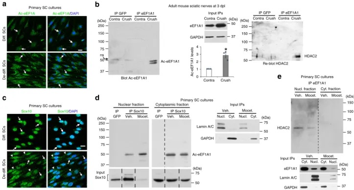
EEF1A1 is deacetylated by HDAC1/2 in SCs. Sox10 is robustly
downregulated at 1 day post sciatic nerve crush lesion (dpl) in
adult mice (Fig.
1
a), a well-characterized experimental model of
PNS lesion where SCs rapidly demyelinate and convert into repair
cells to promote axonal regrowth. At 12dpl, once axons have
regrown, Sox10 is recruited to and activates its target gene
Krox20
33,41, another major promyelinating factor
45,46, to induce
the remyelination process, which is almost complete at 60dpl. We
previously showed that HDAC1 and HDAC2, which can
com-pensate for the loss of each other, are necessary for Sox10
func-tions in the PNS, and that HDAC2 is primarily involved in this
process
38,39,41. To identify direct targets of HDAC1/2, we
col-lected at 1dpl sciatic nerves of adult mice treated with the
HDAC1/2 inhibitor mocetinostat or its vehicle and analyzed the
levels of acetylated proteins by mass spectrometry
41. Interestingly,
analysis by Protein Match Score Summation identified eEF1A1 as
a putative HDAC1/2 deacetylation target (Supplementary
Fig. 1a). To validate this, we ablated Hdac1/2 specifically in SCs
by crossing mice expressing the Cre recombinase under control of
the Dhh promoter (Dhh-Cre
47) with mice expressing
floxed
Hdac1 and Hdac2 (Hdac1fl/fl;Hdac2fl/fl
38,48). We found strongly
increased levels of Ac-eEF1A in SCs of postnatal day (P)4
Dhh-Cre;Hdac1fl/fl;Hdac2fl/fl knockout (dKO) nerves as compared to
Dhh-Cre-negative control littermates (Fig.
1
b, c), while total
eEF1A1 levels were similar in control and dKO nerves (Fig.
1
d),
indicating that eEF1A deacetylation depends on HDAC1/2 in
SCs. Consistently, HDAC1/2 inhibition using the HDAC1/2
inhibitor mocetinostat in primary SC cultures led to strongly
increased levels of Ac-eEF1A1, but not of Ac-eEF1A2 (Fig.
1
e, f
and Supplementary Fig. 2). Mass spectrometry analyses revealed
that all detected acetylated peptides of eEF1A1 were significantly
more abundant in samples of mocetinostat-treated SCs as
com-pared to vehicle-treated SCs (Supplementary Data 1). eEF1A1 has
20 putative acetylation sites
49. Mass spectrometry analyses
allowed us to detect 4 of these sites on K386, K219, K408, and
K273, which are located in different domains of eEF1A1
49. In
addition, the antibody we used in Fig.
1
b, c, e, f showing strongly
increased levels of Ac-eEF1A in the absence of HDAC1/2 activity
is directed against eEF1A acetylated at K41. These
findings
suggest that eEF1A1 is globally acetylated in the absence of
HDAC1/2 activity and that other lysine in addition to the ones
detected by mass spectrometry and by the acetylK41-eEF1A
antibody that we used here are most likely simultaneously
acetylated in the absence of HDAC1/2 activity.
Ac-eEF1A1 drags Sox10 out of the nucleus. We found that
eEF1A1 acetylation is increased in SCs upon de-differentiation in
culture (Fig.
2
a) and in sciatic nerves after lesion (Fig.
2
b and
Supplementary Fig. 1b). While eEF1A1 is mainly localized in the
SC cytoplasm (Supplementary Fig. 3), we show that Ac-eEF1A1 is
localized in both nuclear and cytoplasmic fractions (Fig.
1
e, f).
We found decreased nuclear levels of Sox10 in de-differentiated
as compared to differentiated SCs and partial re-localization of
Sox10 in the cytoplasmic compartment (Fig.
2
c, d). In addition,
Ac-eEF1A co-immunoprecipitated with Sox10 in both
compart-ments, an interaction that was potentiated by HDAC1/2
inhibi-tion in the nuclear compartment, which also led to a strong
decrease of Sox10 levels in the nucleus (Fig.
2
d). In line with its
eEF1A1 deacetylating function, HDAC2 interacted with eEF1A1
in the nucleus of de-differentiated SCs in culture (Fig.
2
e) and in
sciatic nerves after lesion (Fig.
2
b). We show that Sox10 is
tar-geted to the proteasome in de-differentiated SCs (Fig.
3
a and
Supplementary Movie 1) and that inhibition of the proteasome
results in increased Sox10 levels (Fig.
3
b). Taking these data
together, we hypothesized that eEF1A1 can translocate to the
nucleus to interact with Sox10, drag it out of the nucleus and
target it to the proteasome for degradation. Indeed, overexpressed
eEF1A1, which interacts with Sox10 in both cytoplasm and
nucleus of de-differentiated SCs (Fig.
3
c), leads to increased
cytoplasmic re-localization of Sox10 (Fig.
3
d) and increased
co-localization of Sox10 with the proteasome in the cytoplasmic
compartment (Supplementary Fig. 4) within 24 h, and to strongly
decreased Sox10 levels within 3 days (Fig.
3
e). In SCs cultured
under de-differentiating conditions, approximately 25% of
eEF1A1 is localized in the nucleus (Fig.
4
a). To determine
whe-ther acetylation of eEF1A1 promotes its nuclear localization, we
mutated individual lysine (K) into glutamine (Q) residues, which
mimic acetylation. We chose K41, K179, and K273, which have
been experimentally verified and several times described to be
acetylated
49. We found by subcellular fractionation that each
mutation leads to increased nuclear localization of eEF1A1
(Fig.
4
a). Among those, K273Q mutation was the most efficient.
Consistently, the mutation of K273 into arginine (R), which
prevents acetylation, was the most efficient to reduce nuclear
localization of the protein (Fig.
4
b). Interestingly, K273 is the only
K residue specific to eEF1A1
49. Indeed, in eEF1A2, K273 is
replaced by R, thereby preventing acetylation at this position.
Consistent with decreased nuclear localization of K273R mutant,
overexpression of this mutant resulted in a decreased percentage
of low Sox10-expressing SCs as compared to overexpression of
wild-type eEF1A1 (Fig.
4
c). Surprisingly, K179R mutation had a
a
b
c
d
e
f
75 37 50 50 250 150 100 75 50 37 50 75 50 37 (kDa) (kDa) (kDa) Sciatic nerves at 1 dpl P4 sciatic nerves P4 sciatic nerves P4 sciatic nerves Primary SC cultures Primary SC cultures Input IPs Veh. Mocet. IP eEF1A1 IP eEF1A1 eEF1A1 Ac-eEF1A1 Ac-eEF1A1 Blot Ac-eEF1A1 Input GAPDH ns GAPDH Lamin A/C Contra Ac-eEF1A Ac-eEF1A Ac-eEF1A/S100 Merge/DAPIAc-eEF1A/DAPI Contra
Control
Nuclear fraction
Veh. Veh.
Cyt.Nucl.Cyt.Nucl. Mocet. Mocet. Cytopl. fraction dKO Ctrl Ctrl dKO dKO Sox10 eEF1A1 Blot Ac-eF1A1 GAPDH Sox10 levels Vehicle Mocetinostat eEF1A1 levels Ctrl dKO GAPDH 100 75 50 25 0 1.5 1 0.5 0 Crush Crush (kDa) 37 250 150 100 75 50 37 (kDa) 37
Fig. 1 EEF1A1 is deacetylated by HDAC1/2 in SCs. a Sox10 Western blot and quantification normalized to GAPDH showing low Sox10 levels at 1dpl in crushed as compared to contralateral sciatic nerves of adult mice. Paired two-tailed Student’s t-tests, p value = 0.008938, n = 8 animals per group. b, c Co-immunofluorescence of Ac-eEF1A and S100 (SC marker) and DAPI (nuclei) labeling (b) and immunoprecipitation (IP) of eEF1A1 and Western blot of Ac-eEF1A (c) in sciatic nerves of P4 control (Ctrl) and DhhCre;Hdac1;Hdac2 knockout (dKO) mice showing increased levels of Ac-eEF1A in the absence of HDAC1/2 in SCs. n.s.= non-specific. d Western blot of total eEF1A1 and GAPDH (loading control) in sciatic nerves of P4 Ctrl and dKO mice and quantification of eEF1A1 levels normalized to GAPDH levels showing no significant difference between the two groups. Paired one-tailed Student’s t-tests, p value= 0.297774, n = 4 animals per group. e IP eEF1A1 followed by Western blot of Ac-eEF1A in nuclear and cytoplasmic fractions of primary SCs cultured under de-differentiating conditions and treated with the HDAC1/2 inhibitor mocetinostat (Mocet.) or its vehicle (Veh.) for 3 days. Lamin A/C (nuclear marker), GAPDH (cytoplasmic marker) and eEF1A1 Western blots on lysates used for IP show the inputs.f Immunofluorescence of Ac-eEF1A and DAPI labeling in primary SCs cultured under de-differentiation conditions and treated with mocetinostat or its vehicle for 3 days. Arrowheads (b) and arrows (f) show SC nuclei. Z-series projections (b) or single optical sections (f) are shown. b, f Representative images of 3 different animals per group or of 3 independent experiments are shown.b, d Data are presented as mean values ± SEM. Scale bars: 10µm. Source data are provided as a Source Data file.
similar effect as K273R on Sox10 levels (Fig.
4
c). By analyzing
Sox10 levels only in cells where eEF1A1 or the mutants were
present in the nuclear compartment, we found that K179R
pre-vents the decrease of Sox10 levels even when localized in the
nucleus (Fig.
4
d). In SCs cultured under proliferating conditions,
overexpressed eEF1A1 is in a large majority found in the
cyto-plasmic compartment (Fig.
4
e). We show in these conditions that
the three K to Q mutations lead to increased nuclear localization
of eEF1A1 (Fig.
4
e) and to decreased Sox10 levels (Fig.
4
f). We
next tested which region of Sox10 protein is responsible for its
decreased stability in the presence of Ac-eEF1A1. To this aim, we
co-transfected HEK293 cells, which do not express Sox10, with
either wild-type Sox10 of different Sox10 mutants together with
K273Q eEF1A1 or with GFP as control. We show that expression
of K273Q eEF1A1 leads to decreased levels of wild-type Sox10,
Sox10-95 (mutant containing an insertion of 2 amino acids in the
HMG domain, which impairs DNA binding
50), Sox10-MIC
(mutation resulting in a shorter protein containing the
first 188
aminoterminal residues of Sox10, which include the HMG
domain
50), and Sox10-HMG (containing only the HMG
domain
51) (Supplementary Fig. 5a, b), indicating that the HMG
domain is sufficient for Ac-eEF1A1 to decrease Sox10 stability.
Similar to SCs, K273Q eEF1A1 co-immunoprecipitated with
wild-type Sox10 in HEK293 cells (Supplementary Fig. 5c). In
addition, K273Q eEF1A1 also co-immunoprecipitated with
Sox10-95 (Supplementary Fig. 5c). These data show that
Ac-eEF1A1 interacts with Sox10 in HEK293 cells such as in SCs and
that the two additional amino acids in the HMG domain do not
impair this interaction.
Taken together, these data indicate that acetylation of eEF1A1
at any of the three residues K41, K179 or K273 increases eEF1A1
nuclear localization and further decreases Sox10 levels and show
that K179 and K273 are key acetylation sites for eEF1A1 nuclear
localization and decrease of Sox10 levels.
Tip60 associates with Stat3 to acetylate eEF1A1. To understand
the mechanism that leads to eEF1A1 acetylation after sciatic
nerve crush lesion, we analyzed by mass spectrometry eEF1A1
binding partners in sciatic nerves at 2dpl compared to unlesioned
nerves. EEF1A1 is a major translation elongation factor
42,44;
consistent with this, we detected several proteins involved in RNA
transport and metabolism as eEF1A1 putative binding partners.
Also consistent with a function of eEF1A1 interacting with the
proteasome
52and targeting proteins to the proteasome for
degradation, we identified the proteasome regulatory proteins
Proteasome activator subunits 1 and 2 as eEF1A1 putative
binding partners (Supplementary Data 2). In addition, this
ana-lysis suggested that putative binding partners were recruited to
eEF1A1 upon sciatic nerve crush lesion. Among those, Stat3
appeared as an interesting candidate to further investigate
because it has been shown to interact with eEF1A1 in other cells
53and with the histone acetyltransferase Tip60 in the cytoplasmic
compartment
54, and is known to be expressed in SCs and
acti-vated after a sciatic nerve lesion
55–61. We thus decided to further
investigate the potential involvement of Stat3 and Tip60 in
eEF1A1 acetylation. Consistent with previous
findings
53,54, Tip60
and Stat3 co-immunoprecipitated with eEF1A1 (Fig.
5
a) and
Stat3 co-immunoprecipitated with Tip60 (Fig.
5
b) in SCs. Tip60
eEF1A1 eEF1A1 GAPDH 4 3 2 1 0 Ac-eEF1A1 Ac-eEF1A1 HDAC2 HDAC2 Lamin A/C Input Sox10 GAPDH Lamin A/C GAPDH Re-blot HDAC2 Blot Ac-eEF1A1 ns De-diff. SCs Ac-eEF1A1 levels Diff. SCs De-diff. SCs Diff. SCs
Ac-eEF1A Ac-eEF1A/DAPI
Sox10 Sox10/DAPI
Primary SC cultures
Primary SC cultures
Primary SC cultures Adult mouse sciatic nerves at 3 dpl
Primary SC cultures 50 75 50 50 50 150 150 250 75 50 100 75 50 37 100 75 37 (kDa) (kDa) (kDa) (kDa) (kDa) 50 150 100 75 250 (kDa) (kDa) 37 75 37 37 50 37 150 100 75 250 (kDa)
a
b
c
d
e
IP eEF1A1 IP eEF1A1 IP GFP IPGFP Veh. Mocet. GFP Veh.
Veh. Mocet. Veh. Mocet. Mocet. IP IP Sox10 IP Sox10 Input IPs Contra Crush Contra Crush Contra Crush Contra Crush IP eEF1A1 IP GFP
Contra Crush Contra Crush
Input IPs
Input IPs Veh.
Nucl. Nuclear fraction
Nucl. fraction Cyt. fraction Cytoplasmic fraction
Cyt. Nucl.
Nucl. Cyt.
Cyt. Cyt.Nucl. Mocet.
Veh. Mocet.
Fig. 2 Sox10 re-localizes to the cytoplasm with Ac-eEF1A1 in de-differentiated SCs. Immunofluorescence of Ac-eEF1A (a) and Sox10 (c), and DAPI (nuclei) labeling in differentiated and de-differentiated SCs.b Immunoprecipitation (IP) of eEF1A1 or GFP (control) carried out on the same pool of two adult mouse crushed or unlesioned contralateral sciatic nerves at 3dpl and Western blot of Ac-eEF1A followed by HDAC2. EEF1A1 and GAPDH Western blots on lysates show the input, n = 3 (6 animals). The graph shows the quantification of Ac-eEF1A levels (measured on a longer exposure to obtain a value for the contra IP eEF1A1) normalized to eEF1A1 input. Data are presented as mean values ± SEM. Paired two-tailed Student’s t-tests, p value = 0.03376. IP Sox10 or GFP (d) or IP eEF1A1 (e) followed by Ac-eEF1A (d) or HDAC2 (e) Western blot in nuclear and cytoplasmic fractions of primary SCs cultured under de-differentiating conditions and treated with the HDAC1/2 inhibitor mocetinostat or its vehicle for 3 days. Lamin A/C (nuclear marker), GAPDH (cytoplasmic marker), Sox10 and eEF1A1 Western blots on lysates used for IP show the inputs. Dashed lines indicate that samples were run on the same gel but not on consecutive lanes. Arrows point to SC nuclei (a, c) and arrowheads (c) to SC cytoplasm. a–e Representative images of 3 independent experiments are shown. Scale bars: 10µm. Source data are provided as a Source Data file.
has a molecular weight of 60 kDa and the higher molecular
weight isoforms of Tip60 from ~75 to ~100 kDa are sumoylated
isoforms, which have been shown to have increased
acetyl-transferase activity (Supplementary Fig. 6 and Refs.
62,63). We
show that eEF1A1 and Stat3 interact with sumoylated Tip60 in
SCs (Fig.
5
a, b). We found that Tip60 is mainly localized in the
cytoplasmic fraction of SCs cultured under de-differentiating
conditions (Fig.
5
c). In addition, Tip60 is strongly upregulated
after a sciatic nerve crush lesion during SC de-differentiation
(Fig.
5
d). We thus tested whether Tip60 is involved in eEF1A1
acetylation and Sox10 regulation in de-differentiated SCs. Indeed,
inactivating Tip60 with the specific Tip60 inhibitor TH1834
robustly decreased eEF1A1 acetylation (Fig.
5
e) and Tip60
binding to eEF1A1 (Fig.
5
a), and increased Sox10 levels (Fig.
5
f).
Interestingly, downregulation of Stat3 by shRNA led to decreased
levels of sumoylated Tip60 (Fig.
5
g), decreased interaction of
Tip60 with eEF1A1 (Fig.
5
h) and decreased levels of Ac-eEF1A
(Fig.
5
i). Taken together, these results indicate that Tip60
associates with Stat3 to acetylate eEF1A1 in the cytoplasm of
de-differentiating SCs early after a PNS lesion.
Theophylline enhances PNS remyelination. The mechanism of
action of eEF1A1 on Sox10 and its regulation by HDAC2 are
summarized in Fig.
6
. We next aimed at using this mechanism to
enhance remyelination in vivo. We show here that overexpression
of wild-type eEF1A1 or of K273Q eEF1A1 in SCs cultured under
differentiation conditions robustly decreases the percentage of
Krox20-expressing SCs as compared to GFP-expressing SCs, and
that expression of the K273Q mutant decreases further this
per-centage as compared to wild-type eEF1A1-overexpressing cells
(Supplementary Fig. 7). Krox20 is a Sox10 target gene and a major
transcription factor for myelination in developing and adult
SCs
45,46. These data thus suggest that eEF1A1 interferes with the
myelination program in SCs and shows that mimicking
con-stitutive acetylation of K273 increases this function. We
hypo-thesized that increasing the deacetylation rate of eEF1A1 by
increasing HDAC2 expression and activity would prevent Sox10
cytoplasmic re-localization and degradation and thereby maintain
the activation of Sox10 target genes (Fig.
6
). Sox10 directly binds
to and activates the promyelinating factor genes Oct6 and Krox20
in SCs and Myrf in OLs and myelin genes such as P0 in SCs and
Mbp in both SCs and OLs
30,32–34,36. We previously showed that
downregulation of HDAC2 in SCs by shRNA leads to decreased
levels of Sox10, Krox20, and P0, while overexpression of HDAC2
increases Sox10, Krox20, and P0 levels
38. Theophylline is a
known pharmacological activator of HDAC2 at low dose,
Sox10 levels
Sox10 levels
GFP
eEF1A1-GFP
% of low Sox10-expressing cells
a
b
c
d
e
20S Sox10/20S (3D view) GFPSox10 Sox10/GFP/DAPI
Sox10 75 75 37 50 100 150 250 50 25 37 75 37 37 75 50 75 75 37 (kDa) (kDa) 50 (kDa) (kDa) (kDa) (kDa) 75 50 75 50 37 25 75 50 37 Primary SC cultures Cytoplasmic Sox10 eEF1A1-GFP Sox10 GFP GFP GFP 0 25 50 75 Cyt. only Nucl. + Cyt. Cytoplasmic Nuclear eEF1A1 eEF1A1 eEF1A1-GFP GFP ns GFP GAPDH GAPDH Lamin A/C Lamin A/C
GFPeEF1A1-GFP GFPeEF1A1-GFP GFPeEF1A1-GFP GFPeEF1A1-GFP
Nuclear Cytoplasmic Nuclear Sox10 eEF1A1-GFP eEF1A1-GFP GFP eEF1A1 -GFP GFP Lamin A/C GAPDH GFP Sox10 Blot Sox10 Cytoplasmic IP GFP Input Nuclear Cytoplasmic Nuclear + + + + – – + + + + – – – – – – GAPDH Primary SC cultures Primary SC cultures Vehicle 0 0.5 1 1.5 2 2.5 Proteasome inhib. Primary SC cultures Primary SC cultures 2 1.5 1 0.5 0
Fig. 3 EEF1A1 re-localizes Sox10 to the cytoplasm and reduces its expression. a Sox10 and 20 S co-immunofluorescence in SCs cultured under de-differentiation conditions. Representative images (z-series projections) are shown. The right image is a magnified 3D view of the region highlighted by a dashed box on the left images. Arrows point to proteasome structures containing Sox10.b Sox10 and GAPDH Western blot in lysates of SCs induced to de-differentiate for 1 day and treated with the proteasome inhibitor MG132 (proteasome inhib.) or vehicle (Veh.) for 12 h, and Sox10 quantification normalized to GAPDH (n = 3 independent experiments). c GFP immunoprecipitation (IP) and Sox10 Western blot in nuclear and cytoplasmic fractions of SCs transfected with eEF1A1-GFP or GFP and induced to de-differentiate for 1 day, and input GFP, LaminA/C (nuclear fraction) and GAPDH (cytoplasmic fraction) on lysates used for IPs.d Sox10 and GFP Western blots in nuclear and cytoplasmic fractions of SCs transfected with a construct expressing eEF1A1-GFP or control GFP and induced to de-differentiate for 1 day, and Sox10 quantification (normalized to GAPDH or Lamin A/C) in each fraction. n.s.= non-specific. e Sox10 immunofluorescence with GFP fluorescence and DAPI (nuclei) labeling in SCs expressing eEF1A1-GFP or control GFP and cultured under de-differentiating conditions for 3 days, and % of low Sox10-expressing cells among cells expressing GFP or eEF1A1-GFP in the cytoplasm only (Cyt. only) or in both nucleus and cytoplasm (Nucl.+ Cyt.). Arrows point to transfected SCs (e). Orange arrows indicate transfected SCs with low Sox10 levels and/or with eEF1A1 localized in the nucleus (e). Dashed lines indicate that samples were run on the same gel but not on consecutive lanes. Data are presented as mean values ± SEM. Unpaired (b, e) or paired (d) two-tailed Student’s t-tests. P values: 0.001725 (b), 0.045156 (d, cytoplasmic), 0.024327 (d, nuclear), 0.02473 (e). N = 3 independent experiments (b, d, e), 15–25 (GFP) and 48–58 (eEF1A1-GFP) transfected cells counted per n (e). a–e Representative images of 3 independent experiments are shown. Scale bars: 10 µm. Source data are provided as a Source Data file.
increasing both HDAC2 activity and expression, while at higher
concentrations theophylline acts as an antagonist of adenosine
receptors and as an inhibitor of phosphodiesterases
64,65. We show
here that a low concentration of theophylline (1 µM) increases
HDAC2 and Sox10 levels in primary SCs, whereas 1 µM CGS
15943 (antagonist of adenosine receptors) or 500 µM IBMX
(inhibitor of phosphodiesterases) or 10 µM Rolipram (inhibitor of
type IV phosphodiesterases) either have no effect or decrease
HDAC2 and/or Sox10 levels (Supplementary Fig. 8a, b). At these
concentrations, CGS 15943, IBMX, and Rolipram are fully active
on their targets. These data show that theophylline does not
increase Sox10 levels through inhibiting phosphodiesterases or
antagonizing adenosine receptors but is very likely to induce its
effect on Sox10 through its third target HDAC2. Theophylline is
already used as a drug in humans to treat asthma and chronic
obstructive pulmonary disease
65. Its activity, mode of
adminis-tration and toxicity are thus very well described in mice and
humans. To test whether theophylline enhances PNS
remyeli-nation, we carried out sciatic nerve crush lesions and treated mice
with theophylline at 10dpl, just before SCs start remyelinating
regenerated axons, for 2 or 4 consecutive days. We found that
theophylline treatment indeed increases HDAC2 expression
(Fig.
7
a) and decreases Ac-eEF1A1 levels (Fig.
7
b, c) as compared
to vehicle-treated mice. Consistently, Sox10 levels were also
% of protein in nucleus
and cytoplasm
% of cells
% of cells
% of low Sox10- expressing cells % of low Sox10- expressing cells % of low Sox10- expressing cells
a
b
c
d
e
f
75 100 75 (kDa) 37 Primary SC cultures eEF1A1 WT K273QNucl. Nucl. Nucl. Nucl.
Nucl. +Cyt. Cyt. only Nucl. +Cyt. Cyt. only Nucl. +Cyt. Cyt. only Nucl. +Cyt. Cyt. only Nucl. +Cyt. Cyt. only
Nucl. Cyt.Nucl.Cyt.Nucl.Cyt.Nucl.Cyt.
eEF1A1 K41Q K179Q K273Q eEF1A1K41RK179R K273R eEF1A1 K41R K179R K273R eEF1A1K41RK179R K273R eEF1A1 eEF1A1-GF K41R1-GFP K179R-GFP K273R-GFP K179Q K273Q K41Q eEF1A1K41QK179Q K273Q Cyt. only Cyt. only Cyt. only Nucl. +Cyt. Nucl. +Cyt. Nucl. +Cyt. GFP eEF1A1-GF K41Q-GFP GFP GFP Sox10 Sox10 / GFP / DAPI
GFP Sox10 Sox10/GFP/DAPI
K273Q-GFP
All transfected cells Transfected cells with eEF1A1or mutants in nucleus
K179Q-GFP Lamin A/C GAPDH 100 75 50 25 0 100 75 50 25 0 100 75 50 25 0 30 80 60 40 20 0 60 40 20 0 20 10 0
Cyt. Cyt. Cyt. Cyt.
K179Q K41Q
Fig. 4 EEF1A1 acetylation increases eEF1A1 nuclear localization and decreases Sox10 levels. a GFP Western blot in nuclear (Nucl.) and cytoplasmic (Cyt.) fractions of primary SCs cultured 1 day under de-differentiating conditions and transfected with eEF1A1-GFP, K41Q-GFP, K179Q-GFP or K273Q-GFP and % of protein localized in nucleus or cytoplasm normalized to Lamin A/C and GAPDH (n = 3 independent experiments per group). b–d Sox10 immunofluorescence with GFP fluorescence and DAPI (nuclei) labeling in SCs overexpressing eEF1A1-GFP, K41R-GFP, K179R-GFP or K273R-GFP and cultured under de-differentiating conditions for 3 days, and % of cells with eEF1A1 or mutants localized in cytoplasm only (Cyt. only) or in nucleus and cytoplasm (Nucl.+ Cyt., b) or % of low Sox10-expressing cells (c, d). N = 3 independent experiments per group, 18-84 cells counted per group per n. e, f Sox10 immunofluorescence with GFP fluorescence and DAPI (nuclei) labeling in SCs overexpressing eEF1A1-GFP, K41Q-GFP, K179Q-GFP or K273Q-GFP and cultured under proliferating conditions for 2 days, and % of cells with eEF1A1 or mutants localized in cytoplasm only or in nucleus and cytoplasm of SCs (e) or % of low Sox10-expressing cells (f). The lower images are magnifications of the dashed white boxes on the upper images. N = 3 independent experiments per group, 17–71 cells counted per group per n. Arrows show transfected SCs. Orange arrows indicate SCs where eEF1A1 or the mutants are present in the nucleus of SCs. Scale bars: 10µm (b), 20 µm (e). Data are presented as mean values ± SEM. Unpaired one-tailed (gray asterisks) or two-tailed (black asterisks) Student’s t-tests, p values = 0.029415 (a, K41Q), 0.04884 (a, K179Q), 0.0329 (a, K273Q), 0.023698 (b, K41R), 0.008772 (b, K179R), 0.00946 (b, K273R), 0.189827 (c, K41R), 0.027448 (c, K179R), 0.018314 (c, K273R), 0.405588 (d, K41R), 0.03836 (d, K179R), 0.406686 (d, K273R), 0.011923 (e, K41Q), 0.019373 (e, K179Q), 0.021749 (e, K273Q), 0.009059 (f, K41Q), 0.009399 (f, K179Q), 0.02223 (f, K273Q). Source data are provided as a Source Datafile.
increased by theophylline treatment (Fig.
7
a), which also led to
increased levels of Krox20 and P0 (Fig.
7
a). Sox10 is recruited to
its target genes Krox20 (at the myelinating SC element, MSE) and
P0 (at the intron 1) at the SC redifferentiation stage to induce
remyelination (Fig.
7
d). We show here that theophylline rapidly
promotes the recruitment of Sox10 to its target genes (Fig.
7
e),
whereas the HDAC1/2 inhibitor mocetinostat impairs Sox10
recruitment (Fig.
7
f), consistent with our previous
findings
showing impaired remyelination after lesion in sciatic nerves
lacking HDAC1/2 in SCs
41. To test whether theophylline also
increases Sox10 levels during developmental myelination, we
treated mouse pups with theophylline from P1 to P3 and
col-lected their sciatic nerves at P4. While theophylline had no effect
on the levels of Sox10, P0 or HDAC2 in Dhh-Cre dKO mice
lacking HDAC1/2 in SCs, we show that theophylline increases the
levels of Sox10, P0, and HDAC2 in control littermate (Dhh-Cre
negative) mice (Fig.
7
g), indicating that theophylline can increase
Sox10 and P0 levels also during development and suggesting that
Nucl. Cyt. (kDa) 50 250 75 100 150 0 0.5 1 250 75 50 100 150 100 75 37 100 75 75 50 37 50 37 37 (kDa) (kDa) (kDa) (kDa) (kDa) (kDa) 250 75 50 100 150 (kDa) (kDa) 37 75 100 37 75 50 75 75 100 100 IP eEF1A1 IP Ctrl IP Stat3 eEF1A1 eEF1A1 GAPDH Stat3 Stat3 Stat3 Reblot Stat3 Input Input Input Veh. Tip60i (kDa) (kDa) IP eEF1A1 Veh. Veh. Co Cr Co Cr Co Co 1 dpl 3 dpl 5 dpl 12 dpl Cr Co Cr 4 3 2 1 0 0 0.5 0.8 0.4 2.5 2 1.5 1 0.5 0 0 1 Tip60i Tip60i IP eEF1A1 Ctrl sh Ctrl sh Stat3 sh Stat3 sh Stat3 sh Stat3 sh Ctrl sh Sumoylated Non-sumoylated Ctrl sh IP eEF1A1 Veh. Tip60i 250 75 50 100 150 (kDa) 250 75 50 100 150 Vehicle Tip60 inhibitor Tip60 Tip60 Tip60 Tip60 Tip60 Tip60 sumo-Tip60 GAPDH GAPDH GAPDH GAPDH Lamin A/C Tip60 levels Control shRNA Stat3 shRNA Stat3
a
b
c
d
e
f
g
h
i
Ac-eEF1A Stat3/Ac-eEF1A Stat3/Ac-eEF1A/DAPITip60 levels Ac-eEF1A1 levels Tip60 levels Sox10 levels Blot Tip60 Blot Tip60 Blot Ac-eEF1A Ac-eEF1A1 Sox10 GAPDH Vehicle Vehicle Tip60 inhibitor Tip60 inhibitor Blot Tip60 1 dpl 3 dpl 5 dpl 12 dpl
Fig. 5 Tip60 acetylates eEF1A1 in a Stat3-dependent manner. a, e eEF1A1 immunoprecipitation (IP) in lysates of SCs induced to de-differentiate for 1 day and treated with TH1864 (Tip60 inhibitor, Tip60i) or vehicle, and Tip60, Stat3 (a) or Ac-eEF1A (e) Western blot and quantification (normalized to eEF1A1) of Tip60 co-immunoprecipitated with eEF1A1 (a) or of Ac-eEF1A1 (e) in Tip60 inhibitor- compared to vehicle-treated SCs. b IP Stat3 or Flag (Ctrl) in lysates of SCs induced to de-differentiate for 1 day, Tip60 Western blot and Stat3 inputs.c Tip60, GAPDH (cytoplasmic marker) and Lamin A/C (nuclear marker) Western blots in cytoplasmic and nuclear fractions of SCs induced to de-differentiate for 1 day. Dashed lines: samples run on the same gel, but not on consecutive lanes.d Tip60 and GAPDH Western blots in lysates of crushed (Cr) and contralateral (Co) mouse sciatic nerves at 1-3-5-12dpl, and Tip60 quantification normalized to GAPDH in Cr compared to Co. f Sox10 and GAPDH Western blots in lysates of SCs induced to de-differentiate and treated with Tip60i or vehicle for 3 days and Sox10 quantification normalized to GAPDH. g Stat3 and Tip60 Western blots in SCs transduced with Stat3-specific shRNA (Stat3 sh) or non-targeting control shRNA (Ctrl sh) lentiviruses and quantification of sumoylated (higher molecular weight) and non-sumoylated (lower molecular weight) Tip60 isoforms normalized to GAPDH.h eEF1A1 IP and Tip60 Western blot in SCs transduced with Stat3 sh or Ctrl sh lentiviruses.i Stat3 and Ac-eEF1A co-immunofluorescence and DAPI labeling in primary SCs transduced with Stat3 sh or Ctrl sh lentiviruses and induced to de-differentiate for 2 days. Scale bar: 20µm. Data presented as mean values ± SEM. Unpaired (f) or paired (a, d, e, g) one-tailed (gray asterisk) or two-tailed (black asterisks) Student’s t-tests. P values = 0.029197 (a), 0.139771 (d, 1dpl), 0.047935 (d, 3dpl), 0.030298 (d, 5dpl), 0.061845 (d, 12dpl), 0.001796 (e), 0.033551 (f), 0.047979 (g, sumoylated), 0.300207 (g, non-sumoylated). N = 3 independent experiments or 3 animals per time-point. For each panel, representative images of 3 independent experiments are shown. Source data are provided as a Source Datafile.
this effect may require the presence of HDAC2 and/or HDAC1,
although we cannot exclude a potential inability of the Dhh-Cre
dKO mutants to express Sox10 and P0 with or without
theo-phylline or that theotheo-phylline may act through additional or other
pathways to increase Sox10 and P0 in control mice. In addition to
increasing the expression of HDAC2, Sox10, Krox20, and P0,
theophylline also increased myelin thickness (Fig.
8
a) and led to
faster motor and sensory functions recovery (Fig.
8
b), while there
was neither a difference in the density of SCs or of inflammatory
cells (Supplementary Fig. 9a) nor a difference in the percentage of
proliferating cells (Supplementary Fig. 9b). Consistently,
theo-phylline treatment did not alter the density of SCs in culture
(Supplementary Fig. 9c). Remyelinating sheaths are known to
remain thinner as compared to myelin sheaths of unlesioned
nerves, however, myelin in the injured nerve of
theophylline-treated mice, but not of vehicle-theophylline-treated mice, recovered a similar
thickness as in uninjured nerves (Fig.
8
c), indicating that
theo-phylline allows full remyelination after lesion. Taken together,
these data show that theophylline improves PNS remyelination
efficiency after lesion.
Ac-eEF1A1 regulates Sox10 levels in OLs. Similar to SCs, eEF1A
deacetylation in OLs depends on HDAC1/2 activity, as shown by
a robust increase of Ac-eEF1A in OLs of mouse spinal cords 24 h
after intrathecal injection of the HDAC1/2 inhibitor mocetinostat
(Fig.
9
a) and in primary OLs treated with mocetinostat (Fig.
9
b).
In addition, inactivating Tip60 with the specific Tip60 inhibitor
TH1834 strongly reduced the increase of Ac-eEF1A due to
mocetinostat treatment (Supplementary Fig. 10), suggesting that
Tip60 acetylates eEF1A1 also in OLs. Such as in SCs, treatment
with mocetinostat led to cytoplasmic relocalization of Sox10 and
to strongly decreased Sox10 levels in primary OLs (Fig.
9
c).
Consistently, ablation of HDAC1/2 in mature OLs using the
tamoxifen-inducible PLP-CreERT2 mouse line led to increased
interaction of Ac-eEF1A with Sox10 (Fig.
9
d) and to decreased
Sox10 levels (Fig.
9
e, f). To test whether eEF1A1 has a similar
effect on Sox10 levels in OLs as in SCs, we overexpressed
wild-type eEF1A1 or K273Q mutant in the oligodendroglial Oli-neu
cell line
66that can be easily transfected. We and others have
shown that Oli-neu cells express high levels of MBP when
cul-tured in differentiating conditions for 3 days and acquire a
complex morphology after 10 days (Supplementary Fig. 11a and
Ref.
66). Consistent with our
findings in SCs, K273Q mutant was
localized in both nuclear and cytoplasmic compartments of
vir-tually all transfected cells, while wild-type eEF1A1 was either
localized exclusively in the cytoplasm or in both nucleus and
cytoplasm of Oli-neu cells cultured under proliferating conditions
(Supplementary Fig. 11b). We show here that overexpression of
wild-type eEF1A1 or expression of K273Q mutant both decrease
Sox10 levels as compared to control GFP transfection
(Supple-mentary Fig. 11c, d) and that expression of K273Q mutant further
decreases Sox10 levels as compared to overexpression of
wild-type eEF1A1 (Supplementary Fig. 11c). Consistently,
over-expression of wild-type eEF1A1 or over-expression of K273Q mutant
both decreased MYRF levels as compared to control GFP
trans-fection (Supplementary Fig. 11e, f) and expression of K273Q
mutant further decreased MYRF levels as compared to
over-expression of wild-type eEF1A1 (Supplementary Fig. 11e). Of
note, K273Q mutant is re-localized to the cytoplasm of Oli-neu
cells upon induction of differentiation (Supplementary Fig. 11b),
possibly by a compensatory mechanism to recover Sox10
expression in the nucleus and thereby allow the induction of
differentiation. To test whether K273Q mutant indeed impairs
the induction of differentiation, we carried out luciferase gene
reporter assays of the Mbp promoter 5 min after the induction of
differentiation, where the activity of the Mbp promoter is already
increased as compared to proliferating conditions
(Supplemen-tary Fig. 11g), in Oli-neu cells expressing K273Q mutant or GFP
or overexpressing wild-type eEF1A1. While overexpression of
wild-type eEF1A1 led to a trend of decreased activity of the Mbp
promoter, expression of K273Q mutant decreased very
sig-nificantly the activity of the Mbp promoter (Supplementary
Fig. 1h), indicating that K273Q mutant impairs the activation of
the Sox10 target gene Mbp and thus suggesting that K273Q
mutant indeed impairs the induction of differentiation. At a later
time-point in the differentiation process when MBP is already
robustly expressed in Oli-neu cells (Supplementary Fig. 11a), we
show that overexpression of wild-type eEF1A1 also strongly
decreases the activation of the Mbp promoter (Supplementary
Fig. 11i). Taken together, these data show that Ac-eEF1A1 is
deacetylated by HDAC2 and negatively regulates the expression
of Sox10 and of the Sox10 target genes Myrf and Mbp in OLs.
Ac Ac Ac eEF1A1 Proteasome Acetylation Cytoplasm Nucleus Remyelination Myelin genes Deacetylation eEF1A1 Stat3 Tip60 eEF1A1 eEF1A1 eEF1A1 eEF1A1 HDAC2 Sox10 Sox10Fig. 6 Proposed mechanism of Ac-eEF1A1-dependent regulation of Sox10. Schematic representation of mechanisms controlling eEF1A1 acetylation-dependent Sox10 degradation and suggesting deacetylation-acetylation-dependent remyelination.
Theophylline enhances CNS remyelination. To test whether
theophylline enhances CNS remyelination, we carried out focal
demyelinating lesions in the spinal cord of adult mice using
lysolecithin injection, a widely used and well-characterized
experimental model of CNS demyelinating lesion where the
remyelination process starts at 14 days post demyelinating lesion
(dpdl). We show that a short treatment with theophylline from 10
to 14dpdl leads to a strong increase of HDAC2, Sox10 and MBP
expression (Fig.
9
g) and to improved remyelination efficiency at
14 and 30dpdl in young adults (Fig.
9
h) and also in old mice
(Fig.
9
i), where remyelination efficiency is lower as compared to
young adults
15. Theophylline induced remyelination in the lesion
site but did not affect myelination outside the lesion site, as
shown by unchanged g ratio (Fig.
9
i, j). These data indicate that
theophylline improves CNS remyelination efficiency after lesion.
Discussion
Demyelination occurs in various cases in humans. In many cases,
the remyelination process is not efficient enough and the loss of
myelin leads to permanent loss of function. For this reason, it is
crucial to better understand how remyelination is controlled in
order to identify strategies that improve remyelination. In this
study, we have focused our analyses on the remyelination process
that occurs in the PNS after a traumatic lesion and in the CNS
after a demyelinating lesion. In particular, we identified a
mechanism that controls the activity and expression of Sox10, a
a
b
c
d
e
f
g
Ac-eEF1A Ac-eEF1A S100 Ac-eEF1A DAPI Ac-eEF1A1 levels Sox10 levelsFold enrichment Fold enrichment
Sox10 levels
P0 levels
HDAC2 levels
Fold enrichment
Fold enrichment
Fold enrichment Fold enrichment
HDAC2 levels P0 levels Krox20 levels 37 75 75 250 1 0.5 0 150 100 50 37 50 50 37 75 37 37 50 75 50 37 75 37 25 37 25 37 25 37 37 37 75 50 37 25 37 37 75 50 37 37 37 37 (kDa) (kDa) (kDa) (kDa) (kDa) 2 1.5 1 0.5 0 2 1.5 1 0.5 0 2 4 3 2 1 0 1.5 1 0.5 0 (kDa) 2 1.5 1 0.5 0 (kDa) Control dKO Veh. Theo Veh.Theo
75 50 75 50 75 50 12 dpl HDAC2 GAPDH GAPDH GAPDH GAPDH Sox10 Krox20 P0 HDAC2 GAPDH GAPDH GAPDH GAPDH Sox10 Krox20 GAPDH P0 GAPDH Krox20 P0 Vehicle Co Co Co Co Cr Cr Cr Cr Vehicle Co Cr Co Cr Theo Vehicle Co Cr Co Cr Theo Theo Vehicle GFP Veh. Theo
Veh. Mocet. Veh. Mocet. Veh. Theo Sox10 GFP Sox10 Veh. IP eEF1A1 Control dKO Veh. Sox10 GAPDH GAPDH GAPDH HDAC2 P0 Veh. Theo Theo Krox20 MSE, 12 dpl Krox20 MSE, 12 dpl Krox20 MSE, 12 dpl P0 intron 1, 12 dpl P0 intron 1, 12 dpl P0 intron 1, 12 dpl Vehicle Theo Blot Ac-eEF1A Input GAPDH 4 3 2 1 0 4 2 1.5 1 0.5 0 3 2 1 0 1.25 1 0.75 0.5 0.25 0 1 0.75 1.5 0 0.6 1.2 1.8 2.4 1 0.5 0 0.5 0.25 0 2 1 0 Theo Theo Veh. Theo 12 dpl 12 dpl 12 dpl 12 dpl 14 dpl 14 dpl 14 dpl 14 dpl 30 dpl 30 dpl Veh. Theo Crush Crush 14 dpl 30 dpl (kDa) Control dKO Veh. Veh. Veh. Veh. Theo Theo Theo Control Control Control Veh. Theo Theo 12 dpl Veh. Veh. Veh. Theo
Veh.TheoVeh.TheoVeh.Theo
Theo Theo
Veh. Theo Veh. Theo
Fig. 7 Theophylline increases Sox10, Krox20, and P0 expression and eEF1A1 deacetylation. a HDAC2 (12dpl: n = 3, p = 0.025883, 14dpl: n = 5, p = 0.011697), Sox10 (12dpl: n = 3, p = 0.031422, 14dpl: n = 3–4, p = 0.032671), Krox20 (12dpl: n = 3, p = 0.030486, 14dpl and 30dpl: n = 7, p = 0.033493 and 0.022041) and P0 (12dpl: n = 3, p = 0.022239, 14dpl: n = 4-5, p = 0.019084, 30dpl: n = 6, p = 0.0456) Western blots at 12, 14 and 30dpl in lysates of crushed (Cr) or contralateral (Co) sciatic nerves of mice treated at 10dpl with theophylline (Theo) or vehicle (Veh.) for 2 days (12dpl) or 4 days (14 and 30dpl), and quantification normalized to GAPDH and Co. b, c EEF1A1 IP and Ac-eEF1A Western blot (b) and Ac-eEF1A and S100 (SC marker) co-immunofluorescence and DAPI labeling (c) at 12dpl in Cr (c) or in Cr and Co (b) 20 h after one theophylline or vehicle injection, and (b) Ac-eEF1A1 quantification normalized to GAPDH input and Co in theophylline- compared to vehicle-treated groups. N = 3 animals per group (b, c), p = 0.000597 (b). Representative images are shown.d–f GFP (d) and/or Sox10 (d–f) chromatin immunoprecipitation at 12dpl on Krox20 MSE or P0 intron 1 in (d) Cr and fold enrichment normalized to Sox10 input compared to GFP (n = 3 animals, Sox10 and GFP IPs on the same nerve lysate, pKrox20-MSE = 0.032616, pP0-intron-1= 0.027702) or in Cr (e) collected 20 h after theophylline (Theo) or vehicle (Veh.) treatment or (f) after mocetinostat (Mocet.) or Veh. treatment for 2 days.e N = 5–6 animals per group, pKrox20-MSE = 0.030466, intron-1 = 0.04003. f N = 3 animals per group, pKrox20-MSE = 0.037396, pP0-intron-1= 0.023672. g Sox10, P0, HDAC2, and GAPDH Western blots in lysates of P4 DhhCre;Hdac1fl/fl;Hdac2fl/fl dKO and control littermate sciatic nerves treated at P1 with Theo or Veh. for 3 days, and quantification normalized to GAPDH in Theo compared to Veh. N = 6 animals per group, pSox10 = 0.04567, pP0= 0.033358, pHDAC2 = 0.02891. Dashed lines: samples loaded on the same gel but not on consecutive lanes. Data are presented as mean values ± SEM. Paired (a: HDAC2 and Sox10-12dpl, Krox20-14dpl-30dpl; b, d, e, and f: P0-intron-1; g) or unpaired one-tailed (gray asterisks) or two-tailed (black asterisks) Student’s t-tests. Scale bar: 20 µm (c). Source data are provided as a Source Data file.
a
b
c
Vehicle Vehicle Vehicle 5 Vehicle Theo 600 450 300 150 0 Theo 4 3 2 1 0 0.8 0.7 0.6 14 dpl 30 dpl 14 dpl 30 dpl Vehicle No lesion 2 mpl Vehicle Theo Theo 0.85 0.75 0.65 0.85 0.75 0.65 Theo Theo Theo 14 dpl RotarodToe pinch test 30 dpl 14 dpl 30 dpl No lesion 2 mpl, V e hicle 2 mpl, Theo g ratio T ime (s)
Number of sensitive toes
g ratio
g ratio
Vehicle
Fig. 8 Theophylline accelerates and enhances PNS remyelination and functional recovery. a, c Electron micrographs of Cr ultrathin sections at 14dpl, 30dpl (a) and 2mpl (c) from mice treated with theophylline or vehicle at 10dpl for 4 days, and myelin thickness quantification (g ratio) of remyelinated axons (n = 3 animals per group per time-point, 14dpl: p = 0.000046, 30dpl: p = 0.048585, 2mpl: pVehicle/No-lesion=0.013591,
pTheo/No-lesion=0.076193, pTheo/Vehicle=0.034469). b Quantification of motor (Rotarod) and sensory (toe pinch) function recovery at 14 and 30dpl in theophylline- and vehicle-treated mice (4-day treatment at 10dpl). 14dpl: n = 9–10, pRotarod=0.02518, pToe-pinch=0.002327, 30dpl: n = 6 per group, pRotarod= 0.489629, pToe-pinch=0.000363. Data are presented as mean values ± SEM. Unpaired one-tailed (gray asterisks) or two-tailed (black asterisks) student’s t-tests. Scale bars: 5 µm (a, b), 10 µm (c). Source data are provided as a Source Data file.
major transcription factor of PNS and CNS myelination and
remyelination. We previously demonstrated that HDAC1/2 act as
co-factors of Sox10
38-41, but the direct deacetylation mechanism
underlying this function remained unclear. We show here that
HDAC1/2 deacetylate eEF1A1, which prevents interaction of
eEF1A1 with Sox10 and re-localization of Sox10 to the cytoplasm,
thereby allowing Sox10 to activate its target genes and enabling
remyelination. We found that after a sciatic nerve crush lesion,
a
b
c
d
e
f
g
h
j
i
Ac-eEF1A Ac-eEF1A HDAC2 Sox10 Sox10 MBP Sox10 Ac-eEF1A/CC1/DAPI HDAC2 Sox10/CC1/DAPI HDAC2/CC1/DAPI Sox10/DAPI Sox10/DAPI Merge/DAPI 37 37 50 75 100 150 250 2.5 2 1.5 1 0.5 0 1 0.5 0 37 (kDa) 75 50 Spinal cords Ac-eEF1A/CC1 Spinal cords Old mice Vehicle Vehicle 14 dpdl 30 dpdl Vehicle Vehicle Theo Theo 14 dpdl 30 dpdl Vehicle Theo Vehicle TheoTheo Theo Lesion site Lesion site Unlesioned site 100 80 60 40 20 0 0.77 ns ns ns 0.73 0.69 0.65 Unlesioned site Spinal cords Ac-eEF1A Sox10 Ctrl Ctrl dKO dKO GAPDH IP Sox10 Ctrl dKO input GAPDH 14 dpdl 30 dpdl 90 60 30 0 Young adult mice, lesion site
14 dpdl 30 dpdl
0.77 0.73 0.69 0.65 Young adult mice, unlesioned site
Spinal cords Spinal cords
Primary oligodendrocyte cultures Primary oligodendrocyte cultures
Vehicle Mocetinostat Ac-eEF1A co-immunoprecipitated with Sox10 Sox10 levels Vehicle Vehicle Vehicle Theo Theo Control Vehicle Vehicle Theo Theo 14 dpdl g ratio g ratio % of remyelinated axons % of remyelinated axons 30 dpdl PLPCreERT2 dKO Mocetinostat Vehicle Mocetinostat (kDa)
Fig. 9 Theophylline promotes CNS remyelination in young and old adults. a Co-immunofluorescence of Ac-eEF1A and CC1 (marker of mature OLs) and DAPI labeling in white matter of adult mouse spinal cord 24 h after treatment with mocetinostat or its vehicle.b, c Co-immunofluorescence of Ac-eEF1A (b) or Sox10 (c) with CC1 and DAPI labeling in differentiated primary OLs treated with mocetinostat or its vehicle for 24 h. d–f Co-immunoprecipitation (IP) of Ac-eEF1A with Sox10 (d) or Sox10 Western blot (e) and quantification normalized to GAPDH, or co-immunofluorescence of HDAC2 or Sox10 with CC1 and DAPI labeling (f) in spinal cords of PLPCreERT2;Hdac1fl/fl;Hdac2fl/fl (dKO) mice or control littermate (Ctrl) at 14 days post tamoxifen injections. g Co-immunofluorescence of HDAC2, Sox10 and MBP and DAPI labeling in the spinal cord lesion site of adult mice (3–4-month old) at 14dpdl after a 4-day treatment with theophylline (Theo) or its vehicle. Arrows indicate OLs.h–j Electron micrographs of spinal cord ultrathin sections in the lesion site (h, i) or unlesioned site (i, j) at 14 and/or 30dpdl in young adult (4-month old) (h, j) or old (19-month old) (i) mice, and % of remyelinated axons in the lesion site (delineated by axons with thin myelin, g ratio≥ 0.835) or g ratio in unlesioned site. Data are presented as mean values ± SEM. Unpaired (h, i, j) or paired (d, e) one-tailed (gray asterisk) or two-tailed (black asterisks) Student’s t-tests, p values: 0.047387 (d), 0.036845 (e), 0.043182 (h, 14dpl), 0.018162 (h, 30dpl), 0.127958 (i, unlesioned), 0.03517 (i, lesion), 0.328856 (j, 14dpl), 0.447458 (j, 30dpl). d n = 5 animals per group, (e) n = 3 animals per group, (h–j) n = 3 animals per group per time-point. Representative images of 3 animals per group (a, f, g) or of 3 independent experiments (b, c) are shown. Scale bars: white= 10 µm, black = 5 µM. Source data are provided as a Source Data file.
Tip60 is upregulated and acetylates eEF1A1 in the SC cytoplasm,
in a Stat3-dependent manner. Consistently, Stat3 has been
pre-viously shown to interact with cytoplasmic Tip60
54and with
eEF1A1
53in other cells. We show here that Stat3 promotes the
interaction of Tip60 with eEF1A1 and eEF1A1 acetylation.
eEF1A1 is a major translation elongation factor, which is mostly
found in the cytoplasmic compartment. However, eEF1A1 has
been shown to have non-canonical functions and to be localized
in the nuclear compartment in some cases
42–44. EEF1A1 can be
submitted to various post-translational modifications, including
phosphorylation,
acetylation,
methylation,
ubiquitination
49.
Phosphorylation, which is the most studied post-translational
modification of eEF1A1, has been shown to regulate eEF1A1
activity
42. In contrast, the function of eEF1A1 acetylation is not
or poorly described. We show here that acetylation of eEF1A1
induces its re-localization to the nuclear compartment of SCs.
There are 20 putative acetylation sites in eEF1A1
49. We studied
the functions of the three acetylation sites K41, K179, and K273
because they have been several times verified experimentally. In
addition, K273 is found only in eEF1A1 and not in eEF1A2
49.
Our results indicate that acetylation at any of these sites promotes
nuclear localization of eEF1A1 and the decrease of Sox10 levels in
SCs, and suggest that K179 and K273 are key acetylation sites for
these functions. By using mocetinostat, a specific inhibitor of
HDAC1/2, or by ablating HDAC1/2 specifically in SCs or in OLs,
we show that HDAC1/2 are necessary to deacetylate eEF1A1 in
SCs and OLs, and to maintain high Sox10 expression in the
nuclear compartment of these cells.
In the aim of identifying a treatment that promotes this
mechanism and thereby potentially improves remyelination
effi-ciency, we tested the effect of theophylline treatment in vivo.
Although theophylline inhibits phosphodiesterases and
antag-onizes adenosine receptors when used at high concentration, it is
also a known potent activator of HDAC2, increasing HDAC2
activity and expression when used at low concentration
64,65. We
show here that while theophylline increases the levels of HDAC2
and Sox10 in SCs, compounds that specifically inhibit
phospho-diesterases or antagonize adenosine receptors either have no
effect on HDAC2 and Sox10 levels or decrease the levels of
HDAC2 and Sox10, indicating that theophylline does not increase
Sox10 levels through its effect on phosphodiesterases or
adeno-sine receptors but is highly likely to act on Sox10 through
HDAC2. Interestingly, a short treatment with theophylline in
mice increased HDAC2 expression, decreased Ac-eEF1A1 levels,
increased the expression of Sox10 and its target genes and
improved remyelination efficiency in both PNS and CNS. Of
major importance, theophylline also increased the efficiency of
CNS remyelination in old mice, thereby suggesting a potential
beneficial effect on the remyelination process in aged individuals,
where remyelination is strongly impaired.
We previously showed that in differentiated primary SCs where
Sox10 levels are already high and which express the major
pro-myelinating factor Krox20 and the myelin protein P0, the
over-expression of Sox10 results in a 5-fold induction of the Krox20
MSE
38, a critical enhancer for Krox20 expression. In addition,
overexpression of HDAC2 alone leads to a 3-fold induction of the
Krox20 MSE and co-overexpression of Sox10 and HDAC2 has a
synergistic effect on the activation of the Krox20 MSE, with a
25-fold increase
38. These data indicate that increased Sox10 levels in
differentiated SCs can further increase the activation of its target
gene Krox20 and that this is potentiated by simultaneous increase
of HDAC2 expression. In the case of remyelination, Sox10 is
upregulated at the remyelination stage to induce the
remyelina-tion program by the upregularemyelina-tion of Krox20 and of myelin
pro-teins. We show here that theophylline treatment at 10dpl before
remyelination starts allows to reach faster a high upregulation of
Sox10, faster recruitment of Sox10 to its target genes Krox20 and
P0, faster upregulation of Krox20 and P0, faster remyelination
and faster functional recovery. We also showed that theophylline
treatment does not change the density of SCs or of inflammatory
cells nor the percentage of proliferating cells in the sciatic nerve
after lesion. Upregulating Sox10 to a high level faster is thus very
likely to induce faster remyelination and faster functional
recovery. In addition, raising Sox10 to a higher expression level is
also likely to help sustain the remyelination process for a longer
time, as shown by the high expression of Krox20 and P0 at 30dpl
and more efficient remyelination in theophylline-treated mice at
2 months post lesion. Thus, our previous work
38and current
findings suggest that Sox10 levels and activity including the
timing of Sox10 upregulation and the levels of nuclear Sox10 are
rate-limiting for the speed and efficiency of remyelination, and
that increasing Sox10 levels and activity in conjunction with
HDAC2 appears as a very promising strategy to accelerate and
improve remyelination after lesion.
In summary, this study identifies Ac-eEF1A1 as a critical
negative regulator of the remyelination process and shows that
theophylline, by activating HDAC2, promotes eEF1A1
deacety-lation, increases Sox10 levels and activity and remyelination speed
and efficiency after lesion of the PNS and CNS, thus appearing as
a very promising compound to test in future translational studies
to accelerate and promote remyelination after traumatic lesions
or in the context of demyelinating disorders.
Methods
Statistical analyses. For each data set presented, experiments were performed at least three times independently or with at least three animals and p values were calculated in Microsoft Excel (Mac version 16.34) using two-tailed (black asterisks) or one-tailed (gray asterisks) Student’s t-tests. P values: *<0.05, **<0.01, ***<0.001, data are presented as mean values ± SEM. Individual data points are represented in the graphs by scatter points. Sample size was determined by the minimal number of animals or individual experiment required to obtain statisti-cally significant results and increased in some cases to improve confidence in the results obtained. No animal or data point was excluded from the analysis. Animals. We ablated HDAC1/2 specifically in SCs by crossing mice expressing the Cre recombinase under control of the Dhh promoter47with mice expressingfloxed
Hdac1 and Hdac248. To induce ablation of HDAC1/2 in OLs of adult mice, mice
expressingfloxed Hdac1 and Hdac2 were crossed with mice expressing tamoxifen-inducible Cre recombinase under control of the OL (and SC)-specific Plp pro-moter67. To ablate HDAC1/2, mice received daily injections of 2 mg tamoxifen
(Sigma) forfive consecutive days. Genotypes were determined by PCR on genomic DNA.
For all surgical procedures, we used isoflurane (3% for induction, 1.5-2% for narcosis during the operation) for anesthesia. For analgesia, 0.1 mg/kg/body weight buprenorphine (Temgesic; Essex Chemie) was administered by i.p. injection 1 h before surgery, a second time in the evening on the day of surgery and afterwards every 12 h during 3 days. The mice were placed on a heat pad during the entire procedure until waking up from anaesthesia. To prevent dehydration of the eyes, a carbomer liquid eye gel (e.g. Viscotears, Novartis) was used preoperatively. Mice were shaved either at the height of the hip for sciatic nerve crush lesion or on their back for lysolecithin lesion of the spinal cord and thefield of operation was cleaned and disinfected. Sciatic nerve crush lesions were carried out on 3 to 4-month old adult mice (males and females) as follows (by a procedure that we have previously described41): An incision was made at the height of the hip and the sciatic nerve
was exposed on one side. The nerve was crushed (5 ×10 sec with crush forceps: Ref. FST 00632-11). The wound was closed using Histoacryl Tissue Glue (BBraun). After the operation, mice were wrapped in paper towels until recovery from anaesthesia. Lysolecithin injections were carried out at T8 level as follows (by a procedure that was previously described68): to prevent dehydration, a single i.p.
injection of 500 µl electrolyte solution with glucose (e.g. Aequifusine, B. Braun Medical) was administered preoperatively under anesthesia. A 1 cm long skin incision was made through the lower thoracic spine of the animal, followed by the separation of the paravertebral muscles from their insertion points at the processi spinosi over a length of ~3 mm to allow access to the spinal cord. The ligamentum flavum, which connects the dorsal lamella of two adjacent vertebrae, was then incised and the underlying dura mater was pierced with a needle at the injection site (right of the central vein). Two microliters of lysolecithin (1% in saline) were injected focally into the dorsal funiculus and the ventral horn of the spinal cord with a glass capillary. We used a stereotactic three-way micromanipulator to hold the syringe and insert it into the spinal cord at a defined angle (15 degrees from