Critical Window in Autism: A Study on Shank3
By
Yuan (Karen) Mei
B.A. Cognitive Neuroscience
Rice University, 2006
SUBMITTED TO THE DEPARTMENT OF BRAIN AND COGNITIVE SCIENCE IN
PARTIAL FULFILLMENT OF THE REQUIREMENT FOR THE DEGREE OF
DOCTOR OF PHILOSOPHY IN MOLECULAR AND CELLULAR NEUROSCIENCE
AT THE
MASSACHUSETTS INSTITUTE OF TECHNOLOGY
December 2015 Cfjr er 'Lo\Cj
C 2015 Massachusetts Institute of Technology. All righ s reserved.
Signature of Author:
Signature redacted
Department of Brain and/Cognitive Sciences
Certified by:
Signature redacted
Guoping Feng, Profesor'odf Braiifaiid Cognitive Sciences Thesis Advisor
Acceptedb
-
Signature redacted
Matthew A.
ilson, S
an Fair
ProfeYfs'or of Neuroscience and Picower Scholar
Director of Gra uate Educat
r Brain and Cognitive Sciences
MASAHSET ISITT
ASTS ILNSTITUTE OF TECHNOLOGY
JUL 2
0
2015
MlThibraries
77 Massachusetts Avenue
Cambridge, MA 02139 http://Iibraries.mit.edu/ask
DISCLAIMER NOTICE
Due to the condition of the original material, there are unavoidable
flaws in this reproduction. We have made every effort possible to
provide you with the best copy available.
Thank you.
The images contained in this document are of the
Acknowledgements:
I would like to thank all of the people who have supported and guided me through my
years at MIT. First and foremost, I thank Guoping Feng, who has been a phenomenal Ph.D. advisor who has not only provided critical scientific guidance but also personal guidance. His patient and generous mentorship helped me persevere in my Ph.D. pursuits. I am very grateful to my committee members Troy Littleton, Yingxi Lin, and Kay Tye for sharpening my critical thinking and challenging me to think a little bit harder about each scientific problem. They were always very generous with their time and resources in guiding my intellectual goals.
I also want to thank the entire Feng lab for cultivating a fun and warm environment for
doing science. In particular, I'd like to thank Patricia Monteiro, Yang Zhou, Jin-Ah Kim, Xian Gao, Zhanyan Fu, Michael Wells, Congyi Lu, Qian Chen, and Qiangge Zhang for discussing
science with me and/or for generously providing their help in completing projects together. I'm also very grateful for the help provided by Triana Dalia, Bailey Clear, and Heather Zaniewski for managing the animal colonies. During my Ph.D., I was lucky to have worked with some very talented and motivated undergraduate students including Alexander Lim, Shijing Feng, Kasey Han, and William Stockton. They contributed significantly to various projects within the lab, and their intellectual curiosity constantly inspired me and reminded me of how exciting the scientific process can be.
I would like to express my gratitude towards my family and friends outside of the lab for
their support and encouragement. I could always depend on coffee breaks with Shaiyan Keshvari. His barista skills never failed to turn my frown upside down. Gerald Pho, Annie Chen, and John Liu have been incredible food buddies. They provided such comfort not only through their
home-made food, but also their amazing generosity and genuine spirits. I will never forget the lunch/dinner times with Franny Zhang, Christopher Lam, and Ted Dobie during which they consistently reminded me that there was life beyond Ph.D. I am forever grateful to Robia Pautler, who was the first person to give me the opportunity to work in a lab. Even though I had no prior research experience, she believed in my potential and gave me my first exposure to biological research.
I have always considered myself to be extremely lucky because I have a very supportive
family. My parents are my biggest heroes. They overcame such hardships and instilled in me the value of optimism. They are my biggest source of inspiration and my most reliable cheerleaders. I also thank my cousin Hao and his wife Wenzhao, who have been like a brother and sister to me. Finally, I thank Leon Bergen, my partner in life. Being a graduate student is hard, and being the
partner of a graduate student is in some ways much harder. Through the last 5.5 years, he had
been both. I am constantly in awe of his intelligence and love. Our endless discussions about science and life helped me gain focus and perspective during my Ph.D. I am very excited for the next chapter in our lives.
The following work is based on two manuscripts in collaboration with Patricia Monteiro, Xian Gao, Yang Zhou, Jin-Ah Kim, Zhanyan Fu, and Qiangge Zhang.
Table of Contents
ht rrnet---7
Chapter 1: Introduction 9
Chapter 2: Full
Methods-Chapter 3: Adult Restoration of Shank3 Rescues Selective Behaviors --- 51
Chapter 4: Adult Restoration of Shank3 Rescues Striatal Synaptic Network and
Neurotransmission---82
Chapter 5: Novel Viral Method for Sparse Neuronal Labeling and Gene Therapy---102
Chapter 6: Conclusions, Limitations, and Future Directions. -122
135 References --- --- ---34
ABSTRACT:
Autism and autism spectrum disorders (ASDs) are clinically defined by the symptoms of social impairment and repetitive behavior, affecting 1 in 68 children in the United States. Because patients with ASDs typically display symptoms before the age of three, the ASDs are classically categorized as developmental disorders. One of the key questions in autism research is whether the pathology is reversible in adults. Many studies of simple sensory systems have reported that there is a distinct critical period for synaptic plasticity. This is most famously supported by the monocular deprivation studies in young kittens, which resulted in irreversible visual impairment in adulthood (Hubel and Wiesel, 1970). However, it is not clear whether this principle extends to more complicated multi-modal behavioral systems.
Here we demonstrate that adult rescue can lead to improvements in selective phenotypes of ASD by generating and using a novel Shank3 conditional knock-in mouse model. Estimated to contribute to about 1% of all ASD cases, Shank3 is one of the most prominent genes associated with autism. It is a master postsynaptic scaffolding protein that mediates synaptic plasticity and remodeling by regulating many neurotransmitter
receptors including NMDAR, AMPAR, and numerous actin-binding regulators.
Disruptions of Shank3 in mouse models have robustly recapitulated the cardinal
compulsive/stereotyped behavior. By specifically expressing Shank3 in adult mice that were initially born as Shank3 knockouts, we show that deficits in the synaptic protein composition and striatal neurotransmission can be fully recovered. We developed a novel neuronal tracing technique to study the dendritic spine density, and found that the dendritic spine number is also significantly increased in the rescue condition after development. In addition, we show that while anxiety and motor coordination are not improved, social interaction and repetitive behavior can be significantly rescued. This suggests that plasticity for certain neural circuits persist into adulthood in the diseased brain, and that the underlying mechanisms for different autistic-like phenotypes have distinct properties.
CHAPTER 1: INTRODUCTION
The Excitatory Synapse
The brain is a complex organ whose proper function depends on regulated communication between billions of neurons wired in intricate circuits. The synapse is the location of information transmission from neuron to neuron, and it is a highly plastic structure that remodels in response to activity. At the excitatory synapse, glutamate is the primary neurotransmitter released from the presynaptic terminal. After the presynaptic neuron fires an action potential and releases glutamate, the neurotransmitter trafficks across the synaptic cleft and binds to a variety of glutamate receptors on the postsynaptic neuron. These receptors are both abundant in number and heterogeneous in class. Upon ligand binding, these diverse receptors activate multiple downstream signaling cascades that eventually lead to molecular, morphological, and electrophysiological responses (Chua et al, 2010; Scannevin and Huganier, 2000).
The site of glutamate binding in most excitatory synapses occurs on mushroom-like projections from dendrites classically known as the dendritic spines. The tips of these spines contain an electron-dense compartment called the postsynaptic density (PSD), which is formed by vast networks of protein. These dense protein clusters serve several critical functions: 1) tether glutamate receptors to the synapse; 2) recruit signaling molecules; 3) couple receptor activity to signaling pathways; and 4) couple these
molecular players to the cytoskeleton (Chua et al, 2010). The ability of the PSD to mediate such complex protein interactions is through several large multi-domain scaffolding proteins at the core of the molecular network. Previous studies have identified three main scaffolding protein families including members from the SAPAP, PSD-95, and Shank families (Chua et al, 2010).
Shank in Autism Spectrum Disorders
ASD as synaptic disorder
Because Shank plays such an important role in synaptic plasticity and induction of dendritic spines, it is not surprising that mutations in Shank lead to defects in synaptic function and cause severe neurological disorders. Currently, the psychiatric diseases most intimately linked with Shank mutations are autism and autism spectrum disorders, which are a group of developmental pathologies that affect about 1% of the population (Baird et
al, 2006; Zoghbi and Bear, 2012). Autism is clinically diagnosed by three core
symptoms: deficits in communication, social interaction abnormalities, and stereotyped behavior (DSM, 2000; Peca et al, 2011). Even though the etiology of autism is not currently understood, it is considered as a genetic disorder because it is highly heritable and has much higher concordance rates among monozygotic twins than dizygotic twins (Huguet et al, 2013).
With accumulating evidence from genome-wide association studies, ASD has been increasingly linked to mutations in genes associated with the synapse. Thus far,
mutations in Fmrl (Fragile X Syndrome), Tscl, Tsc2 (tuberous sclerosis), Ube3a (Angelman's syndrome), Pten (hamartoma tumor syndrome), NF1 (Niemann Pick disease), Neuroligin, and Neurexin have all been strongly implicated in the mechanisms leading to autistic-like phenotypes (Zoghbi and Bear, 2012). Because these genes all encode either synaptic proteins or regulators of the synaptic network, it has been hypothesized that the molecular mechanisms underlying the cardinal autistic-like phenotypes share a common pathway (Zoghbi and Bear, 2012; Spooren et al, 2012). Due to the key scaffolding role of Shank at the synapse and its interactions with many of the abovementioned proteins, it is likely that by studying the mechanisms of Shank, we can
shed light on a common synaptic etiology of ASD.
Shank3 as an Autism Candidate Gene
The Shank family proteins have been directly linked to autism through genome-wide association studies, clinical cases, and the generation of animal models of autism (Durand et al, 2007; Moessner et al, 2007; Gauthier et al, 2007; Schnutgen et al, 2009; Welch et al, 2007; Jiang and Ehlers, 2013). Phelan-Mcdermid Syndrome (PMS) is an
ASD caused by microdeletions on 22q13.3 and is characterized by global developmental
delay, hypotonia, and autistic-like phenotypes (Phelan and McDermid, 2012).
Interestingly, studies aimed to identify the causal mutation for the phenotypes in PMS consistently indicate Shank3 as the main contributor (Phelan and McDermid, 2012; Wilson HL et al, 2003; Dhar et al, 2010). Subjects carrying ring form chromosome 22 but functional Shank3 do not show the autistic-like phenotypes, suggesting that only Shank3
mutations account for the cardinal autistic-like symptoms in the patients (Jeffries et al,
2005). Furthermore, genetic reports in recent years found that recurring breakpoints,
deletions, and nonsense mutations in Shank3 can lead to autism that is not PMS (Durand et al, 2007; Moessner et al, 2007; Gauthier et al, 2009). This indicates that mutations in Shank3 alone can cause autism, and that by studying the basic function of Shank3, one could shed light on the pathology of monogenic autism.
Autism Spectrum Disorders
Diagnosis
Autism spectrum disorders (ASD) are a set of neurodevelopmental disorders defined by impairments in social interaction, verbal and nonverbal communication, and repetitive/stereotyped behavior (DSM V). They are often comorbid with intellectual disability, anxiety, motor deficits, sleep disorders, gastrointestinal irregularities, epilepsy, sensorimotor gating deficits, and attention-deficit/hyperactivity disorder (Huguet et al,
2013). Due to recent progress in diagnosis, the ASDs are now known to affect 1 in 68
children, and 1 in 100 people worldwide. This prevalence produces a tremendous social burden, costing about $35 billion annually in the US (Ganz, 2006).
Leo Kanner and Hans Asperger initially identified autism in the 1940s. They both described cases of young children displaying deficits in social integration and intense interest in specific action sequences (Asperger, 1944; Kanner, 1943). In the 70 years since their discovery, significant progress has been made in diagnosis and social
awareness. It is now recognized that the symptom onset for autism is in early childhood, typically within the first three years of life (Lord, 2000). Numerous patient studies show that the symptom profile for autism patients is quite heterogeneous (Miles, 2011). Even though the core deficits of social impairment and restricted interest are present in all patients, the spectrum of severity and specific symptoms are highly variable. The Diagnostic and Statistical Manual of Mental Disorders has categorized ASD into several main conditions including autism, Asperger, and pervasive developmental disorder, not otherwise specified (PDD-NOS). In Asperger, the symptoms do not include delay in early language development. In PDD-NOS, the patients do not display symptoms that are easily categorized into the other conditions.
Clinical Features
The symptoms of ASD begin within the first three years of life. Newborns do not display affinity for physical touch; they do not reach out to be held or rocked. They are typically prone to bouts of intense crying and are difficult to console. They are not comforted by hugs and are more easily calmed by being left alone. They do not hold or initiate eye contact. However, these symptoms are subtle and are easily missed by parents. The first blatant signs of abnormality are typically related to delays in language development within the second year (Miles, 2011).
For most ASD patients, the symptoms manifest gradually. However, for about
30% of ASD children, they undergo normal development during the first few months of
suddenly lose their language ability. They initially have the ability to maintain eye contact, but stop this action as well. They do not usually engage in spontaneous imaginative games. While healthy children like to role-play and engage in extended sequences of being a nurse or firefighter, ASD children do not typically participate in such pretend games. They do not assign personality to toys, but rather treat them as props. For example, many ASD children tend to line their toys up in a specific order or sort them in particular sequences. There is little to no evidence of theory of mind in their voluntary games (Miles, 2011).
It is possible that the impairment in social interaction contributes to the delay in language development. A prevalent characteristic in ASD patients is the failure to practice turn taking in conversations, to properly sustain the logical flow of communication with others. ASD children have trouble learning that language can be used to name objects, make requests, and gain attention. The inability to understand how language can be applied could contribute to their delayed speech acquirement. When they do begin verbal communication, their initial speech is typically marked by pronoun reversal, echolalia, and aberrant intonation and inflections. In addition, their first utterances are usually fragments of sentences rather than single words. They do not understand the meaning of each word in these fragments (Miles, 2011; Lord et al, 2004). Thus not only is the language development delayed, the manner in which it develops in
The other ASD cardinal symptom of repetitive movements can develop quite early on or not until about 4 years later after the children learn to engage in relatively complex and independent physical movements (Stefanatos, 2008; Lord et al, 2004; Volkmar et al, 2005; Werner and Dawson, 2005; Ozonoff et al, 2010; Miles, 2011). Early symptoms of repetitive movement can include subtle behaviors such as body rocking and uninterrupted staring. Other manifestations can include licking, page flicking, stereotypical finger movements, body spinning, or constant running. The duration of the stereotypes varies, but can last up to hours. The cause of the repetitive movements is not known. However, a salient characteristic of ASD patients is their affinity for stable routines. Many of them adopt elaborate routines comprised of specific sequences of words, objects, events, and people. It is common for children with autism to display aggressive and self-injurious outbursts when their daily routines are changed. It is possible that when under stress, repetitive behaviors reassure the patients of the lack of change and provide a source of comfort (Miles, 2011)
The deficits in sensory gating in many ASD patients may contribute to their lack of desire for change. Children with autism often show extreme sensitivity to certain sounds such as vacuum cleaners or distant chatter. Many sounds that are typically ignored
by healthy controls may cause great discomfort to autism patients. The same deficits in
hypersensitivity apply to other senses including touch, smell, taste, and vision. For example, the texture of certain fabrics may trigger great distress while physically harmful stimuli such as lacerations and burns are ignored. Thus, it is likely that these abnormalities in sensory integration may incentivize autism patients to develop a specific
routine to avoid the sensory discomfort associated with new activities (Miles, 2011). It should be noted that due to these aberrant sensory responses to physical stimuli, autism patients have a significantly increased risk of harming themselves and may require assistance to avoid injury.
Unfortunately, for most ASD children, recovery in pathology is limited. Many patients will need stable familial and societal assistance (Seltzer et al, 2004; Howlin et al, 2004; Howlin et al, 2000; Farley et al, 2009). A study that followed ASD patients over a period of 20 years showed that over 50% of the patients lived with their parents and only about 10% lived independently (Farley et al, 2009). According to other longitudinal studies, fewer than 5% of children with autism make complete recovery (Nordin and
Gillberg, 1998).
Etiology
Genetic Contribution
Genetic susceptibility plays a large component in ASD etiology. Twin studies show that in monozygotic twins, the heritability of autism is -90% (Bailey et al, 1995; Piven et al, 1997; Chakrabarti et al, 2001), whereas the concordance rate for dizygotic twins is ~24% (Ritvo et al, 1985). This significant increase in risk in monozygotic twins compared to dizygotic twins has been robustly replicated across multiple twin studies across decades (Abrahams and Geschwind, 2008; Devlin and Scherer, 2012; Freitag,
2007). Moreover, the risk of ASD is ten times greater for a sibling of an ASD patient than
chromosomal cause or predisposition in up to 40% of young patients of ASD (Schaffer and Mendelsohn, 2008; Miles, 2011). Large-scale genome analyses have identified novel robust copy number variants (CNV) and single nucleotide variants (SNP) associated with
ASD for 5-15% of the patients (Huguet et al, 2013). Multiple exome sequencing studies
have also revealed new deleterious de novo mutations in 3.6 to 8.8% autism patients (Sahin and Sur, 2015; Huguet et al. 2013; Kong et al, 2012; Neale et al, 2012; O'Roak BJ et al, 2011; O'Roak et al, 2012; Sanders et al, 2012).
Additional evidence that strengthen the genetic basis to ASD are the syndromic autism disorders with known genetic causes. Patients with syndromic autism display a cluster of other distinct phenotypes and have autism only as a secondary diagnosis. Examples include Fragile X, Rett, and Tuberous Sclerosis. Each of these disorders is caused by mutations in a single known genetic variant. DNA modifications mimicking these mutations in cultured cells and animal models have largely recapitulated the human patient pathology and bolstered the causal gene role in these conditions.
Non-Genetic Contribution
Because the heritability for ASDs is not 100%, it is important to note the contribution of non-genetic factors. Large bodies of evidence suggest that stochastic, epigenetic, and environmental influences increase the risk for ASD (Huguet et al, 2013; Sahin and Sur, 2015). Stochastic factors in autism refer to the contribution of noise, random variation in large populations. For example, across many individual samples, it is
possible that noisy variability in gene transcription or other processes involved in gene expression could result in phenotype manifestation (Person et al, 2011).
Advances in neurobiology have revealed the importance of epigenetic
understanding in psychiatric disease mechanisms. Genomic imprinting, a canonical epigenetic process, has been cited as a candidate mechanism for autism in multiple cases. The paternal copy of the gene UBE3A is imprinted or silenced in neurons in healthy individuals. However, abnormal imprinting or lack thereof causes nonphysiological expression levels of UBE3A and has been linked to autistic-like pathology in human patients and mouse models (Chamberlain and Lalande, 2010). DNA methylation is another epigenetic process that controls gene expression and is heavily dependent on both the paternal and maternal experiences. REELIN is a gene regulated by methylation. It has been shown to be a critical gene in neuronal migration and maturation. Studies show that
REELIN concentrations are significantly decreased in autism patients, highlighting the
possible causal role of methylation irregularity (Schanen, 2006; D'Arcangelo, 2014). Other candidate genes in ASD have also been shown to be heavily methylated or involved in methylation mechanisms including MECP2 and FMR1.
Even though past environmental theories of cold maternal care in ASD have been debunked, new environmental theories have arisen from a large collection of clinical cases and mouse models. Studies of premature infants show that perinatal cerebellar hemorrhagic injury is linked to significantly higher risk of developing long-term
Limperopoulous et al, 2010; Sahin and Sur, 2015). They show that injured infants have a ten times greater risk of developing neurological abnormalities and almost 40 times greater risk of displaying autistic-like phenotypes compared to healthy control infants (Limperopoulous et al, 2007).
There is also a host of growing literature on the contributing role of the immune system in ASD pathogenesis. Tissue from autism patients showed significantly higher activation of microglia and astrocytes in the brain. Neuroinflammation was found in multiple brain regions including the cerebellum, cerebral cortex, and white matter. Increased concentrations of cytokines were found in the brain and cerebral spinal fluid of both children and adults with ASD (Vargas et al, 2005). This suggests that neuroinflammation is a stable lifelong symptom. In addition, activation of the maternal immune system during pregnancy also has been significantly linked to ASD development in the fetus. Reports show that exposure to viruses such as rubella or cytomegalovirus prenatally multiplies the risk for disease (Hyman et al, 2006; Moy and Nadler, 2008; Patterson, 2009). It is possible that activation of the maternal immune affects the development of the fetal immune system in the brain. The elevated cytokine profile in the fetus could be an amendatory response to the maternal infection (Patterson, 2009). It is also possible for the infected maternal immune system to affect the peripheral immune system of the fetus because the placenta is the source of hematopoetic stem cells for the
growing fetus (Gekas et al, 2005; Patterson, 2009). In support of this theory, some
studies indicate that the placenta mediates stress during pregnancy and can lead to neurodevelopmental disorders (Bronson and Bale, 2015).
Another growing area of research on non-genetic contributions to ASD focuses on the interactions between the gut microbiome and autism. Studies show that the gastro-intestinal tracts are initially sterile upon birth (Koenig et al, 2011; Breitbart et al, 2008). Gut microbes then colonize the tracts immediately after birth, and evolve into the adult microbiome within the first three years (Koenig et al, 2011). Basic research demonstrate that the gut microbiome may be important for nutrient absorption and immune system development (Mull et al, 2013). It is important to note that the human genome does not encode enzymes necessary for breaking down many plant polysaccharides, whereas the gut microbiome encodes a spectrum of such enzymes (Gill et al, 2006; Mulle et al, 2013). It is also worth noting that the infant microbiome contains a significantly higher proportion of genes necessary for degrading lactate, which is an important source of energy for newborns (Koenig et al, 2011). These evidence collectively suggest an active role in nutrient intake for the gut microbiome. Other studies show that the gut microbiome is actively involved in training the immune system. Animal studies indicate that the presence of specific bacteria species is essential for the proper development of certain immune cell types (Gaboriau-Routhiau et al, 2009; Mazmanian et al, 2005).
More direct evidence linking the microbiome to ASD show that children with
ASD display a significantly different microbiome profile compared to healthy controls.
The concentrations of Clostridium or Desulfovibrio in stool samples from ASD patients are significantly higher compared to children with normal neural development (Finegold et al, 2011; Parracho et al, 2005; Song et al, 2004). The level of Bifidobacterium species was significantly decreased in ASD patients compared to same-age healthy children. In
addition, the concentrations of volatile organic compounds and free amino acids were also found to be significantly different in the stool samples from ASD patients and healthy controls (De Angelis et al, 2013). It is possible that the altered microbial profile in the ASD patients is a contributing factor to abnormal nutrient intake and immune system adaptation, and thus causes aberrant neural development. However, it is also likely that the different microbiome is merely a product of the rigid eating habits of the
ASD patients and only reflects the consequences of already existing neural phenotypes.
Future studies are necessary to elucidate the exact relationship between the gut microbiome and ASD pathogenesis.
The Genetics of Autism
Even though the contribution of non-genetic factors is important for ASD pathology, research over the last seven decades strongly indicate that genetics still plays the main causal role. Early large-scale efforts at identifying the common variants contributing to risk failed to provide clear candidates (Iyengar and Elston, 2007). Results from many additional large-scale studies that take advantage of both rapidly advancing technological developments and increased population samples demonstrated the vast heterogeneity and complexity of ASD genetics.
To date, high-resolution karyotype analyses showed that fewer than 5% ASD patients have microscopically visible cytogenetic abnormalities. Fluorescence in situ
hybridization (FISH) revealed that another 3-5% patients have chromosomal
abnormality in ASD, with a few chromosomes that have significantly higher frequency than others (Reddy, 2005; Vorstman et al, 2006; Lintas and Persico, 2009; Miles, 2001; Huguet al, 2013). The most frequent chromosomal abnormalities have been detected at
2q37, 7q11, 15q11-13, 16pl1, 22q11.2, and 22ql3.3 (Vorstman et al, 2006).
Increasingly, the use of array comparative genomic hybridization (aCGH), which is also known as chromosomal arrays (CMA), is replacing the use of karyotype analyses in genetic screening for ASD. The first such study in the 2000s tested 29 ASD patients and found that 8 patients had copy number variants linked to ASD (Jacquemont et al,
2006). A second aCGH study also showed a significantly higher frequency of de novo
copy number variants (CNV) in ASD patients. This study used a denser CGH array with a larger cohort size and found CNV in 10% of the ASD patients and only 1% in the
healthy controls (Sebat et al, 2007). Many similar studies using aCGH or single
nucleotide polymorphism (SNP) arrays were conducted (Christian et al, 2006; Cooper
GM et al, 2011; Gilman et al, 2011; Glessner et al, 2009; Itsara et al, 2010; Marshall et al, 2008; Pinto et al et al, 2010; Sanders et al, 2011; Szatmari et al, 2007). Taken all
together, about 4000 ASD patients, 1000 unaffected siblings, and 600 healthy controls were screened. While only about 1.9 % of control individuals and 1.4% unaffected siblings showed rare de novo CNVs, about 7% of ASD patients were shown to have such mutations. The data consistently showed that ASD patients have significantly more de novo CNVs in contrast to the individuals in the control group (Huguet et al, 2013).
In addition to investigating the burden of chromosomal abnormalities and CNVs in ASD patients, studies have also been performed to analyze the role of de novo coding-sequence variants. The optimization of next-generation sequencing technologies and decreasing cost of large-scale sequencing platforms have facilitated whole-exome sequencing and whole-genome sequencing in patients. Multiple studies analyzed the exome in about 1000 ASD patients and 800 unaffected siblings (Iossifov et al, 2012; Kong et al, 2012; Neale et al, 2012; O'Roak et al, 2012; Sanders et al, 2012). The results showed that when the exome was analyzed globally, there was no significant difference between the ASD patients and healthy controls. However, when the analysis was performed on only brain-expressing genes, the difference between ASD probands and undiagnosed sibalings became quite dramatic, indicating that the causative genes reside within the central nervous system (Sanders et al, 2012; Huguet et al, 2013).
Further analyses showed that the majority of the de novo mutations originated in the paternal chromosome. The de novo mutations were three times more likely to be attributed to the paternal allele than the maternal allele (Kong et al, 2012; Kumar et al,
2008). A study on the genome-wide mutation rates in a cohort of 78 parent-offspring trios
reported that the age of the father at conception increases the frequency of mutations in the offspring, with about 2 mutations per year. Based on the data, an exponential model predicts that the total number of mutations on the paternal chromosome would double
Even though these large-scale studies revealed many novel gene candidates for autism pathogenesis, they also demonstrated that the contribution of each mutation to the disease is quite small. None of the uncovered hits can account for more than 1% of the
ASD patient population, further highlighting the genetic complexity of autism (Huguet et
al, 2013). It is worth noting that more sophisticated genomic technology and analyses may provide more useful information in the future. One possible strategy for advancing our understanding is to overcome some technical difficulties associated with sequencing certain difficult-to-sequence regions such as GC-rich stretches on the genome. Because current exome sequencing is unable to completely cover all coding regions, it is possible that more penetrant mutations have gone undetected (O'Roak et al, 2012; Huguet et al,
2013). Adopting different criteria for subject recruitment could also lead to novel
findings. For example, categorizing patient samples based on distinct phenotypes and linking significant hits to these specific isolated symptoms may advance our understanding of the relationship between mutations and behaviors (Huguet et al, 2013).
Due to the genetic heterogeneity in ASD and the relatively low contributions by single gene mutations, one of the dominant hypotheses posits that there are "multiple hits" for many ASD patients. Several large-scale studies have found a significant proportion of ASD individuals harboring more than one genetic mutation (Girirajan et al, 2012; Girirajan et al, 2010; Leblond et al, 2012). In 2010, Girirajan et al found that in more than 2000 children who were diagnosed with congenital defects and intellectual disability, about 10% of them harbored two CNVs. They observed that patients with two large CNVs were at eight times more risk for developmental abnormalities compared to
their healthy controls (Girirajan et al, 2010). In addition, in a study characterizing patients with 16p 11.2 microdeletion, the results showed that about 25% of them harborered a second large CNV. This frequency in the patient group was significantly higher than the second-hit frequency in the control group (Girirajan et al, 2012). Also, in Sato et al, 2012, they demonstrated that in all patients with SHANK1 deletion, there was also a mutation in the protocadherin gamma 11-encoding gene. It is possible that the interaction between the two mutations caused the development of ASD (Sato et al, 2012).
Consistent with the multiple hits hypothesis of ASD is the common variant common disease model, which claims that common single nucleotide polymorphism variants cause the disease. Common SNPs are defined as the variants found in at least 5% of the population (Voineagu, 2012). Genome-wide association studies (Wang et al, 2009; Weiss et al, 2009; Anney et al, 2010) showed that the effect of single common variants is small. No common SNPs were found with an odds ratio of greater than 1.5 (Berg and Geschwind, 2012). Due to the lack of significant single common variant hits from GWAS studies, Klei et al used a novel method to consider all common SNP variants simultaneously and estimate the contribution of all such common variants to ASD. They screened a large cohort of samples including 2000 families with ASD and about 4000 healthy controls. The results showed that the SNPs have an additive effect on ASD symptoms, accounting for about 60% of the variance for ASD patients from multiplex families and 40% for the patients from simplex families (Klei et al, 2012). Even though the effect of common variants on ASD pathogenesis is evident, due to their small individual effect, it remains difficult to identify these common SNPs.
Another etiological hypothesis for ASD is the rare variant common disease model, which states that rare variants including copy number variants and rare SNPs contribute significantly to ASD development (Zhao et al, 2007). To that end, many studies have been conducted to identify both inherited and rare mutations in ASD individuals (Berg and Geschwind, 2012). Multiple themes emerged from the compiled results. First, the results consistently showed that the number of inherited rare copy number variants did not differ significantly between ASD patients and healthy controls (Levy et al, 2011; Sanders et al. 2011; Szatmari et al, 2007; Marshall et al, 2008). Second, the results showed that in ASD patients, the size of the rare copy number variants was significantly larger than those in control samples (Levy et al, 2011; Sanders
et al, 2011; Szatmari et al, 2007; Marshall et al, 2008). Two studies published in 2012
showed that for SNPs, the total number of mutation was also not significantly different between patients and healthy siblings. However, when the analysis was restricted to only gene-disrupting SNPs, the effect became significant with the mutation rate being statistically higher in the ASD probands compared to matched controls (Iossifov et al, 2012; Sanders et al, 2012). Iossifov et al showed that AD patients harbored twice as many splice-site, nonsense, and frame-shift mutations (Issofov et al, 2012). Consistently, Sanders et al also reported that the number of non-synonymous and nonsense de novo
SNPs was significantly higher in ASD individuals (Sanders et al, 2012).
Interestingly, identifying rare variants, much like identifying common variants, proves to be a difficult task despite the challenges being different. One hurdle is posed by the typically multi-genic nature of copy number variations, which renders it ambiguous
which gene is the causal contributor. Sophisticated analyses of the intersection of these
GWAS results with data obtained from other methods including systems biology
approaches and animal models have generated a small list of ASD candidate genes including Shank2 and Shank3 (Levy et al, 2011; Sanders et al, 2011; Berg and Geschwind, 2012). Another challenge stems from the sheer rarity of these rare mutations. Because the rate of rare mutations is by definition quite low, it is possible for the rare variants to have only one occurrence in a typical genomic sample size. Such low frequencies do not provide enough evidence for robust statistical conclusions regarding disease association and causality. Much larger sample sizes are thus required for validating the contributions of rare variants (Freimer and Sabatti, 2004).
Currently, due to the heterogenous etiology of ASD, there are many possible models of ASD risk. The identification of monogenic syndromic ASDs support the Mendelian model, which claims that at least a small proportion of ASD pathogenesis is due to single causative genetic mutations. For the rest of the ASD patient population, a spectrum of polygenic models may apply. In addition to the common variant common disease and rare variant common disease models, it is likely that in some patients, a combination of the two models to varying degrees may be the culprit (Berg and Geschwind, 2012). Despite our incomplete understanding of ASD genetic etiology, current data have identified and replicated some key genetic contributors including Shank3. Additional functional characterizations and animal model studies have significantly illuminated interesting genetic pathways and networks of ASD.
Shank3 Contributes to Syndromic and Non-Syndromic ASD
One of the prominent genetic contributors associated with ASD is Shank3, which is a gene encoded by the q terminal end of chromosome 22. Studies show that it contributes to about 1% of all ASD cases, which is quite striking considering the thousands of genes currently linked to the heterogeneous disease. Importantly, genetic studies have linked Shank3 mutations to both idiopathic and syndromic ASD, highlighting its prevalent role in autistic phenotypes. Shank3 mutations were first discovered to be linked to Phelan-McDermid syndrome, a neurodevelopmental disorder frequently associated with autism. Patients with Phelan-McDermid syndrome suffer from severe developmental delay, intellectual disability, hypotonia, language development
aberrations, and autistic phenotypes (Phelan, 2007; Jiang and Ehlers, 2013). The
syndrome is caused by deletions of 22q13.3, with the deletion size ranging from 0.1 to 10
Mb (Wilson et al, 2003; Dhar et al, 2010). Despite the variability in deletion size, almost
all of the cases of Phelan-Mcdermid syndrome have included deletion of Shank3 except for one report in which two children harbor deletions proximal to Shank3 (Wilson et al,
2008). Case studies of patients with deletions specific to Shank3 or balanced
translocations within Shank3 reported that they have symptoms indistinguishable from Phelan-Mcdermid patients with much larger deletions (Wong et al, 1997; Anderlid et al
2002; Bonaglia et al, 2006; Jiang and Ehlers et al, 2013). This indicates that Shank3 is a
major key contributor to the pathogenesis of Phelan-Mcdermid syndrome. It is important to note that Shank3 also contributes significantly to non-syndromic or idiopathic ASD. Microdeletions and point mutations in Shank3 have been discovered in patients of
non-syndromic ASD in multiple studies over the last decade (Moessner et al. 2007; Durand et al, 2007; Marshall et al, 2008; Gauthier et al, 2010; Waga et al, 2011; Gong et al. 2012; Boccuto et al, 2013).
Many different types of molecular defects in Shank3 have been identified in human patients. These include microdeletions (Dhar et al, 2010; Boccuto et al, 2013), microduplication (Okamoto et al, 2007), intragenic deletions (Bonaglia et al. 2011), point mutations (Boccuto et al. 2013; Durand et al. 2007), translocations with breakpoints (Bonaglia et al, 2006), and terminal deletion of 22q13.3 (Wilson et al, 2003), and ring form of chromosome 22 (Jeffries et al, 2005). Different types of de novo sequence mutations in Shank3 have also been documented in ASD patients. These include splice site, missense, and frameshift mutations (Waga et al, 2011; Schaaf et al, 2011; Moessner et al, 2007; Hamdan et al, 2011; Gong et al, 2012; Gauthier et al, 2009; Gautheier et al, 2010; Boccuto et al, 2013; Durand et al, 2007). Examinations of the patients indicate that the different Shank3 mutations give rise to a spectrum of clinical severity (Jiang and Ehlers, 2013). It is possible that the symptom severity depends on Shank3 dosage (Jiang and Ehlers, 2013). It is also possible that the specific isoforms disrupted by the mutations play a key role in symptom presentation. Further studies are needed to determine the cause of clinical heterogeneity.
Shank3 and the Synapse
Previous literature indicate that at the excitatory synapse, the Shank family of proteins serve as key scaffolding proteins that help mediate synapse remodeling by
recruiting important signaling molecules. The Shank family of proteins, Shankl-3, characteristically contain five protein-protein interaction domains including the N-terminal ankyrin repeats, SH3 domain, PDZ domain, proline-rich region, and a
C-terminal SAM domain (Boeckers et al, 1999; Ehlers, 1999; Lim et al, 1999; Sheng and
Kim, 2000). Shank 1-3 use these domains to interact with and regulate numerous proteins and receptors at the PSD. Through the PDZ domain, Shank binds to guanylate
kinase-associated protein (GKAP or SAPAP), which interacts with PSD95. The
PSD95/SAPAP/Shank postsynaptic complex regulates the function of NMDA receptors, which are essential to long-term potentiation6. Through the proline-rich region, Shank binds to Homer, which interacts with group 1 mGluRs, which play a key role in long-term depression (Tu et al, 1999). In addition to neurotransmitter receptors, evidence also indicate that Shank interacts with voltage-gated L-type calcium channels through both the PDZ domain and the Src homology 3 domain (Zhang et al, 2005). Therefore, due to its ability to assemble a wide range of proteins critical for synaptic function, the Shank family proteins are considered to be "master" scaffolding proteins (Baron et al, 2006;
Kreienkamp, 2008; Sheng and Kim, 2000; Hayashi et al, 2009). Many studies, both in
vitro and in vivo, indicate that Shank plays a key role in dendritic spine. It has been biochemically shown that Shank interacts with many actin-binding proteins including
Cortactin (Du et al, 1998; Naisbitt et al, 1999), Abpl (Qualmann et al, 2004), and Abi-1
(Proepper et al, 2007). In addition, studies have reported that when Shankl is overexpressed in hippocampal neurons, it causes significant spine head enlargement and maturation through the recruitment of IP3 receptors to ER (Sala et al, 2001).
Furthermore, reports indicate that artificial expression of Shank3 in normally aspiny cerebellar granule cells leads to the formation of dendritic spines. This suggests that the mere presence of Shank3 is sufficient signal for the generation of functional dendritic spines (Roussignol et al, 2005).
Shank3 and Mouse Model of ASD
Based on the plethora of evidence linking Shank3 to autism, our lab generated a mouse model of autism by deleting exons 13-16 in the PDZ domain of Shank3. This eliminated two major isoforms of Shank3. The resulting animal (heretofore referred to as Shank3B) captured two cardinal phenotypes of autism: social interaction deficits and repetitive behavior. We compared social interaction between wildtype and knockout by using the three-chamber assay. While the wildtype mice preferred to interact with another animal, the knockout mice preferred to spend time in the empty chamber alone. We quantified the time that the test animal spent in all of the chambers, and found that there was a significant deficit in social interaction in the knockout mice (Peca et al, 2011). Through video surveillance, we also found that the knockout mice showed repetitive behavior such as compulsive grooming, which was severe enough to cause visible skin lesions. We quantified the time that the animals spent grooming and found that the knockout mice groomed almost twice as much as the wildtype controls. This phenotype has about 35% penetrance in the general colony and 100% penetrance in females that have given birth to 4-6 colonies (Peca et al, 2011). Additional Shank3 knockout models generated in other labs show similar phenotypes (discussed in Chapter 3). The ability of
the Shank3 knockout to capture key phenotypes of autism render it a novel and attractive animal model for studying the molecular and circuit mechanisms of the disease. It further solidifies the link between Shank3 and autism, and strongly suggests that by understanding the basic functions and properties of Shank3, we can provide insight on the etiology of autism.
Mouse Models as a Readout for Gene Function
Genetically modified mouse models have become a prevalent tool for studying gene function at the molecular, cellular, circuit, and behavioral level. The use of transgenic mouse lines provides several advantages for neurobiologists. First, the human
genome shares about 85% homology with the mouse genome at both the nucleotide and
protein level (Makalowski et al, 1996; Batzoglou et al, 2000). Second, the mouse genome has been well characterized for over a century, providing a rich platform for future genetic studies. Third, many different technologies have been developed and applied for manipulating the mouse genome, facilitating mutation experiments for gain-of-function and loss-of-function studies. Fourth, the short generation time and ease of handling of these small animals are ideal for relatively fast-paced research projects.
However, there are important caveats to consider when interpreting results from animal models. Despite the similarity between the mouse and human genomes, the two species have dramatic differences in their neurobiology and anatomy. It is difficult to directly correlate mouse behaviors with human behaviors. For example, social interaction in human subjects is immensely complex with multiple layers of nuances and facets.
Even though mice are highly social animals, one cannot draw conclusive parallels between aspects of their social interaction with those of humans. In our study, we use the behavior of the mice as a functional readout of the mechanistic impact of the target gene. The presence or lack of differences in behavioral data indicate the neurobiological consequence of gene expression in different circuits. These studies give us insight on the native properties of the gene in the context of the whole organism. It is important to note that we do not equate our animal behavioral results with their exact counterparts in humans.
Chapter 2: FULL METHODS
Animal Model
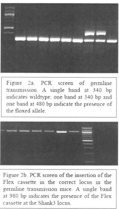
Generation of Shank3CKI: The Shank3 targeting vector was designed by inverting the PDZ domain (exons 13 to 16) and flanking it with the FLEx cassette, which is composed of one pair of LoxP sites staggered with one pair of Lox2722 sites. Shank3If conditional knock-in mice were generated by homologous recombination in RI embryonic stem cells and implanting the correctly targeted cells in C57 blastocysts using standard procedures. Correct locus insertion of the targeting construct into the genomic
DNA was determined by PCR genotyping using two primers EndF
(5'-GGCAGACTCCACACAGTTCCTG-3') and LoxR
(5'-GTATCCTATACGAAGTTATTCCGGGTCGAC-3'). Subsequent mouse genotyping
was determined by PCR of mouse tail or ear DNA using three primers. For the wildtype
(WT) allele, primer FuncF2 (5'-CGTTTGACACACATAAGCACC-3') and primer FuncFlipR4 (5'-CTCCACCTAGCTGAATTTCCC-3') were used to produce a band of
340 bp. For the knockout (Fx) allele, primer FuncF2
(5'-CGTTTGACACACATAAGCACC-3') and primer Gen_Flx_RI
(5'-GCTGACATCACATTGCTGCC-3') were used to produce a band of 481 bp. For the
rescue allele, primer FuncF2 (5'-CGTTTGACACACATAAGCACC-3') and primer FuncFlipR4 (5'-CTCCACCTAGCTGAATTTCCC-3') were used to produce a band of 408 bp.
Chimeric males were crossed to C57BL/6J females from Jackson Labs. The Fl hybrids were crossed with C57BL/6J P-Actin Flp to remove the Neomycin cassette. All progeny were bred onto the pure C57BL/6J (Jackson Labs) for at least two generations before being bred onto a mixed background with 129S1/SvlmJ (Jackson Labs). Heterozygotes were initially bred with heterozygotes to produce experimental animals. All germline
Shank3x/A (KO) and germline rescue (GR) mice along with their respective wildtype
littermates were produced by breeding heterozygotes with heterozygotes. For the adult
Shank3 rescue experiments, the Shank3 conditional knock-in line was crossed with
CAGGS-CreER (Guo et al, 2012). In order to produce enough animals for all necessary experiments, breeding strategy was switched to heterozygotes crossed with homozygotes and homozygotes crossed with homozygotes for all conditions (Shank3f'x+.:CreER+1 bred
with Shank3x/f:CreERK; Shank3l,: CreER+I bred with Shank3ft<I:CreER- ; Shank3+'1 :CreER+I bred with Shank3+1+: CreER1; Shank3*1:CreERI- bred with Shank3+1+:CreER~
/). It should be noted that all animals in the rescue condition were produced from the
same litters as the animals in the knockout condition. The animals were randomly assigned to different conditions. No computerized randomization program was used.
Animals were housed by genotype at a constant 23'C in a 12 h light/dark cycle (lights on at 07:00, lights dark at 19:00) with ad libitum food and water. Rescue treatment i.e. tamoxifen feeding was initiated on mice at 2-4.5 months. All electrophysiological and behavioral experiments were done at least 6 weeks after treatment in adult mice with the
experimenter being blinded to the genotypes. Only age-matched male mice were used for behavioral assays. All experimental procedures were inspected and approved by the MIT Committee on Animal Care.
Behavior
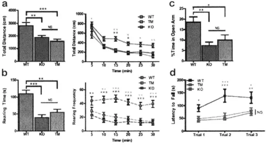
Open Field: An automated Omnitech Digiscan apparatus (AccuScan Instruments) was
used to assess spontaneous locomotion as previously described (Welch et al, 2007).
Anxiety-like behaviors were assessed by the following parameters: time spent rearing
and frequency of rearing. Locomotion was evaluated by the total distance traveled. The first 30 minutes were evaluated for all parameters. Statistical analysis was done using one-way ANOVA with Bonferroni multiple comparison tests.
Zero Maze: An elevated zero maze was illuminated such that the open arm was lit by 60 lux, and the dark arm was lit by 10-20 lux. Animals were habituated with 10-20 lux for at
least one hour before test. The animal was introduced into the closed arm and allowed to
freely explore the maze for 5 minutes, which was videotaped. An observer blinded to the
genotype performed analysis using an automated tracking software Noldus Ethovision. Anxiety-like behavior was assessed by the percentage of time spent by the animal in the open arm during the 5-min interval. Statistical analysis was done using one-way ANOVA with Bonferroni multiple comparison tests.
Rotarod: Animals were placed on a rotarod apparatus (Med Associates) that accelerates
4-40 rpm for 5 minutes. Each animal was tested for three trials with 1-2 hours between trials in a single day. All trials were videotaped. Latency to fall was manually analyzed for each trial by a blinded observer on Noldus Observer. The change in the latency to fall over the course of three trails indicates the quality of motor coordination. Statistical analysis was done using two-way repeated measures ANOVA with Bonferroni post-hoc tests.
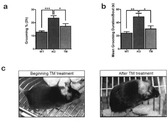
Grooming: Animals were individually placed into a novel cage and allowed to habituate.
Grooming behavior was videotaped for 2 hours from 19:00 to 21:00h with red light (2 lux). An observer blinded to the genotype manually quantified grooming behavior using Noldus Observer. All instances of face-wiping, scratching/rubbing of head and ears, and full-body grooming were counted as grooming behavior. Statistical analysis was done using one-way ANOVA with Bonferroni multiple comparison tests.
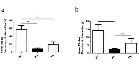
Novel Object Phobia: Animals were habituated in the behavior room (6 lux) for at least one hour prior to test. After test begins, each animal was placed into a 30 cm x 30 cm chamber (6 lux) containing a thin layer of bedding. After 15 minutes of the animal habituating to the chamber, a novel object was introduced to the center of the arena. The animal was allowed to explore the object for 5 minutes. An observer blinded to the genotype quantified the interaction of the animal with the object using NoldusEthovision.
counted as close interaction.Statistical analysis was done using one-way ANOVA with Bonferroni multiple comparison tests.
Social Interaction: A modified version of the three-chamber social interaction assay was used as previously described (Peca et al, 2011; Yang et al, 2012; Sheng and Kim, 2000). Only age-matched males were used for all tests. S129 males were used as stranger mice and were habituated to the test chamber for 3 sessions (20 minutes each) one or two days prior to the behavioral assay. On the day of the test, both test and stranger animals were habituated to the test room for at least one hour before the start of the assay. The left and right chamber of the three-chamber apparatus were both lit by 4-6 lux during the test session. Each test animal was first placed into the center chamber with open access to both the left and right chamber, each of which contained an empty wired cup placed upside down. This allowed the animal to habituate to not only the social apparatus, but also the cups that will eventually contain the stranger mice. After 15 minutes of habituation, the test animal was moved back to the center chamber briefly before the next session. During the social phase, an age-matched stranger mouse was placed randomly into one of the two side chambers while a novel object was placed into the other side chamber. The test animal was allowed to freely explore the social apparatus and demonstrate whether it prefers to interact with the novel object or the novel mouse. This social phase was also 15 minutes. The placement of the stranger mouse and the object was alternated between test mice to eliminate any confounds due to chamber bias. Time
stranger or the object was calculated. Analysis was done by an observer blinded to the genotype on Noldus Ethovision. One-way ANOVA with Bonferroni post hoc test was used for statistical analysis.
Oral Gavage
Tamoxifen Preparation and Feeding: Tamoxifen (Sigma #T5648) was dissolved in
corn oil at 20 mg/ml through vortexing. Freshly prepared tamoxifen was protected from light by aluminum foil and kept for 2-3 days at room temperature. Animal feeding needles from Harvard Apparatus (cat #52-4025) were used for oral gavage. To avoid toxicity of tamoxifen, the following dosages were used for adult animals:
Mice at 17-21 g body weight were fed 5 mg/day Mice at 22-25g body weight were fed 6 mg/day Mice at 26-29g body weight were fed 7 mg/day Mice at 30-35g body weight were fed 8 mg/day
The adult animals were fed for 5 consecutive days followed by two weeks of rest. Then the animals were fed for 5 more consecutive days followed by another two weeks of rest. Corn oil was fed as a control. The mice fed with tamoxifen and mice fed with corn oil were housed separately to avoid contamination.
For induction of Shank3 expression in P20-P21 animals, mice that weighed 7-9 g received 0.1 ml of tamoxifen or corn oil per day for two to three consecutive days. Mice
that weighed 10-12g received 0.15 ml of tamoxifen or coil per day for two to three consecutive days.
Mechanistic Studies:
Western blot:
PSD and synaptosomal fractions of the striatum, cortex, and cerebellum were prepared as
previously described (Welch et al, 2007; Peca et al, 2011). Purified fractions were
separated on SDS-PAGE and quantified using Odyssey Licor.
P-Actin
and Tubulin wereused as loading controls. Specific primary antibody for SAPAP3 was prepared as previously described (Welch et al, 2007; Peca et al, 2011). Commercial antibodies used
include SHANK3 (Santa Cruz SC-30193), GluRI (Millipore MAB2263), GluR2 (Neuromab 75-002), NR1 (BD Biosciences 556308), NR2A (Millipore 07-632), NR2B (Millipore 05-920), Homerl (Chemicon AB5877, Synaptic Systems 160022), Homer3 (Synaptic Systems 160303), mGLUR5 (Upstate 06-451), CaMKIla (Millipore 05-532), ShankI (Synaptic Systems 162002), Shank2 (Cell Signaling 12218S), B-Actin (Sigma
A5441), and Tubulin (Sigma T5168). Statistical analysis was done using two-tailed
Students' t-tests.
Dendritic Spine Analysis:
Mice from WT (N=5), KO (N=4, 1 had to be euthanized due to lesion development), and TM (N=5) conditions at 6 to 12 months old, age-matched males were used. The pAAV-hSyn 1 -EGFP-P2A-EGFPf-WPRE-HGHpA construct was designed in-house and sent for
commercial viral packaging by Upenn Viral Core with serotype 2/9. To achieve sparse labeling, -8-30 uL of this virus was injected through the retro-orbital route into each mouse. Three weeks after viral injection, the mice were transcardially perfused and sectioned into 200 micron slices. Immunohistochemistry was performed by staining the slices with anti-GFP antibody (Invitrogen A 11122) for 48 hours and 24 hours of secondary antibody incubation. The stained slices were then surrounded by a 240-um depth spacer (Electron Microscopy Sciences) and mounted with Vecta Shield Mounting
Media.
Confocal images were taken with a 60X objective of the dorsal striatum. Spine count on intact neurons began 30-40 um away from the soma and was extended for 10-60 um from the origin. Spine density was analyzed automatically by Neuron Studio. All virus injections, imaging, and software analysis were done with the experimenter blinded to the mouse genotypes. Statistical analysis was done using one-way ANOVA, Newman-Keuls post-hoc test.
Electrophysiology
Dorsal and ventral striatum: Acute striatal slices were prepared from 3-7 months old
age-matched mice. Animals were anesthetized by avertin intraperitoneal injection (tribromoethanol, 20mg/ml, 0.5mg/g body weight) and transcardially perfused with ice-cold oxygenated NMDG-based cutting aCSF solution (mM): 92 N-methyl-D-glucamine
Na-ascorbate, 3 Na-pyruvate, 0.5 CaCl2, 10 MgSO4 (-300mOsm, 7.2-7.4pH). Following
decapitation, brains were removed for sectioning in the same ice-cold cutting aCSF using a Vibratome 1000 Plus (Leica Microsystems, USA). For all dorsal striatal recordings, 300 tm coronal slices were prepared, unless otherwise stated. For all NAc core recordings,
300 pm parasagittal slices were prepared and NAc core was identified by the presence of
anterior commissure. Slices were recovered in the same cutting aCSF solution at 32'C for 12 min and transferred to room-temperature carbogenated regular aCSF(mM): 119 NaCl, 2.5 KCl, 1.2 NaH2PO4, 24 NaHCO3, 12.5 glucose, 2 MgSO4.7H20, 2 CaCl2.2H20 (~300mOsm, 7.2-7.4pH). Slices were allowed to recover at least >lh and transferred to a recording chamber (RC-27L, Warner Instruments) prior to recordings. Stimulations were performed using a platinum iridium concentric bipolar electrode (CBAPC75, FHC). For dorsolateral striatum coronal slices, electrode was placed at the corpus callosum to mainly stimulate corticostriatal axons. For dorsolateral striatum parasagittal slices, electrode was placed at the cortex between layer V and VI. For ventral striatum parasagittal slices, electrode was placed dorsally to the anterior commissure at the border between NAcc core and the cortex. Afferents were stimulated with 0. 1ms stimulation step
(Isoflex, AMPI) delivered at 0.05Hz frequency (unless otherwise stated). Slices were
visualized under IR-DIC (infrared-differential interference contrast) using a BX-51WI microscope (Olympus). All slice preparations, recordings and data analysis were performed with experimenter blinded to the genotypes.