XX. CUTANEOUS SENSORY MECHANISMS
Prof. R. Melzack J. Parchesky
P. D. Donahue F. L. Knighton
A. NEUROPSYCHOLOGICAL EFFECTS OF EARLY SENSORY RESTRICTION 1. Introduction
Severe restriction of the early perceptual experience of animals produces profound disturbances in their development.l' 2 Perhaps the most striking feature of the behavior of dogs reared in a restricted environment is the low level of behavioral disruption after burn and pinprick. Seven of 10 Scottish terriers that were observed made no escape responses (apart from reflexive movements) when the experimenter hit their noses with flaming matches. Three dogs repeatedly poked their noses into the flame and sniffed at it as long as it was present. The other four did not sniff at the match, but made no attempt to get away after stimulation. Only three of the restricted dogs squealed after making contact with the flame, and two of these made no attempt to escape.
The behavior of the restricted Scotties to repeated jabs with a dissecting needle was almost identical to that produced by the flame. Moreover, they were observed to bang their heads on low water pipes, one dog having done this more than 30 times in a single hour, without any response to indicate pain perception. Strong electric shock, however, elicited highly disturbed behavior in all of the dogs, their characteristic response being a diffuse, unpatterned, emotional excitement.
2. Hypothesis and Objectives of the Experiment
The cause of the low level of response to noxious stimuli is not clear. One hypoth-esis is that in the absence of earlier experience with the normal environment the dogs are unable to select relevant information from the total sensory input. All stimuli are equally novel, have equal importance, and receive the same degree of attention. The high level of activity shown by these dogs, including "whirling fits"3 that resemble some aspects of an epileptic seizure, lends plausibility to the hypothesis. It suggests that the dogs are unable to respond adaptively to selective information and respond instead with a diffuse, undifferentiated excitement to the total sensory input.
In a sense, this might be called a "confusion" hypothesis: the dogs are so aroused and distracted by the whole unfamiliar environment surrounding them that they fail to attend to the noxious stimulus. It is possible, then, that the absence of facilitation pro-vided by central nervous system activities involved in attention could prevent the normal
This research was supported in part by the National Institutes of Health (Grant M-4235-(CZ)).
central selection and transmission of the nerve-impulse patterns evoked by the noxious stimulus.
This hypothesis can be examined experimentally. (a) Electrical activity can be recorded from brainstem pathways transmitting nerve impulses after noxious stimula-tion4 , 5 to see whether or not the normal patterns are evoked when restricted dogs are in their usual excited state or when they are under the influence of drugs that sedate or "tranqualize," which should lower the excitement level. (b) The dogs' EEG can be examined for physiological signs of the high level of behavioral excitement; we predict that there would be a higher percentage of low-voltage, fast EEG activity (typical of behavioral arousal) in the restricted dogs than in the normal controls.
3. The Present Restriction Experiment
Beagles are being used as subjects in the present experiment, since a recent stereotaxic atlas for the beagle permits accurate placement of recording and stimulating
6
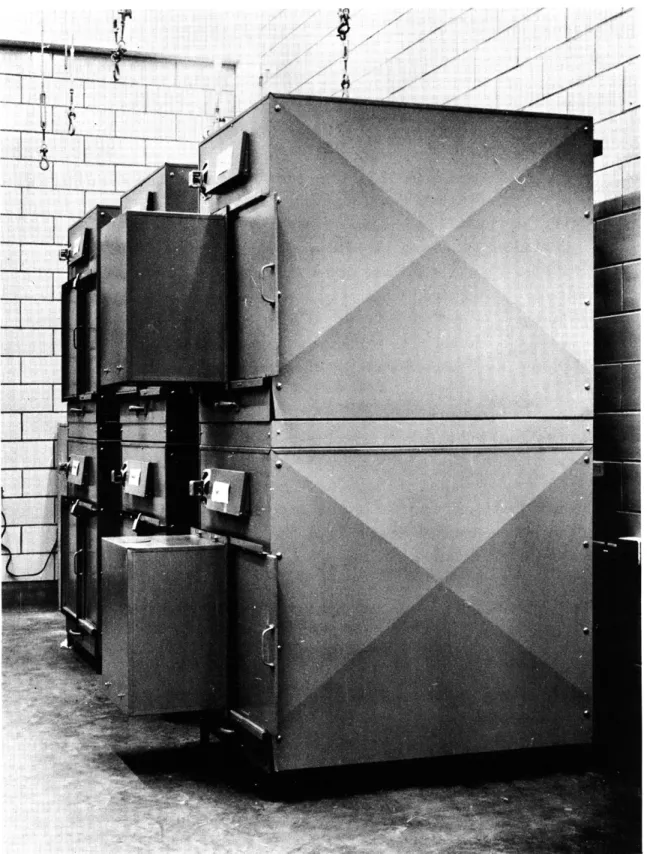
electrodes in their brains. During the past year and a half, 3 litters of purebred beagles were split into 2 groups: an experimental group of 8 dogs was placed in restriction cages at the age of 3 weeks, and a control group of 6 dogs was raised on a farm. The restric-tion cages (Fig. XX-1) are specially designed to permit care and feeding of the dogs without their being able to see out of the cages or coming into contact with the experi-menter. Each dog is raised alone in the cage from 3 weeks to at least 9 months of age, that is, from time of weaning to adolescence. Continuous light is provided in the cage, as well as auditory and olfactory stimulation. The variety of patterned stimulation, however, is drastically restricted by the rearing procedure.
Two of the restricted dogs were removed from the cages at 10 months of age and have undergone preliminary observations and tests during the past 6 months. Two have been in the cages for 18 months and will be removed when they are 20 months of age. Four others have been in restriction for 6 months. All of the control dogs, except one, spent the first 8 months of life on a farm. They were brought to our laboratory, are being kept 2 in a cage, and are frequently allowed to run about in a large outdoor area.
4. Preliminary Behavioral Observations
The behavior of the 2 beagles removed from restriction is strikingly similar, in most respects, to the behavior of the Scotties. One of them crashed violently into the wall of a large wooden test box (after running 25 feet from his cage) every day for more than a month without ever giving any indication of pain. This dog, during a testing pro-cedure described below, also struck a door in the apparatus with great force at least
~II,
Fig. XX-1.
Restriction cages for dogs.
~"9~ -1 d -. -Y t
head without ever wincing or squealing.
The other dog accidentally cut both her rear
paws, tearing the muscle until bone was exposed, without showing any signs of pain.
Response to burn and pinprick will be investigated systematically when the physiological
and behavioral recording apparatus (described below) becomes operational.
Other unusual forms of behavior have been observed.
It was noticed, for example,
that some of the restricted dogs, at approximately 6 months of age, suddenly stopped
eating or ate only a portion of the food. This continued for as long as 5 days, although
they appeared otherwise to be alert, got up on their hind legs, and frequently shifted
position.
Their peculiar stretching movements, however, revealed that these dogs had
their hind paws caught in the wire mesh floor of the cages.
Only when the cage door
was opened so that they could be anesthetized did they become violent and try to escape.
The two restricted dogs that have been observed closely during the past 6 months
also show a high level of excited activity.
At moments of peak excitement, they have
"whirling fits"
3in which they turn in narrow circles at great speed. This "whirling"
behavior may even occur in the home cage when a new kind of food, such as a dog biscuit,
is presented.
Another observation of interest was the failure by these dogs to jump a distance of
1 foot from their cage to the floor. One of the dogs teetered at the edge of the cage with
great excitement, making abortive jumping movements, and then turning back, as though
at the edge of a precipitous fall. After 2 or 3 minutes the dog was lifted down.
This
apparent anomaly in space perception continued for 12 days, when the dog finally fell
out of the cage.
There was one-trial learning, however, for the dog subsequently jumped
every time the door was opened, even from heights of 5 feet.
The other dog, during the
6-month observation period, has jumped only once from the 2-foot height.
Finally, these 2 dogs still ignore other dogs.
They have been heard to bark only
once even though they now live in a room full of barking dogs.
They still struggle when
they are held or patted and show none of the play behavior observed in their normal
littermates.
All of these observations, taken together, indicate a remarkably infantile level of
behavior compared with their normally reared littermates.
5.
Current Activities
In accordance with the aims of the experiment, the dogs are currently undergoing
surgical procedures in which electrodes are implanted at a number of different
subcorti-cal and cortisubcorti-cal points. Specifisubcorti-cally, the electrodes are implanted in the medial
lemnis-cus, the spinothalamic tract, the mesencephalic central gray, and the central tegmental
tracts, all of which carry at least a major part of the impulse patterns signalling noxious
(XX. CUTANEOUS SENSORY MECHANISMS)
area,7 together with electrodes at the auditory and visual cortex, will permit recording of the EEG. Behavioral apparatus for classical conditioning and avoidance learning is now complete, and the dogs will be trained to press two panels to stop electric shock and burn. Concurrently, studies will be made of other problems such as development of sleep patterns and the effects of drugs on the dogs' behavior. Systematic experimenta-tion is scheduled to begin in approximately 2 months, when the telemetering system (described below) becomes operational.
6. Four-Channel EEG Radio Telemetering System
Because of the high activity level of the restricted dogs, including such behavior as the "whirling fits, " the conventional cable-transmission methods for physiological
recording have been found inadequate. The cables twist badly, and often cause artifacts. Even more serious are the physical restrictions imposed by the cables. It is virtually
impossible to record electrical activity when the dogs are in their home cages or moving freely in the outdoor enclosure. For these reasons it became essential to have a method for telemetering the EEG at a distance.
Kamp and Storm van Leeuwen8 have already described a two-channel EEG radio telemetering system. By making radical revisions in their original design, and intro-ducing a tunnel diode FM transmitter, we could replace the HF transmitter in their original circuit, and thereby produce a more compact unit. This revision, which was made in close collaboration with D. F. O'Brien, F. T. Hambrecht, and R. Horter, provides a small unit that should be capable of transmitting 4 EEG channels.
The unit is comprised of two separate systems, each of which contains two identical differential amplifiers, a submodulator, and a tunnel diode FM transmitter. Eash sys-tem is tuned to a different specific frequency. Individual power supplies for the syssys-tems are enclosed in the package. Each of the 4-stage EEG amplifiers has a gain greater than 2000. Emitter-follower input stages provide for an input impedance of 300 kohm. Figure XX-2 shows a block diagram of the complete system, including the receiver. The submodulators control both pulse frequency and pulse duration. In each system the outputs of the two amplifiers are connected to a multivibrator and a monostable trigger. The pulse duration of the monostable trigger varies linearly with the output voltage of amplifier I. The output voltage of amplifier II affects frequency modulation of the multivibrator square wave. As the monostable trigger is started by the multi-vibrator, the output pulses of the monostable trigger vary in duration, as well as in repetition rate, in accordance with the EEG amplifier output voltages. The output pulses of the modulator, which have a mean repetition rate of 4000 cps, thus vary simultane-ously in frequency and duration. These output pulses are coupled to an FM transmitter by means of a G. E. 1N2939 tunnel diode. This circuit was chosen for its simplicity;
ANTENNEA
Fig. XX-2.
Block diagram of the four-channel EEG radio
telemetering system.
it is less bulky and requires fewer components than the original HF transmitter. Tested
on the bench, this transmitter has an operating range in excess of 100 feet.
The simplified block diagram of the EEG telemetering demodulator (Fig. XX-3)
shows that the output of channel II is obtained by measuring the peak voltage of a
capaci-tor that is periodically charged during a time equal to the duration of the ratio-deteccapaci-tor
output pulses.
The leading edge of these pulses also starts the monostable trigger.
Integration of the trigger output voltage by means of a lowpass filter gives the EEG of
channel 1. The complete system has a bandwidth from 1. 5 cps to 200 cps.
With a
10 kohm resistor across the amplifier inputs, the equivalent noise level of the entire
(XX. CUTANEOUS SENSORY MECHANISMS)
T__jL NOISE BISTABLE T MONOSTABLE LOWPASS
DISCR. SUPPRESSOR TRIGGER TRIGGER FILTER (PROPORTIONAL TO
OUTPUT REPETITION RATE)
RECEIVER
PEAK CHARGINGCIRCI T
VOLT OUTPUT II
CIRCUIT METER (PROPORTIONAL TO
PULSE DURATION)
Fig. XX-3. Block diagram of the demodulator for the EEG telemetering system.
approximately 400 grams. The receivers used are Harman Kardon Citation III Profes-sional FM Broadcast Tuners.
The apparatus will soon be used for telemetering the EEG of two control dogs.
7. Thermal Recording Methods
It is assumed that the EEG of the restricted dogs will contain prolonged periods of low voltage and fast activity (the "arousal pattern") that parallel the high level of behavioral excitement. It is possible, however, that the EEG will fail to differen-tiate between levels of brain activity in the two groups. Since heat production pro-duced by metabolic processes is another index of central-nervous-system activity, it is possible that records of thermal changes in the brain may provide better dis-crimination than the EEG.
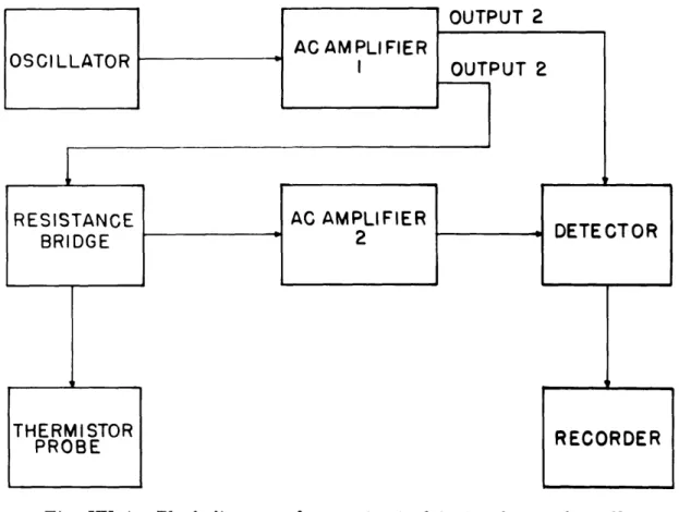
For this reason it was decided to develop a method for recording small changes in brain temperature. The purpose was to develop an instrument capable of detecting and recording changes of temperature in the brains of animals on the order of 0. 001 C in the region 35°-40°C. The most practical solution to the problem appeared to be a thermistor in an ac resistance bridge.
An ac bridge was designed and is under development. A block diagram is shown in Fig. XX-4. The oscillator produces a sine wave at a frequency of approximately 2 kc. The signal from the oscillator is amplified by amplifier no. 1, and output no. 1 is fed into the detector to produce a reference voltage. Output no. 2 is fed to the resistance bridge. A zero temperature level is chosen for the thermistor probe and the bridge is balanced for a null at that temperature; that is, there is no input to amplifier no. 2 and consequently no change in the voltage at the detector. If the temperature of the thermistor probe should be changed, the balance of the bridge would be upset and there would then be an input to amplifier no. 2 which in turn would produce a change in the reference voltage at the detector which would then
recorded.
Fig. XX-4.
Block diagram of apparatus to detect and record small
temperature changes.
developed except for possible future improvements.
The resistance bridge is at present
under development, and work on the thermistor probe will soon begin.
8.
Studies in Visual Discrimination
The frequent failure of the restricted dogs to discriminate noxious stimuli from the
total environmental input raises an important question: Are other sensory systems
simi-larly affected?
None of the earlier investigators studied visual or auditory perception
in these animals. An experiment was therefore undertaken to study the effects of early
restriction on the discrimination of simple visual stimuli.
The subjects of the experiment were two restricted dogs and three control
litter-mates. They were tested in a two-choice visual-discrimination apparatus, in which the
positive and negative stimuli were presented randomly on one or the other side of the
choice point.
The alley from the start box to the choice point was 4 feet long. The
positive stimulus was carried on a door that could be pushed open, giving the dog access
to a food reward.
The door holding the negative stimulus was locked, although there
(XX. CUTANEOUS SENSORY MECHANISMS)
start box. The dogs were given 10 trials per day until they ran to the correct door 18 times on 2 successive days.
Shaping of behavior in the test apparatus was carried out with a light-on-light-off discrimination. The dogs were trained to run to the lighted area for food and to avoid the dark area containing the locked door. The doors carried no stimulus cards during the test, so that the discrimination was based on luminous flux rather than patterned visual stimuli. There was no difference between the two groups in the number of errors made in the course of learning this simple problem.
I0
9
8
7
-
RESTRICTED DOGS
a
DULLA
o
SIN
----
CONTROL
DOGS
2
4
6
8
10
DAYS
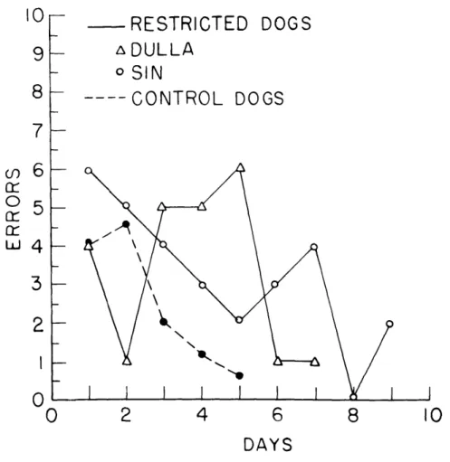
Fig. XX-5.
Error scores made by restricted and control dogs in solving
a white (+)-black
(-)
discrimination problem.
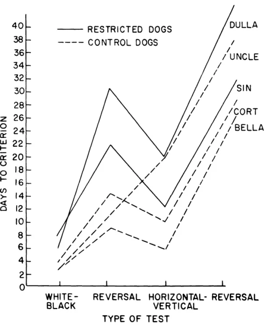
Striking differences were obtained, however, when the dogs had to discriminate
between a white card (positive stimulus) and a black card (negative stimulus) on the
doors (Fig. XX-5).
The response patterns of the two restricted dogs were almost
identical: each showed rapid initial learning of the problem, presumably on the basis
of luminous flux alone, which was followed by a sharp rise in errors before he finally
achieved criterion performance.
The sudden increase in errors was accompanied by
vicarious trial and error behavior at the choice point, in which the dogs appeared
suddenly to become aware of the cues provided by the cards on the doors. The control
dogs, on the other hand, showed the typical smooth fall in errors.
0
n
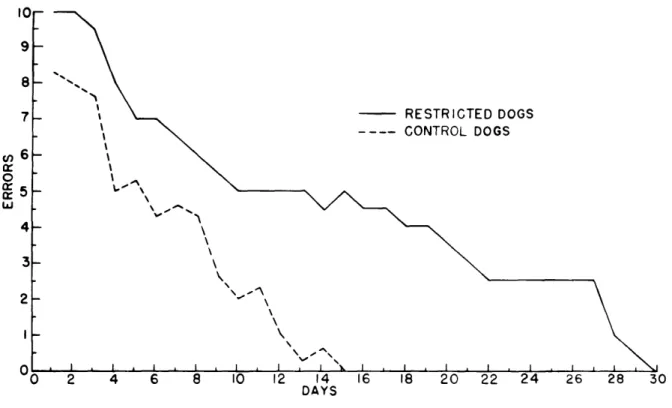
O cr5 i - RESTRICTED DOGS CONTROL DOGS \ -6 8 10 12 14 DAYS 16 18Fig. XX-6.
Error scores made by restricted and control dogs in solving
a reversal black (+)-white
(-)
discrimination problem.
The differences between the two groups were even more marked in reversal training, in which the black card now signalled food and the white card was on the locked door (Fig. XX-6). The control dogs showed a gradual decrease in errors, while both restricted dogs made high error scores for a prolonged period.
The next discrimination, between horizontal and vertical lines, failed to show a clear-cut difference between the two groups. But the primary reason for the failure was totally unexpected: one of the "normal" control dogs ("Uncle") underwent a remark-able change in "personality" and, in terms of both general behavior and error scores, became indistinguishable from the restricted dogs. He showed a continuously high level of behavioral excitement and struggled violently when he was picked up. At the same time he developed a strong position habit, as well as "superstitious" behavior patterns such as turning two complete circles before responding to the stimuli. His shift in error scores from one group to the other is seen clearly in Fig. XX-7.
L/ \
(XX.
CUTANEOUS SENSORY MECHANISMS)
S24
/
/BELLA
S22-S20
/
0
/
/
16-
/
I
0I
I
I
2-WHITE-
REVERSAL HORIZONTAL- REVERSAL
BLACK
VERTICAL
TYPE
OF TEST
Fig.
XX-7.
Scores made by restricted
and control dogs in
solving four
visual discrimination problems.
These results have interesting implications for earlier experiments that show that
animals raised without patterned visual stimulation fail to discriminate between simple
visual patterns at maturity. These effects have been attributed to a simple absence of
memory traces ("cell assemblies") corresponding to lines, angles, arcs, and so forth.
Since the restricted dogs received patterned visual stimulation in their cages, yet had
similar difficulty in performing a simple visual discrimination, other factors, such as
attention and level of EEG arousal, must also be considered.
It is possible to account
nervous system to select (or abstract) the relevant patterns from the total input; that is,
in terms of mechanisms described above to account for the frequent failure to respond
to noxious stimuli.
R. Melzack, P. D. Donahue, J. Parchesky
References
1. R. Melzack and T. H. Scott, The effects of early experience on the response to
pain, J. Comp. Physiol. Psych. 50, 155-161 (1957).
2.
W. R. Thompson and R. Melzack, Early environment, Sci. Am. 194 38-42
(1954).
3.
W. R. Thompson, R. Melzack, and T. H. Scott, "Whirling behavior" in dogs as
related to early experience, Science 123, 939 (1956).
4.
D. I. B. Kerr, F. P. Haugen, and R. Melzack, Responses evoked in the
brain-stem by tooth pulp stimulation, Am. J. Physiol. 183, 253-258 (1955).
5.
R. Melzack, W. A. Stotler, and W. K. Livingston, Effects of discrete brainstem
lesions in cats on perception of noxious stimulation, J.
Neurophysiol. 21, 353-367
(1958).
6.
R. K. S. Lim, C. N. Lin, and R. L. Moffitt (eds.), A Stereotaxic Atlas of the
Dog's Brain
(C.
C. Thomas Publishing Company, Springfield, Ill., 1960).
7.
R. Melzack and F. P. Haugen, Responses evoked at the cortex by tooth
stimula-tion, Am. J. Physiol. 190, 570-574 (1957).
8.
A. Kamp and W. Storm van Leeuwen, A two-channel EEG radio telemetering
system, EEG Clin. Neurophysiol. 13, 803-806 (1961).
9.
A. H. Riesen, Functions of Varied Experience, edited by R. Fiske and S. Maddi
(The Dorsey Press, Homewood,
1T1.,
?
61), p. 57.
B.
PROBLEMS OF SOMESTHESIS
1.
Introduction
In addition to the specific problems raised by the abnormal behavior of the restricted
dogs, there is a more general question concerning the whole problem of somesthesis. It
has long been assumed that pain is a specific sensory modality in which there is a
one-to-one relationship between stimulus, sensation, and response. Yet the responses of
the restricted dogs to noxious stimuli are clearly not proportional to stimulus intensity.
Complex central nervous system activities, based in part at least on early experience,
intervene between stimulus and response.
The assumption of a one-to-one relationship dates back to the discovery made,
ca. 1885, that the skin has a punctate sensitivity to warm and cold stimuli applied with
small tips (usually of 1-mm diameter). In 1895, von Frey integrated this observation
with histological demonstrations of specialized end organs in the skin and with the widely
held theory of Miller that psychological quality is determined by the terminations of
(XX. CUTANEOUS SENSORY MECHANISMS)
sensory pathways in the brain. Thus the finding of "warm spots" and "cold spots" led to the assumption that beneath each spot lay a specific "warm" or "cold" receptor that was presumed to make direct connection with a warm or cold "center" in the brain. Von Frey extended these concepts to include touch and pain, and his specificity theory remains the dominant theory in the field.
The inadequacies of current theories of somesthesis, often based on eroneous obser-vations, led us: (a) to make a critical evaluation of the current concepts and to attempt to propose an alternative formulation (with P. D. Wall); (b) to carry out an experimental
study of skin sensitivity (with G. Rose and D. McGinty); and (c) to attempt to develop a questionnaire for evaluating the variety of perceptual experiences that we normally cate-gorize under the single heading of "pain" (with Dr. Warren Torgerson of Lincoln Labo-ratory, M.I.T.).
2. Theoretical Consideration of Skin Sensory Mechanisms
An examination of the problems of somesthesis carried out by P. D. Wall and the writer has led to a new theoretical formulation. These ideas have been described in two papers that are to be published. The main points covered in these
papers are:
(i) Skin receptors have specialized physiological properties for the transduction of particular kinds and ranges of stimuli into patterns of nerve impulses rather than modality-specific information.
(ii) Certain aspects of temporal and spatial patterns of impulses produced by stimulation of the skin may be filtered out presynaptically by the properties of terminal arborizations.
(iii) Central cells can detect some characteristics of stimuli from the impulse pat-terns arriving from the skin by their properties of threshold, temporal summation, and adaptation.
(iv) Central cells can detect some characteristics of stimuli from the impulse patterns arriving from the skin by their property of spatial summation.
(v) Central cells can detect some characteristics of stimuli from the impulse pat-terns arriving from the skin as a result of the special connections of afferent systems.
(vi) More than one nerve impulse from a single afferent fiber, or more than one fiber carrying single nerve impulses, is essential for central cells to detect the charac-teristics of a sensory stimulus.
(vii) The somesthetic system is a unitary, integrated system comprised of special-ized component parts.
(viii) Every discriminably different somesthetic perception is produced by a unique pattern of nerve impulses.
3.
Fluctuation of Skin Sensitivity to Thermal Stimuli
Since the concept of the "skin spot" is the basis of most current theories of skin
sensory mechanisms, a study was carried out with G. Rose and D. McGinty on the
dis-tributions of skin sensitivity in large areas of skin. The following summarizes the main
results of the study:
a.
Limitations of the Technique of Punctiform Stimulation
It became clear at the outset that the scaling of the signal provided by the traditional
1-mm stimulator tips was frequently a difficult task for the subjects. In any mapping
session perhaps one-half the stimulations elicited a prompt, confident report; but the
remainder presented the subjects with a difficult decision.
The warm stimuli in
partic-ular felt unnatural. Temperatures below 42
0C were difficult to detect as "warmth" when
applied with small tips. With higher temperatures, however, ranging between
420
and
45°C, tips of 1-mm diameter frequently produced reports of faint pricking pain. Larger
tips of 2. 5 mm and 5 mm produced unpleasant stinging pain. Some of the subjects
actu-ally objected that they were being jabbed with a pin, although they were being stimulated
as gently as possible with the warm flat tip. Even with tips as large as 10 mm there
were reports of burning, stinging sensations, although brief immersion of the fingers
or the whole hand in water in this temperature range was perceived unequivocally as
pleasant warmth.
These difficulties were not encountered with the cold stimuli.
Cold applied with
small diameter tips was perceived as having a "natural" quality, without any biting,
painful concomitants.
With a tip diameter of 2.
5
mm, the perceptions varied primarily
in intensity and the subjects had little difficulty in making the intensity judgments.
b.
Patterns of Skin Sensitivity to Small Thermal Stimuli
More than a hundred maps of various sizes were made of distributions of cold
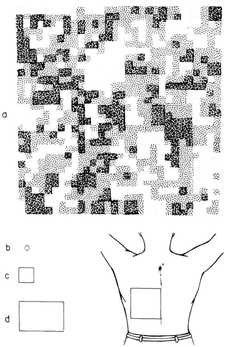
sensi-tivity on the back of the hand, on the upper forearm, and on the back.
Figure XX-8
shows a map of cold sensitivity made on approximately a quadrant of the back.
The
results are essentially the same in all of the maps: instead of randomly distributed
spots, they contain one or more large sensitive areas whose stimulation produces
reports of strong cold, surrounded by less sensitive areas that elicited moderate and
mild cold.
The characteristics of these sensitive areas are observed clearly only when
maps are made of sufficiently large areas of skin. They demonstrate that the thermally
sensitive surface of the skin consists of large fields having a circumscribed area with
distinct boundaries. Since the size of some of these fields is larger than the total areas
of the maps made by earlier investigators,1,2 it becomes obvious why they have not
been found in previous studies.
b
o
d
Fig. XX-8.
Distribution of sensitivity to cold on a quadrant of the back. P shows
the sensitivity distribution; the square drawn on the back (lower right)
represents the area stimulated. b represents the size of the
stimu-lator tip used in this experiment.
c and d represent areas studied
by earlier investigators.
White areas represent reports of no cold,
darkest stipple represents reports of strong cold. Intermediate shades
of stipple represent mild and moderate cold.
c.
Fluctuation of Thermal Sensitivity Distributions
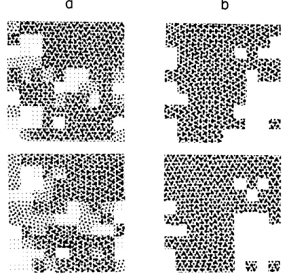
Fluctuation of thermal sensitivity from map to map of the same area has long been 3
known. Our study shows that these fluctuations generally take two forms: (a) marked changes occurred primarily at the boundaries of the large sensitive fields while the cen-tral portions remained stable, and (b) the fields fragmented or coalesced in successive maps, forming new distribution patterns that remained in the same general portion of the map.
a
b
..
..
sx
..
.
. .
I
[tro,,',,
AV W..•0 ,,° t , -. .• ..
of cold sensitivity remain strikingly similar.
Fig. XX-10. The first three maps of cold sensitivity were made in the morning and the... XX-9. s ce t, . oa Af ey WA &,oW; T.. m . . s
,he criteri. of up &.V oi., " A•iat .,!. ... s v .
Fig. XX-9. Successive maps of sensitivity to cold. a shows two maps made by using
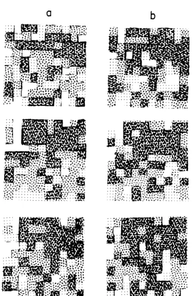
(ii) The fragmentation and coalescence of sensitive areas are illustrated in Fig. XX-10. The first three maps of cold sensitivity were made in the morning and the last three were made four hours later. The maps made in each session were separated
(XX.
CUTANEOUS SENSORY MECHANISMS)
a
b
. *.
*
.
4:
..
...
Fig. XX-10.
Six successive maps of cold sensitivity. The maps in column a were
made in the morning; the maps in column b, of the same skin area, were made in the afternoon.
by 10-minute intervals. It is seen that the sensitive fields in the upper right-hand corner fragment and coalesce from map to map, producing continually changing patterns. Never-theless, the general distribution of sensitivity to cold has a similar over-all configura-tion in all of the maps.
d. Effects of Adaptation on Thermal Sensitivity Distributions
It is well known that skin sensitivity adapts to continuous thermal stimulation.3 The effects of adaptation brought about by 10 minutes of continuous cooling or warming on the distribution patterns of cold sensitivity is shown in Fig. XX-11. It is seen
::: ..
::
::
::::.
u
: ....
...
..
..
. ... , - .
. . . . . . . . . . . . . 4 .. .. , . . .. .. " ... .... .... . • .: ., -... .. . . ... ...
..
......
. o .. t.. . . . .. . . . . . . . . •, .. ... .._ .. .....': . 5.:.i
... ..
...
..
. . . .
..
i::*
... : ..'. .'
.rI'
U"-,
. . .... .. .5 ,, . ... . ' " ' • ll . . . . I d • " '' "'" .. ... 4 ' • 'r 1,. . .. . . . .. . . ., ? i
!!!i
_ ::
!
• - ,'.'p ,,. . .e ::.... ... ,: .. .. ... ....: T :Fig. XX-11.
~s~t. .. (' : j ~ (~ ~~ ~. .L ; ~Lf.)'. IU~j . ;-~~YI' ' r.t t T;fi .~:r :rr .I. 4' . ;Z S:18 ...
"''
c
~,tr,-i
,, ".'tir,. r ; -. .~~ ,, . r. r r fWA RM ING
Effects of cooling and warming of the skin on distributions of cold sensitivity.
that cold adaptation produces a marked narrowing in the sensitive fields while warm
adaptation produces a small (but consistent) expansion of the fields.
e.
Implications of the Results
The difficulties encountered with small warm stimulators underscore the artificiality
of our present methods for studying skin sensitivity. More important, however, is the
implication that, for warmth at least, the "spot" is not the basic unit of skin sensation.
The "punctate" concept presumes that the quality of sensory experience is independent
(XX. CUTANEOUS SENSORY MECHANISMS)
of stimulator size. The results show that such is not the case.
The anomalous perceptions produced by warmth stimulation, taken together with the observations that thermal sensitivity is distributed in the form of large fields and that these distributions are in continual flux, takes us away from the traditional concept of a static, straight-through transmission system from receptor to brain center. The data suggest a picture of great plasticity in which it is reasonable to assume that the skin never has the same sensititivy distribution twice. How the central nervous system copes with the continuous flux of such a system presents an intriguing problem.
4. Questionnaire for Evaluation of Pain Perceptions
The assumption of a one-to-one relationship between stimulus intensity and sensation has promoted methods for evaluation of pain intensity but none for variations in quality. Yet the English language contains more than a hundred words that are used to describe the nuances and subtleties of the many different perceptions that we categorize under the single label of "pain." Dr. Warren Torgerson has long been interested in psychometric scaling methods, and a collaborative study was undertaken to categorize words describing the different qualities of pain and their variations in intensity. At present, we have a preliminary check-list of words that fall into the following classes (with some sample words presented for each):
a. What kind of a pain is it? (It is, or feels like, a(n) .) ache stab cramp burn, etc.
b. The area or organ or tissue involved: (i) Active: (It .)
aches smarts stings burns, etc. (ii) Passive: (It is .)
tender sore inflamed numb, etc. (iii) Nonpainful sensations: (It -- .)
twitches pulses throbs pounds, etc. c. The pain itself:
(i) Active: What the pain does (It- .) flutters spreads pierces flashes, etc.
(ii) Passive: What are its static qualities? (It is .) steady constant diffuse sharp, etc.
d. Causal description or analogy:
(i) The pain feels as if the area is being . burned crushed wrenched tickled, etc. (ii) The pain feels as if the area has been __ .
e.
How does the person feel?
(i) I am
.
dizzy
faint
feverish
nauseated, etc.
(ii) I feel .
dizzy
faint
feverish
nauseated, etc.
(iii) I feel
.
calm
jittery
alert
sleepy, etc.
(iv) I feel - .