Selective Neurofilament (SMI-32, FNP-7
and N200) Expression in Subpopulations of
Layer V Pyramidal Neurons In Vivo and
In Vitro
Courtney C.J. Voelker1, Nathalie Garin2, Jeremy S.H. Taylor1, Beat H. Gähwiler3, Jean-Pierre Hornung4 and Zoltán Molnár1,4 1Department of Human Anatomy and Genetics, University of Oxford, South Park Road, Oxford, OX1 3QX, UK, 2Swiss Institute for Experimental Cancer Research, Ch. Boveresses 155, 1066 Epalinges, Switzerland, 3Brain Research Institute, University of Zurich, Winterthurerstrasse 190, CH-8057, Zurich, Switzerland and 4Institut de Biologie Cellulaire et de Morphologie, Rue Du Bugnon 9, 1005 Lausanne, Switzerland
There are two main types of layer V pyramidal neurons in rat cortex.
Type I neurons have tufted apical dendrites extending into layer I,
produce bursts of action potentials and project to subcortical targets
(spinal cord, superior colliculus and pontine nuclei). Type II neurons
have apical dendrites, which arborize in layers II–IV, do not produce
bursts of action potentials and project to ipsilateral and contralateral
cortex. The specific expression of different genes and proteins in
these two distinct layer V neurons is unknown. To distinguish
between distinct subpopulations, fluorescent microspheres were
injected into subcortical targets (labeling type I neurons) or primary
somatosensory cortex (labeling type II neurons) of adult rats. After
transport, cortical sections were processed for
immunohistochem-istry using various antibodies. This study demonstrated that antigens
recognized by SMI-32, N200 and FNP-7 antibodies were only
expressed in subcortical (type I) — but not in contralateral (type II)
— projecting neurons. NR1, NR2a/b, PLC
β1
, BDNF, NGF and TrkB
antigens were highly expressed in all neuronal subpopulations
exam-ined. Organotypic culture experiments demonstrated that the
devel-opment of neurofilament expression and laminar specificity does not
depend on the presence of the subcortical targets. This study
suggests specific markers for the subcortical projecting layer V
neuron subpopulations.
Keywords: cell differentiation, corpus callosum, intracortical and
intercortical connections, spinal cord, superior colliculus
Introduction
The cerebral cortex is comprised of many different neuronal types (Peters and Jones, 1985), which can be classified according to distinct morphology, connectivity, and neuro-chemical and electrophysiological characteristics; these char-acteristics can be related to the laminar location of the cell body. Although there is basic structural similarity across the neocortex, specific functions are clearly localized to distinct areas, which are characterized by microcircuitry, input and output connectivity, local cytoarchitecture and proportions of cell types (Brodmann, 1909). The subtle variations in cyto-architecture reflect important differences in the computational role of numerous cortical areas (Kaas, 1993). Although much research has been conducted on brain development, funda-mental questions remain about how specific neuronal subpopulations differentiate and form functional circuits.
Layer V pyramidal neurons provide an excellent model for addressing these important questions. In adult rodent cortex, there are two major subpopulations of layer V pyramidal neurons that have distinct projection targets, somatodendritic morphologies and electrophysiological properties (Larkman
and Mason, 1990; Koester and O’Leary, 1992; Kasper et al., 1994). Type I subcortical projecting neurons (projecting to the spinal cord, superior colliculus and pontine nuclei) have tufted apical dendrites terminating in layer I and fire bursts of action potentials in response to depolarizing current. In contrast, type II contralateral cortex projecting neurons have, non-tufted apical dendrites, which arborize in layers II–IV and never fire bursts of action potentials (Kasper et al., 1994). Our main ques-tion is, are there molecules that characterize the different somatodendritic morphologies of these two neuronal subpop-ulations?
The aim of this study was to identify proteins that are differ-entially expressed in layer V pyramidal neuron subpopulations in the adult rat. Several proteins have been reported to be strongly expressed in layer V, including: (i) neurotransmitter receptors — N-methyl-D-aspartate receptor subunit 1 (NR1; Aoki et al., 1994) and NR2a/b (Conti et al., 1999); (ii) neuro-trophin receptors — tyrosine kinase receptor type B (TrkB; Tongiorgi et al., 1999; Miller, 2000); (iii) neurotrophins — nerve growth factor (NGF; Miller, 2000) and brain-derived neurotrophic factor (BDNF; Murer et al., 1999); and (iv) neuro-filaments — Sternberger monoclonal incorporated antibody 32 (SMI-32; Hof et al., 1995; Gabernet et al., 1999), medium-sized neurofilament clone (FNP-7; Hornung and Riederer, 1999) and neurofilament 200 (N200; Sasaki and Maruyama, 1994). However, these studies did not investigate the protein expres-sion in layer V pyramidal neuron subpopulations based on axonal projection. Therefore, for most of these proteins, it is not clear if all — or only a subset of pyramidal cells — express these molecules. The medium-sized neurofilament proteins (SMI-32, N200 and FNP-7) were of particular interest because previous studies in primate (monkey and human) have shown that these proteins are expressed in ∼30% of cortical pyramidal neurons (Campbell et al., 1991; Hof and Morrison, 1995; Hof et al., 1996b; Bussière et al., 2003a). These studies have described the overall regional and laminar distributions of cells containing these neurofilaments, but have only examined corti-cocortical neurons. This paper further characterizes the neuronal pyramidal populations that express these neurofila-ment proteins by examining not only the corticocortical — but also the subcortical — projecting neurons in layer V. The only layer V pyramidal cell subpopulation marker identified is the transcription factor Otx1, which is specific for subcortical projecting neurons (Weimann et al., 1999). In this study, layer V pyramidal neurons were divided into subpopulations based on their axonal projection site, which were identified by retro-grade labeling. Immunohistochemistry was then used to screen the identified subpopulations using a panel of antibodies
against proteins expressed in layer V. Three proteins were identified that are selectively expressed in subcortical (superior colliculus and spinal cord projecting type I neurons) — but not contralateral cortex (type II neurons) — projecting layer V pyramidal neurons in adult rats. Their specific and selective expression pattern was maintained in early postnatal cortical slice cultures, suggesting that the development and maintenance of these neurofilament protein expression patterns are not dependent on their target.
Materials and Methods
Animals
Nine young adult wistar rats (120–180 g) were used for this study (n = 3, cortical injections; n = 3, spinal cord injections; n = 3, superior colliculus injections). Embryonic day 16 (E16) rats (n = 11) and post-natal day 4 (P4) rats (n = 12) were used for the organotypic cultures. All experimental protocols were approved by and in accordance with the regulations and guidelines of the University of Oxford (UK), the Home Office (UK) and the University of Lausanne (Switzerland).
Retrograde Tracing with Fluorescent Latex Microspheres
Green fluorescent latex microspheres (Lumafluor, Naples, FL) were used to identify subpopulations of cortical layer V pyramidal cells based on their axonal targets.
Adult rats were anesthetized with 2.7 mg/kg Hypnovel (Roche, Basel, Switzerland), Hypnorm (Janssen, Titusville, NJ) and distilled H2O (1:1:2 volume ratio), which was delivered i.p. and placed in a
stereotaxic frame. After the skin was disinfected and incised, a micro-drill was used to perform a craniotomy. Glass micropipettes (Clark Electromedical Instruments, Reading, UK) and a binocular stereo-microscope (Zeiss, Germany) were used to inject 0.3–1.0 µl of micro-spheres into one of three pyramidal cell targets: primary somatosen-sory cortex (n = 3; 3.0 mm posterior to bregma, 3.5 mm lateral of sagittal suture, 1 mm deep; Welker et al., 1996), superior colliculus (n = 3; 6.5 mm posterior to bregma, 1.5 mm lateral of sagittal suture, 3.5 mm deep; Paxinos et al., 1985) or spinal cord [n = 3; between thoracic 1 (T1) and (T2)]. Each target received two or three injections ∼100 µm from each other. The micropipette was kept in place for 1–2 min before retraction. During the postoperative period, animals were kept under a heating lamp before returning them to their cages. All animals recovered quickly and resumed normal behavior following the procedure. Animals were allowed to survive for 24–48 h to permit adequate retrograde transport of the microspheres to the pyramidal soma (Fig. 1A). For the organotypic culture experiments, P3 rats were anesthetized by hypothermia and injections into either the somatosen-sory cortex (n = 6; 2 mm posterior to bregma, 2–3 mm lateral of sagittal suture, approximately 150–200 µm deep) or the center of the superior colliculus (n = 6; under direct visual guidance since it is not yet covered by the occipital pole of the cortex) were made. The rest of the retrograde-labeling procedure for P3 animals followed the adult protocol above.
Immunohistochemistry
After 1–2 days, animals were anesthetized with sodium pento-barbitone (25 mg/kg) delivered i.p. and perfused transcardially with cold saline followed by 4% paraformaldehyde in 0.1 M phosphate buffer (PB; pH 7.4). Brains were removed and post-fixed for 6 h to overnight in 4% paraformaldehyde in 0.1 M PB (pH 7.4) at 4°C. Brains were sectioned at 60 µm in the coronal plane using a vibroslicer (VT1000S; Leica, Heidelberg, Germany) and serial sections through the primary somatosensory and motor cortices were collected. Free-floating sections were washed in 0.05 M tris buffer containing 0.9% NaCl (TBS; pH 7.4) and incubated for 2 h in TBS containing 10% normal goat serum (NGS) and 0.1% Triton X-100 to mask non-specific binding sites. Sections were then incubated overnight in the primary antibody diluted in TBS containing 1% NGS and 0.1% Triton X-100 (see Table 1 for dilutions). After rinsing in TBS, sections incubated with polyclonal primary antibodies were further incubated in cyanine (CY3)-conjugated goat anti-rabbit (Jackson Immunoresearch
Labora-tories, West Grove, PA) in TBS with 1% NGS for 2 h (see Table 1 for dilutions). Sections incubated with monoclonal primary antibodies (Table 1) were initially incubated in biotinylated-conjugated goat anti-mouse antibody (Vector Laboratories, Burlingame, CA) in TBS with 1% NGS (all diluted 1:100) for 2 h, rinsed in TBS and incubated in CY3-conjugated to streptavidin (Jackson Immunoresearch Laboratories, West Grove, PA) for 2 h (all diluted 1:500). After a final rinse in TBS, sections were counterstained with bisbenzimide Hoechst trihydro-chloride (2.5 µg/ml PBS; Sigma-Aldrich, St Louis, MO), mounted on gelatin-coated slides, air-dried and cover slipped with PBS. Slides were stored in 4°C and protected from light. The immunohistochemistry controls were negative (data not shown).
Organotypic Cortical Cultures
To determine if targets of layer V pyramidal neurons are responsible for the maintenance and the selective expression of neurofilament antigens, two types of organotypic culture experiments were performed (Fig. 1B,C).
Cortical slices were prepared from the parietal cortex of E16 rats (n = 11) and maintained for 2 weeks in culture conditions according to the methods described in Molnár and Blakemore (1991, 1999; see also Fig. 1B). A tissue chopper was used to cut 350 µm thick cortical slices from E16 rats. The tissue blocks were transferred to a Petri dish containing Hanks balanced salt solution (Sigma) supplemented with glucose to a final concentration of 6.5 mg/ml. Explants were then placed on Transwell-COL culture chamber membranes (pore size 0.4µm, 24.5 mm diameter inserts; Costar, Cambridge, MA). Cultured explants were maintained in N2 medium [1:1 mixture of Dulbecco’s modified Eagle’s medium (DMEM) and Ham’s F12, supplemented with insulin (5 µg/ml), transferrin (100 µg/ml), progesterone (20 nM), putrescine (100 mM) and selenium (30 nM, as Na2SeO4)]. These
constituents were made up into stock solutions, filter-sterilized (pore size 0.2 µm) and then added to the medium individually. In most cases, we used N2 stock solution (Gibco BRL, Grand Island, NY). The
Figure 1. Schematic representation of fluorescent latex microsphere injection and
co-culturing methods. (A) Fluorescent microsphere injection locations into the (a) adult rat spinal cord, (b) superior colliculus, or (c) contralateral cortex. Microspheres were retrogradely transported from the target tissue location to the cell bodies of layer V pyramidal neurons. Brains were sectioned coronally and prepared for immunohistochemistry with the various antibodies tested. (a′) Bilateral distribution of layer V spinal cord projecting neurons. (b′) Location of layer V neurons projecting to the ipsilateral superior colliculus. (c′) Distribution of the contralateral cortex projecting neurons, which were located in all layers except layer I. Layers II, III and V showed a particularly strong labeling. (B) Cortical sections were cultured from E16 rats for 2 weeks. Sections were then fixed in 4% paraformaldehyde and then immunostained with SMI-32 and FNP-7. (C) Layer V neurons were pre-labeled before culturing. Microsphere injections were made into the (a) superior colliculus or (b) contralateral cortex of P3 animals. Cortical slices containing back-labeled layer V neurons were cultured at P4 for 2 weeks, fixed and immunostained with SMI-32 and FNP-7 as in B.
cultures were maintained in Transwell-COL culture chambers (Costar) under standard culturing conditions with continuous flow of humidi-fied carbogen (5% CO2 and 95% air, 100% humidity, 40°C) in a
modular incubator chamber (Flow Laboratories). After culturing, the slices were fixed in 4% paraformaldehyde in 0.1 M PB (pH 7.3) and the entire slice (without resectioning) was immunostained using SMI-32 and FNP-7 antibodies (for details, see above).
Organotypic culture experiments with cortical slices containing pre-labeled pyramidal cells were used to examine whether the subpopulations of layer V neurons maintain their antigenicity in the absence of the target tissue (Fig. 1C). At P3, after the corticofugal projections have reached the superior colliculus and the contralateral cortex, fluorescent latex microspheres were injected under direct visual guidance into either the somatosensory cortex or the superior colliculus in rats as described above. At P4, animals were anesthetized by hypothermia and cortical slice cultures (40 slices in total) were prepared (Porter et al., 1999) from regions containing labeled cells (Fig. 1C). The brains were quickly removed from the skull and placed into cold, sterile MEM (Life Technologies, Gaithersburg, MD) with 200 mM Tris buffer (dissecting medium). A McIlwain tissue chopper was used to cut sections at a thickness of 250 µm and placed into dissecting medium. The slices were then placed into a Millicell-CM (Millipore, Bedford, MA) insert that was immersed in dissecting medium. Dissecting medium was replaced with culture medium, which included: DMEM supplement with 25% normal horse serum (NHS; Life Technologies) and 20 nM glutamine. Medium levels were maintained so that there was an air-medium interface. Medium was refreshed every 2–3 days by removing half the volume and replacing it with fresh medium. Slices were cultured for 2 weeks after which they were fixed and immunostained without resectioning using FNP-7 and SMI-32 (see above). Individual microsphere-labeled layer V neurons were examined for immunohistochemistry using fluores-cence and confocal laser scanning microscopy.
Quantification of Double-labeled Cells using Fluorescence Microscopy
Immunolabeled sections from adult, embryo and neonate animals were analyzed with a Leitz Diaplan fluorescence microscope (Wetzlar, Germany) equipped with appropriate barrier filters for the various fluorophores. Quantification of double-labeled cells was restricted to the somatosensory cortex for superior colliculus and contralateral cortex projecting cells and to the motor cortex for the spinal cord projecting neurons. Two to three cortical sections for each injection
paradigm were selected that contained numerous fluorescent back-labeled pyramidal cells within S1 and M1. All layer V pyramidal neurons per section that contained fluorescent microspheres were examined for immunostaining by changing the fluorescence filter under 100× oil immersion objective. True color images were captured using a Leica DC 500 digital camera (Leica, Bensheim, Germany). The present study is based on the examination of 6501 layer V pyramidal neurons. An average of 240 cells per layer V subpopulation was counted for each antibody (n = 9) used. All layer V neurons per post-natal organotypic culture that contained fluorescent microspheres were examined for neurofilament immunoreactivity. In total, 40 cultures were prepared from somatosensory cortex injections (n = 6 rats; SMI-32 = 320 cells counted; FNP-7 = 286 cells counted) and supe-rior colliculus injections (n = 6 rats; SMI-32 = 196 counted; supesupe-rior colliculus = 361 cells counted).
Statistical Analysis
A total of nine adult rats were used for the immunohistochemical and back-labeling protocol (Fig. 1A); three for each injection paradigm (n = 3, somatosensory cortex; n = 3, superior colliculus; n = 3, spinal cord). A total of 12 P3 rats were used for the culture experiments (Fig. 1B,C); six for each injection paradigm (n = 6, somatosensory cortex; n = 6, superior colliculus). The percentage of double-labeled cells identified relative to the total number of cells counted (fluor-escent-labeled only plus double-labeled) was calculated for each animal. Percentages for each subpopulation (contralateral cortex, superior colliculus or spinal cord projecting cells) were compared for each antibody using a student’s t-test, with P < 0.05 being considered statistically significant.
Confocal Microscopy
Laser-scanning confocal microscopy (Leica TCS NT, Germany) vali-dated the quantification conducted under the fluorescence micro-scope. Excitation was obtained with an argon–krypton laser, with lines set at 488 nm for fluorescein isothiocyanate (FITC) and 568 nm for tetramethyl rhodamine isothiocyanate (TRITC). Between six and eight optical sections (z distance between each: 0.2–0.5 µm) were scanned through a single pyramidal cell. Images were taken in a 1024 × 1024 pixel format using a 100×/1.4 N.A. oil immersion objective. Individual optical sections and the Z-axis reconstructions were exam-ined before images were compiled into a single image. The single images were then processed using Adobe Photoshop 6.0.
Table 1
Sources, technical details and concentrations of antibodies used
Abbreviations: BDNF, brain derived neurotrophic factor; bGM, biotinylated goat mouse antibody; C.I.I. Chemicon International Inc.; Cy3-R, Cy3 goat rabbit antibody; Cy3-St, Cy3 goat anti-streptavidin antibody; FNP-7, medium-sized neurofilament clone; M, mouse; mAb, monoclonal antibody; N200, neurofilament 200 kDa; NF, neurofilament; NGF, nerve growth factor; NMDA, N-methyl-D-aspartate; NR1, N-methyl-D-aspartate receptor subunit 1; NR2a/b, N-methyl-D-aspartate receptor subunit 2a/b; pAb, polyclonal antibody; PAb, primary antibody; PLCb1, phospholipase C isoenzyme beta1; R, rabbit; S.A. Sigma-Aldrich Corp. (St Louis, MO); S.C.B.I. Santa Cruz Biotechnology Inc. (Santa Cruz, CA); S.M.I. Sternberger Monoclonal Inc. (Lutherville, MD); SMI-32, Sternberger Monoclonal Inc. antibody 32; TrkB, tyrosine protein kinase receptor type B (Temecula, CA); V.L. Virginia Lee (Philadelphia, PA).
Antigen Source Type Specificity Dilutions
PAb bGM Cy3-St Cy3-R
NR1 C.I.I. R IgG pAb R1-1a, -1b, -2a, -2b subunits of the NMDA receptor 1:500 1:100 NR2a/b C.I.I. R IgG pAb R2a and R2b subunits of the NMDA receptor 1:1000 1:500 PLCb1 S.C.B.I. R IgG pAb PLCb1 enzyme of the PLC isoenzyme family 1:1000 1:100
BDNF S.C.B.I. R IgG pAb Amino terminus of BDNF 1:500 1:100
NGF S.C.B.I. R IgG pAb Amino terminus of NGF 1:1000 1:500
TrkB S.C.B.I. R IgG pAb TrkB receptor of the Trk family 1:200 1:500
SMI-32 S.M.I. M IgG mAb Non-phos. NF-H (200 kDa) 1:5000 1:100 1:500
N200 S.A. M IgG mAb Phos. and non-phos. NF-H (200 kDa) 1:400 1:100 1:500
Results
Distribution of Labeled Cells from the Fluorescent Microsphere Injections into Different Pyramidal Cell Targets
Spinal Cord Projecting Neurons
Spinal cord injections (Fig. 2A) revealed retrogradely labeled cells in the somatosensory and motor cortices (Fig. 2B,C). Cells were more numerous in the dorso-medial part of the frontal poles. The labeled neurons were limited to layer V (Fig. 2B,D). The soma size of the spinal cord projecting neurons appeared similar to that of the superior colliculus projecting neurons, both of which were larger than those projecting to the contral-ateral hemisphere (Fig. 2C,G,K).
Superior Colliculus Projecting Neurons
All cells labeled after superior colliculus injections (Fig. 2E) were located exclusively in layer V (Fig. 2F,H). The majority of back-labeled cells were found in the upper part of layer V. A minority of labeled cells were scattered in the lower portion of layer V as well; however, no sharp boundary between the upper and lower layer V cells was observed. The band of labeled cells extended throughout the primary somatosensory
cortex, while more posterior regions (i.e. occipital cortex) contained a slightly greater number of labeled neurons. Contralateral Cortex Projecting Neurons in the Primary Somatosensory Area
Although it is known that primary cortical areas have fewer callosal connections (Akers and Killackey, 1978), numerous contralateral projecting neurons were found in the primary somatosensory cortex after cortical injections (Fig. 2I). The labeled cells were distributed in all cortex layers, with the exception of layer I (Fig. 2J,L). Layers II, III and V had the greatest number of labeled cells, whereas layer IV contained relatively few labeled cells. The callosal projecting neurons were smaller than the spinal cord or superior colliculus projecting neurons (Fig. 2K).
Immunohistochemical Analysis of Layer V Projection Neuron Subpopulations
Markers Ubiquitously Expressed in all Three Subpopulations Examined
Most neurons in the neocortex were stained with the antibody against the glutamate receptor, NR-1. However, layers II, III, V and VI contained especially large numbers of heavily labeled cells bodies. The apical dendrites, as well as some parts of the terminal tufts, exhibited strong immunoreactivity. NR2a/b was also expressed in all cortical layers, with cell bodies most strongly labeled in layers II–V. Similar to NR-1 staining, NR2a/b also stained the proximal dendrites. The isozyme, phospho-lipase Cβ1 (PLCβ1) was diffusely expressed in layers II, III, IV, V and VI on neuronal apical dendrites. Staining for the neuro-trophin BDNF showed expression in cortical layers II, III, V and VI throughout the cortex with labeling of the soma, prox-imal dendrites and axons of pyramidal cells. NGF was expressed in almost every cell in the cortex with a particularly high density in layers II, III and V. Pyramidal cell bodies and dendrites were most strongly immunolabeled. The BDNF receptor, TrkB was expressed in large numbers of neurons in all cortical layers. Layers V and VI contained the greatest number of immunoreactive cells. Only the initial segment of the layer V neuronal apical dendrites and primary dendrites were labeled. The apical dendrites and the apical tufts were not immunoreactive for TrkB.
Markers Specific for Subcortical Projecting Pyramidal Neurons (SMI-32, FNP-7 and N200)
SMI-32, which reacts with non-phosphorylated epitopes in neurofilament-M (150 kDa) and -H (200 kDa; Lee et al., 1988), was intensely expressed in the cortical white matter. Layers II, III and V cells were strongly stained in the medial somato-sensory cortex; however, the immunoreactivity in layers II and III decreased when progressing laterally, while layer V staining remained the same (Fig. 3A,B). SMI-32 was expressed in the soma and the dendrites, but only some thick axons of pyra-midal neurons (Fig. 3C,D). FNP-7, which reacts exclusively with non-phosphorylated NF-M, is most prominently expressed in layers III, V and VI (Fig. 3E,F). FNP-7 also stained the somata and apical and basal dendrites, as well as the prox-imal axon extending into the white matter (Fig. 3G,H). N200, which reacts with both the phosphorylated and non-phos-phorylated NF-H, most strongly stained layers III, V and VI (Fig. 3I,J). Like SMI-32 and FNP-7, N200 stained apical and basal
Figure 2. Labeling of layer V pyramidal cells by fluorescent latex microsphere
injections into the spinal cord, superior colliculus and contralateral cortex hemisphere. Injection sites into the (A) spinal cord, (E) superior colliculus and (I) primary somatosensory cortex on coronal sections (indicated by arrows; white-green appearance) counterstained with bisbenzimide (blue appearance). Coronal cortical sections show microsphere labeling of pyramidal neurons after retrograde transport (B,
C, F, G, J, K). Sections were counterstained with bisbenzimide to reveal the
cytoarchitecture for cortical layer identification (D, H, L). (B) Neurons projecting to the spinal cord were labeled and (D) were restricted to layer V. (C) A confocal image of fluorescent microbeads contained in layer V pyramidal neurons taken from B after a spinal cord injection. (F) superior colliculus injections label pyramidal neurons in the ipsilateral cortex and (H) were also exclusively found in layer V. (G) A confocal image of fluorescent microbead labeled neurons taken from F after a superior colliculus injection. (J) Pyramidal neurons projecting to the contralateral cortex hemisphere through the corpus callosum and (L) were located in all cortical layers except layer I. Layers II, III and V showed the greatest number of cells projecting to the contralateral cortex. (K) A confocal image of layer V neurons labeled with fluorescent microbeads taken from J after a contralateral cortex injection. (D, H, L) Images were taken from the same field shown in B, F and J with a UV filter. Scale bars = 2 mm (A, E, I); 1 mm (B, D, F, H, J,
dendrites as well as a few thick axons (Fig. 3K,L). Immuno-staining from all three neurofilament markers indicated that the majority of stained fibers that exited the cortex entered the cortical white matter and turned laterally toward the internal capsule (not toward the corpus callosum). Nevertheless, strongly immunoreactive fibers were observed within the corpus callosum itself and layers II–III consistently showed a very small percentage of contralateral cortex projecting cells which contained both green microspheres and stained with one of the three neurofilament antibodies (average ± SEM; SMI-32 = 2.31 ± 0.58; N200 = 8.89 ± 2.27; FNP-7 = 1.74 ± 0.39). Other fiber tracts — including the cerebral peduncle and the optic tract — as well as the thalamic reticular nucleus were also heavily stained. A sizable percentage of corticocortical neurons projecting to ipsilateral cortical areas showed neurofilament reactivity (SMI-32 = 21.8% and FNP-7 = 61%). This finding suggests that a large proportion of the cells that establish intra-cortical connections also express SMI-32 and FNP-7; therefore, these markers are not restricted to the longer-range inter-cortical and subinter-cortical projection neurons.
Quantification of Immunoreactive Layer V Pyramidal Cells Based on their Projection Sites
Markers Ubiquitously Expressed in all Three Subpopulations Examined
Layer V pyramidal cells containing fluorescent microspheres were quantified based on their immunoreactivity. Figure 4 shows confocal images of the three cell populations (contralat-eral cortex, superior colliculus and spinal cord projecting) stained with NR1, NR2a/b, PLCβ1, BDNF, NGF and TrkB anti-bodies. The panels illustrate that all six antibodies co-localized with the pyramidal cells that contained fluorescent micro-spheres labeled from the spinal cord, superior colliculus and contralateral cortex. The percentage of double-labeled cells compared to the total number of cells counted (fluorescent-labeled only plus double-(fluorescent-labeled) was calculated. Regardless of their projection site, a very high percentage of back-labeled
layer V pyramidal cells expressed NR-1, NR2a/b, PLCβ1, BDNF, NGF and TrkB (Table 2). There was no significant difference between the expression of these six proteins in the three pyramidal cell populations.
Figure 3. Laminar distribution of neurofilament protein immunoreactivity in the
primary somatosensory cortex. (A–D) SMI-32, (E–H) FNP-7 and (I–L) N200 all showed
an intense staining pattern in layers II–III, V, and VI. (C, D) Higher magnifications of layer V pyramidal cells showed SMI-32 immunoreactivity on the soma and apical and basal dendrites. (G, H) High power confocal images of layer V neurons immunoreactive for FNP-7. (K, L) Higher magnification of N200 immunoreactive layer V neurons. Scale bars = 1 mm (A, B, E, F, I, J); 100 µm (C, G, K); 20 µm (D, H, L).
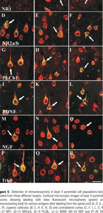
Figure 4. Detection of immunoreactivity in layer V pyramidal cell populations
back-labeled from three different targets. Confocal microscopic images of layer V pyramidal neurons showing labeling with latex fluorescent microspheres (green) and immunostaining (red) for various antigens after labeling from the spinal cord (A, D, G, J,
M, P), superior colliculus (B, E, H, K, N, Q) and contralateral cortex (C, F, I, L, O, R).
(A–C) NR1, (D–F) NR2a/b, (G–I) PLCβ1, (J–L) BDNF, (M–O) NGF and (P–R) TrkB antibodies. All six of these antibodies produced staining on layer V neurons containing retrogradely transported microspheres (arrows). In the adult rat neocortex, these selected proteins were expressed in all three subpopulations of layer V pyramidal cells: that is, neurons with projections to the spinal cord, superior colliculus and contralateral cortex. Scale bar = 20 µm.
Markers Specific for Subcortically Projecting Pyramidal Neurons (SMI-32, FNP-7 and N200)
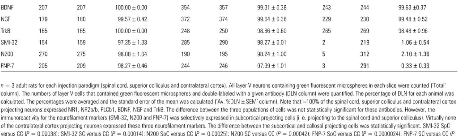
Figure 5 shows confocal images of the three cell populations (contralateral cortex, superior colliculus and spinal cord projecting) stained with three antibodies (SMI-32, N200 and FNP-7) that are specific for different neurofilament epitopes. The panels demonstrate that SMI-32, N200 and FNP-7 were expressed in neurons projecting to the spinal cord and to the superior colliculus, but these three antigens were not expressed in the neurons projecting through the corpus callosum to the contralateral hemisphere. Sections stained with these three antibodies were analyzed using the same quantification technique described above. Most of the subcor-tical projecting pyramidal cells expressed SMI-32 (mean ± SEM; spinal cord = 97.35 ± 1.33%; superior colliculus = 98.27 ± 0.007%), N200 (spinal cord = 98.08 ± 1.04%; superior colliculus = 98.24 ± 1.00%) and FNP-7 (spinal cord = 98.27 ± 0.46%; supe-rior colliculus = 97.99 ± 1.01%; Table 2). There was no signifi-cant difference between the percentage of cells expressing these three neurofilament markers in the subcortical projecting neurons. However, the contralateral cortex projecting cells barely expressed a quantifiable level of these neurofilament markers (SMI-32 = 1.06 ± 0.54%; N200 = 2.10 ± 1.36%; FNP-7 = 0.33 ± 0.33%; Table 2). The percentage differ-ence between neurofilament expression in callosal — compared to subcortical — projecting neurons was statistically significant (Table 2).
Neurofilament Protein Expression in Organotypic Culture
We performed two experiments to determine the likelihood that the neurofilament immunoreactivity in layer V projection neurons is elicited and maintained by their subcortical targets through their specific axonal projections. We prepared cortical
slices from the parietal cortex of E16 embryos and maintained them in organotypic cultures for two weeks (Fig. 1B) to test if layer V neurons express SMI-32 and FNP-7 while developing Table 2
Proportion of the double-labeled layer V projection neurons back-labeled from different targets and stained with various antibodies
n = 3 adult rats for each injection paradigm (spinal cord, superior colliculus and contralateral cortex). All layer V neurons containing green fluorescent microspheres in each slice were counted (‘Total’
column). The numbers of layer V cells that contained green fluorescent microspheres and double-labeled with a given antibody (DLN column) were quantified. The percentage of DLN for each animal was calculated. The percentages were averaged and the standard error of the mean was calculated (‘Av. %DLN ± SEM’ column). Note that ∼100% of the spinal cord, superior colliculus and contralateral cortex projecting neurons expressed NR1, NR2a/b, PLCb1, BDNF, NGF and TrkB. The difference between the three populations of cells was not statistically significant for these antibodies. However, the immunoreactivity for the neurofilament markers (SMI-32, N200 and FNP-7) was selectively expressed in subcortical projecting cells (i. e. projecting to the spinal cord and superior colliculus). Virtually none of the contralateral cortex projecting neurons expressed these three neurofilament markers. The difference between the subcortical and callosal projecting cells was statistically significant. SMI-32 SpC versus CC (P = 0.00038); SMI-32 SC versus CC (P = 0.00014); N200 SpC versus CC (P = 0.00025); N200 SC versus CC (P = 0.00042); FNP-7 SpC versus CC (P = 0.0000024); FNP-7 SC versus CC (P = 0.00016). Abbreviations: Av. %DL, average percentage of double-labeled layer V neurons quantified; CC, contralateral cortex projecting neurons; DLN, double-labeled layer V neuron; Total, total number of layer V neurons quantified; SC, superior colliculus projecting neurons; SEM, standard error of the mean; SpC, spinal cord projecting neurons.
Spinal cord Superior
colliculus
Contralateral cortex
Antigen DLN Total Av. %DLN ± SEM DLN Total Av. %DLN ± SEM DLN Total Av. %DLN ± SEM
NR1 218 222 98.67 ± 1.33 208 212 98.16 ± 1.11 223 224 99.37 ± 0.66 NR2a/b 197 198 99.73 ± 0.27 261 266 97.87 ± 0.70 273 275 99.28 ± 0.36 PLCb1 165 168 98.65 ± 1.53 248 255 97.37 ± 1.78 223 227 98.21 ± 0.96 BDNF 207 207 100.00 ± 0.00 354 357 99.31 ± 0.38 243 244 99.63 ±0.37 NGF 179 180 99.57 ± 0.42 372 374 99.64 ± 0.36 229 230 99.48 ± 0.52 TrkB 165 165 100.00 ± 0.00 248 250 98.86 ± 0.60 265 269 98.48 ± 0.96 SMI-32 154 159 97.35 ± 1.33 285 290 98.27 ± 0.01 2 219 1.06± 0.54 N200 270 275 98.08 ± 1.04 190 195 98.24 ± 1.00 5 312 2.10± 1.36 FNP-7 205 209 98.27 ± 0.46 244 246 97.99 ± 1.01 3 291 0.33± 0.33
Figure 5. Confocal microscopic images of layer V pyramidal neurons showing that the
three neurofilament antibodies (red) specifically stained neurons that project subcortically, but they did not stain cells with projections to the contralateral cortex (all populations contain green microspheres). SMI-32 (A, B), N200 (D, E) and FNP-7 (G, H) were specifically located in the spinal cord (A, D, G) and superior colliculus (B, E, H) projecting layer V neurons (arrows indicate examples of double-labeling). (C, F, I) contralateral cortex projecting neurons (green microspheres indicated by arrowheads), did not express SMI-32 (C), N200 (F) or FNP-7 (I). The open arrow heads indicate neurons expressing the neurofilaments, but do not contain green microspheres and thus do not project to the contralateral cortex. Scale bar = 20 µm.
without contact with their target tissues. The slices derived from the embryonic cortex did, in fact, express SMI-32 (Fig. 6A) and FNP-7 (Fig. 6B). In high power images, layer V pyram-idal neurons were clearly identified by strong SMI-32 (Fig. 6C) and FNP-7 (Fig. 6D) immunoreactivity. Layers II and III did not develop as well as the other layers, which is a typical observa-tion in embryonic explants grown in culture (Molnár and Blakemore, 1999).
To further investigate whether or not subpopulations of layer V neurons maintain their SMI-32 and FNP-7 immunoreac-tivity specificity in the absence of contact with their target tissue, pyramidal cells were pre-labeled and cortical slices were cultured. At P3, after the corticofugal projections had reached the superior colliculus and the contralateral cortex, fluores-cent latex microspheres were injected into one of these targets. At P4, cortical slice cultures were prepared from regions containing labeled cells. These slices were cultured in isolation for two weeks, then fixed and immunostained for FNP-7 and SMI-32 without resectioning (Fig. 7). In 40 cultures, individual microsphere-labeled layer V neurons (n = 1163) were examined for immunohistochemistry. Of the 320 layer V cells that were labeled from the contralateral hemisphere (Fig. 7), only a small percentage (mean ± SEM; 1.69 ± 1.40) expressed SMI-32. In contrast, of the 196 layer V cells that were labeled from the superior colliculus (Fig. 7), a much higher percentage (27.11 ± 4.67) of labeled cells were SMI-32 immunoreactive. Of the 286 layer V neurons that were labeled from the contralateral cortex (Fig. 7), only 4.2% were FNP-7 immunoreactive, whereas 44.6% of layer V neurons (n = 361) that were labeled from the superior colliculus were reactive (Fig. 7). Laminar specificity of neurofilament immuno-reactivity was also maintained with stronger staining in layers II, III, V and VI (Fig. 7). Although the absolute number of double-labeled neurons in the cultures was decreased, the overall pattern of neurofilament expression was consistent with the in vivo observations.
Discussion
Differential Protein Expression in Layer V Pyramidal Cell Subpopulations
In this study, a combination of retrograde labeling and immu-nostaining techniques were used to determine whether or not subpopulations of layer V pyramidal cells projecting to distinct
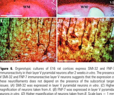
Figure 6. Organotypic cultures of E16 rat cortices express SMI-32 and FNP-7
immunoreactivity in their layer V pyramidal neurons after 2 weeks in vitro. The presence of SMI-32 and FNP-7 immunoreactive layer V neurons suggests that the expression of these neurofilaments does not depend on the presence of the subcortical target tissues. (A) SMI-32 was expressed in layer V pyramidal neurons in vitro. (C) Higher magnification of neurons taken from A. (B) FNP-7 was expressed in layer V pyramidal neurons in vitro. (D) Higher magnification of neurons taken from B. Scale bars = 1 mm (A, B); 100 µm (C, D).
Figure 7. The subpopulation-specific expression of SMI-32 and FNP-7 immunoreactivity is maintained in organotypic cortical cultures from postnatal rats after 2 weeks in vitro.
Layer V pyramidal neurons were back-labeled from the contralateral cortex (see inserts in A, C) or from the superior colliculus (see inserts in E, G) at P3 (both populations contain green microspheres). At P4, the somatosensory cortex of both groups was cultured for 2 weeks. SMI-32 and FNP-7 maintained a laminar (A, C, E, G) pattern of expression similar to that found in vivo. This suggests that the presence of specific targets is not necessary for the maintenance of the particular layer V populations after P4. (B, D) Confocal images of pyramidal cells taken from A and C respectively. Both SMI-32 (B) and FNP-7 (D) failed to co-localize with contralateral cortex projecting neurons (green microspheres indicated by the arrowheads). (F, H) Confocal images of pyramidal cells taken from E and G respectively. A large percentage of both SMI-32 (F) and FNP-7 (G) co-localized with superior colliculus projecting neurons (indicated by the arrows). P, postnatal day. Scale bars = 1 mm (A, C, E, G); 20 µm (B, D, F, H).
targets show differential expression of antigens known to be located in layer V of adult rats. These antigens include the following: NR1, NR2a/b, PLCβ1, BDNF, NGF, TrkB, SMI-32, FNP-7 and N200. Staining patterns found in this study confirm previous observations with the same antibodies: NR1 (Aoki et al., 1994), NR2a/b (Conti et al., 1999), TrkB (Tongiorgi et al., 1999; Miller, 2000), NGF (Miller, 2000), BDNF (Murer et al., 1999), SMI-32 (Hof and Morrison, 1995; Gabernet et al., 1999), FNP-7 (Hornung and Riederer, 1999) and N200 (Sasaki and Maruyama, 1994). However, these experiments show for the first time a striking difference in neurofilament expression (SMI-32, FNP-7 and N200) between subcortical (superior collic-ulus and spinal cord projecting type I neurons) and contralat-eral cortex (type II neurons) projecting neurons. While a large proportion of spinal cord and superior colliculus projecting neurons were heavily immunoreactive for the neurofilament antigens (SMI-32, FNP-7 and N200), a visual assessment indi-cated that none of the callosal projecting layer V cells had significantly detectable immunoreactivity. In contrast, all of the other proteins tested (NR1, NR2a/b, PLCβ1, BDNF, NGF and TrkB) were expressed equally in the layer V pyramidal subpopulations examined.
Differential Neurofilament Expression Layer V Pyramidal Neuron Subpopulations
Neurofilaments (NF), which are part of the intermediate fila-ment family, are one of the earliest recognizable features of the developing central nervous system (Ulfig et al., 1998). Neurofil-aments are heteropolymers consisting of three subunit proteins: NF-L (68 kDa), NF-M (150 kDa) and NF-H (200 kDa) (Hoffman and Lasek, 1975; Nixon and Sihag, 1991; Nixon, 1998). These subunits combine to form filaments (∼10 nm diameter). All triplets consist of two structural domains: an α-helical domain, which is the functional backbone and a carboxy end. The carboxy terminals vary in their amount of phosphorylation, which is responsible for the different molec-ular weights (Shaw, 1991). Decreases in neurofilament expres-sion or abnormal neurofilament phosphorylation have been implicated in normal aging (Vickers et al., 1992; Budinger et al., 2000) as well as in neurological diseases (Bickford et al., 1998) such as: Alzheimer’s disease (Morrison et al., 1987; Hof et al., 1990; Hof and Morrison, 1990; Trojanowski et al., 1993; Vickers et al., 1994; Bussière et al., 2003a,b), Parkinson’s disease (Goldman et al., 1983), Pick’s disease (Perry et al., 1987), amyotrophic lateral sclerosis (Manetto et al., 1988; Munoz et al., 1988; Mizusawa et al., 1989; Tsang et al., 2000), traumatic brain injury (Saatman et al., 1998) and multiple scle-rosis (Trapp et al., 1998).
Neurofilaments have a very specific laminar distribution pattern in the cortex that reflects the functional and anatom-ical brain divisions. Therefore, neurofilament staining is a powerful tool for confirming and extending previous architec-tural observations (Hof et al., 1995; Preuss et al., 1997; Geyer et al., 2000). SMI-32 recognizes the non-phosphorylated NF-H form, N200 recognizes the non-phosphorylated and phosphor-ylated NF-H forms and FNP-7 recognizes the non-phosphor-ylated NF-M form. These protein epitopes initially appear at P7, which interestingly coincides with the divergence of the layer V pyramidal neuronal subpopulations with respect to their somatodendritic morphologies (Riederer and Matus, 1985; Riederer, 1995; Riederer et al., 1995; Kogan et al., 2000).
It is important to relate the onset of neurofilament expres-sion to other aspects of layer V pyramidal cell development. Neurons from layer V subpopulations are born around E15–18 and migrate to layer V by E19–20 (Miller, 1988). Morphologic-ally, they initially appear indistinguishable: all have stout apical dendrites with terminal tufts in layer I and they do not fire action potential bursts in response to depolarizing current (Kasper et al., 1994). Shortly after reaching layer V, they begin extending their axons toward different targets. The callosal axons start to cross the corpus callosum at E16–18, whilst the corticofugal axons enter the internal capsule at E17 and enter the cerebral peduncle at E19. The axons reach the basal pons at E19 and the superior colliculus and spinal cord shortly after birth (De Carlos and O’Leary, 1992). Axonal target invasion occurs postnatally. It is only after axonal outgrowth that the dendritic morphologies begin to diverge (P5; Koester and O’Leary, 1992). The different electrophysiological properties are first detected much later (P14; Kasper et al., 1994). These findings raise the question: how do seemingly similar cell populations differentiate into a heterogeneous group with different targets?
It is not clear what causes these morphological changes, but it has been shown that neurofilaments participate in the struc-tural organization and stabilization of dendrites (Riederer and Matus, 1985; Riederer et al., 1995; Kogan et al., 2000). There-fore, the neurofilament quantity may be important in estab-lishing and maintaining different morphologies of layer V neuronal subpopulations; therefore, playing a role in connec-tions reaching their developmental endpoint (Liu et al., 1994; Kogan et al., 2000).
Previous studies in macaque monkeys have shown that SMI-32 is differentially expressed in certain subpopulations of pyramidal cells. These studies have exclusively examined corticocortical — not subcortical — projecting neurons in layers II, III, V and VI. The corticocortical neuron populations that express low levels of SMI-32 include cells that project from the anterior cingulate to prefrontal cortex; from prefrontal to prefrontal cortex; and from V1, V2 or V3 to V4 (Campbell et al., 1991; Hof et al., 1995, 1996b). Corticocortical neuron populations that express high levels of SMI-32 include cells that project from polysensory association cortices (superior temporal sulcus) to prefrontal cortices and from V1, V2, V3 to MT (Campbell et al., 1991; Hof et al., 1995, 1996b). Equal numbers of corticocortical neurons that send their projections from high level visual association cortex (intraparietal sulcus) to prefrontal cortex express SMI-32 (Campbell et al., 1991). Our results confirm previous findings in rats, which show that somatomotor corticocortical pyramidal neurons in layers III and V projecting contralaterally express very low levels of SMI-32 (Kirkcaldie et al., 2002). One must, therefore, take into account that SMI-32 specificity may vary between primate and non-primate species.
Several studies have correlated the neurofilament amount or type with specific cellular populations in order to better understand the functional significance of each cell type. It is known that the amount of neurofilament increases with the increase of the cell size (Campbell and Morrison, 1989; Tsang et al., 2000); therefore, layer V projection neurons would be expected to have high neurofilament levels compared to other cortical layers. Campbell and Morrison (1989) first showed that non-phosphorylated neurofilaments are found in neurons with long axonal projections extending to cortical and subcortical
areas. This finding is supported by studies that show high neurofilament levels in: long distance ipsilateral and contralat-eral cortex projections (Campbell et al., 1991), long ipsilatcontralat-eral association connections of the visual system (Hof et al., 1996a), large retinal ganglion cells (Straznicky et al., 1992) and long corticofugal projections (Hornung and Riederer, 1999). Few cells having short corticocortical, callosal or limbic projections express neurofilament epitopes (Hof et al., 1995, 1996a; Hornung and Riederer, 1999). Our observations demonstrate that layer V pyramidal neurons with callosal (type II) projec-tions do not express these selected neurofilament epitopes, in spite of their long axons. However, it has been suggested that the neurofilament amount might not be associated with axonal length. Instead, the neurofilament quantity might be associated with the amount of axonal myelination (Kirkcaldie et al., 2002). The degree of myelination corresponds with the projec-tion target and would explain the contradicprojec-tions between the different neurofilaments in ipsilateral connections and callosal connections. Cortical projections extending to subcortical targets and projections from primary sensory cortex extending to association areas (heterotopic) have heavily myelinated axons and are integral for important cognitive and motor processing. Goldstein et al. (1987) found that NF-H protects against proteolysis, thus increasing structural stability, which is critical in large, fast-conducting neurons. In addition to micro-tubules, neurofilaments have been implicated in regulating the nutrient transport rate, which is especially important in main-taining the structural integrity of these large neurons (Lasek, 1988).
We investigated whether these two distinct classes of layer V cells preserve their neurofilament specificity in two different organotypic slice culture paradigms, in which the projection neurons have been axotomized and lack contact with their targets. When E16 cortical slices were cultured for 2 weeks, SMI-32 and FNP-7 immunoreactive cells were still present, suggesting that the subpopulation phenotype is not dependent upon the target and is an inherent property of neurons. Obvi-ously, before the connections reach their target, the back-labe-ling of selective layer V subpopulations is not possible; therefore, the issue of subpopulation specificity cannot be addressed with the techniques used in the current study at this embryonic stage. However, maintenance of subpopulation neurofilament specificity in the absence of target tissue contact can be addressed at later developmental stages. In this study, we demonstrated that after 2 weeks of culturing P4 slices, the overall pattern of SMI-32 and FNP-7 immunoreactivity was consistent with the in vivo observations. The pyramidal neurons labeled from the superior colliculus at P3 maintained specific expression of SMI-32 and FNP-7, but neurons labeled from the contralateral cortex hemisphere did not express these epitopes. These findings suggest that whilst the specific subpopulation phenotype development normally occurs once the axons have innervated their targets, this process is not dependent upon those targets. Therefore, it stands to reason that by P4, the two layer V neuronal subpopulations might already be committed to different differentiation programs which continue in the absence of their target tissue contact.
In summary, this study demonstrates that layer V neuronal subpopulations not only have specific projection targets and somatodendritic morphologies, but also express different structural protein levels recognized by SMI-32, NFP-7 and N200 antibodies. The finding that neurofilament proteins are
expressed in subcortical (superior colliculus and spinal cord type I) projecting neurons (but not in type II, homotopic callosal projecting cells) that have large dendritic tufts supports theories that neurofilaments are associated with dendritic arborizations, long projecting axons and axons with a high degree of myelination. The characterization of molec-ular and cellmolec-ular differences in adult rat layer V neuronal subpopulations sheds light on the mechanisms involved in functional circuit differentiation in mammalian cerebral cortex development. The specific neurofilament expression in certain neuronal populations, even in the absence of long projections and maintenance after distant target removal through slice culturing, suggests that early in corticogenesis, neuronal subpopulations are already committed to distinct protein expression patterns.
Notes
We are indebted to Miss Carole Bezençon and Miss Lynden Guiver for their excellent technical assistance; to Mr Eric Bernardi, Mr Marc Berney and Mr Colin Beesley for their help with photography and Drs Tim Doubell and Anita Scheuber for their help with the initial experi-ments on NR-1 expression. The study was initiated in the laboratories of Professors Colin Blakemore and Egbert Welker and their support is greatly appreciated. We thank Dr Virginia Lee for the generous gift of the FNP-7 antibody. We are also grateful to Dr Amanda Law for helpful discussions and Drs Wei Zhi Wang and Ricardo Reis for their critical comments on an earlier version of the manuscript. Funding for C.C.J.V. was provided by a Rhodes Scholarship. The study was supported by grants from the Human Frontiers Science Program (RGP0107/2001), The European Community (Grant QLRT-1999-30158), Wellcome Trust (Grant 063974/B/01/Z) and Swiss National Science Foundation (Grant 56032.98 to Z.M. and 51036.97, 31-62113.00 to J.P.H.). B.H.G. held the Newton-Abraham Visiting Profes-sorship at the University Laboratory of Physiology and Lincoln College, Oxford in 1997.
Address correspondence to Dr Zoltán Molnár, Department of Human Anatomy and Genetics, University of Oxford, South Parks Road, Oxford, OX1 3QX, UK. Email: [email protected].
References
Akers RM, Killackey HP (1978) Organization of corticocortical connections in the parietal cortex of the rat. J Comp Neurol 181:513–537.
Aoki C, Venkatesan C, Go CG, Mong JA, Dawson TM (1994) Cellular and subcellular localization of NMDA-R1 subunit immunoreactivity in the visual cortex of adult and neonatal rats. J Neurosci 14:5202–5222.
Bickford ME, Guido W, Godwin DW (1998) Neurofilament proteins in Y-cells of the cat lateral geniculate nucleus: normal expression and alteration with visual deprivation. J Neurosci 18:6549–6557. Brodmann K (1909) Vergleichende localisationslehre der
grosshirn-ride in ihren prinzipien dargestellt auf grund des zellenbaues. Leipzig: JA Barth.
Budinger E, Heil P, Scheich H (2000) Functional organization of audi-tory cortex in the Mongolian gerbil (Meriones unguiculatus). III. Anatomical subdivisions and corticocortical connections. Eur J Neurosci 12:2425–2451.
Bussière T, Giannakopoulos P, Bouras C, Perl DP, Morrison JH, Hof PR (2003a) Progressive degeneration of nonphosphorylated neurofila-ment protein-enriched pyramidal neurons predicts cognitive impairment in Alzheimer’s disease: stereologic analysis of prefrontal cortex area 9. J Comp Neurol 463:281–302.
Bussière T, Gold G, Kovari E, Giannakopoulos P, Bouras C, Perl DP, Morrison JH, Hof PR (2003b) Stereologic analysis of neurofibrillary tangle formation in prefrontal cortex area 9 in aging and Alzhe-imer’s disease. Neuroscience 117:577–592.
Campbell MJ, Morrison JH (1989) Monoclonal antibody to neurofila-ment protein (SMI-32) labels a subpopulation of pyramidal
neurons in the human and monkey neocortex. J Comp Neurol 282:191–205.
Campbell MJ, Hof PR, Morrison JH (1991) A subpopulation of primate corticocortical neurons is distinguished by somatodendritic distri-bution of neurofilament protein. Brain Res 539:133–136.
Conti F, Zuccarello LV, Barbaresi P, Minelli A, Brecha NC, Melone M (1999) Neuronal, glial, and epithelial localization of gamma-aminobutyric acid transporter 2, a high-affinity gamma-amino-butyric acid plasma membrane transporter, in the cerebral cortex and neighboring structures. J Comp Neurol 409:482–494. De Carlos JA, O’Leary DD (1992) Growth and targeting of subplate
axons and establishment of major cortical pathways. J Neurosci 12:1194–1211.
Gabernet L, Meskenaite V, Hepp-Reymond MC (1999) Parcellation of the lateral premotor cortex of the macaque monkey based on staining with the neurofilament antibody SMI-32. Exp Brain Res 128:188–193.
Geyer S, Zilles K, Luppino G, Matelli M (2000) Neurofilament protein distribution in the macaque monkey dorsolateral premotor cortex. Eur J Neurosci 12:1554–1566.
Goldman JE, Yen SH, Chiu FC, Peress NS (1983) Lewy bodies of Parkinson’s disease contain neurofilament antigens. Science 221:1082–1084.
Goldstein ME, Sternberger LA, Sternberger NH (1987) Varying degrees of phosphorylation determine microheterogeneity of the heavy neurofilament polypeptide (Nf-H). J Neuroimmunol 14:135–148. Hof PR, Morrison JH (1990) Quantitative analysis of a vulnerable
subset of pyramidal neurons in Alzheimer’s disease: II. Primary and secondary visual cortex. J Comp Neurol 301:55–64.
Hof PR, Morrison JH (1995) Neurofilament protein defines regional patterns of cortical organization in the macaque monkey visual system: a quantitative immunohistochemical analysis. J Comp Neurol 352:161–186.
Hof PR, Cox K, Morrison JH (1990) Quantitative analysis of a vulner-able subset of pyramidal neurons in Alzheimer’s disease: I. Superior frontal and inferior temporal cortex. J Comp Neurol 301:44–54.
Hof PR, Nimchinsky EA, Morrison JH (1995) Neurochemical pheno-type of corticocortical connections in the macaque monkey: quantitative analysis of a subset of neurofilament protein-immuno-reactive projection neurons in frontal, parietal, temporal, and cingulate cortices. J Comp Neurol 362:109–133.
Hof PR, Glannakopoulos P, Bouras C (1996a) The neuropathological changes associated with normal brain aging. Histol Histopathol 11:1075–1088.
Hof PR, Ungerleider LG, Webster MJ, Gattass R, Adams MM, Sailstad CA, Morrison JH (1996b) Neurofilament protein is differentially distributed in subpopulations of corticocortical projection neurons in the macaque monkey visual pathways. J Comp Neurol 376:112–127.
Hoffman PN, Lasek RJ (1975) The slow component of axonal trans-port. Identification of major structural polypeptides of the axon and their generality among mammalian neurons. J Cell Biol 66:351–366.
Hornung JP, Riederer BM (1999) Medium-sized neurofilament protein related to maturation of a subset of cortical neurons. J Comp Neurol 414:348–360.
Kaas JH (1993) Evolution of multiple areas and modules within neocortex. Perspect Dev Neurobiol 1:101–107.
Kasper EM, Lubke J, Larkman AU, Blakemore C (1994) Pyramidal neurons in layer 5 of the rat visual cortex. III. Differential matur-ation of axon targeting, dendritic morphology, and electrophysio-logical properties. J Comp Neurol 339:495–518.
Kirkcaldie MT, Dickson TC, King CE, Grasby D, Riederer BM, Vickers JC (2002) Neurofilament triplet proteins are restricted to a subset of neurons in the rat neocortex. J Chem Neuroanat 24:163–171. Koester SE, O’Leary DD (1992) Functional classes of cortical
projec-tion neurons develop dendritic distincprojec-tions by class-specific sculpting of an early common pattern. J Neurosci 12:1382–1393.
Kogan CS, Zangenehpour S, Chaudhuri A (2000) Developmental profiles of SMI-32 immunoreactivity in monkey striate cortex. Brain Res Dev Brain Res 119:85–95.
Larkman A, Mason A (1990) Correlations between morphology and electrophysiology of pyramidal neurons in slices of rat visual cortex. I. Establishment of cell classes. J Neurosci 10:1407–1414. Lasek RJ (1988) Studying the intrinsic determinants of neuronal form
and function. In: Intrinsic determinants of neuronal form and func-tion (Lasek RJ, Black MM, eds), pp. 1–60. New York: Alan R. Liss. Lee VM, Otvos L Jr, Carden MJ, Hollosi M, Dietzschold B, Lazzarini RA
(1988) Identification of the major multiphosphorylation site in mammalian neurofilaments. Proc Natl Acad Sci USA 85:1998–2002. Liu Y, Dyck R, Cynader M (1994) The correlation between cortical neuron maturation and neurofilament phosphorylation: a develop-mental study of phosphorylated 200 kDa neurofilament protein in cat visual cortex. Brain Res Dev Brain Res 81:151–161.
Manetto V, Sternberger NH, Perry G, Sternberger LA, Gambetti P (1988) Phosphorylation of neurofilaments is altered in amyo-trophic lateral sclerosis. J Neuropathol Exp Neurol 47:642–653. Miller MW (1988) Development of projection and local circuit
neurons in neocortex. In: Cerebral cortex: development and maturation of cerebral cortex (Peters A, Jones E, eds), pp. 133–175. New York: Plenum Press.
Miller MW (2000) Expression of nerve growth factor and its receptors in the somatosensory-motor cortex of Macaca nemestrina. J Neuro-cytol 29:453–469.
Mizusawa H, Matsumoto S, Yen SH, Hirano A, Rojas-Corona RR, Donnenfeld H (1989) Focal accumulation of phosphorylated neurofilaments within anterior horn cell in familial amyotrophic lateral sclerosis. Acta Neuropathol (Berl) 79:37–43.
Molnár Z, Blakemore C (1991) Lack of regional specificity for connec-tions formed between thalamus and cortex in coculture. Nature 351:475–477.
Molnár Z, Blakemore C (1999) Development of signals influencing the growth and termination of thalamocortical axons in organotypic culture. Exp Neurol 156:363–393.
Morrison JH, Lewis DA, Campbell MJ, Huntley GW, Benson DL, Bouras C (1987) A monoclonal antibody to non-phosphorylated neurofilament protein marks the vulnerable cortical neurons in Alzheimer’s disease. Brain Res 416:331–336.
Munoz DG, Greene C, Perl DP, Selkoe DJ (1988) Accumulation of phosphorylated neurofilaments in anterior horn motoneurons of amyotrophic lateral sclerosis patients. J Neuropathol Exp Neurol 47:9–18.
Murer MG, Boissiere F, Yan Q, Hunot S, Villares J, Faucheux B, Agid Y, Hirsch E, Raisman-Vozari R (1999) An immunohistochemical study of the distribution of brain-derived neurotrophic factor in the adult human brain, with particular reference to Alzheimer’s disease. Neuroscience 88:1015–1032.
Nixon RA (1998) Dynamic behavior and organization of cytoskeletal proteins in neurons: reconciling old and new findings. Bioessays 20:798–807.
Nixon RA, Sihag RK (1991) Neurofilament phosphorylation: a new look at regulation and function. Trends Neurosci 14:501–506. Paxinos G, Watson C, Pennisi M, Topple A (1985) Bregma, lambda and
the interaural midpoint in stereotaxic surgery with rats of different sex, strain and weight. J Neurosci Methods 13:139–143.
Perry G, Stewart D, Friedman R, Manetto V, Autilio-Gambetti L, Gambetti P (1987) Filaments of Pick’s bodies contain altered cytoskeletal elements. Am J Pathol 127:559–568.
Peters A, Jones EG (eds) (1985) Cerebral cortex (3): visual cortex (4): association and auditory corticies. New York: Plenum Press. Porter LL, Rizzo E, Hornung JP (1999) Dopamine affects parvalbumin
expression during cortical development in vitro. J Neurosci 19:8990–9003.
Preuss TM, Stepniewska I, Jain N, Kaas JH (1997) Multiple divisions of macaque precentral motor cortex identified with neurofilament antibody SMI-32. Brain Res 767:148–153.
Riederer BM (1995) Differential phosphorylation of MAP1b during postnatal development of the cat brain. J Neurocytol 24:45–54.
Riederer B, Matus A (1985) Differential expression of distinct microtu-bule-associated proteins during brain development. Proc Natl Acad Sci USA 82:6006–6009.
Riederer BM, Draberova E, Viklicky V, Draber P (1995) Changes of MAP2 phosphorylation during brain development. J Histochem Cytochem 43:1269–1284.
Saatman KE, Graham DI, McIntosh TK (1998) The neuronal cytoskel-eton is at risk after mild and moderate brain injury. J Neurotrauma 15:1047–1058.
Sasaki S, Maruyama S (1994) Immunocytochemical and ultrastructural studies of the motor cortex in amyotrophic lateral sclerosis. Acta Neuropathol (Berl) 87:578–585.
Shaw G (1991) Neurofilament proteins. In: The neuronal cytoskeleton (Burgoyne RD, ed.), pp. 185–214. New York: Wiley-Liss.
Straznicky C, Vickers JC, Gabriel R, Costa M (1992) A neurofilament protein antibody selectively labels a large ganglion cell type in the human retina. Brain Res 582:123–128.
Tongiorgi E, Cattaneo A, Domenici L (1999) Co-expression of TrkB and the N-methyl-D-aspartate receptor subunits NR1-C1, NR2A and NR2B in the rat visual cortex. Neuroscience 90:1361–1369. Trapp BD, Peterson J, Ransohoff RM, Rudick R, Mork S, Bo L (1998)
Axonal transection in the lesions of multiple sclerosis. N Engl J Med 338:278–285.
Trojanowski JQ, Schmidt ML, Shin RW, Bramblett GT, Rao D, Lee VM (1993) Altered tau and neurofilament proteins in
neuro-degenera-tive diseases: diagnostic implications for Alzheimer’s disease and Lewy body dementias. Brain Pathol 3:45–54.
Tsang YM, Chiong F, Kuznetsov D, Kasarskis E, Geula C (2000) Motor neurons are rich in non-phosphorylated neurofilaments: cross-species comparison and alterations in ALS. Brain Res 861:45–58. Ulfig N, Nickel J, Bohl J (1998) Monoclonal antibodies SMI 311 and
SMI 312 as tools to investigate the maturation of nerve cells and axonal patterns in human fetal brain. Cell Tissue Res 291:433–443. Vickers JC, Delacourte A, Morrison JH (1992) Progressive trans-formation of the cytoskeleton associated with normal aging and Alzheimer’s disease. Brain Res 594:273–278.
Vickers JC, Riederer BM, Marugg RA, Buee-Scherrer V, Buee L, Delacourte A, Morrison JH (1994) Alterations in neurofilament protein immunoreactivity in human hippocampal neurons related to normal aging and Alzheimer’s disease. Neuroscience 62:1–13. Weimann JM, Zhang YA, Levin ME, Devine WP, Brulet P, McConnell
SK (1999) Cortical neurons require Otx1 for the refinement of exuberant axonal projections to subcortical targets. Neuron 24:819–831.
Welker E, Armstrong-James M, Bronchti G, Ourednik W, Gheorghita-Baechler F, Dubois R, Guernsey DL, Van der Loos H, Neumann PE (1996) Altered sensory processing in the somatosensory cortex of the mouse mutant barrelless. Science 271:1864–1867.