International Immunology. Vol. 6, No. 9. pp. 1287- 1295
Homing and in situ differentiation of
resident pulmonary lymphocytes
Gek-Kee Sim, Ramanujam Rajaserkar
1, Mark Dessing and Andrei Augustin
1*
2 Basel Institute for Immunology,. Grenzacherstrasse 487, CH-4005 Basel, Switzerland1 Department of Medicine, National Jewish Center for Immunology and Respiratory Medicine, 1400,
Jackson Street, Denver, CO 80206, USA
2Department of Immunology, University of Colorado Health Sciences Center, Denver, CO 80262, USA
Key words: yS T cells, extrathymic differentiation, lymphoid precursors, RAG-1, RAG-2, resident pulmonary lymphocytes, sckJ reconstitution, selection
Abstract
At birth, T lymphocytes which colonize the lung are mainly of the y6 subset, while a/3 T cells predominate in the spleen. Thus, the lung is a preferred site for the homing of yS T cells in the perinatal period. However, after birth, the pattern of VT gene usage among resident pulmonary
lymphocytes (RPL) changes with age, from a predominance of VT6 at birth to a predominance of
VT4 in older mice. The generation of the V76 fraction appears to be thymus dependent, since In
athymic nude mice, the VT6 population present at birth Is replaced by VT4 T cells. In the
postnatal period, both RAG-1 and RAG-2 genes are expressed at high levels In the RPL population. TCR bearing cells are among those that express RAG genes, Indicating that maturation of T cells takes place In this organ. In addition, transfer experiments reveal that lymphoid precursors are present In the lung. The stage of differentiation of these precursors will be characterized in future studies. The data presented here Indicate that pulmonary T
lymphocytes are derived from both migrants of thymlc origin and from precursors which have undergone differentiation and selection in the lung. The population that Is generated In situ and that has not been selected In the thymus may Include cells that are typical for the pulmonary environment.
Introduction
The selection of some yd T cell subsets occurs in the lung and is dependent on the genetic background of the mouse strain but not on thymic selection (1,2). This suggests that y6 T cells in the lung are not necessarily descendants of thymocytes. Notably, in contrast to other peripheral lymphoid organs, there is an unusually high frequency of CD4~CD8~ double-negative a/3 TCR* T cells among resident pulmonary lymphocytes (3). yh and a/S T cells are also found in the spleen and intestine of athymic nude mice (4,5). Bone marrow reconstitution studies using adult thymectomized mice as recipients have demonstrated the thymic independent origin of intestinal intraeprthelial lympho-cytes (5,6). It is not dear whether these extrathymically generated T cells are derived from stem cells which home to the organ, rearrange their receptor genes and proceed to differentiate into T cells, or whether rearrangement and expression of TCR took place prior to migration into the tissue, and only selection and functional maturation occur upon homing. In support of the latter, rearrangements of TCR y genes have been observed in day 11 fetal liver, before the thymus is colonized with T cells (7).
Conceivably, in normal mice, rearrangements of TCR y genes may precede the homing of pre-T cells to the thymus in some instances and that such cells may already be committed to differentiate along a particular lymphoid lineage. It is also plausible that these pre-committed precursors may be the source of extrathymically generated T cells. In this report, we present data bearing on the origin of resident lymphocytes in the lung, and assess the contribution of the thymic and extrathymic components in the generation and selection of T cells in this micro environment.
Methods
Mice
BALB/c mice and BALB/c nulnu mice were all of Jackson origin and were either purchased from the Jackson Laboratory (Bar Harbor, ME) or from IFFA (L'Arbresle, France). Timed pregnant BALB/c mice were either purchased from Jackson or from Ciba Correspondence to: G.-K. Sim
(Basel, Switzerland). The day of birth was considered day 1 of life. CB-17 SCID mice were bred at the Basel Institute for Immunology.
Preparation of resident pulmonary lymphocytes (RPL), thymocytes and splenocytes
To prepare RPL, regardless of the age of the mice, lungs were extensively perfused through the pulmonary artery to remove circulating blood, as previously described (3). The dissected lungs were then cut into 1 mm strips in PBS without calcium and magnesium and in the presence of 1 mM EDTA at 4°C. Cells were released from the matrix by teasing the tissue on a steel mesh. The debris was allowed to settle and the cell suspension was washed twice in Iscove's modified Dulbecco's medium in the presence of 5% FCS. Viable cells were further purified by Ficoll-Hypaque centrifugation. Single cell suspensions were also prepared from spleens and thymus by disruption of the organ on a steel mesh. In the case of splenocytes, red cells were removed by hypotonic lysis.
RNA preparations
Total cellular RNA was extracted from various cellular populations by the APGC procedure (8). For RNA from a/9+ or y6+ RPL, cells were magnetically sorted by treatment with biotin-conjugated anti-a/9 TCR mAb H57-597 or anti-y^ antibody GL3, followed by streptavidin-coupled Dynal beads. Trace amounts of contaminating DNA was further removed by treatment with RNAse free DNAse (Boehringer, Mannheim, Germany). cDNA synthesis and polymerase chain reaction (PCR) reactions This was performed essentially as published (2,9,10). For PCR analysis of VT gene expression, the sequences of all primers
used were as reported, under conditions previously established (9). The RAG-1 and RAG-2 primers for PCR were as described (10). Each PCR cycle consists of the following: 94°C30s, 52°C 30 s (60°C for the RAG-1 and 2 primers), and 72°C 2 min. A 6 min incubation at 72°C was added to the last cycle and the number of cycles varied according to the nature of the experiment. In estimating the relative levels of V7 gene
expression, to ensure the linearity of the PCR reaction at the time of assay, 5 /d samples were withdrawn at cycles 21, 24, 27 and 30, and the cycles at which the most prominent product became visible was chosen for quantitation.
mAbs
The following mAbs used in this work were purchased from PharMingen (San Diego, CA) anti-a/S TCR, H57-597; anti-76 TCR, GL-3; anti-VT4, UC3-10A6; anti-CD3, 145-2Ci1;anti-H2Kb,
AF6-88.5. M1/70.15 (anti-Mac 1), 187.1 (anti-mouse x) and 3JP (anti-l-A") were prepared in our laboratory.
Assays for GYS versus GxYS Vy4 sequences
JT1 primers (10 pmol) were end-labeled with [-y-^PJATP using
T4 polynucleotide kinase and purified on a Biorad Bio-spin 6 column. Then, 1/1 Oth of this was added to a 30 /d PCR reaction containing as template either 1 ng of boiled plasmid DNA or 104
cell equivalent of RPL cDNA, PCR buffer, 100 /iM dXTP, 0.5 /tM 5' (VT4) and 3' (JT1) primers, and 0.5 units of Taq polymerase.
Amplification was for 25 cycles at 94°C 30 s, 50°C 30 s, 72°C 1 min, followed by an additional 6 min incubation at 72°C.
Amplified products were further purified on a Biorad Bio-spin 30 column and an aliquot containing 104 c.p.m. from each sample
was digested with 5 units of MboW at 37°C for 1 h in the presence of 0.5 fig carrier DNA. Samples were then phenol extracted, ethanol precipitated and solubilized in 5 /d of formamide DNA sequencing gel buffer, heated at 90°C for 1 min, and 1 /d was loaded in each lane.
Quantification of RAG-1 concentrations relative to normalized actin concentrations
Total RNA from spleen, thymus and RPL was prepared by the APGC procedure as described above from pools of at least 10 mice. cDNA was synthesized using hexanucleotide primers, starting with RNA extracted from an equivalent of 5 x 10s cells
per sample. To estimate the concentration of actin in each preparation, we took 1 % of the cDNA, made a series of 3-fold dilutions, and performed 25 cycles of PCR with /3-actin primers CATCACTATTGGCAACGAGC, ACGCAGCTCAGTAACAGTCC which spanned an intron-exon junction. Then, 1 ^Ci of [^PJdCTP was added to each reaction and the amount of radioactivity incorporated into the /3-actin band was determined after gel fractionation of the PCR products. Adjustments were then made to normalize the quantity of mRNA in the various samples, using actin as an internal standard. Sets of three samples, i.e. spleen, thymus, and RPL from the same mice, were normalized to each other at any one time. The normalization was verified by comparing the amount of radioactivity incorporated into amplified materials obtained for three different concentra-tions of actin after 20 cycles of PCR amplification. To compare the level of RAG-1 mRNA expression in the different cellular populations using actin as an internal standard, equivalent amounts of actin-normalized cDNA samples were used in PCR amplifications of RAG-1. Typically, a 100 /d reaction was set up including 100 /tM of each dXTP, 10 /tCi [^PJdCTP, cDNA, PCR buffer, 2 units of Taq polymerase, and 0.5 /iM of each of the 3' and 5' primers. This was split into eight aliquots of 12 /d each, 30 /d of oil was layered over each sample, and an aliquot was removed after each increment of three cycles starting from cycle 15 and incubated for another 6 min at 72°C to ensure completion of chain extension. Samples were analyzed on 8% native pdyacryalmide gels and the radioactivity associated with the RAG-1 bands was quantjtated on the Phosphor Imager. For each sample, the amount of radioactivity incorporated after different numbers of cycles of amplification was graphed (11). Assay for lymphoid precursors in the lung
RPL from 20 2 - 3 week old C57BL/6 mice were isolated after extensive perfusion as described. The cells were then incubated with biotin-conjugated anti-CD3, anti-o/3 TCR, anti-T* TCR and anti-l-Ab at 4°C for 30 min, washed, and incubated with
streptavidin-coupled Dynal beads at a bead:cell ratio of 20:1. After a further incubation of 20 min at 4°C with gentle rocking, the antibody bound T cells, B cells, macrophages and dendritic cells were magnetically removed. A second round of depletion was performed and a small fraction of the cells recovered were monitored for purity after further incubation with the antibody cocktail followed by streptavidin - phycoerythrin. Less than 1 % of the cells were positive for the combination of markers. Then, 105 cells were mixed with 5 x 1 0 * bone marrow cells of SCID
(•) 7 5 4 1.10 , -7 • J 4 J - I-1 ilO * tz 7 5 4 1 -• op cate • 7* + cells G3 B cefls (b) op/jt T cell ratio n new-born (day I) BALOte mice
1 3 5 7
DAYS AFTER BIRTH
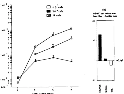
Fig. 1. Lymphocyte colonization of the lung at birth and in the perinatal period, (a) Kinetics of a/3 and yd T cell colonization, (b) Ratio of a/3 versus
yi T cells in the thymus, spleen and lung at birth, a/3 and yd T cells were quantitated by flow cytometric analysis using anti-aj3 TCR (H57-597) and anti-yi TCR (GL-3) antibodies. For each time point, two to four independent measurements were made on pools of RPL Isolated from five to nine mice. The results are calculated as the number of cells recovered per animal.
previously tested for the absence of serum Ig. The SCID mice were 3 months old at the time of transfer and were irradiated (350 rad) before injection. Control mice received only SCID bone marrow. Four months later, the mice were killed, and cells were isolated from their spleens, thymus and lungs for analysis.
Results
T cell colonization of the lung during the first week of life To analyze the kinetics of T cell colonization of the lung during the perinatal period, we used a standard method for the isolation of RPL. On average, immediately after birth, we recovered a total of >103 T$ T cells from the lungs of each mouse, but only
7x10²o^Tcells(Fig. 1a). At the same time, in the thymus and
spleen, the number of afi T cells exceeded that of yS T cells (Fig. 1 b). These observations suggest that either the yd T cells present in the lung at birth are generated in situ and do not come from the thymus or that there is a preferential homing of yd T cells to the lung before birth. However, after birth, between day 1 and 3, the number of a/3 T cells increases by almost 10-fold in the lung, while yd T cells increase less dramatially in number. The y6:a0 ratio is rapidly reversed during the first week of life, such that by day 7, yd T cells represent a minor fraction of RPL (Fig. 1). Thus, in the first week after birth, there is a rapid colonization of the lung by a/J T cells, while the number of yd T cells increases slowly.
Age related changes in the pattern of Vr gene expression The high frequency of yd RPL at birth compared with a/3 T cells prompted us to investigate the VT gene usage in this population.
This was performed by a modified PCR-based assay that has previously been used to define the pattern of VT and V4 genes
expressed in RPL isolated from adult BALB/c mice (1,2,9). Surprisingly, the predominant VT gene that was expressed in the
lung at birth was VT6 (Fig. 2a). We have previously shown that
the major VT mRNA species present in adult BALB/c RPL was
VT4 (2), but that in mice immunized with aerosols of purified
protein derivative, both VT4 and VT6 cells are present (9).
Moreover, a large fraction of the resident lymphocytes isolated from the adult lung reacts to the anti-VT4 antibody UC3-10A6
(12) that has become available (unpublished observation). The conspicuous absence of the VT4 population in the neonatal
lungs prompted us to analyze the kinetics of expression of the various VT genes in RPL isolated from mice of different ages.
As shown in Fig. 2, in very young mice, the pattern of VT gene
expression in RPL is significantly different from that of the adult. It is also different from that of the thymus and spleen in mice of the same age. Previous studies have shown that right before birth and in adult mice, VT4 is the main VT gene that is expressed in
the thymus, lymph node and.spleen (13,14). However, in the lung, from birth until the young adult period, VT6 constitutes the
major fraction of the yd T cells present, although VT4 bearing
Relative expression of Vyi and Vtf In RPL
I-birth D»y»afttr conception
Relative expression of V>5 and Vy7 in RPL
Dayi i f m concipUon
Fig. 2. VT gene expression in RPL changes with time, (a) Relative
expression of VT4 and VT6. (b) Relative expression of V 5 and VT7. Each
data point was obtained from RNA from a pool of at least five mice. Different data points for mice of the same age were derived from different batches of mice. For each sample of RPL, equal amounts of the same preparation of y cDNA were used as templates for the amplification of the four different V genes under conditions of linear amplification (see Methods). The PCR products of V 4, 5, 6 and 7 were fractionated on a 1.4% agarose gel, transferred to Genescreen membrane (Dupont), and hybridized to a ^P-labeted JT1 probe. The amount of radioactivity
associated with each V7 gene was directly quantitated on a Phosphor
Imager (Molecular Dynamics) or an AMBIS Bioanalysis Imaging System. The relative level of expression of each V gene was estimated as a percentage of all VT genes assayed for that time point. The y-axis
represents the percentage gene expression on a linear scale. The x-axts represents the time elapsed after conception of the fetus on a tog10
scale. The curves representing the kinetics of relative expression for each V gene were obtained by interpolation. The four graphs were plotted on two separate figures to allow better discrimination of the data points.
C/5 > i > X
O O
>2
- 5 9
- 5 6
- 2 0
Fig. 3. The first and second waves of VT4 cells which colonize the
lungs differ in their predominant TCR sequences: GYS versus GxYS. Autoradiogram of an 8% denaturing polyacrylamide gel showing the patterns of Mtooll cleaved PCR amplified products from (a) a plasmid carrying a VY4 cDNA clone with a GYS junction, (b) a plasmid carrying
a V 4 cDNA clone with a GxYS junction, (c) total cDNA prepared from day 5 BALB/c RPL and (d) total cDNA prepared from 3 month old BALBfc RPL. An anti-sense ^P-end-labeled J 1 primer (5'-CCTTCTGCAAAT-ACCTTGTG) was used in the amplification together with unlabeled VY4
primer. Mtooll does not cut at the recognition site itself but 8 bases downstream of it. Due to two ctosety spaced Mtooll recognition sites, the expected Mtooll fragments for GYSkype junctions are 56 and 59 bases respectively, and those for GxYS-type junctions are 59 and 62 bases. The 20 nudeotide band is due to contaminating JT1 primers.
There is a first peak of VT4 expression right after birth. This
drops to a very low level in 2 - 3 week old mice, but increases steadily thereafter such that the majority of 76 T cells in older BALB/c mice express V74 by 5 months of age. The data on VT4
have been verified by surface staining using a VT4-specific
antibody. There is a strong correlation between the RNA and the immunoflorescence data (data not shown). VT5 and VT7
expression each emerges as a wave that peaks respectively on day 7 and 10 after birth, but constitutes a very low proportion of 7« RPL later in life. In this study, the relative levels of VT1CT4
mRNA are barely detectable throughout. The data plotted indicate that changes in the ratios between these populations occur as a function of time and that, in genera), waves of y& T cells expressing distinct VT genes appear in the lung at various
time points after birth.
Why does the first peak of VT4 decline while the second wave
of VT4 continues to expand? One possibility is that the first set
of cells is qualitatively different from the second, late-appearing population. To test this, we took advantage of the fact that the predominant VT4JT1 rearrangement is represented in older
BALB/c RPL by GxYS sequences, while GYS is predominant among 'fetal' V74 rearrangements (2,15). To determine the type
of TCR sequence that prevails among the VT4 rearrangements
in the two populations, we measured the sizes of the VT4JT1
junctions. Using a ^P-end-labeled JT1 primer, we amplified the
VT4 cDNA from RNA extracted from day 5 (representing peak
1) and 90 (representing peak 2) RPL, digested the PCR product with the diagnostic restriction enzyme MboW, and analyzed the labeled fragments. It is evident from Fig. 3 that the first peak consisted mainly of GYS-type junctions which are three nucleo-tides shorter than that of the GxYS type that predominates in the
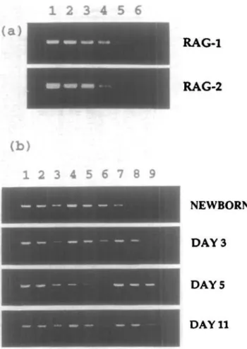
\o
BALQc
^ ~ 3 4 5 6
RAG-1
RAG-2
BALB/C + /+
Fig. 4. Comparison of V gene expression in newborn BALB/c and BALB/c nulnu RPL. cDNAs corresponding to 105 non-adherent cells
isolated from newborn BALB/c or newborn BALB/c nude mice (verified by their lack of the thymus) were divided into four aliquots and used as templates for PCR amplification of V <1, 5, 6 and 7 in conjunction with the C71 primer. Thirty cycles of PCR were performed, and the PCR
products were fractionated by gel electrophoresis and hybridized to a
32P 5'-end-labeled JT1 primer.
second peak. Thus, the variation in VT4 expression in early life
is due to the appearance of a peak of 'fetal type' VT4 bearing
cells, several days after the shift to the 'adult' type of rearrange-ments had already occurred in the thymus. These data could be explained, if one assumes that some of these T cells are not thymus derived or that the pulmonary environment early in life does not allow the selective expansion of adult VT4 T cells, but
transiently favors the expansion of cells bearing the 'fetal' GYS receptor. Alternatively, a combination of both factors may be responsible for our observation.
Thymic origin of Vy6 in RPL in newborn mice
Data from previous work have shown that Vy4 RPL can be
generated in the absence of the thymus (2). However, it has also been shown that the generation of dEC of the skin which bore the VT5 canonical receptor is strictly thymus dependent (16). It
was therefore of interest to determine whether the V76 T cells
found in the lung at birth require the presence of the thymus for their appearance. Therefore, we compared the nature of the yd TCR genes expressed in RPL isolated from newborn BALB/c mice with those of newborn BALB/c nulnu mice. As shown in Fig. 4, in the absence of the thymus, the VT6 population is not
detectable in the lung at birth and that the few yb T cells present at this time are mainly of the VT4 subset. This establishes that
at least one major subset of yb T cells in the lung of normal mice is of thymic origin and that the lung is also a preferred site where yb T cells home to during ontogeny.
High levels of RAG-1 and RAG-2 expression in the lung The marked asynchrony in the TCR phenotype between cells that are produced in the thymus and those that home to the lung may be explained as follows. The lung is populated in early life with T cell precursors or immature T cells of thymic or non-thymic origin, all of which are programmed to generate subsets of lymphocytes in a preset sequence upon receiving the appropriate signals. These hypothetical precursors, at the time of their
1 2 3 4 5 6 7 8 9
NEWBORN
DAY 3
DAY 5
DAY 11
Fig. 5. RAG-1 and RAG-2 are present in RPL of young BALB/c mice, (a) Detection of RAG-1 and RAG-2 expression. In lanes 1 - 6 , RNAsfrom 6.25x10⁵, 1.25X10⁵, 2.5x10⁴, 5X10³, 103 and 2X10² thymocytes
from 4 week old mice were analyzed using 30 cycles of PCR. One third of the amplified products were shown in the ethidium bromide stained gels, (b) RAG-2 expression in thymocytes, RPL and splenocytes from mice of various ages. Lanes 1 - 3 represents the RAG-2 PCR products from 10", 3 x 10³ and 103 thymocytes; lanes 4 - 6 and 7 - 9 represents
the RAG-2 PCR products from 106, 3 x 10⁵, and 105 splenocytes and
RPL respectively.
migration to the lung, might not have completed gene rearrange-ment, or have not yet undergone selection, and might express RAG-1 and RAG-2 genes while in the lung. To investigate this possibility, we examined total RNA from newborn, day 3, day 5 and day 11 thymocytes, spleen and RPL for RAG-1 and RAG-2 activities. To begin with, we performed a series of 5-fold dilutions of newborn thymocyte cDNA beginning with 105 cell equivalent,
and determined that we could easily detect both RAG-1 and RAG-2 activities among 103 thymocytes on ethidium bromide
stained gels after 30 cycles of PCR amplification (Fig. 5). In this population, the level of RAG-1 RNA was ~ 5-fold higher than that of RAG-2. We then proceeded to determine whether the RAG-1 and RAG-2 genes were expressed in RPL, and discovered that, indeed, such mRNA species were found in RPL preparations from newborn, day 3, day 5 and day 11 mice, but at lower abundance than in the thymocyte population (Fig. 5b).
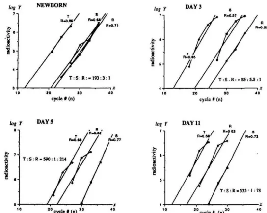
To assess the significance of this observation, we used a quantitative PCR method to compare the amount of RAG-1 RNA expressed in thymocytes, splenocytes and RPL at various ages
log Y NEWBORN DAY 3 10 10 cycle #(n) zo 10 cycle # (n) DAY 11 1 0 cycle # ( n ) " " c y c l e # ( n ) "
R g . 6. Relative levels of RAG-1 expression in thymocyte, splenocytes and RPL populations at various time after birth. Splenocytes (S), thymocytes (T) and RPL (R) were isolated from mice of different ages and the quantitative PCR performed as described in Methods. The extent of amplification (Y) is given by the formula V-/A(1 +Rf, where A is the initial amount of RAG-1 transcript, R is the efficiency of the amplification procedure and n is the number of PCR cycles (11). / c a n be estimated by the amount of radioactivity incorporated into the RAG-1 band. The plot log V-tog/4+ n log(1 +) with log V as a function of n yields a slope value - log(1 + R) from which R is derived. Thus the initial value A can be calculated and the relative initial quantity of RAG-1 mRNA estimated. The ratio of the A value is shown for each set of comparison.
in the perinatal period. The results are plotted in Fig. 6. Since the absolute amount of RAG-1 mRNA per cell cannot be measured, the relative level of RAG-1 expression is calculated using 0-actin as an internal standard. As expected, at all ages tested, thymocytes show the highest level of RAG-1 activity. Surprisingly, although RAG-1 was expressed in RPL at levels lower than in splenocytes at days 1 and 3 after birth, the level of RAG-1 mRNA in RPL was significantly higher than in spleno-cytes at day 5 and 11: by 200- and 80-fold respectively (Fig. 6). Furthermore, RAG-1 and RAG-2 transcripts can both be found in the TCR o/S+ as well as the TCR yd* populations isolated
from the lung (Fig. 7). Since mature T cells that have undergone selection are expected to shut off RAG gene expression (17,18), these data support the idea that the T cell repertoire in RPL is shaped at a significant extent by local diversification and selection. Precursor potential of antigen receptor negative cell in RPL The T cells in the lung which express RAG could be either thymic migrants that have not yet undergone selection or they could be derived from T cell precursors present in the lung. To further investigate these possibilities, we injected 3 month old SCID mice with cells isolated from C57BL/6 RPL that were depleted of T and B lymphocytes, and asked whether there were any changes in the lymphoid compartment of the recipients 4 months later. Each irradiated CB-17 SCID mice ( H - ^ received 105 antigen
receptor negative cells from 2 - 3 weeks old C57BL/6 (H-213)
RPL
RAG-1
RAG-2
Fig. 7. RAG-1 and RAG-2 are expressed in y& and a/3 surface TCR
positive cells. RPL (2X10⁶) from 3 week old BALB/c mice were
pre-incubated with the anti-Fc block antibody from PharMingen for 15 min at 4°C, divided into three fractions and either treated with Wotin-conjugated anti-r8 TCR antibody (GL-3), anti-a£ TCR antibody (H57-597) or medium only for 30 min. TCR+ cells were isolated using
streptavidin-coated Dynal beads and RNA was prepared from the bead bound cells. RAG-1 and RAG-2 expression were analyzed in the same reaction using
MP 5'-end-labeled primers, and the reaction products were fractionated
on a 6% polyacrylamide gel A PCR reaction using a thymocyte derived template was performed in parallel.
Table 1. Phenotypic analysis of splenocytes and RPL from SCID
mice which received precursors from the lung
Mouse Control SCID1 SCID 2 SCID 3 SCID 4 SCID 5 SCID 6 RPL H-2" 1.2 79 2.0 53 40 34 71 H-26 CD3 1.0 74 3.2 42 7.3 6.4 59 a/S 0.7 67 1.3 33 3.2 1.7 49 0.5 2.3 0.8 8.8 4.8 8.2 5.0 Spleen H-2" 0.8 76 37 25 57 54 40 CD3 1.2 64 4.2 24 4.7 5.0 19 «P 0.6 60 1.1 20 1.6 1.7 13 7* 0.5 4.6 4.1 4.0 3.8 4.5 5.4 l g * 1.2 6.5 31 2.3 50 39 26 S C I D n o s 1 - 3 a n d 4 - 6 a r e from two sets of independent experiments. The control is representative of the six SCID mice which received only SCID bone marrow cells after irradiation. The data were collected on a FACScan. The anti H-213 antibody was conjugated to biotin and revealed
with streptavidin - phycoerythrin. The other reagents were FITC conjugates. Wherever applicable, the antMgx staining was always followed by a 30 min incubation with 30% mouse serum before the addition of another antibody. The data presented represent percentage of total cells analyzed.
donors, together with 5X10⁵ SCID bone marrow cells as a
source of hematopoietic precursors. Control mice were given only SCID bone marrow cells. Two independent experiments were performed, each consisting of groups of three recipients. As shown in Table 1, cells of H-26 donor origin were present in
each of the six recipients which received cells purified from RPL, while no H-2" cells were found in mice which received only SCID bone marrow. The presence of both T and B cells of donor origin indicates that more than one lineage of lymphocytes can be generated from the cells transferred.
The thymus, spleen and RPL from each reconstituted SCID mouse were individually analyzed. We recovered 0 . 5 - 1 x 10⁶
cells from the thymus, but there were no tymphoid cells present according to forward and side scatter flow cytometry analysis, and no H-2*1 positive cells were detected in the thymic
prepara-tions. From the spleens, we obtained 0 . 6 - 2 X 1 07 cells and
H-2b positive lymphocytes were present in all cases. From the
lungs, 0.6-1.5X10⁵ RPI were obtained. With one exception
(no. 2), all mice displayed a significant fraction of H-2b positive
RPL. In spite of the undetectable level of H-26 positive cells in
the lung of SCID no. 2, 37% of the splenocytes in this mouse were of donor origin. A summary of the phenotypic analysis is presented in Table 1. In the recipient SCID no. 1, practically all the H-26 positive cells were T cells, both in the spteen and in the
lung. On the other hand, in SCID no. 2, there were no T cells in either the spleen or the lung, but donor derived B cells were present in the spleen. The other four mice had varying numbers of T and B cells in their spleens and lungs. Moreover, since the presence of a large number of allogeneic cells in the recipient SCID mice do not appear to induce clinically detectable graft versus host disease, one can further argue that these cell are descendants of immature precursors. Considering the low number of cells transferred and the fact that they have been depleted of TCR and cell surface lg bearing cells, these data show that a very good level of lymphoid reconstitution was achieved from the precursor enriched cells isolated from the lung epithelia. It is evident from Table 1 that not all mice have the same
number of mature, antigen receptor bearing lymphocytes. Most likely, the number of cells transferred is close to the lower limit at which single precursors can be detected by this assay.
Among the CD3+ cells, it should be noted that both yd and
a/3 T cells were present; however, their proportion varies from mouse to mouse. The relative levels of yd and a/3 T cells among RPL is represented in Table 1. It appears that yd T cell repres-entation is higher when there are fewer total CD3+ cells. It also
appears that the development of yd and a/3 T cells occurred independently. Taken together, these data support the idea that at least part of the T cell repertoire in the lungs of normal mice is generated and selected locally.
Discussions
One of the most intriguing observations in the field of 76 T cell biology is the presence of distinct T cell subsets marked by specific TCR rearrangements in different epithelia. While the partitioning of 76 T cells appears to be a function of the fine structures of their TCR, the nature of the elements which maintain this specific partition is not evident. In the first series of experi-ments, we attempt to identify a dynamic pattern in the homing of resident 76 T lymphocytes to the lung epithelia with the hope that it would indicate whether the localization of certain yd T cells in the lung is due to preferential homing or whether it is the product of site specific proliferation.
At birth, VT6 expression predominates in RPL (Fig. 2), but
between day 1 and 5 of extra-uterine life, the relative representa-tion of V74, V75 and VT7 transcripts increases, such that,
proportionately, VT6 drops to a minimum around day 7. Since
in this period there is only a small increase in the number of 76 T cells in the lung (Fig. 1), these changes could be due to a local selective expansion of the resident population rather than to new arrivals from the thymus. Surprisingly, further measurements revealed patterns of VT gene expression which were not
predicted by this hypothesis or by other views on the preferen-tial yd T cell homing in various epithelia. Specifically, Vy4
expression describes a first peak scon after birth, drops again in the young adult animal but becomes predominant again in older mice (Fig. 2). VT6 expression displays an inverse
correla-tion with VT4. This is particularly evident in older mice, where
VT6 is progressively replaced by VT4. However, there is a major
difference between the VY4 and VT6 T cells: while both the early
and the late VT6 peaks consist of fetal canonical 75 TCR, the two
peaks of Vy4 differ in their TCR composition. The early peak contains mainly fetal-type VT4JY1 rearrangements of GYS
junctions (2), while the later peak consists mainly of adutt-type GxYS rearrangements (Fig. 3). The unexpected peak of fetal VT4 rearrangements which appears among RPL after birth
could be due to 'left-over' fetal thymocytes that were produced between E15 and E18 but only expressed lung-specific homing receptors 5 - 1 0 days after they were generated. Alternatively, one could consider the possibility that a slightly delayed wave of TCR rearrangements might have taken place among immature lymphocytes in the lung epithelia. Thus, the first wave of VT4
expression in the lung could be explained by the extrathymic production of a pool of 'fetal' VT4 bearing cells during
extra-uterine life, at a moment when the shift to 'adult'-type TCR rearrangements has already occurred in the thymus.
that the lung is populated in early life with T cell precursors of thymic or non-thymic origin, programmed to generate T cells in a preset temporal sequence. These precursors might not have completed TCR gene rearrangements at the time of migration to the lung and might express the recombination activating genes, RAG-1 and RAG-2, while in the lung. Indeed, these mRNA were found in RPL preparations at surprisingly high levels early in life. To assess the significance of this observation, we compared the quantity of RAG-1 mRNA expressed in thymocytes and spleno-cytes with RPL of the same age at various times in the perinatal period. In essence, RAG-1 levels in RPL were lower than in splenocytes at days 1 and 3 after birth, but increased dramatically to levels of 200- and 80-fold higher than in splenocytes at day 5 and 11 respectively (Fig. 6). Such data support the idea that the structure of the T cell repertoire in RPL is shaped in situ rather than by differential homing of individual clonotypes preselected in the thymus. Thus, the existence of the short-lived waves of perinatal 76 RPL and the data on RAG expression in the lung during this period suggest that colonization of the lung with pre-T cells could occur.
Surprisingly, RAG-1 and RAG-2 transcripts were found in both yS+ and a/3+ RPL (Fig. 7), suggesting that colonization of the
lung with T cell precursors or with T cells still capable of performing TCR rearrangements in situ occurred for both lineages. Two explanations of this observation should be considered: (i) that T cell precursors in which rearrangements are still spontaneously taking place are preferentially attracted to the lung epithelium or (ii) that T cells which already exhibit a functional TCR home to this organ, but that upon encounter with the local antigenic environment, their RAG-1 and RAG-2 activity can be modulated. In this second case, their selection could occur by deviating, subsequent to secondary rearrangements, their original antigen specificity. In either case, one wonders whether such cells have the potential to diversify and proliferate efficiently, so that they could serve as lymphocyte precursors in in vivo reconstitution experiments.
The transfer of allogeneic TCR and Ig receptor negative 'RPL' into irradiated SCID mice was informative for assessing the precursor potential of these cells. It appears that, over a long period of time (4 months), they can generate not only a T cell population which contains both the o/9+ and the yS* subsets,
but also B cells. The lack of thymic reconstitution in these SCID mice contrasts with the substantial colonization of lymphoid organs and of the lung epithelium with mature lymphocytes. This could be interpreted as an indication of the extrathymic differen-tiation potential of the T cell precursors isolated from the lung epithelia. The low number of cells (10s) transferred and the
generation of significant numbers of lymphocytes of allogeneic origin indicate that these lymphocytes developed from immature precursors that have a high differentiation and proliferation potential. This pattern of 'active reconstitution' of SCID mice with T cells is in contrast with the relatively low level of in vivo expansion of peripheral adult T cells transferred into SCID mice documented by Sprent ef a/. (19). In essence, the extrathymic differentiation of some lymphoid precursors in the lung is a reasonable explanation for our data on the early waves of y6 T cell colonization of the pulmonary epithelia, as well as the high levels of RAG-1 and RAG-2 expression that we observed. In this regard, one should note that the ability of the murine intestine
to generate and select its own T cell repertoire is now well documented (20,21).
A recent quantitative investigation of the ordered rearrange-ments at the CT1 locus indicates that for individual VT genes
there is a strong correlation between transcription and rearrange-ment frequency (22). Moreover, there are strong indications that the rearrangement machinery in fetal thymocytes favors the production of canonical Vy5JT1 dendritic-epidermal cell
junctions (23). In the perspective of previous work (16) which demonstrated that the high frequency of productive VT5 fetal
canonical rearrangement requires both fetal T cell precursors and the fetal thymic environment, these more recent data suggest that T cell precursors are not intrinsically prone to rearrange a particular type of VT gene, but that this event is selectively
induced by the thymic stroma. Our data favor the notion that some T cell rearrangements can occur extrathymically, but that, at the same time, they are coordinated with thymic development. This could be achieved if a specific thymic soluble factor and not a thymic stromal cell ligand is responsible for inducing the ordered and coordinated transcription and rearrangement of various VT genes. On the other hand, recent reports showing
that the engraftment of fetal skin and intestine promote T cell development (24,25) further demonstrate the thymus-like activity of these epithelial tissues. Conceivably, the lung epithelium may have similar thymopoietic potential.
The role of the thymus in generating part of the pulmonary repertoire is not to be denied. Figure 1 clearly illustrates that the lung is a preferred site for the homing of 76 T cells during the perinatal period. However, this first set of VT6-specific RPL is
undetectable at birth in the absence of the thymus, as in nude mice, where the VT4 subset is present (Fig. 4). It is also evident
that the total number of T cells is much lower in the lungs of adult nu/nu mice, as is the case for the intestinal epithelial lymphocytes (5), when compared with euthymic mice. Thus, it is possible that in euthymic mice certain thymic factors may enhance extrathymic T cell differentiation.
From the data presented and discussed here, it appears that the resident pulmonary T cell population consists of cells which are generated and selected locally, as well as of cells which are generated in the thymus but may be further selected and expanded in the pulmonary environment. Our finding that RAG-1 and RAG-2 are expressed in cells which already have TCRs on their surface is in line with recent reports showing that a significant fraction of peripheral yS and a/S T cells in human blood express two different TCRs (26,27). It is possible that some thymic derived T cells may undergo further TCR mediated selection in the pulmonary environment as part of their adjustments to the requirements of this micro environment. On the other hand, resident pulmonary T cells that are generated extrathymically, and hence have not undergone thymic selection, may include cells carrying specificities that are absent among the thymus selected set but which are unique to the pulmonary environment. In this way, thymic and extrathymically derived RPL may complement each other in the defense of the host.
Acknowledgements
We are grateful to A. Lanzavecchia, K. Karjalainen and F. Ronchese for critical reading of the manuscript and M. McCarty and A. SBT for technical