www.elsevier.com / locate / cardiores www.elsevier.nl / locate / cardiores
Update review
ST-segment elevation in the electrocardiogram: a sign of myocardial
ischemia
*
`
´
Andre G. Kleber
Department of Physiology, University of Bern, Bern, Switzerland
Abstract
In 1972 Kjekshus et al. published the seminal article ‘Distribution of myocardial injury and its relations to epicardial ST-segment changes after coronary occlusion in the dog’ in Cardiovascular Research. In this article it was shown that the ST-segment elevation occurring early after occlusion of the left descending coronary artery was closely related to the depletion of the necrotic cells from creatine kinase and to flow reduction at a later stage (24 h). This correlation was especially prominent if the infarction was transmural. Starting from these phenomenological relationships, this article briefly describes and summarizes the experimental research which was carried out in other laboratories after the publication of Kjekshus et al. Special emphasis is laid on the discussion of the main basic mechanisms which underly the clinically observed ST-segment elevation and its evolution after the acute stage of ischemia, i.e. the changes in the transmembrane action potential and the alteration in electrical cell-to-cell coupling. 2000 Elsevier Science B.V. All rights reserved.
Keywords: ECG; Infarction; Ischemia
1. Introduction be discussed. Finally, further complexities inherent to the interpretation of ST-segment elevation in vivo will be In 1972 Kjekshus et al. [1] published an article entitled outlined in Section 5.
‘Distribution of myocardial injury and its relation to epicardial ST-segment changes after coronary occlusion in
the dog’ in Cardiovascular Research. This and other 2. ST-segment elevation as a marker of acute
related publications [1–5], were aimed at the definition of myocardial ischemia
early markers of infarct size. ST-segment elevation was
already used in clinical electrocardiography as a direct The term ischemic injury was originally derived from indicator of acute ischemia at that time. Nevertheless, the the observation that mechanical injury to the heart articles published by these authors are considered seminal, produces changes of the extracellular electrogram closely because they marked the beginning of a period which similar to ischemia. In 1879, Burdon-Sanderson and Page brought considerable insight into the electrophysiological [6] already described the effects of mechanical injury to alterations in myocardial ischemia and the mechanisms of the surface of the frog heart and noted that, during activity, infarct formation. In Section 2 of this brief review, devoted the injured site was charged positively with respect to the to the publication by Kjekshus et al., the early work on non-injured surface. In the past four decades, many ST-segment changes as a sign of myocardial ischemia will investigators have studied the effects of mechanical or be discussed. Section 3 will focus on the relation between ischemic injury on local extracellular electrograms in the the alterations of the ST-segment in the electrocardiogram, heart. Thus, it was gradually established that ST-elevation the underlying early changes in metabolism and extension described in the surface electrocardiogram corresponded in of necrosis. In Section 4, the cellular electrophysiologic reality to a combination of a TQ-segment change and a mechanisms leading to the changes of the intracardiac real ST-segment change. Samson and Scher [7] provided
extracellular electric field and the electrocardiogram will first evidence that the diastolic baseline depression in the extracellular electrogram was associated with a change in resting membrane potential of ischemic cells, a finding
*Tel.: 41-31-631-8740; fax: 41-31-631-8785. ´
E-mail address: [email protected] (A.G. Kleber) which was confirmed by Prinzmetal et al. [8]. Samson and
0008-6363 / 00 / $ – see front matter 2000 Elsevier Science B.V. All rights reserved. P I I : S 0 0 0 8 - 6 3 6 3 ( 9 9 ) 0 0 3 0 1 - 6
Scher [7] postulated that the real ST-segment elevation ischemia and early predictors of infarct size. Thus, is was was mostly due to shortening of the ischemic action shown that myocardial depletion of creatinephosphokinase potential, a view that was later partially disputed. At the (CPK) provided an index of ischemic cell injury and time of the publication of the work by Maroko, Kjekshus, infarct size [3]. In addition, it was demonstrated that the Sobel, Ross, Braunwald and others, it was generally degree of CPK depletion was closely correlated to the accepted that ST-segment elevation was an indicator of extent of myocardial flow reduction after coronary occlu-ischemic injury. However, most of the basic cellular sion [3] and to the extent of myocardial necrosis assessed mechanisms underlying the alterations of the ischemic histologically [5]. The results reported in the article by electrocardiogram were as yet inappropriately described, as Kjekshus et al. are illustrated and summarized in Fig. 1. In
discussed below. this article, the goal was to correlate the changes in
myocardial blood flow and CPK depletion to the changes in the ST-segment of the electrocardiogram. Accordingly,
3. ST-segment elevation, changes in ischemic in Fig. 1, extracellular electrograms, recorded with
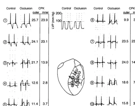
non-metabolism and infarct size polarizable electrodes before and after occlusion of the anterior interventricular coronary artery of a dog heart in The main goal of the articles by Maroko, Kjekshus, situ were compared to the corresponding local levels of Sobel, Ross and Braunwald was to define markers of CPK taken from the subepi- (‘outer’) and subendocardial
Fig. 1. Epicardial ST-segment changes 15 min after coronary occlusion and CPK activity 24 h later in corresponding subepicardial and subendocardial samples from a representative dog heart. LVP5left ventricular pressure. Locations of numbered sample sites and the occlusive coronary tie are indicated on the central diagram (reproduced from Ref. [1] with permission).
(‘inner’) muscle layers. From this article, a close correla- 4. Cellular electrophysiologic and ionic changes
tion between the ST-segment changes recorded early (15 explaining ST-segment elevation
min after coronary occlusion), the later sign of cell
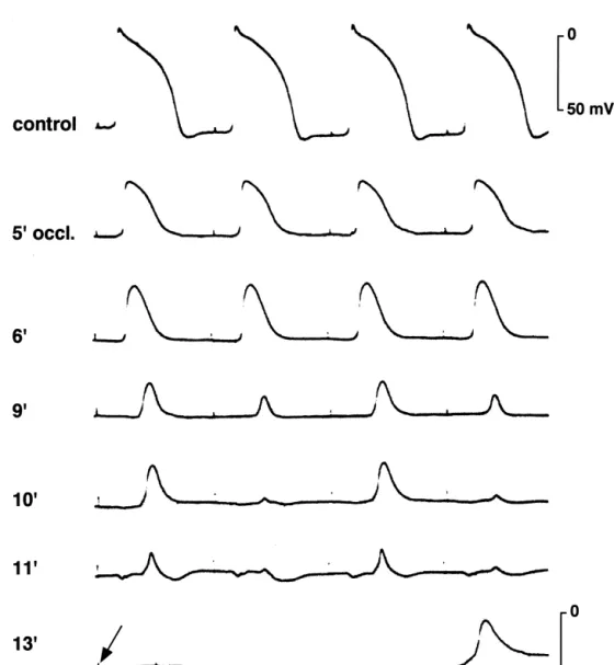
necrosis (CPK content after 24 h), and the remaining Recording transmembrane action potentials from the cononary blood flow, became evident. Moreover, there was surface of whole, arterially-perfused heart made it possible a consistent gradient between the more severe subendocar- to establish the basic relationship between changes in the dial CPK depletion and the subepicardial CPK depletion extracellular unipolar electrograms and the cellular electri-and the correlation between epicardial ST-segment changes cal changes. In a seminal paper Downar et al. [12] showed was closer to the changes in CPK in the subepicardium that regional ischemia was associated with rapid changes than in the subendocardium. These transmural gradients in the transmembrane action potentials. A rapid loss of among electrical, metabolic and flow changes after cor- membrane polarization during the resting phase (shift of onary occlusion appeared later to be specific for the dog the resting potential to more positive values) was followed heart. In dog hearts, a variable collateral circulation by a loss of amplitude, a shortening and a decrease in interferes with the flow reduction from the primarily upstroke velocity of the transmembrane action potential, occluded vessel [9]. In the pig heart, collateral flow is until the ischemic tissue became totally inexcitable after absent and an ischemic border zone in which coronary about 10 min of ischemia (Fig. 2). In addition to the flow changes from normal to very low extends over only a characterization of the basic changes in the action po-few mm [10]. The border zone itself is composed of tential, this work showed major cellular electrophysiologic interdigitating healthy and necrotic tissue with a sharp features of the acutely ischemic tissue, which later became separation between necrotic and surviving tissue [10]. The relevant for our understanding of the malign ventricular gradual change in the average concentration of ischemic arrythmias: (1) electrical alternans, which was described as metabolites and creatine phospohate across this border a harbinger of ventricular tachycardia and fibrillation, and zone is therefore due the changing ratio of necrotic to (2) the prolonged recovery of the action potential upstroke, surviving cells. Similarly to the observations by Kjekshus termed post-repolarization refractoriness, a phenomenon
1 11
et al., the experiments in porcine hearts confirmed that the due to time-dependent recovery of Na and Ca chan-correlation between ST-segment changes during early nels from inactivation [13]. This initial study on the ischemia with the later indicators of necrosis is only valid changes in the transmembrane action potential of ischemic if the alterations of the ST-segment are measured before tissue stimulated a number of further experimental in-the first 15–20 min of acute ischemia. At later stages, in-the vestigations aimed at: (1) a more close definition of the metabolites continue to change while the alterations in the nature of the cellular electrical changes; or (2) the relation-ST-segments become reduced [11] because of cell-to-cell ship between the extracellular and transmembrane
po-uncoupling (see below). tentials. To separate the real ST-segment change from the
Fig. 2. Transmembrane action potentials recorded from the subepicardium of the left ventricle of an in situ pig heart before and after occlusion of the left anterior descending coronary artery (modified from Ref. [12] with permission).
TQ-segment change, DC electrograms, recorded by non- the extracellular compartment), a special arrhythmogenic polarizable electrodes, were recorded from multiple in- role was attributed to injury current in the situation of tramural and epicardial sites in whole hearts [11]. As electrical alterans [14].
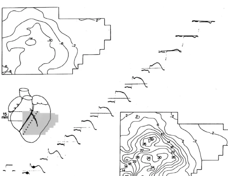
shown in Fig. 3A and B, regional ischemia produced a The work on the cellular electrophysiological basis of marked shift of both the ST-segment and the TQ-segment. ST-segment elevation and TQ-segment depression also In the case of the porcine heart, where ischemic zones are revealed a further important alteration of electrical be-transmural, the changes were relatively homogenous havior associated with ischemia, namely the electrical throughout the ventricular wall. Therefore, the gradient in uncoupling of cardiac cells [11]. In the work of Kjekshus extracellular potential during the ST- and the TQ-segment et al., ST-segment elevation was postulated as a valid could be attributed to flow of injury current between the marker of myocardial ischemia only in the minutes imme-extra- and intracellular compartments of normal and diately following coronary occlusion. Assessment of real ischemic tissue. The ischemic injury current was estimated ST-segment elevation and TQ-segment depression revealed to be of a strength which theoretically could exert a that both became maximal after approximately 10–15 min stimulatory effect [14]. Since a stimulatory effect of of coronary occlusion and declined afterwards until they diastolic current flow can only occur at sites where current almost vanished in the center of the ischemic zone after 2 is flowing in an outward direction (from the intracellular to h of maintained coronary occlusion [11]. This general
Fig. 3. Epicardial potential distribution in diastole (top) and systole (bottom) after 15–25 min of occlusion of the left anterior descending artery. Asterisks on the lowest electrogram indicate the moments during the cycle at which the potentials were measured. Signals were recorded from the shadowed area under the anterior aspect of the heart shown in the inset, at sites 3 mm apart. The extracellular complexes shown were recorded from sites along the line of steep potential gradients indicated. Square wave pulse indicates a 30 mV calibration. Isopotential lines in both maps represent 4 mV steps (modified from Ref. [11] with permission).
decrease of injury current was best explained by an exercise [19]. Further hypotheses included shifts of ion increase of resistance within the current loop, the obvious concentrations associated with osmotic swelling [25]. A candidates for this change being the gap junctions. Uncou- major difficulty with the definition of the mechanism of
1 1
pling in ischemia was also suggested by direct measure- cellular K loss during ischemia relates to the fact that K ments of alterations in tissue resistance in whole hearts accumulates in the narrow intercellular clefts.
Consequent-1
[15]. ly, the large change in [K ]o reflects a very minor
A closer definition of the two basic processes determin- imbalance of the unidirectional transmembrane fluxes [26]. ing the electrocardiographic changes in ischemia: (1) the In contrast to the mechanisms responsible for the shift in change in the transmembrane action potential; and (2) the resting membrane potential, the changes in the trans-change in electrical cell-to-cell coupling became possible membrane action potential seem to be relatively well with the development of more sophisticated techniques to understood. Thus, the positive shift in membrane potential
1
determine changes in ion activity in the extra- and intracel- and increased [K ] lead to a progressive inactivation ofo 1
lular spaces and cell-to-cell resistance. In the work of Na channels with a decrease in upstroke velocity and Harris [16] it was demonstrated that ischemic cardiac amplitude of the action potential. Comparison of the tissue loses K ions from its intracellular space, a change different components of ischemia, acidification, elevated
1
which was associated with arrhythmogenesis. The intro- [K ]o and hypoxia has shown that acidification and
1
duction of ion-sensitive electrodes into whole hearts hypoxia each add to the effect of elevated [K ] to altero 1
[17,18] made it possible to directly monitor extracellular the action potential [27], whereby the ATP-sensitive K
1 1 1
[H ], [K ], and [Na ] in the ischemic region and to current may play an additional role [28].
correlate these values with the changes in the transmem- A striking feature of the electrical changes in myocardial brane action potential. Very similar to ST-segment eleva- ischemia is the dissociation between the very early changes tion, TQ-segment depression and the associated changes in in the transmembrane action potentials and the more
1
transmembrane potential, [K ] increased rapidly in the delayed electrical cell-to-cell uncoupling. As aforemen-ischemic zone towards a plateau level, and showed a tioned, it is only this dissociation which allows for the flow secondary increase after 15–20 min. The change in resting of injury current and the generation of the early
ST-1
potential followed the change in extracellular [K ] closely segment changes in the electrocardiogram to occur: the
1
and estimates of the change in [K ] equilibrium potential changes in transmembrane potential build up the driving suggested a total balance between the depolarized resting force for injury current flow, the current flow itself requires
1
potential and the distribution of [K ] ions [19]. The low resistance pathways between the ischemic and non-mechanisms governing the change in resting potential and ischemic region, i.e. intact cell-to-cell coupling. Cable the cellular loss of potassium became a matter of a long analysis in a specially developed arterially-perfused rabbit lasting controversy among several groups partially offering papillary muscle has shown that electrical cell-to-cell divergent experimental findings and / or interpretations. The uncoupling develops rapidly after about 12–15 min after main issue was related to the fact that the changes in coronary occlusion and is completed after about 30–40
1
resting potential and the changes in extracellular [K ], min [29]. This rapid onset can be modified by precondi-[K1] , can be mutually interactive, i.e. an increase ino tioning [30], acidification [31] and by measures affecting [K1] can explain the change in membrane potential, ando energy metabolism [32]. Its exact mechanism is not fully inversely, the change in membrane potential can explain clarified, because a number of changes, all known per se to
1
the loss of intracellular [K ]. The observation that hypoxic affect gap junction resistance in vivo, occur almost simul-and ischemic cells lose potassium [20] simul-and that action taneously with ischemic cell-to-cell uncoupling:
acidifica-11
potential shortening during hypoxia is largely due to tion [33,34], increase in intracellular [Ca ], [33,34] and
1
opening of ATP-sensitive K channels [21,22] was taken accumulation of lipid metabolites [35]. The observation
1 11
as argument favoring the opening of [K ] channels as the that the increase in intracellular [Ca ] slightly precedes primary mechanism [23]. As a second hypothesis, it was the onset of ischemic cell-to-cell uncoupling [36] has led
1 11
argued that an increase of [K ] was simply reflecting ano to the hypothesis that [Ca ] would be the initiator of
1 1
inhibition of energy-dependent K / Na pumping. This electrical uncoupling and that all other aforementioned hypothesis was at least partially corroborated by the fact changes occur as a consequence of the rapidly developing
1 1
that the K / Na pump was shown in early ischemia to energy imbalance and the breakdown of ionic homeostasis.
1
react to a Na load [19,24] and that the methods to Several observations indicate that the decrease in the
1
determine intracellular Na in ischemia showed contro- cytosolic thermodynamic driving force, the so-called free
1
versial results. A third hypothesis related the cellular K energy change of ATP-hydrolysis [37], leads to a depletion
11
loss to anaerobic glycolysis, intracellular and metabolic of [Ca ] from sarcoplasmic reticulum as the primary
1
acidification. It was postulated that K might redistribute event of these afterwards self-perpetuating ionic and consequent to an electrogenic anion movement (e.g. lac- metabolic changes [38].
1
tate), a theory that appears to explain the loss of K from A further level of complexity describing the deter-skeletal muscle during fatigue and strenuous, anaerobic minants of ischemic ST-elevation in the electrocardiogram,
relates to the observation that in whole heart, even in fully 5. ST-segment elevation: a quantitative marker of
developed (no flow) ischemia, the electrical changes are regional ischemia?
heterogeneous. The distribution of the ST- and TQ
seg-ment isopotential lines in Fig. 3 shows: (1) a continuous The mechanisms underlying acute ST-segment changes decrease from the center of the ischemic zone towards the in the electrocardiogram, as briefly discussed in the above ischemic border; and (2) locally irregular isopotential lines sections, indicate a high level of complexity. Furthermore, indicating electrical heterogeneity [11]. These gradients there is a certain degree of uncertainty in the interpretation were carefully studied and compared to changes in ex- of experimental findings which is mainly related to
meth-1 1
tracellular K and H in intact porcine hearts [39–41]. odological difficulties in assessing cellular and molecular Interestingly, gradients were found to be present in fully mechanisms in whole heart tissue with occluded vessels. ischemic tissue, i.e. in absence of local oxygen (Fig. 4). Albeit complex, the mechanisms described above were Since these gradients developed rapidly over several investigated in relatively simple and partially reductionistic
1 1
millimeters, diffusion of K and H from the ischemic to experimental models. There are a variety of further vari-the non-ischemic myocardium could not fully account for ables which may be important to the explanation of the electrical heterogeneity. A further diffusible and vola- ischemic ST-segment elevation. Firstly, clinical ischemia tile substance which may explain this heterogeneity over a may often be related to a limited but not fully interrupted relatively large scale is carbondioxide, which accumulates blood supply to the heart. The discrepancy between the to .300 mmHg in the center of the ischemic zone. In an sharply demarcated necrotic zone in porcine infarcts and isolated ischemic rabbit papillary muscle, it was demon- the presence of electrically conducting tissue in human strated that carbon dioxide accumulation and diffusion infarcts suggests that the flow pattern in human infarct exerted a major effect on the cellular loss and the zones might be complex. Low flow ischemia cannot be
1
concomitant accumulation of extracellular K and that considered as pathophysiologically equivalent to total, no-diffusion of carbon dioxide may explain the centrifugal flow ischemia. For example, important ionic changes such
1 1
decrease in extracellular K and H in the ischemic region as extracellular potassium accumulation are only observed
[42]. at coronary flow ,30% of normal [43]. Furthermore,
1
Fig. 4. Lower panel: Diagrammatic representation of the changes in [K ] , pH and PO in the ischermic zone from the center to the border. Upper panel:o 2 Schematic depiction of action potentials typical for the various ischemic zones shown on lower panel. Reproduced from Ref. [39] with permission.
time course of S-T and T-Q segment changes during acute regional
anoxic perfusion is associated with a considerably larger
1 myocardial ischemia in the pig heart determined by extracellular and
cellular K loss than no flow ischemia [44]. Thus,
intracellular recordings. Circ Res 1978;42:603–613.
relatively small changes in flow reduction are likely to [12] Downar E, Janse MJ, Durrer D. The effect of acute coronary artery have an impact on the ionic and the associated electrical occlusion on subepicardial transmembrane potentials in the intact changes. Secondly, clinical ST-segment elevation after porcine heart. Circ 1977;56:217–224.
[13] Gettes LS, Reuter H. Slow recovery from inactivation of inward
myocardial infarction often persists after the acute phase of
currents in mammalian myocardial fibres. J Physiol (Lond)
ischemia, especially in the case of ventricular aneurysm.
1974;240:703–724.
Persistent ischemic damage in the border zone of myocar- [14] Janse MJ, van Capelle FJ, Morsink H et al. Flow of ‘injury’ current dial infarction, combined with a low electrical impedance and patterns of excitation during early ventricular arrhythmias in of scar tissue in the center of the infarction may partially acute regional myocardial ischemia in isolated porcine and canine hearts. Evidence for two different arrhythmogenic mechanisms. Circ
explain this phenomenon [45,46]. As a third factor
affect-Res 1980;47:151–165.
ing ST-segment elevation, the influence of the autonomous
[15] Van Oosterom A. Cardiac potential distributions. In: Medical
nervous system should be mentioned. Increased
sympa-physics and experimental cardiology, Amsterdam: University of
thetic tone affects the amount of ST-segment elevation Amsterdam, 1976.
observed early in ischemia [47], and metabolism-related [16] Harris S, Bisteni A, Russell RA, Brigham JC, Firestone JE. Excitatory factors in ventricular tachycardia resulting from
myocar-depletion of noradrenaline stores in ischemic myocardium
dial ischemia. Potassium a major excitant. Science 1954;119:200–
importantly contributes to the electrophysiologic changes
203.
observed after coronary occlusion [48,49]. In summary, the
[17] Hill JL, Gettes LS. Effect of acute coronary artery occlusion on
1
work by Kjekshus et al. has demonstrated an important local myocardial extracellular K activity in swine. Circulation relationship between ST-segment elevation in acute 1980;61:768–778.
¨
[18] Hirche H, Franz C, Bos L, Lang R, Schramm M. Myocardial
myocardial ischemia and the extent of later necrosis and
1 1
extracellular K and H increase and noradrenaline release as
coronary flow reduction. However, ST-segment elevation
possible cause of early arrhythmias following acute coronary artery
as a quantitative marker of acute ischemia should be used
occlusion in pigs. J Mol Cell Cardiol 1980;12:579–593.
with caution, because of the multiple variables contributing [19] Kleber AG. Resting membrane potential, extracellular potassium` to this electrocardiographic change. activity, and intracellular sodium activity during acute global ischemia in isolated perfused guinea pig hearts. Circ Res 1983;52:442–450.
[20] Vleugels A, Carmeliet EE. Hypoxia increases potassium efflux from
References mammalian myocardium. Experientia 1976;32:483–484.
1
[21] Venkatesh N, Lamp ST, Weiss JN. Sulfonylureas, ATP-sensitive K
1
[1] Kjekshus JK, Maroko PR, Sobel BE. Distribution of myocardial channels, and cellular K loss during hypoxia, ischemia, and injury and its relation to epicardial ST-segment changes after metabolic inhibition in mammalian ventricle. Circ Res coronary artery occlusion in the dog. Cardiovasc Res 1972;6:490– 1991;69:623–637.
499. [22] Wilde AAM, Escande D, Schumacher CA et al. Potassium
accumu-[2] Kjekshus JK, Blix AS, Grottum P, Aasen AO. Beneficial effects of lation in the globally ischemic mammalian heart. Circ Res vagal stimulation on the ischaemic myocardium during beta-receptor 1990;67:835–843.
blockade. Scand J Clin Lab Invest 1981;41:383–389. [23] Noma A. ATP-regulated K channels in cardiac muscle. Nature [3] Kjekshus JK, Sobel BE. Depressed myocardial creatine phosphoki- 1983;305:147–148.
1
nase activity following experimental myocardial infarction in rabbit. [24] Weiss J, Shine KI. Extracellular K accumulation during myocardial Circ Res 1970;27:403–414. ischemia in isolated rabbit heart. Am J Physiol 1982;242:H619– [4] Maroko PR, Radvany P, Braunwald E, Hale SL. Reduction of infarct H628.
size by oxygen inhalation following acute coronary occlusion. [25] Yan GX, Chen J, Yamada KA, Kleber AG, Corr PB. Contribution of
1
Circulation 1975;52:360–368. shrinkage of extracellular space to extracellular K accumulation in [5] Maroko PR, Libby P, Sobel BE et al. Effect of glucose-insulin- myocardial ischaemia of the rabbit. J Physiol (Lond) 1996;490:215–
potassium infusion on myocardial infarction following experimental 228.
coronary artery occlusion. Circulation 1972;45:1160–1175. [26] Johnson EA. First electrocardiographic sign of myocardial ischemia: [6] Burdon-Sanderson J, Page FJM. On the time relation of the an electrophysiological conjecture. Circulation 1976;53(Suppl.
excitatory process in the ventricle of the heart of the frog. J Physiol I):82–84.
(London) 1879;2:384–429. [27] Morena H, Janse MJ, Fiolet JW et al. Comparison of the effects of [7] Samson WE, Scher AM. Mechanism of ST-segment alteration regional ischemia, hypoxia, hyperkalemia, and acidosis on intracel-during acute myocardial injury. Circ Res 1960;8:780–787. lular and extracellular potentials and metabolism in the isolated [8] Prinzmetal MH, Toyoshima A, Ekmekci Y, Mizumo Y, Nagaya T. porcine heart. Circ Res 1980;46:634–646.
Myocardial ischemia. Nature of ischemic electrocardiographic pat- [28] Shaw RM, Rudy Y. Electrophysiologic effects of acute myocardial terns in the mammalian ventricles as determined by intracellular ischemia: a theoretical study of altered cell excitability and action electrographic and metabolic changes. Am J Cardiol 1961;8:493– potential duration. Cardiovasc Res 1997;35:256–272.
`
503. [29] Kleber AG, Riegger CB, Janse MJ. Electrical uncoupling and
[9] Janse MJ, Opthof T, Kleber AG. Animal models of cardiac increase of extracellular resistance after induction of ischemia in arrhythmias. Cardiovasc Res 1998;39:165–177. isolated, arterially perfused rabbit papillary muscle. Circ Res [10] Janse MJ, Cinca J, Morena H et al. The ‘border zone’ in myocardial 1987;61:271–279.
ischemia. An electrophysiological, metabolic, and histochemical [30] Tan HL, Mazon P, Verberne HJ et al. Ischaemic preconditioning correlation in the pig heart. Circ Res 1979;44:576–588. delays ischaemia induced cellular electrical uncoupling in rabbit [11] Kleber AG, Janse MJ, van Capelle FJ, Durrer D. Mechanism and myocardium by activation of ATP sensitive potassium channels
[published erratum appears in Cardiovasc Res 1993 Jul; changes during acute myocardial ischemia in the isolated perfused 27(7):1385]. Cardiovasc Res 1993;27:644–651. porcine heart. Circulation 1988;77:1125–1138.
`
[31] Yan G-X, Kleber AG. Changes in extracellular and intracellular pH [41] Coronel R, Fiolet JWT, Wilms-Schopman FJG et al. Distribution of in ischemic rabbit papillary muscle. Circ Res 1992;71:460–470. extracellular potassium and electrophysiologic changes during
two-`
[32] Cascio WE, Yan G-X, Kleber AG. Passive electrical properties, stage coronary ligation in the isolated, perfused canine heart. mechanical activity, and extracellular potassium in arterially per- Circulation 1989;80:165–177.
`
fused and ischemic rabbit ventricular muscle. Effect of calcium [42] Cascio WE, Yan G-X, Kleber AG. Early changes in extracellular entry blockade or hypocalcemia. Circ Res 1990;66:1461–1473. potassium in ischemic rabbit myocardium. The role of extracellular [33] Noma A, Tsuboi N. Dependence of junctional conductance on carbon dioxide accumulation and diffusion. Circ Res 1992;70:409–
proton, calcium and magnesium ions in cardiac paired cells of 422.
guinea-pig. J Physiol 1987;382:193–211. [43] Watanabe I, Johnson TA, Buchanan J, Engle CL, Gettes LS. Effect [34] Firek L, Weingart R. Modification of gap junction conductance by of graded coronary flow reduction on ionic, electrical, and me-divalent cations and protons in neonatal rat heart cells. J Mol Cell chanical indexes of ischemia in the pig. Circulation 1987;76:1127–
Cardiol 1995;27:1633–1643. 1134.
`
[35] Wu J, McHowat J, Saffitz JE, Yamada KA, Corr PB. Inhibition of [44] Yan G-X, Yamada KA, Kleber AG, McHowat J, Corr PB.
Dissocia-1
gap junctional conductance by long-chain acylcarnitines and their tion between cellular K loss, reduction in repolarization time, and preferential accumulation in junctional sarcolemma during hypoxia. tissue ATP levels during myocardial hypoxia and ischemia. Circ Res
Circ Res 1993;72:879–889. 1993;72:560–570.
21
[36] Dekker LR, Fiolet JW, Van Bavel E et al. Intracellular Ca , [45] Cinca J, Warren M, Rodriguez-Sinovas A et al. Passive transmission intercellular electrical coupling, and mechanical activity in ischemic of ischemic ST segment changes in low electrical resistance rabbit papillary muscle. Effects of preconditioning and metabolic myocardial infarct scar in the pig. Cardiovasc Res 1998;40:103–
blockade. Circ Res 1996;79:237–246. 112.
[37] Fiolet JW, Baartscheer A, Schumacher CA, Coronel R, ter Welle HF. [46] Cinca J, Bardaji A, Carreno A et al. ST segment elevation at the The change of the free energy of ATP hydrolysis during global surface of a healed transmural myocardial infarction in pigs. ischemia and anoxia in the rat heart. Its possible role in the Conditions for passive transmission from the ischemic peri-infarc-regulation of transsarcolemmal sodium and potassium gradients. J tion zone. Circulation 1995;91:1552–1559.
Mol Cell Cardiol 1984;16:1023–1036. [47] Cinca J, Bardaji A, Figueras J et al. Effects of regional denervation `
[38] Kleber AG, Oetliker H. Cellular aspects of early contractile failure on epicardial DC electrograms during coronary occlusion in pigs. in ischemia. In: Fozzard HA, Haber E, Jennings RB, Katz AM, Am J Physiol 1987;253:H138–146.
editors, The heart and cardiovascular system, New York: Raven [48] Wilde AAM, Peters RJG, Janse MJ. Catecholamine release and Press, 1991, pp. 1975–1996. potassium accumulation in the isolated globally ischemic rabbit [39] Coronel R. Distribution of extracellular potassium during myocar- heart. J Mol Cell Cardiol 1988;20:887–896.
dial ischemia. Thesis, University of Amsterdam, The Netherlands, [49] Schomig A, Fischer S, Kurz T, Richardt G, Schomig E.
Non-1988. exocytotic release of endogenous noradrenaline in the ischemic and
[40] Coronel R, Fiolet JWT, Wilms-Schopman FJG et al. Distribution of anoxic rat heart: mechanism and metabolic requirements. Circ Res extracellular potassium and its relation to electrophysiological 1987;60:194–205.
![Fig. 4. Lower panel: Diagrammatic representation of the changes in [K ] , pH and PO in the ischermic zone from the center to the border](https://thumb-eu.123doks.com/thumbv2/123doknet/14926954.664146/6.918.116.798.589.1058/lower-panel-diagrammatic-representation-changes-ischermic-center-border.webp)