HAL Id: inserm-00365055
https://www.hal.inserm.fr/inserm-00365055
Submitted on 2 Mar 2009
HAL is a multi-disciplinary open access
archive for the deposit and dissemination of
sci-entific research documents, whether they are
pub-lished or not. The documents may come from
teaching and research institutions in France or
abroad, or from public or private research centers.
L’archive ouverte pluridisciplinaire HAL, est
destinée au dépôt et à la diffusion de documents
scientifiques de niveau recherche, publiés ou non,
émanant des établissements d’enseignement et de
recherche français ou étrangers, des laboratoires
publics ou privés.
Heterotrimerization of heat-shock factors 1 and 2
provides a transcriptional switch in response to distinct
stimuli.
Anton Sandqvist, Johanna Björk, Malin Akerfelt, Zhanna Chitikova, Alexei
Grichine, Claire Vourc’H, Caroline Jolly, Tiina Salminen, Yvonne Nymalm,
Lea Sistonen
To cite this version:
Anton Sandqvist, Johanna Björk, Malin Akerfelt, Zhanna Chitikova, Alexei Grichine, et al..
Het-erotrimerization of heat-shock factors 1 and 2 provides a transcriptional switch in response to distinct
stimuli.. Molecular Biology of the Cell, American Society for Cell Biology, 2009, 20 (5), pp.1340-7.
�10.1091/mbc.E08-08-0864�. �inserm-00365055�
Vol. 20, 1340 –1347, March 1, 2009
Heterotrimerization of Heat-Shock Factors 1 and 2 Provides
a Transcriptional Switch in Response to Distinct Stimuli
Anton Sandqvist,*
†
Johanna K. Bjo¨rk,*
‡
Malin Åkerfelt,*
‡
Zhanna Chitikova,*
‡
Alexei Grichine,
§
Claire Vourc’h,
§
Caroline Jolly,
§
Tiina A. Salminen,
†
Yvonne Nymalm,
†
and Lea Sistonen*
‡
*Turku Centre for Biotechnology, University of Turku, Åbo Akademi University, 20520 Turku, Finland;
Departments of
†Biochemistry and Pharmacy and
‡Biology, Åbo Akademi University, 20520 Turku, Finland;
and
§Institut National de la Sante´ et de la Recherche Me´dicale, U823, Institut Albert Bonniot; Universite´
Joseph Fourier, Institut Albert Bonniot, Grenoble 38700, France
Submitted August 22, 2008; Revised December 19, 2008; Accepted December 23, 2008
Monitoring Editor: M. Bishr Omary
Organisms respond to circumstances threatening the cellular protein homeostasis by activation of heat-shock
transcrip-tion factors (HSFs), which play important roles in stress resistance, development, and longevity. Of the four HSFs in
vertebrates (HSF1-4), HSF1 is activated by stress, whereas HSF2 lacks intrinsic stress responsiveness. The mechanism by
which HSF2 is recruited to stress-inducible promoters and how HSF2 is activated is not known. However, changes in the
HSF2 expression occur, coinciding with the functions of HSF2 in development. Here, we demonstrate that HSF1 and HSF2
form heterotrimers when bound to satellite III DNA in nuclear stress bodies, subnuclear structures in which HSF1
induces transcription. By depleting HSF2, we show that HSF1-HSF2 heterotrimerization is a mechanism regulating
transcription. Upon stress, HSF2 DNA binding is HSF1 dependent. Intriguingly, when the elevated expression of HSF2
during development is mimicked, HSF2 binds to DNA and becomes transcriptionally competent. HSF2 activation leads
to activation of also HSF1, revealing a functional interdependency that is mediated through the conserved trimerization
domains of these factors. We propose that heterotrimerization of HSF1 and HSF2 integrates transcriptional activation in
response to distinct stress and developmental stimuli.
INTRODUCTION
Cells react to stressful conditions by activation of
heat-shock factors (HSFs), of which there are three mammalian
isoforms: HSF1, HSF2, and HSF4 (Pirkkala et al., 2001).
Activated HSFs bind to heat-shock elements (HSEs)
within the promoters of their target genes and induce
synthesis of protective molecular chaperones called
heat-shock proteins (Hsps). Hsps prevent protein misfolding
and are required for stress resistance and healthy cell
growth, development, and aging (Bukau et al., 2006,
Mo-rimoto, 2008). They also protect against metabolic,
neuro-degenerative and cardiovascular disorders (Balch et al.,
2008). In addition to Hsps, HSFs are associated with
ex-pression of noncoding satellite III (sat III) RNA in nuclear
stress bodies (nSBs) (Jolly et al., 2004, Rizzi et al., 2004), to
which HSF1 and HSF2 translocate upon stress (Jolly et al.,
1997, Alastalo et al., 2003). The nSBs form on the locus
9q12 consisting of pericentromeric heterochromatin, and
the sat III transcripts provide scaffolds for docking of
other nSB components, as shown for the splicing factor
ASF/SF2 (Chiodi et al., 2004, Metz et al., 2004). Besides
ASF/SF2, the RNA processing factors heterogeneous
nu-clear ribonucleoprotein (hnRNP) A1-associated protein
(HAP), hnRNPM, Sam68, and SRp30c localize to the nSBs
(Weighardt et al., 1999, Denegri et al., 2001).
HSF1 is activated by classical stresses such as heat shock
and heavy metals and responds to elevated temperatures in
vitro (Ahn et al., 2001, Anckar and Sistonen, 2007). HSF1 is
also involved in development and has critical roles in
lon-gevity and cancer (Xiao et al., 1999, Hsu et al., 2003, Morley
and Morimoto, 2004, Dai et al., 2007). Unlike HSF1, HSF2
lacks intrinsic stress responsiveness (Ahn et al., 2001).
An-other major difference between these factors is that while
HSF1 is evenly expressed, the levels of HSF2 fluctuate.
These changes in expression coincide temporally with
HSF2 DNA binding activity during developmental
pro-cesses (Rallu et al., 1997, Min et al., 2000). The function of
HSF2 in development was revealed by hsf2
⫺/⫺mice, which
display neurological and reproductive abnormalities in both
genders (Kallio et al., 2002, Wang et al., 2003, Chang et al.,
2006, Åkerfelt et al., 2008). In addition, the stress-induced
expression of hsps in hsf2
⫺/⫺cells is altered (O
¨ stling et al.,
2007). However, the mechanism by which HSF2 is recruited
to stress-inducible promoters is not known. How HSF2 is
activated, and the functional relationship between HSF1 and
HSF2 also remain to be elucidated.
In this study, we use the nSBs as a model system to
show that HSF1 and HSF2 interact as DNA-bound
hetero-trimers. When HSF1-HSF2 heterotrimerization is
inhib-ited by depletion of HSF2, the transcription of sat III DNA
is enhanced, indicating that HSF1-HSF2
heterotrimeriza-This article was published online ahead of print in MBC in Press
(http://www.molbiolcell.org/cgi/doi/10.1091/mbc.E08 – 08 – 0864)
on January 7, 2009.
Address correspondence to: Lea Sistonen (lea.sistonen@btk.fi).
Abbreviations used: HSF, heat-shock factor; Hsp, heat-shock
pro-tein; nSB, nuclear stress body; sat III, satellite III.
tion regulates transcription. We mimic the elevated HSF2
concentration during development and demonstrate that
increased HSF2 expression induces transcription of sat III
DNA and localization of both HSF1 and HSF2 to the nSBs.
In testis, where HSF2 is abundantly expressed and plays a
role in spermatogenesis (Sarge et al., 1994, Fiorenza et al.,
1995, Kallio et al., 2002, Wang et al., 2003, Åkerfelt et al.,
2008), we show interaction between HSF1 and HSF2.
In-creased HSF2 expression also induces transcription of the
classical HSF target hsp70, suggesting that HSF2 is
acti-vated by its eleacti-vated expression. Importantly, although
the stress-induced DNA binding of HSF2 is dependent on
HSF1 activity, induced HSF2 expression converts HSF1 to
a transcriptionally competent state. We propose that
het-erotrimerization is a transcriptional switch at the interface
of activation by either HSF1 or HSF2.
MATERIALS AND METHODS
Cell Culture, Treatments, Plasmids, and Transfections
HeLa and human embryonic kidney (HEK) 293T cells were cultured in Dulbecco⬘s modified Eagle’s medium and K562 cells in RPMI 1640 medium in 5% CO2at 37°C. The media were supplemented with 10% fetal calf serum, 2 mM l-glutamine, and penicillin and streptomycin. Heat shocks were performed at 42°C in a water bath for the indicated times. HeLa and HEK293T cells were transfected by electroporation (975F, 220 V; Gene Pulser; Bio-Rad, Hercules, CA). Plasmid DNA and 5⫻ 106cells in 0.4 ml of Opti-MEM (Invitrogen, Carlsbad, CA) were added to a 0.4-cm-gap electroporation cuvette (BTX, San Diego, CA) and subjected to a single electric pulse. The mouse HSF1-yellow fluorescent protein (YFP) was generated as the mouse HSF1-cyan fluorescent protein (CFP), described in Jolly et al., 2002. The human HSF2-YFP, containing amino acids 1-214, was constructed by polymerase chain reaction (PCR) and cloned into the BamHI and XhoI sites of pEYFP-N1 (Clontech, Mountain View, CA). The tandem CFP-YFP construct was a kind gift of Richard I. Morimoto (North-western University, Evanston, IL). The mHSF2␣ and mHSF2 plasmids used for over-expression of HSF2 were described in Alastalo et al., 2003. All constructs were sequenced.RNA Interference (RNAi)
Transient down-regulation of HSF1 was performed by electroporation of Scramble and HSF1 RNAi plasmids in HEK293T (O¨ stling et al., 2007). The cells were harvested after 72-h incubation. The stable scrambled and HSF1– down-regulating cell lines were generated by transfection of the pSuper shRNA Scrambled and HSF1 RNAi plasmids (O¨ stling et al., 2007) to HeLa cells, and single clones were established after selection with neomycin. For down-regulation of HSF2, small interfering RNA (siRNA) against HSF2 or AllStars negative control siRNA was transfected using HIPerFect Transfection reagent (all from QIAGEN, Hilden, Germany).
Chromatin Immunoprecipitation (ChIP)
ChIP was performed as in O¨ stling et al., 2007. K562 cells were cross-linked with 1% formaldehyde. Chromatin was sonicated and immunoprecipitated with antibodies against HSF1 (SPA-901; Nventa Biopharmaceuticals, San Diego, CA), HSF2 SFI58 (O¨ stling et al., 2007), and normal rabbit serum (NS; Jackson ImmunoResearch Laboratories, West Grove, PA). The following primers were used for ChIP: sat III (based on clone17 in Valgardsdottir et al., 2005), For 5⬘-AAT GAA CCC GAT GCA AT-3⬘, Rev 5⬘-CCA TTC TTG TTG AAT CCA TT-3⬘; and-actin, For 5⬘-AAC TCT CCC TCC TCC TCT TCC TC-3⬘, Rev 5⬘-GAG CCA TAA AAG GCA ACT TTC GG-3⬘.
Immunofluorescence
For immunofluorescence analysis, HeLa cells cultured on coverslips were fixed with⫺20°C methanol for 6 min or with 3% paraformaldehyde in phosphate-buffered saline (PBS) for 15 min. After three washes with PBS-0.5% Tween 20, the cells were incubated in blocking solution (1% bovine serum albumin PBS-0.5% Tween 20) for 1 h. Rabbit anti-HSF1 (Holmberg et al., 2000), rat anti-HSF1 (NeoMarkers, Fremont, CA), rabbit anti-HSF2 (Sarge et al., 1993), or rat anti-HSF2 (NeoMarkers) antibodies were diluted in blocking solution and added for 1 h. Secondary antibodies, anti-rabbit Alexa 488 and anti-rat Alexa 568 (Invitrogen), were incubated for 1 h. The coverslips were mounted and DNA was visualized using VECTASHIELD mounting medium with 4,6-diamidino-2-phenylindole (DAPI) (Vector Laboratories, Burlingame, CA). The cells were analyzed with an LSM510-Meta scanning confocal microscope (Carl Zeiss, Jena, Germany) equipped with the SP2 (version 3.2) software. The images were
acquired using a Plan-Apochromat 63⫻/1.4 oil differential interference contrast objective and further processed using Adobe Photoshop (Adobe Systems, Mountain View, CA) and CorelDRAW software.
Western Blot Analysis
Soluble cell extracts were prepared and subjected to SDS-polyacrylamide gel electrophoresis (PAGE) followed by transfer to nitrocellulose membrane (Pro-tran nitrocellulose; Whatman Schleicher and Schuell, Dassel, Germany). HSF1 was detected by polyclonal anti-HSF1 antibodies (Sarge et al., 1993; Holmberg et al., 2000), HSF2 by polyclonal anti-HSF2 antibodies (Sarge et al., 1993; O¨ stling et al., 2007) and Hsc70 by SPA-815 (Nventa Biopharmaceuticals). Secondary antibodies were horseradish peroxidase conjugated and purchased from Promega (Madison, WI) or GE Healthcare (Little Chalfont, Buckingham-shire, United Kingdom). The immunoblots were developed with an enhanced chemiluminescence method (ECL kit; GE Healthcare).
Immunoprecipitation
Male hybrid mice of the B6129SF2/J strain were used in coimmunopre-cipitation experiments. HSF2 knockout mice were obtained by matings of heterozygous mice that have been described previously (Kallio et al., 2002) and were maintained in a C57BL/6N background. The pathogen-free mice were housed under controlled environmental conditions and fed with complete pellet chow and allowed tap water. The mice were killed by CO2 asphyxiation. All mice were handled in accordance with the institutional animal care policies of the Åbo Akademi University (Turku, Finland). For coimmunoprecipitation experiments, testes were isolated from 60- to 80-d-old mice and lysed in 2 ml of lysis buffer (Alastalo et al., 2003). The precleared cellular lysate was incubated with anti-HSF1 (NeoMarkers), anti-HSF2 (NeoMarkers), or anti-FLAG M2 (Sigma-Aldrich, St. Louis, MO) antibodies at 4°C for 1 h under rotation, after which 40l of a 50% slurry of protein-G/Sepharose was added to the reaction mixture and incubated for 1 h at 4°C under rotation. After centrifugation, the Sepharose beads were washed with supplemented TEG buffer, and the immunoprecipitated proteins were run on 8% SDS-PAGE and transferred to nitrocellulose filter for immunoblotting as described above.
Semiquantitative Reverse Transcription (RT)-PCR
and Real-Time RT-PCR
RNA was isolated with the RNAeasy kit (QIAGEN). Contaminating genomic DNA was removed with two DNase I treatments according to the RNAeasy protocol (QIAGEN). Of each sample, 1g of RNA was subjected to reverse transcription using the High Capacity cDNA Reverse Transcription kit (Ap-plied Biosystems, Foster City, CA). For semiquantitative RT-PCR, ABsolute Rox mix (Advanced Biotechnologies, Epsom, United Kingdom) was used and the PCR was run 40 cycles. The same sat III primers as for ChIP were used. The glyceraldehyde-3-phosphate dehydrogenase (GAPDH) primers were GAPDH For 5⬘-ACC CAC TCC TCC ACC TTT GA-3⬘, GAPDH Rev 5⬘-TTG CTG TAG CCA AAT TCG TTG T-3⬘. Real-time RT-PCR analyses were per-formed with ABsolute cybrgreen mix (Advanced Biotechnologies) and the ABI Prism 5700 and 7900 (Applied Biosystems). Relative RNA quantities were normalized to GAPDH. For real-time RT-PCR, the following primers and probes were used: sat III For 5⬘-AAT GGA ATG CAA TGG AAT GG-3⬘, sat III Rev 5⬘-CCT GTA CTC GGG TTG ATT CC-3⬘, GAPDH For 5⬘-ACC CAC TCC TCC ACC TTT GA-3⬘, and GAPDH Rev 5⬘-CTG TTG CTG TAG CCA AAT TCG T-3⬘ (Shumaker et al., 2006); and hsp70 Probe 5⬘-FAM TTACACACCT-GCTCCAGCTCCTTCCTCTT TAMRA-3⬘, hsp70 For 5⬘-GCCGAGAAGGAC-GAGTTTGA-3⬘, hsp70 Rev 5⬘-CCTGGTACAGTCCGCTGATGA-3⬘, GAPDH Probe 5⬘-FAM ACCAGGCGCCCAATACGACCAA TAMRA-3⬘, GAPDH For 5⬘-GTTCGACAGTCAGCCGCATC-3⬘, and GAPDH Rev 5⬘-GGAATTTGC-CATGGGTGGA-3⬘.
Structural Modeling
The structural model of the human HSF heterotrimer of two HSF1 (amino acids [aa] 16-205) molecules and one HSF2 (aa 8-194) was done in three steps. First, a template of the DNA binding domain of six Kluyveromyces lactis HSF monomers bound to a 32-base pair DNA was generated using SYBYL 7.3 (Tripos, St. Louis, MO) by aligning three dimers of the crystal structure of K. lactis HSF bound to DNA next to each other as suggested by Littlefield and Nelson (1999). Second, the HR-A domain was aligned against the Escherichia coli Lpp-56 x-ray structure (Shu et al., 2000), and the HR-B domain was aligned against the mH38-P1 GCN4 Leucine Zipper x-ray structure (Shu et al., 1999), resulting in the template structure for the HR-A/B trimerization domain. The alignments were done according to the characteristic heptad repeat sequence (abcdefg)nseen in coiled coil struc-tures (Supplemental Figure 3). Third, the final template used for modeling the heterotrimer of the DNA binding and HR-A/B domain was generated by linking the two domains by using the x-ray structure of human GABP␣ protein (Batchelor et al., 1998). In the final model of the heterodimer, HSF2 makes both head-to-head and tail-to-tail contacts with HSF1. For sequence alignments, MALIGN and MALFORM (Johnson and Overington, 1993) were used within the Bodil visualization and modeling package (Lehtonen
HSF1-2 Heterotrimers and Transcription
et al., 2004). Ten models were generated with Modeler (Sali and Blundell, 1993), and the model with the lowest objective function was chosen. Sequence alignment in Supplemental Figure 3 was done with ALSCRIPT (Barton, 1993), and Figure 2, A and B, were created with the PYMOL Molecular Graphics System (Delano Scientific, Palo Alto, CA).
Confocal Microscopy and Two-Photon Fluorescence
Lifetime Imaging
The two-photon and confocal microscopy on HeLa cells was performed with an inverted two-photon laser scanning microscope Axiovert 200M (LSM510 NLO META; Carl Zeiss). During the experiment, cells were maintained at 37°C in a humidified atmosphere containing 5% CO2by using an on-stage incubator (PeCon, Frankfurt, Germany). All measurements were performed using a 63⫻/1.4 oil immersion plan-apochromat objective. In the fluorescence lifetime imaging (FLIM) experiments, the fluorescence decays were measured by the time-correlated single photon counting technique. Fluorescence de-cays were fitted using a biexponential model and the corresponding mean decay time in each pixel was color coded to obtain FLIM images (SPCIm-age software; Becker & Hickl, Berlin, Germany). Fluorescence resonance energy transfer (FRET) was identified by the shorter lifetime of donor (CFP) in the presence of acceptor (DA) as compared with that (D) in the control donor-only cells. The FLIM/FRET efficiency was calculated as EFILM/FRET⫽ 1 ⫺DA/D.
Additional acceptor photobleaching experiments were carried out on the same cell and completed with FLIM measurements to confirm FRET. At least five cells were measured for each experimental condition.
RESULTS
Stress-induced Translocation of HSF2 in the nSBs Is HSF1
Dependent
The colocalization of HSF1 and HSF2 into the nSBs
(Alastalo et al., 2003) prompted us to further investigate
their functional relationship. HSF1 binds to DNA in the
nSBs (Jolly et al., 2002), and we performed ChIP in K562
cells to examine the binding of HSF2 to sat III DNA. Heat
shock-induced binding of HSF1 and HSF2 was detected
(Figure 1A), indicating that both HSFs occupy the same
sat III DNA sequences (also see Supplemental Figure 1).
To investigate how HSF1 affects the localization of HSF2,
we generated a HeLa cell line stably down-regulating
HSF1 by using vector-based RNAi (Figure 1C). In
HSF1-depleted cells, the localization of both HSF1 and HSF2
into nSBs was abrogated (Figure 1B; for additional times,
see Supplemental Figure 2A), inferring that
stress-in-duced HSF2 DNA binding activity is dependent on HSF1.
This result is supported by the finding that transient
down-regulation of HSF1 by shRNAs in HEK293T cells
(Figure 1E) abolishes sat III transcription (Figure 1D and
Supplemental Figure 2B).
HSF1 and HSF2 Interact as DNA-bound Heterotrimers
The dependence of HSF2 on HSF1 activity for localization to
the nSBs (Figure 1B) raised the question of the underlying
mechanism. Upon activation, both HSF1 and HSF2 form
homotrimers through their trimerization domain consisting
of the heptad repeats A and B (HR-A/B) (Sarge et al., 1993).
Because HSF2 interacts with HSF1 via the HR-A/B domain
(Alastalo et al., 2003), we studied whether HSF1 and HSF2
can form heterotrimers. We aligned the HR-A/B sequences
of HSF1 and HSF2 and found that the amino acids involved
in trimerization are conserved, especially well within the
midsection of the HR-A/B domains of HSF1 and HSF2
(Supplemental Figure 3). Using two HSF1 HR-A/B helices
and one HSF2 HR-A/B, we made a structural model of the
left-handed coiled coil that constitutes the trimer interface
(Figure 2A). All buried polar residues, which have been
suggested to play a role in partner verification, are
con-served in the HSF1-HSF2 coiled coil structure (Figure 2A).
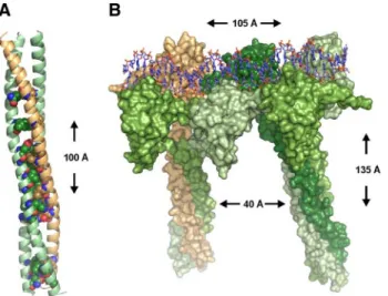
Based on the crystal structure of DNA-bound K. lactis
(KLUELA) HSF (Littlefield et al., 1999), we generated a
model of a human HSF1-HSF2 heterotrimer. The
heterotri-mer is bound to DNA and composed of the DNA binding
and HR-A/B domains of HSF1 and HSF2 (Figure 2B).
Ad-jacent to the HSF1-HSF2 heterotrimer, an HSF1 homotrimer
is shown. The trimers are bound to a 32-base pair DNA
double-stranded helix, with two nucleotide spacers as in a
canonical HSE. We measured a distance of
⬃40 Å between
the HR-A/B coiled coils of two HSF trimers (Figure 2B).
Noncovalent contacts require proximities of
⬍4 Å (Laberge,
1998), and at a distance of 40 Å, electrostatic interactions are
unlikely to occur (Creighton, 1993), excluding interactions
between separate trimers. This suggests that interactions
between HSF1 and HSF2 on DNA are mediated through
heterotrimerization.
FLIM-FRET was used to investigate whether HSF1 and
HSF2 interact on DNA within the nSBs. We could not detect
FRET when fusing CFP and YFP to the C termini of
full-length HSF1 and HSF2, presumably because the C termini of
HSF1 and HSF2 are highly mobile, preventing FRET.
There-fore, to facilitate the proper positioning of the CFP and YFP
moieties, we used HSF1 and HSF2 C-terminal deletion
con-structs (Figure 2C), that translocate into nSBs spontaneously
(Supplemental Figure 4A; Jolly et al., 2004). These C-terminal
deletion constructs contain the DNA binding and HR-A/B
domains of HSF1 and HSF2 and should be apt for the study
of interaction between the proteins as HSF1 and HSF2
inter-act through the HR-A/B domains (Alastalo et al., 2003).
Upon transfection of HeLa cells with the HSF1 and HSF2
constructs, the fluorescence lifetime of the donor
(HSF1-CFP) was shorter in the presence of acceptor (HSF2-YFP)
compared with that in cells with only HSF1-CFP (Figure
2D), indicating that FRET occurs. The FRET signal was
pre-dominantly localized to the nSBs with a mean FRET
effi-ciency of 10%, a significant number considering that the
FRET efficiency of an HSF1-CFP/HSF1-YFP pair
represent-ing HSF1 homotrimers was 14% (Figure 2E). Additional
FACS-FRET (Supplemental Figure 4B) and acceptor
photo-bleaching experiments (data not shown) confirmed the FRET
results, showing that HSF1 and HSF2 interact as
DNA-bound heterotrimers.
Stress-induced HSF Activity Is Regulated through
HSF1-HSF2 Heterotrimerization
To investigate the impact of HSF1-HSF2 heterotrimerization
on stress-induced transcription, we abrogated heterotrimer
formation by depleting HSF2. HSF2-specific siRNAs were
transfected to HEK293T cells (Figure 3B), and the
transcrip-tion of sat III DNA was measured by real-time RT-PCR. A
robust increase in the accumulation of sat III transcripts was
evident when HSF2 was depleted (Figure 3A),
demonstrat-ing that HSF1-HSF2 heterotrimerization regulates
HSF-me-diated transcription. As expected, HSF2 knockdown did not
alter the stress-induced relocalization of HSF1 to the nSBs
(Figure 3C).
Elevated HSF2 Expression Induces HSF1-HSF2
Heterotrimerization and Activates Transcription
HSF2 is involved in development, and several reports
show a correlation between increased HSF2 expression
and DNA binding activity (Murphy et al., 1994, Rallu et al.,
1997, Min et al., 2000, Chang et al., 2006, Åkerfelt et al.,
2008). Abrogation of HSF2 DNA binding in the nSBs was
associated with reduced HSF2 protein levels (Figure 1C),
indicating that HSF2 is regulated in a
concentration-de-pendent manner. We investigated the impact of elevated
HSF2 expression on sat III transcription in HeLa and
HEK293T cells by real-time RT-PCR, and we found that
increased HSF2 concentration (Figure 4C and
Supplemen-tal Figure 5C) led to a prominent induction (⬃500-fold in
HeLa cells;
⬃150-fold in HEK293T cells) of sat III
tran-scription in the absence of stress (Figure 4A and
Supple-mental Figure 5A). To extend the study to other HSF
targets, we measured the impact of HSF2 overexpression
on the transcription of hsp70. In HeLa cells, transcription
of also hsp70 was induced approximately twofold (Figure
4B), suggesting that HSF2 is activated when abundantly
expressed. No similar induction of hsp70 was detected in
HEK293T cells (Supplemental Figure 5B), which is
prob-ably due to the constitutive HSF activity in these cells,
caused by the adenoviral transactivator E1A (Phillips et
al., 1991).
The effect of elevated HSF2 expression on the localization
of HSF1 was studied with immunofluorescence in HeLa and
HEK293T cells. When abundantly expressed, HSF2
translo-cated to the nSBs (Figure 4D and Supplemental Figure 5D),
whereas it remained dispersed in the nucleoplasm of
mod-erately over-expressing HeLa cells (Figure 4E), further
sug-gesting that HSF2 is regulated by its concentration.
Intrigu-ingly, HSF2 recruited also HSF1 to the nSBs (Figure 4D),
implying that the increased expression of HSF2 seen during
development leads to activation of HSF1.
To provide more evidence for HSF1-HSF2
heterotrimer-ization as a regulatory mechanism of transcription during
developmental processes, we chose to investigate interaction
between HSF1 and HSF2 in mouse testis, a tissue
undergo-Figure
1.
Stress-induced
localization
of
HSF2 into the nSBs is HSF1 dependent. (A)
HSF1 and HSF2 binding to sat III in untreated
(C) and 1-h heat-shocked (HS) K562 cells was
analyzed with ChIP.
-actin was used as a
control promoter. Input represents 1% of the
total material and a nonspecific antibody (NS)
was used as a negative control. The ChIP
as-say on sat III was performed on four
biologi-cal samples. (B) HSF1 was down-regulated in
HeLa cells (HSF1 RNAi) and the nSB
forma-tion followed by staining of HSF1 (green) and
HSF2 (red). As a control, a scrambled cell line
(Scr.) was used. Note that the settings used for
image acquisition of HSF1 and HSF2 in Scr.
cells were reused for HSF1 RNAi cells. For
staining, polyclonal and monoclonal
antibod-ies were used against HSF1 and HSF2,
respec-tively. The nucleus is shown in blue by
stain-ing of DNA with DAPI. Heat shock and
control are indicated as HS and C,
respec-tively. For additional time points, see
Supple-mental Figure 2A. (C) Western blot analysis of
HSF1 and HSF2 in the stable Scr. (⫺) and
HSF1-RNAi (
⫹) cell lines. The retarded
mo-bility of HSF1 in heat-shocked (HS) samples is
due to hyperphosphorylation (Sarge et al.,
1993). Hsc70 serves as a loading control. (D)
Real-time RT-PCR analysis of sat III
transcrip-tion in Scr. (⫺) and HSF1-RNAi (⫹) cells. The
results are shown as fold induction upon 30
min and 1 h of HS. Fold induction was
calcu-lated by comparing the induction in HSF1
RNAi samples to the induction in scrambled
samples, which were arbitrarily set to 1. The
data represent three biological samples, and
relative quantities of sat III RNA were
normal-ized to GAPDH. Error bars indicate SD. (E)
Western blot analysis of HSF1
down-regula-tion in HEK293T cells. HS indicates a 1-h heat
shock.
HSF1-2 Heterotrimers and Transcription
ing active differentiation. Moreover, HSF2 is abundantly
expressed in testis and has been shown to be active during
spermatogenesis (Sarge et al., 1994, Fiorenza et al., 1995,
Alastalo et al., 1998, Kallio et al., 2002, Wang et al., 2003,
Åkerfelt et al., 2008). We performed coimmunoprecipitation
of HSF1 and HSF2 in both wild-type and HSF2 knockout
testis, and we found that HSF2 could be
coimmunoprecip-tiated with HSF1 antibodies and vice versa (Figure 4F).
These results indicate that HSF1 and HSF2 form
heterotri-mers during spermatogenesis.
Figure 2.
HSF1 and HSF2 interact as DNA-bound heterotrimers.
(A) Structural model of a heterotrimer formed by the HR-A/B
domains of two HSF1 molecules (green) and one HSF2 molecule
(beige). The conserved amino acids found at positions a and d in
the heptad repeat (see Supplemental Figure 3) are shown as
spheres. (B) Surface representation of the structural model of an
HSF1-HSF2-HSF1 heterotrimer and an HSF1 homotrimer bound to
DNA. HSF2 is colored beige and the different HSF1 molecules are colored in different shades of green. The HSF trimers are separated
on DNA by two nucleotides as in a consensus HSE. The height and width of the complex as well as the distance between the two coiled
coils are indicated. (C) Schematic presentation of the HSF1 and HSF2 C-terminal deletion constructs used for FRET. The position of
amino acids is shown, and the DNA-binding domain (DBD) and trimerization domains HR-A/B are indicated. The deleted C-terminal
regions are illustrated by dashed lines. (D) Interaction between HSF1 and HSF2 on DNA in the nSBs was detected with FLIM-FRET on
live cells. HeLa cells were transfected with C-terminal HSF1 and HSF2 deletions fused to CFP and YFP, and the fluorescence lifetime
of the donor (HSF1-CFP) was measured before or after photobleaching of the acceptor (HSF2-YFP). The fluorescence lifetime after
photobleaching of the acceptor is indicated by a color scale bar. (E) A mean FLIM-FRET efficiency was calculated for the FRET pairs
HSF1-CFP
⫺ HSF1-YFP and HSF1-CFP ⫺ HSF2-YFP. The data for each experimental condition represents measurements from at least
five cells, and the SD is indicated.
Figure 3.
Stress-induced HSF activity is regulated through
HSF1-HSF2 heterotrimerization. (A) Real-time RT-PCR was used to assess
the impact of HSF2 down-regulation on the transcriptional activity
during heat shock (HS) in the nSBs. The fold induction was calculated
by comparing the induction in HSF2 RNAi (
⫹) samples to the
induc-tion in Scr. (⫺) samples, which were arbitrarily set to 1. The data
represent three biological samples, and relative quantities of sat III
RNA were normalized to GAPDH. Error bars indicate SD. (B) Western
blot analysis of Scr. (
⫺) and HSF2 RNAi (⫹) HEK293T cells. HS
indicates a 1-h heat shock. (C) The localization of HSF1 (green) and
HSF2 (red) to nSBs in Scrambled (Scr.) or HSF2– down-regulating cells
(HSF2 RNAi) was followed by staining. The settings used for
acquir-ing images of HSF1 and HSF2 in Scr. cells were reused for HSF2 RNAi
cells. For staining, polyclonal and monoclonal antibodies were used against HSF1 and HSF2, respectively. The nucleus is shown in blue by
staining of DNA with DAPI. Heat shock and control are indicated by HS and C, respectively.
DISCUSSION
Integration of HSF Activity in Response to Stress
and Developmental Stimuli
Trimerization is a crucial step in the activation process of
HSFs that greatly increases the affinity for DNA (Xiao et al.,
1991). In this study, we show that HSF1 and HSF2 form
heterotrimers and propose that HSF1-HSF2
heterotrimeriza-tion provides a switch that integrates transcripheterotrimeriza-tional
activa-tion in response to stress and developmental stimuli (Figure
5). HSF1 is known to be activated by stress and responds to
elevated temperatures in vivo and in vitro. We suggest that
when activated, HSF1 does not only form homotrimers but
also trimerizes with HSF2, which itself is inert to stress. This
model explains why the translocation of HSF2 into the nSBs
is abrogated when HSF1 is depleted and also the previously
suggested dependence of HSF2 on HSF1 for stress-induced
DNA binding activity (O
¨ stling et al., 2007). Moreover, we
show that elevated expression of HSF2 leads to its
activa-tion, suggesting that concentration regulates HSF2 during
development (Figure 5). When activated, HSF2 incorporates
HSF1 into a transcriptionally competent heterotrimer,
illus-trating the capacity of HSFs to initiate heterotrimerization.
This shows, for the first time, an interdependent
cooperat-ivity between HSF1 and HSF2 that is mediated through the
conserved HR-A/B domains. We propose that HSF1 needs
cooperation from HSF2 in responding to developmental
stimuli and that some of the functions during development
earlier ascribed to HSF1 are in fact a consequence of
HSF1-HSF2 heterotrimer activity.
Implications of HSF1-HSF2 Heterotrimerization
Our results show that HSF1-HSF2 heterotrimerization
reg-ulates stress-induced transcription, as demonstrated by
in-creased expression of sat III transcripts when HSF2 is
de-pleted. Also, the positive impact on hsp70 and hsp25, and the
negative impact on hsp40 and hsp110 transcription seen in
hsf2
⫺/⫺cells (O
¨ stling et al., 2007), can now be explained by
HSF1-HSF2 heterotrimerization. HSF1 and HSF2 prefer
ar-chitecturally different HSEs (Kroeger et al., 1993). This
sug-gests that HSF1-HSF2 heterotrimers bind DNA differently
than homotrimers and that the distinct regulation of HSF
Figure 4.
Elevated HSF2 expression activates transcription and
in-duces HSF1-HSF2 heterotrimerization. (A) Real-time RT-PCR analysis
of sat III transcription upon HSF2 over-expression (
⫹) in untreated
HeLa cells. To exclude the possibility of unspecific activation of the
heat shock response, GFP was over-expressed as a control (
⫺). (B)
hsp70 transcription upon HSF2 overexpression (
⫹) was measured in
untreated HeLa cells by real-time RT-PCR. GFP was over-expressed as
a control (⫺). In A and B, fold induction was calculated by comparing
transcription in the HSF2-over-expressing cells to the control cells, in
which transcription was arbitrarily set to 1. The data represent three
(A) and five (B) biological samples, and the relative quantities of RNA
were normalized to GAPDH. Error bars indicate SD. (C) Western blot
analysis of HSF1 (top) and HSF2 (bottom) in control (
⫺) and
HSF2-over-expressing (⫹) HeLa cells. (D) HSF2 was over-expressed in HeLa
cells and the localization of HSF1 (green) and HSF2 (red) was
moni-tored in the absence of stress. The arrow marks a cell over-expressing
HSF2. (E) HSF1 and HSF2 localization in untreated
HSF2-over-express-ing HeLa cells. Moderate and robust HSF2 expression is indicated by a
thick and thin arrow, respectively. In D and E, monoclonal anti-myc
and polyclonal anti-HSF1 antibodies were used for staining. The
nu-cleus is shown in blue by staining of DNA with DAPI. (F)
Coimmu-noprecipitation of endogenous HSF1 and HSF2 in mouse wild-type
testis (HSF2 WT). To show interaction during spermatogenesis,
anti-bodies against HSF1 and HSF2 were used for immunoprecipitation. In
HSF2 knockout testis (HSF2 KO), no interaction could be detected. As
a negative control, anti-FLAG antibodies were used. The asterisk
indi-cates small molecular weight isoforms of HSF2, as described in
Alastalo et al. (1998).
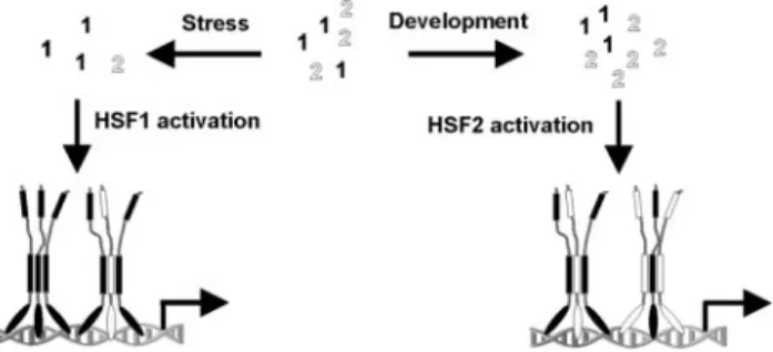
Figure 5.
Schematic illustration of HSF1-HSF2 heterotrimerization as
a mechanism integrating HSF activity. HSF1 and HSF2 are indicated in
black and white, respectively. Upon stress, HSF1 is activated, leading
to formation of HSF1-HSF2 heterotrimers. Stress-induced HSF activity
is regulated through HSF1-HSF2 heterotrimerization, a mechanism
that probably provides also temporal regulation as heat shock
dimin-ishes HSF2 levels, thereby restricting heterotrimerization through
lim-ited availability of HSF2. During development, HSF2 levels are
in-creased at certain stages and in a tissue-specific manner, leading to
activation of HSF2. Elevated HSF2 expression in turn induces
HSF2 heterotrimerization, illustrating the integrating role for
HSF1-HSF2 heterotrimerization in response to distinct stimuli.
HSF1-2 Heterotrimers and Transcription
target genes could arise from binding of HSF1-HSF2
hetero-trimers to specific sites. Another possibility is that
HSF1-HSF2 heterotrimers compete with homotrimers for
com-mon binding sites. For example, the clusterin promoter
contains an HSE which binds only one trimer, and on
which HSF1-HSF2 heterotrimerization has been proposed
to occur (Loison et al., 2006). However, no formal proof for
HSF1-HSF2 heterotrimerization on the clusterin promoter
has yet been provided.
Another example of a protein family using
heterotrimer-ization is the matrilins. The matrilin heterotrimers have a
variable stoichiometry that has been suggested to be
deter-mined by the concentration of the individual monomers
(Frank et al., 2002). Our model does not exclude variations in
the stoichiometry of the HSF1-HSF2 heterotrimers, which
may change similarly to that of the matrilin analogs. Because
the expression of HSF2 varies between different cell types
and tissues (Rallu et al., 1997, Fiorenza et al., 1995), the
HSF1-HSF2 heterotrimer stoichiometry could be modified
accordingly, allowing tissue-specific regulation of
HSF-me-diated transcription.
Interestingly, HSF2 levels are reduced when HSF1 is
down-regulated (Figure 1C), a phenomenon that has been
noted also by others (Rossi et al., 2006) and is not due to
unspecific down-regulation by RNAi (Supplemental Figure
6). Besides HSF1 down-regulation, heat shock reduces the
levels of HSF2 (Figure 3B). It is plausible that the
HSF-mediated stress-induced transcription upon prolonged
stress is determined by the receding amounts of HSF2
avail-able for heterotrimerization with HSF1 and that HSF1-HSF2
heterotrimerization regulates transcription in a temporal
manner.
The nSBs: Versatile Centers for Regulation of Gene
Expression?
The localization of HSF1 and HSF2 to the locus 9q12 is
followed by expression of sat III transcripts and formation of
nSBs. Transcription of sat III DNA is a general response to
stress (Valgardsdottir et al., 2008) and the sat III transcripts
are noncoding and heterogenous in size (Jolly et al., 2004,
Rizzi et al., 2004). The transcripts bind a subset of
mRNA-processing factors that localize to the nSBs (Weighardt et al.,
1999, Denegri et al., 2001). Because the ratio between splicing
factors determines the choice of splicing site, the nSBs are
thought to induce alternative splicing upon stress (Jolly and
Lakhotia, 2006). However, the nSBs may have multiple
func-tions. The sat III transcripts could be involved in genomic
silencing by incorporation into the RNAi system (Biamonti,
2004). Possibly, the sat III transcripts may play a role in the
regulation of gene expression. In Drosophila, noncoding
RNAs (ncRNAs) can activate transcription (Sanchez-Elsner
et al., 2006). Interestingly, this regulation is mediated via
binding of Ash1 to the ncRNA molecules, which bind to the
same sequences from which they are transcribed, a feature
they share with the sat III transcripts in the nSBs. Moreover,
it has been proposed that the expression of human and chick
coding mRNAs containing
␣-like sequences in their
untrans-lated regions is controlled by small and developmentally
expressed ncRNAs derived from
␣-satellite DNA (Li and
Kirby, 2003). In human genes, segments of sat III DNA have
been detected in the flanking regions and introns (Borstnik
et al., 1994). It is tempting to speculate that an analogous
control system of gene expression, involving the sat III
tran-scripts and regulated by HSF1 and HSF2 in response to
distinct stimuli, is used in humans.
ACKNOWLEDGMENTS
We thank Helena Saarento, Pia Roos-Mattjus, Mia Blomqvist, Gunilla Ho¨gna¨s, Jung Hue Ryung, Marianne Suominen, and John Eriksson for valuable contributions and critical review of the manuscript. Perttu Terho, Jouko Sandholm, and Oso Rissanen (Turku Centre for Biotechnology Cell Imaging Core and DNA-chips Facility) are acknowledged for excellent technical assis-tance. This work was supported by the Academy of Finland (L. S., T.A.S.), the Sigrid Juse´lius Foundation (L. S., T.A.S.), the Finnish Cancer Organizations, the Åbo Akademi University (L. S.), the Turku Graduate School of Biomedical Sciences (A. S., J.K.B., M. Å.), and the EpiPro (CLARA/INCa) and ARECA (ARC) programs (C. V., C. J., A. G.).
REFERENCES
Ahn, S. G., Liu, P. C., Klyachko, K., Morimoto, R. I., and Thiele, D. J. (2001). The loop domain of heat shock transcription factor 1 dictates DNA-binding specificity and responses to heat stress. Genes Dev. 15, 2134 –2145. Åkerfelt, M., Henriksson, E., Laiho, A., Vihervaara, A., Rautoma, K., Kotaja, N., and Sistonen, L. (2008). Promoter ChIP-chip analysis in mouse testis reveals Y chromosome occupancy by HSF2. Proc. Natl. Acad. Sci. USA 105, 11224 –11229.
Alastalo, T-P., Hellesuo, M., Sandqvist, A., Hietakangas, V., Kallio, M., and Sistonen, L. (2003). Formation of nuclear stress granules involves HSF2 and coincides with the nucleolar localization of Hsp70. J. Cell Sci. 116, 3557–3570. Alastalo, T.-P., Lo¨nnstro¨m, M., Leppa¨, S., Kaarniranta, K., Pelto-Huikko, M., Sistonen, L., and Parvinen, M. (1998). Stage-specific expression and cellular localization of the heat shock factor 2 isoforms in the rat seminiferous epi-thelium. Exp. Cell Res. 240, 16 –27.
Anckar, J., and Sistonen, L. (2007). Heat shock factor 1 as a coordinator of stress and developmental pathways. Adv. Exp. Med. Biol. 594, 78 – 88. Balch, W. E., Morimoto, R. I., Dillin, A., and Kelly, J. W. (2008). Adapting proteostasis for disease intervention. Science 319, 916 –919.
Barton, G. J. (1993). ALSCRIPT: a tool to format multiple sequence alignments. Protein Eng. 6, 37– 40.
Batchelor, A. H., Piper, D. E., de la Brousse, F. C., McKnight, S. L., and Wolberger, C. (1998). The structure of GABPalpha/beta: an ETS domain-ankyrin repeat heterodimer bound to DNA. Science 279, 1037–1041. Biamonti, G. (2004). Nuclear stress bodies: a heterochromatin affair? Nat. Rev. Mol. Cell Biol. 5, 493– 498.
Borstnik, B., Pumpernik, D., Lukman, D., Ugarkovic, D., and Plohl, M. (1994). Tandemly repeated pentanucleotides in DNA sequences of eucaryotes. Nu-cleic Acids Res. 22, 3412–3417.
Bukau, B., Weissman, J., and Horwich, A. (2006). Molecular chaperones and protein quality control. Cell 125, 443– 451.
Chang, Y. et al. (2006). Role of heat-shock factor 2 in cerebral cortex formation and as a regulator of p35 expression. Genes Dev. 20, 836 – 847.
Chiodi, I., Corioni, M., Giordano, M., Valgardsdottir, R., Ghigna, C., Cobianchi, F., Xu, R. M., Riva, S., and Biamonti, G. (2004). RNA recognition motif 2 directs the recruitment of SF2/ASF to nuclear stress bodies. Nucleic Acids Res. 32, 4127–4136.
Creighton, E. T. (1993) Proteins: Structures and Molecular Properties, New York: W. H. Freeman and Company.
Dai, C., Whitesell, L., Rogers, A. B., and Lindquist, S. (2007). Heat shock factor 1 is a powerful multifaceted modifier of carcinogenesis. Cell 130, 1005–1018. Denegri, M., Chiodi, I., Corioni, M., Cobianchi, F., Riva, S., and Biamonti, G. (2001). Stress-induced nuclear bodies are sites of accumulation of pre-mRNA processing factors. Mol. Biol. Cell 12, 3502–3514.
Fiorenza, M. T., Farkas, T., Dissing, M., Kolding, D., and Zimarino, V. (1995). Complex expression of murine heat shock transcription factors. Nucleic Acids Res. 23, 467– 474.
Frank, S., Schulthess, T., Landwehr, R., Lustig, A., Mini, T., Jeno, P., Engel, J., and Kammerer, R. A. (2002). Characterization of the matrilin coiled-coil domains reveals seven novel isoforms. J. Biol. Chem. 277, 19071–19079. Holmberg, C. I., Illman, S. A., Kallio, M., Mikhailov, A., and Sistonen, L. (2000). Formation of nuclear HSF1 granules varies depending on stress stim-uli. Cell Stress Chap. 5, 219 –228.
Hsu, A. L., Murphy, C. T., and Kenyon, C. (2003). Regulation of aging and age-related disease by DAF-16 and heat-shock factor. Science 300, 1142–1145. Johnson, M. S., and Overington, J. P. (1993). A structural basis for sequence comparisons. An evaluation of scoring methodologies. J. Mol. Biol. 233, 716 –738.
Jolly, C., Konecny, L., Grady, D. L., Kutskova, Y. A., Cotto, J. J., Morimoto, R. I. and Vourc’h, C. (2002). In vivo binding of active heat shock transcription factor 1 to human chromosome 9 heterochromatin during stress. J. Cell Biol. 156, 775–781.
Jolly, C., and Lakhotia, S. C. (2006). Human sat III and Drosophila hsr omega transcripts: a common paradigm for regulation of nuclear RNA processing in stressed cells. Nucleic Acids Res. 34, 5508 –5514.
Jolly, C., Metz, A., Govin, J., Vigneron, M., Turner, B. M., Khochbin, S. and Vourc’h, C. (2004). Stress-induced transcription of satellite III repeats. J. Cell Biol. 164, 25–33.
Jolly, C., Morimoto, R., Robert-Nicoud, M. and Vourc’h, C. (1997). HSF1 transcription factor concentrates in nuclear foci during heat shock: relation-ship with transcription sites. J. Cell Sci. 110, 2935.
Kallio, M., et al. (2002). Brain abnormalities, defective meiotic chromosome synapsis and female subfertility in HSF2 null mice. EMBO J. 21, 2591–2601. Kroeger, P. E., Sarge, K. D., and Morimoto, R. I. (1993). Mouse heat shock transcription factors 1 and 2 prefer a trimeric binding site but interact differ-ently with the HSP70 heat shock element. Mol. Cell Biol. 13, 3370 –3383. Laberge, M. (1998). Intrinsic protein electric fields: basic non-covalent inter-actions and relationship to protein-induced Stark effects. Biochim. Biophys. Acta 1386, 305–330.
Lehtonen, J. V., et al. (2004). BODIL: a molecular modeling environment for structure-function analysis and drug design. J. Comput. Aided Mol. Des. 18, 401– 419.
Li, Y. X., and Kirby, M. L. (2003). Coordinated and conserved expression of alphoid repeat and alphoid repeat-tagged coding sequences. Dev. Dyn. 228, 72– 81.
Littlefield, O., and Nelson, H.C.M. (1999). A new use for the ‘wing’ of the ‘winged’ helix-turn-helix motif in the HSF-DNA cocrystal. Nat. Struct. Biol. 6, 464 – 470.
Loison, F., Debure, L., Nizard, P., Le Goff, P., Michel, D., and Le Drean, Y. (2006). Up-regulation of the clusterin gene after proteotoxic stress. Implication of HSF1/HSF2 heterocomplexes. Biochem. J. 395, 223–231.
Metz, A., Soret, J., Vourc’h, C., Tazi, J., and Jolly, C. (2004). A key role for stress-induced satellite III transcripts in the relocalization of splicing factors into nuclear stress granules. J. Cell Sci. 117, 4551– 4558.
Min, J. N., Han, M. Y., Lee, S. S., Kim, K. J., and Park, Y. M. (2000). Regulation of rat heat shock factor 2 expression during the early organogenic phase of embryogenesis. Biochim. Biophys. Acta 1494, 256 –262.
Morimoto, R. I. (2008). Proteotoxic stress and inducible chaperone networks in neurodegenerative disease and aging. Genes Dev. 22, 1427–1438. Morley, J. F., and Morimoto, R. I. (2004). Regulation of longevity in Caeno-rhabditis elegans by heat shock factor and molecular chaperones. Mol. Biol. Cell 15, 657– 664.
Murphy, S. P., Gorzowski, J. J., Sarge, K. D., and Phillips, B. (1994). Charac-terization of constitutive HSF2 DNA-binding activity in mouse embryonal carcinoma cells. Mol. Cell Biol. 14, 5309 –5317.
O¨ stling, P., Bjo¨rk, J. K., Roos-Mattjus, P., Mezger, V., and Sistonen, L. (2007). Heat shock factor 2 (HSF2) contributes to inducible expression of hsp genes through interplay with HSF1. J. Biol. Chem. 282, 7077–7086.
Phillips, B., Abravaya, K., and Morimoto, R. I. (1991). Analysis of the speci-ficity and mechanism of transcriptional activation of the human hsp70 gene during infection by DNA viruses. J. Virol. 65, 5680 –5692.
Pirkkala, L., Nyka¨nen, P., and Sistonen, L. (2001). Roles of the heat shock transcription factors in regulation of the heat shock response and beyond. FASEB J. 15, 1118 –1131.
Rallu, M., Loones, M., Lallemand, Y., Morimoto, R., Morange, M., and Mezger, V. (1997). Function and regulation of heat shock factor 2 during mouse embryogenesis. Proc. Natl. Acad. Sci. USA 94, 2392–2397.
Rizzi, N., Denegri, M., Chiodi, I., Corioni, M., Valgardsdottir, R., Cobianchi, F., Riva, S., and Biamonti, G. (2004). Transcriptional activation of a constitu-tive heterochromatic domain of the human genome in response to heat shock. Mol. Biol. Cell 15, 543–551.
Rossi, A., Ciafre, S., Balsamo, M., Pierimarchi, P., and Santoro, M. G. (2006). Targeting the heat shock factor 1 by RNA interference: a potent tool to enhance hyperthermochemotherapy efficacy in cervical cancer. Cancer Res. 66, 7678 –7685.
Sali, A., and Blundell, T. L. (1993). Comparative protein modelling by satis-faction of spatial restraints. J. Mol. Biol. 234, 779 – 815.
Sanchez-Elsner, T., Gou, D., Kremmer, E., and Sauer, F. (2006). Noncoding RNAs of trithorax response elements recruit Drosophila Ash1 to Ultrabithorax. Science 311, 1118 –1123.
Sarge, K. D., Murphy, S. P., and Morimoto, R. I. (1993). Activation of heat shock gene transcription by heat shock factor 1 involves oligomerization, acquisition of DNA-binding activity, and nuclear localization and can occur in the absence of stress. Mol. Cell Biol. 13, 1392–1407.
Sarge, K. D., Park-Sarge, O-K., Kirby, J. D., Mayo, K. E., and Morimoto, R. I. (1994). Expression of heat shock factor 2 in mouse testis: potential role as a regulator of heat-shock protein gene expression during spermatogenesis. Biol. Reprod. 50, 1334 –13343.
Shu, W., Ji, H., and Lu, M. (1999). Trimerization specificity in HIV-1 gp 41, analysis with a GCN4 leucine zipper model. Biochemistry 38, 5378 –5385. Shu, W., Liu, J., Ji, H., and Lu, M. (2000). Core structure of the outer mem-brane lipoprotein from Escherichia coli at 1.9 A resolution. J. Mol. Biol. 299, 1101–1112.
Shumaker, D. K., et al. (2006). Mutant nuclear lamin A leads to progressive alterations of epigenetic control in premature aging. Proc. Natl. Acad. Sci. USA 103, 8703– 8708.
Valgardsdottir, R., Chiodi, I., Giordano, M., Cobianchi, F., Riva, S., and Biamonti, G. (2005). Structural and functional characterization of noncoding repetitive RNAs transcribed in stressed human cells. Mol. Biol. Cell 16, 2597–2604.
Valgardsdottir, R., Chiodi, I., Giordano, M., Rossi, A., Bazzini, S., Ghigna, C., Riva, S., and Biamonti, G. (2008). Transcription of Satellite III non-coding RNAs is a general stress response in human cells. Nucleic Acids Res. 36, 423– 434.
Wang, G., Zhang, J., Moskophidis, D., and Mivechi, N. F. (2003). Targeted disruption of the heat shock transcription factor (hsf)-2 gene results in in-creased embryonic lethality, neuronal defects, and reduced spermatogenesis. Genesis. 36, 48 – 61.
Weighardt, F., Cobianchi, F., Cartegni, L., Chiodi, I., Villa, A., Riva, S., and Biamonti, G. (1999). A novel hnRNP protein (HAP/SAF-B) enters a subset of hnRNP complexes and relocates in nuclear granules in response to heat shock. J. Cell Sci. 112, 1465–1476.
Xiao, H., Perisic, O., and Lis, J. T. (1991). Cooperative binding of Drosophila heat shock factor to arrays of a conserved 5 bp unit. Cell 64, 585–593. Xiao, X., Zuo, X., Davis, A. A., McMillan, D. R., Curry, B. B., Richardson, J. A., and Benjamin, I. J. (1999). HSF1 is required for extra-embryonic development, postnatal growth and protection during inflammatory responses in mice. EMBO J. 18, 5943–5952.