HAL Id: hal-01619294
https://hal.sorbonne-universite.fr/hal-01619294
Submitted on 19 Oct 2017
HAL is a multi-disciplinary open access
archive for the deposit and dissemination of
sci-entific research documents, whether they are
pub-lished or not. The documents may come from
teaching and research institutions in France or
abroad, or from public or private research centers.
L’archive ouverte pluridisciplinaire HAL, est
destinée au dépôt et à la diffusion de documents
scientifiques de niveau recherche, publiés ou non,
émanant des établissements d’enseignement et de
recherche français ou étrangers, des laboratoires
publics ou privés.
catheter-related thromboembolism in breast cancer
patients: the CAVECCAS study
Philippe Debourdeau, Marc Espié, Sylvie Chevret, Joseph Gligorov, Antoine
Elias, Pierre François Dupré, Kristell Desseaux, Issa Kalidi, Stephane Villiers,
Sylvie Giachetti, et al.
To cite this version:
Philippe Debourdeau, Marc Espié, Sylvie Chevret, Joseph Gligorov, Antoine Elias, et al.. Incidence,
risk factors, and outcomes of central venous catheter-related thromboembolism in breast cancer
pa-tients: the CAVECCAS study. Cancer Medicine, Wiley, 2017, �10.1002/cam4.1201�. �hal-01619294�
ORIGINAL RESEARCH
Incidence, risk factors, and outcomes of central venous
catheter- related thromboembolism in breast cancer
patients: the CAVECCAS study
Philippe Debourdeau1, Marc Espié2,3, Sylvie Chevret3,4, Joseph Gligorov5,6, Antoine Elias7,
Pierre François Dupré8, Kristell Desseaux3,4, Issa Kalidi9, Stephane Villiers10, Sylvie Giachetti2,3,
Corinne Frere11,12,13 & Dominique Farge3,14
1Department of Medical Oncology, Sainte Catherine Institute, Avignon, France
2Breast Cancer Unit, Saint Louis Hospital, Assistance Publique-Hôpitaux de Paris, Paris, France 3University Paris Diderot, Paris, France
4Biostatistic Department and Medical Informatics, Saint-Louis Hospital, Assistance Publique-Hôpitaux de Paris, Paris, France 5Medical Oncology, Tenon Hospital, Assistance Publique-Hôpitaux de Paris, Paris, France
6Francilian Breast Intergroup, APREC, IUC-UPMC Sorbonne University, Paris, France 7Department of Vascular Medicine, Sainte Musse Hospital, Toulon, France 8Department of Gynecology and Surgery, CHRU, Brest, France
9Department of Biology, Saint Louis Hospital, Assistance Publique-Hôpitaux de Paris, Paris, France
10Department of Anesthesiology and Reanimation, Saint Louis Hospital, Assistance Publique-Hôpitaux de Paris, Paris, France 11Department of Haematology, Pitié-Salpêtrière Hospital, Assistance Publique-Hôpitaux de Paris, Paris, France
12Sorbonne Universités, UPMC Université Paris 06, UMR_S 1166, Paris, France 13Institute of Cardiometabolism and Nutrition, ICAN, Paris, France
14Internal Medicine Unit: Autoimmune and Vascular Diseases, UF 04, Saint-Louis Hospital, Assistance Publique-Hôpitaux de Paris, AP-HP, Paris, France
Keywords
Breast cancer, central venous catheter, chemotherapy, risk factors, venous thromboembolism
Correspondence
Dominique Farge, Internal Medicine Unit: Autoimmune and Vascular Diseases, UF04, Saint-Louis Hospital, Assistance Publique-Hôpitaux de Paris, 1 Avenue Claude-Vellefaux, 75010 Paris, France. Tel: +33 1 42 49 97 64; Fax: +33 1 42 49 94 78;
E-mail: dominique.farge-bancel@aphp.fr
Funding Information
This work was supported by Programme hospitalier de recherche clinique (PHRC AOM 2007 K070104/N° ID RCB : 2007-A01123-5). Received: 3 June 2017; Revised: 16 July 2017; Accepted: 16 August 2017
doi: 10.1002/cam4.1201
pour le Groupe Francophone Thrombose et Cancer (www.thrombose-cancer.com)
Abstract
Previous epidemiologic studies investigating central venous catheter (CVC)- related venous thromboembolism (CRT) were conducted in heterogenous cancer populations and data in breast cancer (BC) remain limited. To investigate the Doppler ultrasound (DUS)- CRT incidence, risk factors and outcomes in BC, we designed a prospective, multicenter cohort of nonmetastatic invasive BC patients undergoing insertion of a CVC for chemotherapy. All patients under-went double- blind DUS before, 7, 30, and 90 days after CVC insertion and a 6 months clinical follow- up. Symptomatic DUS- CRT were treated by antico-agulants. D- Dimers, thrombin generation, and platelet- derived microparticles were measured before and 2 days after CVC placement. In DUS- CRT patients, a nested case–control study analyzed the role of thrombophilia. Among 524 patients, the DUS- CRT (14 symptomatic, 46 asymptomatic) cumulative probabil-ity was 9.6% at 3 months and 11.5% at 6 months (overall incidence rate: 2.18/100 patient- months). Ten/14 symptomatic DUS- CRT were detected on double- blind DUS before the clinical symptoms, and 3/14 had a simultaneous pulmonary embolism. No clinical thrombotic event subsequently occurred in untreated asymptomatic DUS- CRT. Age >50 years (OR, 1.80; 95% CI, 1.01–3.22), BMI >30 kg/m² (OR, 2.64; 95% CI, 1.46–4.76) and comorbidities (OR, 2.05; 95% CI, 1.18–3.56) were associated with DUS- CRT. No biomarkers was found to predict DUS- CRT. In multivariate analysis, BMI >30 kg/m² (OR, 2.66; 95%CI, 1.46–4.84) and lobular carcinoma histology (OR, 2.56; 95%CI, 1.32–4.96) remained the only significant DUS- CRT risk factors. Thrombophilia did not account for DUS- CRT. Only clinical parameters identified high risk DUS- CRT patients who may be considered for thromboprophylaxis.
Cancer Medicine
Introduction
Breast cancer (BC), accounting for 25% of women cancers, is the most frequent cancer and the leading cause of death in the female population worldwide [1]. As com-pared to their age- matched individual counterparts without cancer, BC women have a three to fourfold increased risk of venous thromboembolism (VTE) [2, 3], which was notably demonstrated to be an independent prognostic factor for survival and the second cause of death in all cancer patients [4–7]. A large UK registry study [6, 8] recently showed that the VTE risk in BC patients is spe-cifically higher around the diagnosis period and until 3 months later, particularly on (neo)adjuvant chemotherapy (NAC) [6], meanwhile long- term central venous catheters (CVC) are commonly inserted to facilitate intravenous administration of treatments. While risk factors for VTE in BC women have been extensively investigated [6], data regarding CVC- related thrombosis (CRT) are scarce and factors promoting CRT in this specific population remain poorly understood. Previous epidemiologic cohort studies were conducted only in heterogeneous cancer patient populations. Verso et al. first reported an overall incidence of 4–5% (0% to 28%) for symptomatic CRT and 30% (27% to 66%) for asymptomatic CRT detected by venog-raphy in unselected cancer patients [9]. The incidence of CRT further varies widely between studies, due to many differences in CRT definition and diagnostic procedures. Importantly, symptomatic CRT have been found to result in pulmonary embolism (PE) in 10–15% of unselected cancer patients [9, 10]. On the other side, the clinical consequences of an asymptomatic CRT diagnosed on vari-ous imaging tools still remain uncertain, particularly con-cerning its spontaneous outcome without anticoagulation treatment and if its early diagnosis may predict the onset of symptomatic CRT [11]. In the absence of evidence, anticoagulants are not currently recommended for CRT prophylaxis [12], and the ASCO guidelines on CVC care for cancer patients highlighted the need for additional research in this area [13]. We, therefore, designed the prospective multicenter CAVECCAS (Cathéter VEineux Central et CAncer du Sein) study in a highly selected population of nonmetastatic invasive BC patients to inves-tigate the specific incidence, risk factors and outcomes of both asymptomatic and symptomatic CRT in BC patients within the 6 months after CVC insertion for NAC.
Materials and Methods
Study design
This multicenter observational cohort study was conducted in nine cancer hospitals between September 2008 and
December 2011. All nonmetastatic invasive BC patients were screened. The inclusion criteria were histologically proven BC cancer patients older than 18 years to be treated by adjuvant chemotherapy or NAC necessitating the insertion of a port single central lumen catheter for more than 3 months [14]. All CVC were inserted via the internal jugular, axillary or subclavian veins and terminated in the superior vena cava. We excluded patients receiving NAC via peripherally inserted catheter, tunneled catheters without port and femoral CVC, patients treated by a previous chemotherapy and/or hormonal therapy, patients on curative anticoagulant therapy, patients with a platelet count <80G/L, a INR <1.5, a fibrinogen level < 1 g/L, and a creatinine level >175 μmol/L. All eligible consecu-tive patients were prospecconsecu-tively enrolled after obtaining their written informed consent to participate in the trial. The study was approved by Ethical Committee of Paris (France) and registered on clinical trial.gov (ClinicalTrials. gov Identifier: NCT00714909).
Data collection
Baseline patients’ demographic and clinical characteristics, cancer characteristics, site (jugular or subclavian) and side (right or left) of CVC insertion were recorded as well as the insertion procedure duration and the number of veni-punctures (>2). According to Good Clinical Practices Guidelines [15], Doppler- ultrasonography (DUS) was rec-ommended to guide CVC insertion, which had to be preferably positioned on the right side [10, 14, 16], with CVC distal tip at the superior vena cava and the right atrium junction. In women with right BC, CVC insertion was allowed on the left side.
Outcome measures
All patients underwent double- blind DUS before and 7, 30, and 90 days (D) after CVC insertion, including com-pression, B- mode imaging with the addition of color and pulsed- wave Doppler. The following parameters were recorded on both sides on the humeral, axillary, subcla-vian, internal jugular veins, and when possible on the brachiocephalic and superior cava veins: venous vessels patency, presence or absence of vein compressibility, echo-genicity within the vein lumen, characteristics of venous flow, including presence or absence of cardiac pulsatility transmitted, response to respiratory maneuvers. The main outcome measure was the occurrence of either: (a) asymp-tomatic CRT detected by repeated DUS performed at each investigating site by the same radiologist at day 7 (±2), day 30 (±5) and day 90 (±7) after CVC insertion or (b) symptomatic CRT over the 6 months of patients clinical follow- up diagnosed by any VTE clinical symptoms of
the upper limb, neck, or head (pain, edema, or headache) or PE, and objectively confirmed by DUS, phlebography, angiography, computed tomography, or pulmonary scin-tigraphy. CRT was defined as the occurrence of a mural thrombus extending from CVC into the lumen [15]. For veins accessible to direct insonation, diagnostic criteria of CRT were: noncompressibility, visualization of echogenic intravascular mass and absence of respiratory variation (jugular, axillary or subclavian veins) [17]. For veins inac-cessible to direct insonation (middle part of the subclavian vein, brachiocephalic vein and superior caval vein) the criterion of monophasic flow to detect occlusive throm-bosis was used. All DUS thrombotic events were reviewed by two physicians (DF, PD) unaware of clinical data. When an asymptomatic CRT was diagnosed on DUS as scheduled by systematic examination according to the study protocol, patient and referring physicians were blind to DUS results. CVC dysfunction not related to CRT, such as distal thrombus without mural involvement or a fibrin sleeve around the CVC distal tip, or pinch- off syn-drome were not recorded as an event. According to the study protocol, asymptomatic CRT were not to be treated, and thrombus evolution was assessed upon next DUS examination. Anticoagulant treatment was initiated only at the onset of clinical symptoms for symptomatic CRT [17].
Blood samples and laboratory analysis
Venous blood samples were collected before and 2 days after CVC insertion. D- Dimers levels were measured, using an Enzyme Linked Fluorescent Assay (VIDAS® D- Dimer
Exclusion™, Biomérieux, Marcy l’Etoile, France) on a Mini
Vidas analyzer (Biomérieux). Platelet- derived MPs (Pd- MPs) and Pd- MPs expressing phosphatidyl serine (Pd- MP/ PS+) were measured, using a flow cytometry assay as previously described [18]. Thrombin generation was stud-ied, using the Calibrated Automated Thrombogram assay (CAT®, Stago, Asnieres, France) according to the
manu-facturers’ instructions with PPP reagent 5 pM®
(Thrombinoscope b.v., Maastricht, the Netherlands). Each patient’s plasma was studied in duplicate. In a third well, PPP reagent 5 pM® was replaced with the same volume
of Thrombin Calibrator® (Thrombinoscope b.v.,
Netherlands) to correct thrombin generation curves for substrate consumption and inner filter fluorescence effects. The following thrombogram parameters were analyzed: the lag- time of thrombin generation, the time to reach the peak of thrombin (time to Peak), the thrombin peak (Peak), the endogenous thrombin potential (ETP), which reflects the total amount of thrombin activity. Additional individual heritable or acquired thrombophilia risk factors were analyzed in patients selected for the nested
case- control study. It included antithrombin, protein C and protein S levels measured on a STAR- R analyzer (Stago) using the STACHROM and STACLOT assays (Stago); search for Lupus anticoagulant performed on a STAR- R analyzer, using the STACLOT DRVV and the PTT- LA assays (Stago); dosage of anticardiolipin and antiβ2GP1 antibodies levels measured with an ELISA method (respectively, Stago, Biorad, Phadia); and search for the G1691A polymorphism in the gene encoding fac-tor V (FVL) and the prothrombin G20210A polymorphism identified, using an allele- specific restriction- enzyme analy-sis as previously described [19, 20].
Nested case–control study
After completing recruitment and follow- up, a nested case–control study was performed to analyze additional individual thrombophilia risk factors (Antithrombin, Protein C and Protein S levels, presence of Factor V and Factor II Leiden mutations, presence of antiphospholipid, anticardiolipin and antiβ2GP1 antibodies). Two controls without CRT were matched for TNM status with each symptomatic or asymptomatic CRT patient from the CAVECCAS cohort.
Statistical analysis
Statistical analysis was performed on the open- source software R Version 2.15.2. R foundation, Vienna, Austria Summary statistics are expressed as median and inter- quartile range [IQR] and (minimum, maximum) for quantitative data and numbers and percentages for categorical data. Continuous variables with a skewed distribution were log- transformed. Univariate comparisons used the exact Fisher test or the Wilcoxon rank sum test according to the variable type. In the multivariate models, a step- down selection procedure was used with P- values < 0.10 as the selection criterion. A con-ditional logistic model was used to study the occurrence of thrombosis based on thrombophilia factors testing.
Results
Study population
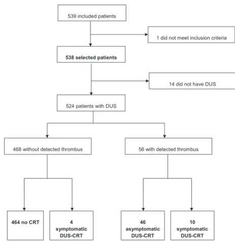
Here, between September 2008 and December 2011, 539 consecutive patients were enrolled in the study. One patient did not meet all the inclusion criteria, and 14 did not attend the DUS examination. Finally, 524 patients were included in the CAVECCAS study cohort. Flowchart is displayed in Figure 1. Baseline patients’ demographic and clinical characteristics, as well as cancer characteristics and treatments are shown in Table 1. At inclusion, patients had a median age of 53 years (interquartile range [IQR],
46–62 years) and 523 were women (99.8%). One hundred forty- five patients (27.7%) presented one or more comor-bidities, including diabetes mellitus, hypertension, liver failure, kidney failure, chronic respiratory failure, or cardiac failure. Twelve patients (2.3%) had a previous VTE his-tory (7 with lower limb DVT, 5 with PE). Other VTE risk factors were obesity with Body Mass Index (BMI) above 30 kg/m² (n = 89), lower limb varicose (n = 50), previous thoracic venous catheter or traumatism (n = 5) or hereditary thrombophilia (n = 1). At enrollment, 85.9% of patients (n = 450) had an invasive ductal carcinoma, 49.2% (n = 230) had node involvement, and 79.5% (n = 415) were positive for either estradiol or progesterone receptors. All CVC were single port CVC from various manufacturers brands, namely Braun (272), Perouse (n = 113), Bard (n = 92), Districath (n = 3), Heliosite (n = 2), Vygon (n = 1), or others (n = 41).
Catheter- related thrombosis
During the 180 days of clinical follow- up, 60 patients developed a DUS- CRT. The final DUS- CRT incidence rate was 2.18 cases per 100 patient- months. The cumulative probability of DUS- CRT was 9.6% after 3 months and
11.5% after 6 months (Fig. 2). DUS- CRT events were symptomatic in 14 patients (2.7%) and remained asymp-tomatic in 46 patients (8.8%) up to 6 months of clinical follow- up. Among the 14 symptomatic CRTs, 10 were detected on DUS before the onset of symptoms (median 6.5 [IQR], 0–17.5 days before the onset of symptoms) and 3 presented simultaneous PE. All symptomatic patients were treated according to the international guidelines [12]. In the 46 asymptomatic patients, presence of mural throm-bus was detected on study protocol repeated DUS at day 8, 30 and 90 after CVC insertion in, respectively, 27 (46%), 10 (22%), and 9 (19%) patients, and thrombus resolved on the next study protocol DUS in 30 patients. Due to protocol deviation, 5 asymptomatic CRTs were treated by anticoagulation. In the 41 patients with asymptomatic CRT who remained untreated, no symptomatic CRT or PE subsequently occurred.
Biological parameters
Among the 524 selected patients, 501 (95.6%) had coagu-lation blood samples before and after catheter insertion (Table 2). The median D- Dimers value was statistically higher after (586 [366; 842] ng·mL−1) than before CVC
Table 1. Breast cancer patients characteristics and venous thrombosis risk factors at enrollment.
All (n = 524) No CRT (n = 464) CRT (n = 60) P
Breast cancer side, n (%) 1.00
Left 271 (52) 240 (52) 31 (51.7) Right 251 (48) 222 (48) 29 (48.3) Missing 2 2 0 Histological type, n (%) 0.016 Ductal carcinoma 445 (85.0) 400 (86.2) 45 (75.0) Lobular carcinoma 64 (12.2) 51 (11.0) 13 (21.7) Ductal and lobular carcinoma 5 (0.9) 3 (0.6) 2 (3.3)
Other 10 (1.9) 10 (2.2) 0 TNM staging, tumour n (%) 0.025 T0 2 (0.5) 0 2 (3.3) T1 209 (47.6) 188 (49.6) 21 (35.0) T2 190 (43.3) 164 (43.3) 26 (43.3) T3 35 (8.0) 33 (8.7) 2 (3.3) T4 3 (0.7) 3 (0.8) 0 Missing 85 85 0 TNM staging, node (%) 1.00 N0 254 (48.6) 224 (48.5) 30 (50.0) N1 207 (39.6) 183 (36.9) 24 (40.0) N2 48 (9.2) 43 (9.3) 5 (8.3) N3 13 (2.5) 12 (2.6) 1 (1.7) Missing 2 2 0 Node involvement, n (%) 0.77 No 223 (49.2) 195 (48.9) 28 (51.9) Yes 230 (50.8) 204 (51.1) 26 (48.2) Missing 74 68 6
Steroid hormone receptors, n (%) 1.00
No 107 (20.5) 95 (20.6) 12 (20.0) Yes 415 (79.5) 367 (79.4) 48 (80.0) Missing 2 2 0 SBR grading, n (%) 0.74 1 57 (11.2) 49 (10.8) 8 (13.8) 2 271 (53.0) 242 (53.4) 29 (50.0) 3 183 (35.8) 162 (35.8) 21 (36.2) Missing 13 11 2 HER2 status, n (%) 0.85 Negative 443 (84.9) 391 (84.6) 52 (86.7) Positive 79 (15.1) 71 (15.4) 8 (13.3) Missing 2 2 0 Chemotherapy, n (%) 0.25 Anthracycline 116 (22.2) 99 (21.4) 17 (28.3) Anthracycline + taxanes 407 (77.8)1 364 (78.6)1 43 (71.7)0 Missing 1 1 0 Herceptin 76 (14.6) 69 (14.9) 7 (11.7) 0.70 Previous DVT, n (%) 0.57 No 516 (98.7) 458 (98.7) 58 (98.3) Yes 7 (1.3) 6 (1.3) 1 (1.7) Missing 1 0 0 Previous PE, n (%) 1.00 No 519 (99.1) 459 (98.9) 61 (100) Yes 5 (0.9) 5 (1.1) 0 Obesity (BMI >30 kg/m2), n (%) 0.044 No 433 (82.9) 389 (84.2) 44 (73.3) Yes 89 (17.1) 73 (15.8) 16 (26.7) Missing 2 2 0
Lower limbs varicose, n (%) 0.82
No 474 (90.5) 420 (90.5) 54 (90.0)
Yes 50 (9.5) 44 (9.5) 6 (10.0)
insertion (454 [294; 757] ng·mL−1; P < 0.0001). No
sig-nificant increase in thrombin generation peak height (257 [194; 307] vs. 253 [201; 308] nmol) nor in endogenous thrombin potential (1304 [1063; 1652] vs. 1322 [1052; 1582] nmol·min) were observed. Both Pd- MPs (982 [518; 2147] vs. 759 [417; 1373] mL−1; P < 0.0001) and Pd-
MPs/PS+ (778 [409; 1851] vs. 730 [381; 1412] mL−1;
P = 0.021) levels were significantly lower after CVC
insertion.
Risk factors for CRT
In univariate analysis, the following patient- related covari-ates were significantly associated with CRT on repeated DUS as per study protocol: increased age (>50 years), obesity and presence of one or more comorbidities (Table 3). The strongest association was observed with obesity (BMI >30 kg/m²) (OR, 2.64; 95% CI, 1.46–4.76;
P = 0.001), both in symptomatic (OR, 3.60; 95% CI,
1.22–10.6; P = 0.021) and asymptomatic (OR, 2.18; 95% CI, 1.11–4.26; P = 0.023) patients (Table 3). Patients
having one or more comorbidities were at higher risk of CRT (OR, 2.05; 95% CI, 1.18–3.56; P = 0.011), as well as women older than 50 years at BC diagnosis (OR, 1.80; 95% CI, 1.01–3.22; P = 0.048). Previous history of VTE was not significantly associated with the detection of CRT (OR, 1.54, 95%CI, 0.87–2.73; P = 0.14). Cancer histologi-cal type was significantly associated with the risk to develop a CRT, and was lower in patients with ductal (OR, 0.55; 95% CI, 0.28–1.07; P = 0.078) compared to lobular car-cinoma (OR, 2.53; 95% CI, 1.32–4.85; P = 0.005) (Table 3). CVC insertion- related factors (such as placement site and side, more than 2 venipunctures, insertion procedure dura-tion >20 min) were not significantly associated with CRT (Table 3), except a trend for CVC jugular insertion (OR, 1.80: 95%CI, 0.93–3.49, P = 0.082). There was no dif-ference between the various original manufacturers brand of inserted CVC and the frequency nor type of VTE event. The association between each biological blood parameters and CRT occurrence was further analyzed. For Pd- MPS and Pd- MPs/PS+, we examined the delta between baseline and D2 values after CVC insertion while other blood
Table 1. (Continued)
All (n = 524) No CRT (n = 464) CRT (n = 60) P
Previous thoracic venous catheter, n (%) 1.00
No 523 (99.8) 463 (99.8) 60 (100) Yes 1 (0.2) 1 (0.2) 0 Known thrombophilia, n (%) 1.00 No 522 (99.8) 462 (99.8) 60 (100) Yes 1 (0.2) 1 (0.2) 0 Missing 1 1 0 Comorbidities ≥1, n (%) 0.014 No 379 (72.3) 344 (74.1) 35 (58.3) Yes 145 (27.7) 120 (25.9) 25 (41.7) Diabetes mellitus, n (%) 0.34 No 498 (95.0) 439 (94.6) 59 (98.3) Yes 26 (5.0) 25 (5.4) 1 (1.7) Hypertension, n (%) 0.006 No 395 (75.4) 359 (77.4) 36 (60.0) Yes 129 (24.6) 105 (22.6) 24 (40.0) Liver failure, n (%) 0.19 No 523 (100.0) 463 (100.0) 60 (100.0) Yes 0 0 0 Missing data 1 1 1 Kidney failure, n (%) 1.00 No 524 (100.0) 464 (100.0) 60 (100.0) Yes 0 0 0
Chronic respiratory failure, n (%) 1.00
No 516 (98.5) 457 (98.5) 59 (98.3) Yes 8 (1.5) 7 (1.5) 1 (1.7) Cardiac failure, n (%) 1.00 No 520 (99.8) 460 (99.8) 60 (100.0) Yes 1 (0.2) 1 (0.2) 0 Missing data 3 3 0
CRT, catheter- related thrombosis; TNM, Tumour, Node, Metastases; SBR, Scarff Bloom et Richardson grading; HER2, human epidermal growth factor receptor 2; DVT, deep vein thrombosis; PE, pulmonary embolism.
parameters were analyzed as a binary variable with a threshold at the 75th percentile of the total study popula-tion as previously described [21]. No statistical difference was observed between patients who developed a CRT and those who did not for any biomarkers (Table 3). Using multivariate analysis and a backward stepwise model, obesity (OR, 2.66; 95%CI, 1.46–4.84, P = 0.001) and lobular carcinoma histological type (OR, 2.56; 95%CI, 1.32–4.96, P = 0.005) remained strongly associated with the occurrence of CRT (Table 4).
Nested case–control study for Thrombophilia factors
None of study protocol DUS- CRT- patients had an antithrombin, protein S nor a protein C deficiency. There was no significant difference between patients (P) and controls (C) regarding the rates of lupus anticoagulant positivity (P = 0.56 for the Rosner positivity and P = 0.43
for DRVVT positivity), anticardiolipin antibodies positivity (P = 0.70), β2GP1 antibodies positivity (C = 1, P = 0), FVL polymorphism (C = 6; P = 1; P = 0.26), and pro-thrombin G20210A polymorphism mutation (C = 3, P = 3, P = 0.68). All patients and controls carrying the FVL or prothrombin G20210A polymorphisms were heterozygous.
Discussion
Women with BC carry a three to fourfold higher risk of VTE compared with women of a similar age without cancer [2, 3, 6, 22], and important insights into time- dependent- related VTE risk factors during BC treatment were recently gained [6]. However, previous studies investigating VTE in BC patients focused on the risk of deep vein thrombosis (DVT) and PE. They did not address the risk of CRT, while the highest VTE absolute rate in BC population was observed during the course of chemotherapy, usually administered via CVC, and within the month after cessation of chemotherapy, with, respectively, 10.8 and 8.4 cases/1000 [6, 8]. The use of CVC to deliver NAC in BC has considerably increased in the past decades and CRT has become a major prob-lem in contemporary oncology practice. Indeed, CRT accounts for significant morbidity with prolonged hos-pitalization [23], direct increase in treatment- related and management costs when CVC replacement is warranted [24]. Therefore, it remains important to identify BC patients at higher risk for CRT.
The CAVECCAS study was specifically designed in a selected population of nonmetastatic BC patients undergo-ing insertion of a sundergo-ingle lumen CVC for at least 3 months to analyze the incidence and outcomes of both asymp-tomatic and sympasymp-tomatic CRT after CVC insertion on repeated DUS, and until 6 months of clinical follow- up. It also aimed to a comprehensive analysis of the various clinical and biological CRT risk factors that may help clinicians to further identify BC patients who may be candidate for thromboprophylaxis.
Figure 2. Incidence of catheter- related thrombosis in the CAVECCAS
study.
Table 2. Coagulation tests before and 2 days after central venous catheter insertion in all CAVECCAS patients.
Biomarkers
Before catheter insertion Median (interquartile range)
After catheter insertion Median (interquartile range)
P- value (sign rank
Wilcoxon test) D- Dimers (ng·mL−1) n = 490 454 (294.2–756.5) n = 465 586 (366–842) <0.0001 Thrombin generation test
Peak high (nmol) n = 488 253.5 (201–308.1) n = 460 257.9 (194.6–307.3) 0.84 ETP (nmol·min) n = 488 1322 (1052–582) n = 460 1304 (1063–652) 0.023 Pd- MPs (number·mL−1)
Total n = 488 981.5 (518–2147) n = 464 758.5 (416.5–373) <0.0001 Annexin V+ n = 488 778 (409–1851) n = 464 730 (380.5–412) 0.021 ETP, endogenous thrombin potential, Pd- MPs, platelet derived microparticles.
Table 3. Catheter- related thrombosis risk factors in univariate analysis. Variable Symptomatic CRT 14 events in 524 pts Asymptomatic CRT 46 events in 524 pts All CRT 60 events in 524 pts Missing on CRT+/CRT 510/14 OR P Missing on CRT+/CRT 478/46 OR P Missing on CRT+/CRT 464/60 OR P Clinical variable Age >50 years 0/0 1.31 (0.43–3.97) 0.63 0/0 1.94 (1.00–3.78) 0.052 0/0 1.80 (1.01–3.22) 0.048 BMI ≥30 kg/m2 0/0 3.60 (1.22–10.6) 0.021 0/0 2.18 (1.11–4.26) 0.023 0/0 2.64 (1.46–4.76) 0.001 Ductal carcinoma 0/0 0.59 (0.16–2.18) 0.43 0/0 0.56 (0.26–1.18) 0.12 0/0 0.55 (0.28–1.07) 0.078 Lobular carcinoma 0/0 2.74 (0.83–8.99) 0.097 0/0 2.28 (1.10–4.73) 0.027 0/0 2.53 (1.32–4.85) 0.005 Previous VTE 0/0 2.08 (0.71–6.11) 0.18 0/0 1.35 (0.71–2.59) 0.36 0/0 1.54 (0.87–2.73) 0.14 Presence of comorbidities 0/0 3.63 (1.24–10.7) 0.019 0/0 1.60 (0.85–3.02) 0.14 0/0 2.05 (1.18–3.56) 0.011 Subclavian CVC 0/0 0.45 (0.06–3.52) 0.45 0/0 0.55 (0.19–1.57) 0.26 0/0 0.51 (0.20–1.32) 0.17 Jugular CVC 0/0 2.60 (0.57–11.7) 0.22 0/0 1.58 (0.77–3.28) 0.22 0/0 1.80 (0.93–3.49) 0.082 Cephalic CVC 0/0 2.38 (0.29–19.3) 0.42 0/0 0.64 (0.08–4.95) 0.67 0/0 1.03 (0.23–4.63) 0.97 Right versus left
CVC 23/0 1.93 (0.63–5.87) 0.25 20/3 0.76 (0.34–1.69) 0.50 20/3 1.00 (0.52–1.94) 0.99 Insertion procedure duration >20 min 126/4 3.00 (0.42–9.56) 0.39 121/9 0.79 (0.39–1.59) 0.51 117/13 0.95 (0.50–1.80) 0.87 Number of venipuncture >2 124/3 2.95 (0.61–14.4) 0.18 112/15 0.87 (0.20–3.82) 0.85 109/18 1.39 (0.46–4.21) 0.56 Distal tip of > junction SVC- RA 99/3 0.64 (0.14–3.01) 0.57 93/9 0.79 (0.35–1.78) 0.56 90/12 0.74 (0.36–1.54 0.40 Biological variable Platelet count 11/1 1.00 (0.99–1.01) 0.60 10/2 1.00 (1.00–1.00) 0.74 9/3 1.00 (1.00–1.00) 0.58 Creatinine level 164/7 0.99 (0.94–1.04) 0.72 148/23 0.99 (0.96–1.02) 0.38 141/30 0.99 (0.96–1.01) 0.34 APPT 23/2 0.98 (0.87–1.10) 0.78 23/2 0.99 (0.96–1.03) 0.66 21/4 0.99 (0.95–1.03) 0.60 Prothrombin time 31/1 1.06 (1.00–1.12) 0.079 29/3 1.00 (0.97–1.03) 0.99 28/4 1.01 (0.98–1.05) 0.45 D- Dimers (ng·mL−1) <Q3 vs. ≥Q3 Before catheter insertion 23/0 2.43 (0.82–7.14) 0.11 29/5 1.09 (0.53–2.23) 0.82 29/5 1.39 (0.76–2.55)) 0.29 After catheter insertion 23/0 1.93 (0.63–5.87) 0.25 52/7 1.35 (0.67–2.73) 0.40 50/9 1.52 (0.83–2.80) 0.18 TGT Peak (nmol) <Q3 vs. ≥Q3 Before catheter insertion 23/0 1.21 (0.37–3.93) 0.75 20/3 1.04 (0.51–2.23) 0.92 20/3 1.08 (0.58–2.03) 0.80 After catheter insertion 23/0 1.86 (0.61–5.66) 0.27 20/3 0.99 (0.47–2.08) 0.99 20/3 1.20 (0.64–2.25) 0.57 TGT ETP (nmol·min) <Q3 vs ≥Q3 Before catheter insertion 23/0 1.70 (0.56–5.17) 0.35 20/3 1.18 (0.59–2.38) 0.64 20/3 1.32 (0.72–2.43) 0.37 After catheter insertion 23/0 2.54 (0.86– 7.48) 0.090 20/3 1.14 (0.56–2.34) 0.72 20/3 1.46 (0.79–2.69) 0.22
During the 6 months of clinical follow- up, we observed 14 symptomatic CRT (2.7%) and 46 asymptomatic CRT (8.8%) corresponding to a CRT incidence rate of 2.18 cases per 100 patient- months. Our results are consistent with previous reports estimating the overall incidence of symptomatic CRT in the general cancer population to be lower than 5% [9]. The rate of asymptomatic CRT in CAVECCAS BC patients was notably lower than previously reported in the general cancer population (between 27% and 66%) [9]. The time of onset for all CRT event did not exceed 3 months after CVC inser-tion. These results are concordant with a large retrospec-tive study of 5447 CVC placed for different malignancies, including 50% BC [25], where the risk of symptomatic CRT was 0.1149/1000 days catheters with 30% of CRT occurring during the first month and 48% within the first 60 days after CVC insertion. The incidence of symp-tomatic CRT was 0.9% at 30 days, 1.36% at 60 days,
1.83% at 90 days, and 2.22% at 120 days [25]. Similarly, the median time to symptomatic CRT was 30 days among 444 consecutive patients with various cancer types pro-spectively followed with CVC in place for at least 4 and up to a maximum of 52 weeks [26], while earlier asymp-tomatic CRT (64% at 8 days and 98% at 1 month) was diagnosed on systematic phlebography in 127 of them. All together, these results indicate that the CRT risk is higher in the 3 months following CVC insertion in cancer patients.
In the CAVECCAS study, 3 out of 14 patients (21%) with symptomatic CRT presented with simultaneous non-fatal PE. In a prospective study, using systematic ventilation- perfusion lung scan performed within 24 h of CRT diagnosis [27], PE was detected in 13/86 (15%) patients with CRT. These data underline that symptomatic CRT is a serious disease necessitating a prompt diagnosis as well as an appropriate anticoagulation [12, 14]. On the contrary,
Table 4. Catheter- related thrombosis risk factors in backward stepwise multivariate analysis.
OR Lower Upper P- value
Symptomatic CRT
Comorbidities ≥1 (including diabetes mellitus, hypertension, liver failure, kidney failure, chronic respiratory failure or cardiac failure)
3.63 1.24 10.7 0.019 Asymptomatic CRT BMI ≥30 kg/m2 2.18 1.11 4.26 0.023 All CRT BMI ≥30 kg/m2 2.66 1.46 4.84 0.001 Lobular carcinoma 2.56 1.32 4.96 0.005
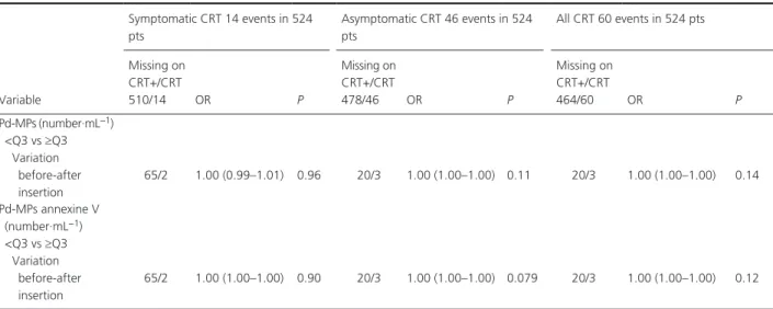
CRT, catheter- related thrombosis; BMI, body mass index. Variable Symptomatic CRT 14 events in 524 pts Asymptomatic CRT 46 events in 524 pts All CRT 60 events in 524 pts Missing on CRT+/CRT 510/14 OR P Missing on CRT+/CRT 478/46 OR P Missing on CRT+/CRT 464/60 OR P Pd- MPs (number·mL−1) <Q3 vs ≥Q3 Variation before-after insertion 65/2 1.00 (0.99–1.01) 0.96 20/3 1.00 (1.00–1.00) 0.11 20/3 1.00 (1.00–1.00) 0.14 Pd- MPs annexine V (number·mL−1) <Q3 vs ≥Q3 Variation before- after insertion 65/2 1.00 (1.00–1.00) 0.90 20/3 1.00 (1.00–1.00) 0.079 20/3 1.00 (1.00–1.00) 0.12 CRT, catheter- related thrombosis; BMI, body mass index,; VTE, venous thromboembolism; CVC; central venous catheter; APTT, Activated Partial Thromboplastin Time; TGT, thrombin generation test; ETP, endogenous thrombin potential; Pd- MPs, platelet derived microparticules; Q3, quartile 3; OR, odds ratio.
none of the 46 patients with asymptomatic CRT experi-enced symptomatic CRT or DVT or PE up to 6 months of clinical follow- up. A major finding of the CAVECCAS study is to point the reassuring clinical outcome of asymp-tomatic CRT diagnosed by DUS within the first 3 months up to 6 months of clinical follow- up, further supporting the current international guidelines.
Much of our knowledge about CRT risk factors derived from a large meta- analysis that included 5636 subjects with various types of unselected cancers [16]. In this meta- analysis, a previous history of DVT (OR, 2.03; 95% CI, 1.05–3.92), a subclavian venipuncture insertion (OR, 2.16; 95% CI, 1.07–4.34) and catheter tip misplacement, that is, malpositioning the tip of the CVC, which distal tip should be placed at the superior vena cava and the right atrium junction (OR, 1.92; 95% CI, 1.22–3.02) were found to increase the risk of CRT [16]. In the CAVECCAS cohort, neither a history of DVT (OR, 1.54, 95%CI, 0.87–2.73; P = 0.14), nor the CVC insertion technique and position- related factors were significantly associated with CRT onset. A recent Cochrane analysis concluded that jugular and subclavian CVCs insertion sites carry the same risk of CRT in long- term recipients [28].
In CAVECCAS highly selected BC patient population, using multivariate analysis, we found that obesity (OR, 2.66: 95%CI, 1.46–4.84, P = 0.001) and lobular carcinoma histological type (OR, 2.56: 95%CI, 1.32–4.96, P = 0.005) were significantly associated with CRT. Obesity has been known as a VTE risk factor for long both in noncancer and more recently in cancer patients. Using the Khorana risk assessment scale, a BMI > 35 kg/m2 is an
independ-ent risk factor for subsequindepend-ent DVT or PE in patiindepend-ents treated with chemotherapy [29] and BMI was recently shown to be a significant predictor of VTE in a large BC population (HR, 3.0; 95%CI, 2.1–4.4) [6]. CAVECCAS results point out the role of obesity as a major contribu-tor to CRT in BC patients. It first highlights the role of invasive lobular carcinoma (ILC) histological type, which has been reported to be less responsive to NAC than ductal carcinoma, probably due to differences in molecular characteristics, particularly HR and HER2 expression [30]. A relationship between ILC and serum estrogen levels has been previously suggested given the increasing fre-quency of ILC among postmenopausal women taking hormone replacement therapy [31, 32]. Interestingly, estrogens have different effects on the coagulation system resulting in a procoagulant state [33].
We also investigated if reliable biomarkers of blood coagulation activation and fibrinolysis help to identify those BC patients at higher risk of CRT and to tailor the need for CRT thromboprophylaxis. Two previous studies supported that D- dimers levels correlate with symptomatic CRT. In a case control study by Jansen et al.
[34], 30 CRT patients with a Hickman catheter undergo-ing allogeneic bone marrow transplantation for hemato-logical malignancies were compared with 30 matched controls. Patients with D- dimers levels >350 μg/L measured three to 5 days after catheter insertion had a 6 times higher risk of developing subclavian CRT [34]. Similar results were found in 48 patients with renal insufficiency, albeit inserted with double lumen catheters for hemodi-alysis [35]. In CAVECCAS study, the significant increase in D- dimers levels after catheter insertion (P < 0.0001), reflecting blood coagulation activation, did not predict CRT occurrence. Measures of the thrombin generation potential provide a global method to quantify the effect of the numerous genetic and environmental factors involved in the coagulation pathway. In the Vienna Cancer and Thrombosis Study (CATS), elevated thrombin peak values were associated with an increased risk of DVT or PE with a hazard ratio of 2.1 (95% CI 1.3–3.3) in multivari-ate analysis [21]. On the contrary, in our study, neither the thrombin generation peak height nor the endogenous thrombin potential predicted CRT.
MPs have emerged as promising biomarkers due to their procoagulant properties related to the exposure of negatively charged phospholipids, mainly phosphatidyl- serine and tissue factor. Several studies showed that increased levels of Tissue Factor positive MPs correlated with VTE or PE in cancer patients [36], but there are no data yet on the role of MPs in the occurrence of CRT in cancer patients. We therefore investigated whether Pd- MPs were predictive of CRT in the CAVECCAS cohort. Paradoxically, we observed that both Pd- MPs and Pd- MPs/PS+ levels significantly decreased after CVC insertion. High levels of Pd- MPS and Pd- MPS/PS+ did not correlate with CRT. Our results do not support routine testing of these biomarkers in BC patients with CVC, but further studies are required to confirm these findings in patients with other malignancies.
Heritable or acquired risk factors for VTE of clinical relevance include antithrombin, protein S, protein C defi-ciencies, the G1691A polymorphism FVL, the prothrombin G20210A polymorphism, presence of lupus anticoagulant, positivity of anticardiolipin and antiβ2GP1 antibodies. We performed a nested case- control study for all these throm-bophilia risk factors and found no difference between cases and matched controls for any of these. Our results appeared controversial with previous studies, which reported significant associations between FV Leiden poly-morphism and CRT in acute leukemia [37], bone marrow transplant [38], unselected cancer patients [39] and in locally advanced or metastatic BC patients, all treated with the same chemotherapy protocol [40].
A limitation of our study is that it was underpowered to demonstrate the association of heritable or acquired
thrombophilia with CRT occurrence. For economical and organizational reasons, it was not possible to measure antithrombin, protein C, protein S, lupus anticoagulant, anticardiolipin and antiβ2GP1 antibodies, and to test FVL and G20210A prothrombin polymorphisms in the whole included population. Blood samples were drawn in patients with CRT and matched- controls only and we used of a nested case control study approach.
The main strength of our study using repeated DUS is to provide current estimates of absolute risk of both asymptomatic and symptomatic CRT and their outcomes in a large sample size of 534 selected BC patients, as well as a comprehensive analysis of the multiplicity of CRT risk factors. According to current identified CRT risk fac-tors [10, 14, 16], we chose to study a highly selected population of nonmetastatic invasive BC patients neces-sitating NAC via a central lumen catheter. Indeed, we aimed to investigate the specific incidence, risk factors and outcomes of both asymptomatic and symptomatic CRT in nonmetastatic BC patients within the 6 months after CVC insertion for NAC, since no study has yet ana-lyzed this frequent clinical setting. Further studies will be necessary in BC patients with metastatic disease to analyze if the risk of CRT is higher, as already shown in metastatic cancer patients with VTE outside the context of CRT.
In summary, the CAVECCAS study provides unique clinical data on a large number of highly selected patients over a relatively long period of follow- up. BC patients treated with NAC are at increased risk of CRT, and obesity and lobular carcinoma histological type appear as major CRT- risk factors. Clinical parameters only allowed to identify high risk DUS- CRT patients. Whether assessment of these risk factors may be clinically useful for an indi-vidual stratification of BC patients who might benefit from CRT prophylaxis deserves further studies.
Existing guidelines recommend not to use anticoagula-tion for routine prophylaxis of CRT [14, 41], Nevertheless, two studies [42, 43] and one meta- analysis [44] suggested a potential benefit in CRT prevention using anticoagulants. In this setting, the potential benefits of new anti- thrombotic drugs should be further evaluated.
Acknowledgements
Other GFTC members significantly contributed to patients inclusion and data collection as co-investigators in the study: Ismail Elalamy (Department of Haematology, Assistance Publique-Hôpitaux de Paris, Hôpital Tenon, Paris, France), Grigorios Gerotziafas (Department of Haematology, Assistance Publique-Hôpitaux de Paris, Hôpital Tenon, Paris, France), Joel Lelong (Department of Gynecology, Clinique des Aubépines, Saint-Aubin-sur-Scie, France), Sandrine Lavau-Denes (Department of
Medical Oncology, CHU Dupuytren, Limoges, France), Pierre Yves Dubois (Department of Surgery, Institut Jean Goudinot, Reims, France), Marie Antoinette Sevestre (Department of Vascular Medicine, Amiens University Hospital, Amiens, France), and Laurent Bastit (Department of Radiotherapy, Clinique Pasteur, Evreux, France).
References
1. Breast Cancer Statistics | World Cancer Research Fund International. Published November 18, 2016. Available at http://www.wcrf.org/int/cancer-facts-figures/data-specific-cancers/breast-cancer-statistics (accessed 18 November 2016).
2. Cronin-Fenton, D. P., F. Søndergaard, L. A. Pedersen, J. P. Fryzek, K. Cetin, J. Acquavella, et al. 2010. Hospitalisation for venous thromboembolism in cancer patients and the general population: a population- based cohort study in Denmark, 1997–2006. Br. J. Cancer 103:947–953.
3. Walker, A. J., T. R. Card, J. West, C. Crooks, and M. J. Grainge. 2013. Incidence of venous thromboembolism in patients with cancer - a cohort study using linked United Kingdom databases. Eur. J. Cancer Oxf. Engl. 49:1404–1413.
4. Timp, J. F., S. K. Braekkan, H. H. Versteeg, and S. C. Cannegieter. 2013. Epidemiology of cancer- associated venous thrombosis. Blood 122:1712–1723.
5. Brand, J. S., E. Hedayati, N. Bhoo-Pathy, et al. 2016. Time- dependent risk and predictors of venous thromboembolism in breast cancer patients: a population- based cohort study. Cancer 123:468–475. 6. Walker, A. J., J. West, T. R. Card, C. Crooks, C. C.
Kirwan, and M. J. Grainge. 2016. When are breast cancer patients at highest risk of venous
thromboembolism? A cohort study using English health care data. Blood 127:849–857; quiz 953.
7. Amir, E., B. Seruga, S. Niraula, L. Carlsson, and A. Ocaña. 2011. Toxicity of adjuvant endocrine therapy in postmenopausal breast cancer patients: a systematic review and meta- analysis. J. Natl. Cancer Inst. 103:1299–1309.
8. Paulus, J. K., and A. S. Rosenberg. 2016. Breast cancer and thrombosis: timing matters. Blood 127:793–794.
9. Verso, M., and G. Agnelli. 2003. Venous
thromboembolism associated with long- term use of central venous catheters in cancer patients. J. Clin. Oncol. 21:3665–3675.
10. Lee, A. Y. Y., and P. W. Kamphuisen. 2012. Epidemiology and prevention of catheter- related thrombosis in patients with cancer. J. Thromb. Haemost. 10:1491–1499.
11. McGee, D. C., and M. K. Gould. 2003. Preventing complications of central venous catheterization. N. Engl. J. Med. 348:1123–1133.
12. Farge, D., H. Bounameaux, B. Brenner, et al. 2016. International clinical practice guidelines including guidance for direct oral anticoagulants in the treatment and prophylaxis of venous
thromboembolism in patients with cancer. Lancet Oncol. 17:e452–e466.
13. Schiffer, C. A., P. B. Mangu, J. C. Wade, et al. 2013. Central venous catheter care for the patient with cancer: American Society of Clinical Oncology clinical practice guideline. J. Clin. Oncol. 31:1357–1370.
14. Debourdeau, P., D. Farge, M. Beckers, et al. 2013. International clinical practice guidelines for the
treatment and prophylaxis of thrombosis associated with central venous catheters in patients with cancer. J. Thromb. Haemost. 11:71–80.
15. Debourdeau, P., D. Kassab Chahmi, G. Le Gal, et al. 2009. 2008 SOR guidelines for the prevention and treatment of thrombosis associated with central venous catheters in patients with cancer: report from the working group. Ann. Oncol. 20:1459–1471.
16. Saber, W., T. Moua, E. C. Williams, et al. 2011. Risk factors for catheter- related thrombosis (CRT) in cancer patients: a patient- level data (IPD) meta- analysis of clinical trials and prospective studies. J. Thromb. Haemost. 9:312–319.
17. van Rooden, C. J., F. R. Rosendaal, R. M. Y. Barge, et al. 2003. Central venous catheter related thrombosis in haematology patients and prediction of risk by screening with Doppler- ultrasound. Br. J. Haematol. 123:507–512.
18. Robert, S., P. Poncelet, R. Lacroix, et al. 2009.
Standardization of platelet- derived microparticle counting using calibrated beads and a Cytomics FC500 routine flow cytometer: a first step towards multicenter studies? J. Thromb. Haemost. 7:190–197.
19. Ridker, P. M., C. H. Hennekens, K. Lindpaintner, M. J. Stampfer, P. R. Eisenberg, and J. P. Miletich. 1995. Mutation in the gene coding for coagulation factor V and the risk of myocardial infarction, stroke, and venous thrombosis in apparently healthy men. N. Engl. J. Med. 332:912–917.
20. Poort, S. R., F. R. Rosendaal, P. H. Reitsma, and R. M. Bertina. 1996. A common genetic variation in the 3’- untranslated region of the prothrombin gene is associated with elevated plasma prothrombin levels and an increase in venous thrombosis. Blood 88:
3698–3703.
21. Ay, C., D. Dunkler, R. Simanek, et al. 2011. Prediction of venous thromboembolism in patients with cancer by measuring thrombin generation: results from the Vienna
cancer and thrombosis study. J. Clin. Oncol. 29:2099–2103.
22. Horsted, F., J. West, and M. J. Grainge. 2012. Risk of venous thromboembolism in patients with cancer: a systematic review and meta- analysis. PLoS Med. 9:e1001275. 23. Joffe, H. V., N. Kucher, V. F. Tapson, and S. Z.
Goldhaber. 2004. Deep vein thrombosis (DVT) FREE Steering Committee. Upper- extremity deep vein thrombosis: a prospective registry of 592 patients. Circulation 110:1605–1611.
24. Kuter, D. J. 2004. Thrombotic complications of central venous catheters in cancer patients. Oncologist 9:207–216.
25. Morazin, F., I. Kriegel, B. Asselain, and M. C. Falcou. 2005. [Symptomatic thrombosis in central venous catheter in oncology: a predictive score?]. Rev. Med Interne. 26:273–279.
26. Lee, A. Y. Y., M. N. Levine, G. Butler, et al. 2006. Incidence, risk factors, and outcomes of catheter- related thrombosis in adult patients with cancer. J. Clin. Oncol. 24:1404–1408.
27. Monreal, M., A. Raventos, R. Lerma, et al. 1994. Pulmonary embolism in patients with upper extremity DVT associated to venous central lines–a prospective study. Thromb. Haemost. 72:548–550.
28. Ge, X., R. Cavallazzi, C. Li, S. M. Pan, Y. W. Wang, and F.-L. Wang. 2012. Central venous access sites for the prevention of venous thrombosis, stenosis and infection. Cochrane Database Syst. Rev. 14:CD004084. 29. Khorana, A. A., N. M. Kuderer, E. Culakova, G. H.
Lyman, and C. W. Francis. 2008. Development and validation of a predictive model for chemotherapy- associated thrombosis. Blood 111:4902–4907.
30. Lips, E. H., R. A. Mukhtar, C. Yau, et al. 2012. Lobular histology and response to neoadjuvant chemotherapy in invasive breast cancer. Breast Cancer Res. Treat. 136:35–43. 31. Chen, C.-L., N. S. Weiss, P. Newcomb, W. Barlow, and
E. White. 2002. Hormone replacement therapy in relation to breast cancer. JAMA 287:734–741. 32. Inoue, M., H. Nakagomi, H. Nakada, et al. 2017.
Specific sites of metastases in invasive lobular carcinoma: a retrospective cohort study of metastatic breast cancer. Breast Cancer 24:667–672.
33. Rosendaal, F. R., F. M. Helmerhorst, and J. P. Vandenbroucke. 2002. Female hormones and thrombosis. Arterioscler. Thromb. Vasc. Biol. 22:201–210.
34. Jansen, F. H., H. M. van der Straaten, M. Roest, F. Haas, P. G. de Groot, and R. Fijnheer. 2005. Elevated levels of D- dimer and fragment 1+2 upon central venous catheter insertion and factor V Leiden predict subclavian vein thrombosis. Haematologica 90:499–504.
35. Kanno, Y., K. Kobayashi, H. Takane, et al. 2007. Elevation of plasma D- dimer is closely associated with venous thrombosis produced by double- lumen catheter in pre- dialysis patients. Nephrol. Dial. Transplant. 22:1224–1227.
36. Geddings, J. E., and N. Mackman. 2013. Tumor- derived tissue factor- positive microparticles and venous
thrombosis in cancer patients. Blood 122:1873–1880. 37. Wermes, C., M. von Depka Prondzinski, R.
Lichtinghagen, M. Barthels, K. Welte, and K. W. Sykora. 1999. Clinical relevance of genetic risk factors for thrombosis in paediatric oncology patients with central venous catheters. Eur. J. Pediatr. 158(Suppl. 3):S143–S146. 38. Fijnheer, R., B. Paijmans, L. F. Verdonck, H. K.
Nieuwenhuis, M. Roest, and A. W. Dekker. 2002. Factor V Leiden in central venous catheter- associated
thrombosis. Br. J. Haematol. 118:267–270.
39. Van Rooden, C. J., F. R. Rosendaal, A. E. Meinders, J. A. Van Oostayen, F. J. M. Van Der Meer, and M. V. Huisman. 2004. The contribution of factor V Leiden and prothrombin G20210A mutation to the risk of central venous catheter- related thrombosis. Haematologica 89:201–206.
40. Mandalà, M., G. Curigliano, P. Bucciarelli, et al. 2004. Factor V Leiden and G20210A prothrombin mutation and the risk of subclavian vein thrombosis in patients with breast cancer and a central venous catheter. Ann. Oncol. 15:590–593.
41. Lyman, G. H., K. Bohlke, A. A. Khorana, et al. 2015. Venous thromboembolism prophylaxis and treatment in patients with cancer: American Society of Clinical Oncology Clinical Practice Guideline Update 2014. J. Clin. Oncol. 33:654–656.
42. Young, A. M., L. J. Billingham, G. Begum, et al. 2009. Warfarin thromboprophylaxis in cancer patients with central venous catheters (WARP): an open- label randomised trial. Lancet Lond. Engl. 373:567–574. 43. De Cicco, M., M. Matovic, L. Balestreri, et al. 2009.
Early and short- term acenocumarine or dalteparin for the prevention of central vein catheter- related
thrombosis in cancer patients: a randomized controlled study based on serial venographies. Ann. Oncol. 20:1936–1942.
44. D’Ambrosio, L., M. Aglietta, and G. Grignani. 2014. Anticoagulation for central venous catheters in patients with cancer. N. Engl. J. Med. 371:1362–1363.