Advance Access publication 21 January 2008
Introduction
Although radiation doses during dental examinations are in general relatively low, they account for nearly one-third of the total number of radiological examinations in the European Union (EU; Janssens et al. , 2004 ). Despite
the fact that according to the offi cial guidelines adopted by the EU ( Isaacson and Thom, 2001 ) lateral cephalometric radiographs (LCRs) should be restricted to severe malocclusions, an average of three panoramic radiographs (PRs) and three LCRs are taken of orthodontic patients ( Hujoel et al. , 2006 ). A reduction in the number of radiographs during orthodontic treatment is supported by the fi ndings that a clinical examination supplemented by study models is often suffi cient for treatment planning ( Han et al. , 1991 ) and that a treatment plan based on clinical examination, study models, and photographs is only altered in 7 per cent of the cases due to an additional radiographic examination ( Bruks et al. , 1999 ).
If the EU guidelines are followed and no LCRs are taken of patients with mild to moderate malocclusion, certain information normally derived from LCRs, such as the vertical jaw base relationship and gonial angles, are missing. These parameters are among others used to predict the mandibular growth pattern ( Björk, 1969 ). It
Assessment of vertical facial and dentoalveolar changes using
panoramic radiography
Nasila Nohadani * and Sabine Ruf **
Departments of Orthodontics, Universities of * Berne, Switzerland and ** Giessen, Germany
SUMMARY The purpose of the study was to analyse longitudinal vertical facial and dentoalveolar changes using panoramic radiographs (PRs) and to compare the results with measurements on lateral cephalometric radiographs (LCRs) in order to determine whether, under certain circumstances, the radiation dose for a patient may be reduced by taking only a PR instead of a PR and a LCR. Pre- and post-treatment PRs and LCRs of 30 (15 females and 15 males) orthodontically treated adolescents (mean age pre-treatment 10.9 years, post-treatment 13.4 years) were analysed using Pearson’s correlation coeffi cients and gender differences using Fisher’s z -transformation.
The results revealed that most variables exhibited larger absolute values on PRs than on LCRs. Comparison of dentoskeletal morphology between the LCRs and the PRs revealed moderate to high, mostly statistically signifi cant, interrelations both before and after orthodontic treatment. The lowest correlations were found for the maxillary jaw base angle (NL/H; r = 0.35***) and the highest for the gonial angle (ML/RL; r = 0.90***). However, when assessing the combined growth and treatment changes from before to after treatment, only weak to moderate, not statistically signifi cant, interrelations were found between LCRs and PRs. Anterior face height (AFH; r = 0.43***), the mandibular plane angle (ML/H;
r = 0.06*), and the distance of the incisal tip of the most extruded mandibular incisor to the ML-line (ii-ML;
r = − 0.21*) were the only statistically signifi cant parameters. The average group differences for growth and treatment changes, however, were small for most parameters.
Analysis of vertical facial and dentoalveolar parameters on PRs delivers a moderate approximation to the situation depicted on LCRs. However, PRs cannot be recommended for the analysis of individual longitudinal changes in vertical facial and dentoalveolar parameters.
would thus be useful if this information could be derived from PRs.
PRs have been used previously to metrically assess gonial angles, condylar and ramus heights, as well as asymmetries ( Mattila et al. , 1977 ; Kjellberg et al. , 1994 ; Raustia and Salonen, 1997 ; Dutra et al. , 2004 ) and showed high correlations for gonial angles, interjaw base angle, and anterior and posterior face height ( Dahan and Jesdinsky, 1968 ; Mattila et al. , 1977 ; Raustia and Salonen, 1997 ). However, all corresponding studies available in the literature have been cross-sectional.
Although there are longitudinal studies that have used PRs to assess long-term changes of bone growth after implant placement ( Roberts, 2005 ) or longitudinal morphological changes of the mandible in patients with hemifacial microsomia ( Sarnas et al. , 2004 ), it is not clear whether such longitudinal changes assessed using PRs represent the true changes.
Thus, the aim of the study was to analyse longitudinal vertical facial parameters and dentoalveolar bone height using PRs and to compare the results to measurements on LCRs in order to determine whether the radiation dose for the patient may be reduced by taking only a PR instead of a PR and a LCR in certain indications.
Subjects and methods
The study comprised 30 orthodontically treated adolescent subjects (15 females and 15 males). The average pre-treatment age of the patients was 10.9 years and post-treatment 13.4 years.
From all patients completing active orthodontic treatment in the Department of Orthodontics of the University of Giessen in 1999, the fi rst 30 subjects fulfi lling the following inclusion criteria were selected: fully erupted fi rst molars and permanent incisors at the time of the initial investigation, no disabilities, syndromes, severe asymmetries, or multiple tooth agenesis, as well as available good-quality LCRs and PRs from before and after treatment taken exclusively by one operator.
The LCRs and PRs had been taken as part of the routine diagnostic procedures for orthodontic treatment and were retrospectively analysed. Both radiographs (LCR and PR) of each subject were required to be taken on the same day before and after treatment, respectively. The average time interval between the before (T1) and after treatment (T2) radiographs was 2.5 years. All radiographs were taken with the same X-ray machine (Orthophos CD, Siemens, Munich Germany) at both examination times.
The LCRs and PRs from both examination times were taken in ideal position according to the manufacturers’ operating instructions. The radiographs were traced and analysed using a modifi ed ‘ cephalometric ’ analysis based on comparable reference points, which could be located on both the LCR and the PR ( Tables 1 and 2 and Figures 1 – 5 ).
Double contours on the LCR were averaged, while on the PR the reference points were located separately for the left and right side. Measurements were performed to the nearest 0.5 mm or 0.5 degrees, respectively.
All registrations were performed twice by one investigator (MAB, see acknowledgements), and the mean value of the duplicate registrations was used in the fi nal evaluation. Before the evaluation, the investigator was calibrated to identify the anatomical points on the PRs. For all variables, the arithmetic mean (mean) and standard deviation (SD) were calculated. No correction for linear enlargement was performed. Possible interrelations between the variables and the treatment changes measured on the LCRs and PRs were assessed by means of Pearson’s correlation coeffi cients. Gender differences were analysed using Fisher’s z -transformation. The following correlation categories were established: weak ( r < 0.30), moderate ( r = 0.30 – 0.70), and strong ( r > 0.70). Statistical signifi cance was determined at the 0.1, 1, and 5 per cent levels of confi dence. A confi dence level larger than 5 per cent was considered not signifi cant.
Repeated measurements were used for the method error (ME) calculation: ME= (∑ d2/2 ,n) where d is the
difference between two registrations of a pair and n is the number of double registrations. The combined ME in locating, superimposing, and measuring the changes of the different landmarks did not exceed 1.0 mm or 1.5 degrees, respectively, for any of the variables investigated. The ME did not differ between LCRs and PRs.
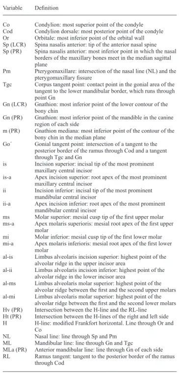
Table 1 Defi nition of the reference points and reference lines used in the analysis of the lateral cephalometric radiographs (LCRs) and panoramic radiographs (PRs).
Variable Defi nition
Co Condylion: most superior point of the condyle Cod Condylion dorsale: most posterior point of the condyle Or Orbitale: most inferior point of the orbital wall Sp (LCR) Spina nasalis anterior: tip of the anterior nasal spine Sp (PR) Spina nasalis anterior: most inferior point in which the nasal
borders of the maxillary bones meet in the median sagittal plane
Pm Pterygomaxillare: intersection of the nasal line (NL) and the pterygomaxillary fi ssure
Tgc Corpus tangent point: contact point in the gonial area of the tangent to the lower mandibular border, which runs through point Gn
Gn (LCR) Gnathion: most inferior point of the lower contour of the bony chin
Gn (PR) Gnathion: most inferior point of the mandible in the canine region of each side
m (PR) Gnathion mediana: most inferior point of the contour of the bony chin in the median plane
Go΄ Gonial tangent point: intersection of a tangent to the posterior border of the ramus through Cod and a tangent through Tgc and Gn
is Incision superior: incisal tip of the most prominent maxillary central incisor
is-a Apex incision superior: root apex of the most prominent maxillary central incisor
ii Incision inferior: incisal tip of the most prominent mandibular central incisor
ii-a Apex incision inferior: root apex of the most prominent mandibular central incisor
ms Molar superior: mesial cusp tip of the fi rst upper molar ms-a Apex molaris superioris: mesial root apex of the fi rst upper
molar
mi Molar inferior: mesial cusp tip of the fi rst lower molar mi-a Apex molaris inferioris: mesial root apex of the fi rst lower
molar
al-is Limbus alveolaris incision superior: highest point of the alveolar ridge in the upper incisor area
al-ii Limbus alveolaris incision inferior: highest point of the alveolar ridge in the lower incisor area
al-ms Limbus alveolaris molar superior: highest point of the alveolar ridge between the fi rst and the second upper molars al-mi Limbus alveolaris molar superior: highest point of the
alveolar ridge between the fi rst and the second lower molars Hv (PR) Intersection between the H-line and the RL-line
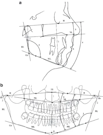
Ht (PR) Intersection between the H-lines of the right and left side H H-line: modifi ed Frankfort horizontal. Line through Or and
Co
NL Nasal line: line through Sp and Pm ML Mandibular line: line through Gn and Tgc
MLa (PR) Anterior mandibular line: line through Gn of each side RL Ramus tangent: tangent to the posterior border of the ramus
Results
Dentoskeletal morphology
The results of the analysis and comparison of the dentoskeletal morphology before and after treatment, are shown in Table 3 . Except for gonial angle (ML/RL), interjaw-base angle (ML/NL), anterior maxillary height (AHMx), and the distance between the root apex of the most extruded upper incisor to the NL-line (isa-NL), all PR variables exhibited larger absolute values.
Comparison of dentoskeletal measurements on LCRs and PRs revealed moderate to high, mostly statistically signifi cant interrelationships. The lowest correlations were found for the maxillary jaw base angle (NL/H; r = 0.35***) and the highest for the gonial angle (ML/RL; r = 0.90***). No systematic gender differences were found for any of the interrelationships analysed.
Growth and treatment changes
The results of the analysis and comparison of the growth and treatment changes occurring from before to after treatment are given in Table 4 . Most parameters exhibited only small average differences [mean( d ) = 0.0 – 0.8 mm and 0.1 – 0.2 degrees, respectively] between the growth and the treatment changes measured on LCRs and PRs. The SDs, however, were large. The variables, anterior face height (AFH), mandibular plane angle (ML/H), interjaw-base angle (ML/NL), and the distance of the incisal tip of the most extruded mandibular incisor to the ML-line (ii-ML), on the other hand, exhibited larger mean differences [mean( d ) = 1.9 – 4.2 mm and 1.7 – 1.9 degrees, respectively] with all growth and treatment changes being smaller on the PRs.
Table 2 Defi nition of the skeletal (1 – 6), alveolar (7 – 10), and dental (11 – 18) variables used in the analysis of the lateral cephalometric radiographs (LCRs) and panoramic radiographs (PRs).
Variable Defi nition
1 AFH (mm) Anterior face height (LCR): vertical distance between Gn and the H-line
AFH (mm) Anterior face height (PR): distance between Ht and m
2 PFH (mm) Posterior face height (LCR): vertical distance between Gò and the H-line
PFH (mm) Posterior face height (PR): distance between Hv and Gò
3 ML/RL (degree) Gonial angle: angle between the reference lines ML and RL
4 ML/H (degree) Mandibular plane angle: angle between the reference lines ML and H
5 NL/H (degree) Maxillary plane angle: angle between the reference lines NL and H
6 ML/NL (degree) Interjaw-base angle: angle between the reference lines ML and NL
7 AHMx (mm) Anterior maxillary height (LCR): vertical distance between al-is and NL
AHMx (mm) Anterior maxillary height (PR): distance between al-is and Sp
8 PHMx (mm) Posterior maxillary height: vertical distance between al-ms and NL
9 AHMn (mm) Anterior mandibular height (LCR): vertical distance between al-ii and ML
AHMn (mm) Anterior mandibular height (PR): distance between al-ii and m
10 PHMn (mm) Posterior mandibular height: vertical distance between al-mi and ML
11 is-NL (mm) Distance of the incisal tip of the most extruded maxillary incisor to NL
12 ii-ML (mm) LCR: distance of the incisal tip of the most extruded mandibular central incisor to ML ii-MLa (mm) PR: distance of the incisal tip of the most
extruded mandibular central incisor to MLa 13 ms-NL (mm) Distance of the mesial cusp tip of the fi rst
permanent upper molar to NL
14 mi-ML (mm) Distance of the mesial cusp tip of the fi rst permanent lower molar to ML
15 isa-NL (mm) Distance of the root apex of the most extruded maxillary central incisor to NL
16 iia-ML (mm) LCR: distance of the root apex of the most extruded mandibular central incisor to ML iia-MLa (mm) PR: distance of the root apex of the most
extruded mandibular central incisor to MLa 17 msa-NL Distance of the root apex of the mesial root of
the fi rst permanent upper molar to NL 18 mia-ML Distance of the root apex of the mesial root of
the fi rst permanent lower molar to ML
Figure 1 Reference points used in the analysis of (a) the lateral cephalometric and (b) the panoramic radiographs.
In contrast to the dentoskeletal measurements, the growth and treatment changes on the LCRs and PRs showed only weak to moderate, mostly not statistically signifi cant, interrelations. Anterior face height (AFH; r = 0.43***), the mandibular plane angle (ML/H; r = 0.06*), and the distance of the incisal tip of the most extruded mandibular incisor to the ML-line (ii-ML; r = − 0.21*) were the only statistically signifi cant parameters.
Discussion
To reduce the infl uence of measurement errors, one calibrated examiner evaluated all radiographs twice and the mean of the duplicate measurements was used in the fi nal evaluation. No adjustment for radiographic enlargement was performed in the present study because the magnifi cation of PRs will vary between 13 and 28 per cent depending on the area imaged and the panoramic machine used ( Philipp and Hurst, 1978 ; McDavid et al. , 1985 ; Thanyakarn et al. , 1992 ). Furthermore, vertical measurements on PRs are more susceptible to projective distortion than vertical measurements on LCRs. While minor antero – posterior shifts and tilts affect vertical measurements only to a limited degree ( Xie et al. , 1996 ), rotations, and, especially lateral tilts, result in left – right asymmetries ( Ruf and Pancherz, 1995 ; Malkoc et al. , 2005 ).
Figure 2 Reference lines used in the analysis of (a) the lateral cephalometric and (b) the panoramic radiographs.
It might be argued that LCRs are not a gold standard and instead a dry skull should have been used ( Dermaut, 2002 ). However, longitudinal changes cannot be measured on dry skulls.
As expected, due to the larger magnifi cation of the PRs (13 – 28 per cent; Philipp and Hurst, 1978 ; McDavid et al. , 1985 ; Thanyakarn et al. , 1992 ) compared with LCRs (10 per cent = average magnifi cation value of the LCR unit), the majority of the PR parameters exhibited larger absolute values. The skeletal parameters showed larger differences between PRs and LCRs than the alveolar and dental parameters. Furthermore, two skeletal parameters (ML/RL and ML/NL) presented smaller values on the PRs. As both ML/RL and ML/NL are angular measurements, varying magnifi cation cannot explain the differences. The largest vertical and horizontal distortions on the PRs were located at the border of the fi lm and thus in the area of the mandibular ramus and the condyles. This distortion is larger in the upper compared with the lower part of the fi lm ( Samawi and Burke, 1984 ). This could explain why, in the present study, skeletal parameters showed more variability than alveolar and dental parameters.
In agreement with the literature ( Dahan and Jesdinsky, 1968 ; Mattila et al. , 1977 ; Raustia and Salonen, 1997 ),
Figure 3 Skeletal variables, AFH (1), PFH (2), ML/RL (3), ML/H (4), NL/H (5), and ML/NL (6) used in the analysis of (a) the lateral cephalometric and (b) the panoramic radiographs.
comparison of the PR and LCR measurements on radiographs from the same day revealed moderate to high ( r = 0.35 – 0.90) in most cases signifi cant ( P < 0.01, P < 0.001) interrelationships for the skeletal, alveolar, and dental parameters. The highest interrelationships ( r =
0.90***) existed for gonial angle (ML/RL). No systematic gender difference was found for the interrelations. Therefore, male and female subjects were pooled in the analysis.
Analysis of the longitudinal facial and dentoalveolar changes and their comparison between LCRs and PRs showed only small mean differences, except for the parameters AFH, ML/H, and ii-ML. The SDs, however, were very large. The interindividual variation was obvious and the direction of changes inconsistent because the correlations for growth and treatment changes measured on PRs and LCRs were only weak to moderate and mostly not statistically signifi cant.
The low correlations are most probably due to the varying degrees of distortion and enlargement within the PRs ( Philipp and Hurst, 1978 ; Samawi and Burke, 1984 ;
Figure 4 Alveolar variables, AHMx (7), PHMx (8), AHMn (9), and PHMn (10) used in the analysis of (a) the lateral cephalometric and (b) the panoramic radiographs.
Thanyakarn et al. , 1992 ), the higher susceptibility for positioning errors ( Philipp and Hurst, 1978 ; Samawi and Burke, 1984 ; Ruf and Pancherz, 1995 ; Xie et al. , 1996 ), as well as the diffi culty in exactly reproducing a PR in case of repeated exposure ( Larheim and Svanæs, 1986 ). Nevertheless, Stramotas et al. (2002) showed that
comparing linear and angular measurements on PRs taken at different times is suffi ciently accurate for measuring changes in root length and root parallelism, to assess sites for implant location, and to measure angulation of developing third molars, provided that the occlusal plane is kept at a similar angulation and is not tilted more than 10 degrees.
Another factor that infl uenced the differences found in the present study was the fact that the PRs and LCRs were obtained in different jaw positions (LCR = habitual occlusion; PR = incisor edge-to-edge). These different positions correspond to the standard imaging procedures (LCR) or standard manufacturers’ instructions (PR). However, some measurements such as anterior face height, posterior face height, and mandibular plane angle can be
Figure 5 Dental variables, is-NL (11), ii-ML (12), ms-NL (13), mi-ML (14), isa-NL (15), iia-ML (16), msa-NL (17), and mia-ML used in the analysis of (a) the lateral cephalometric and (b) the panoramic radiographs.
infl uenced due to the different mandibular position on the radiographs. No variation from these standard positions (e.g. taking the PRs in habitual occlusion) was attempted because this would have compromised the quality of the PR (overlapping of teeth, increased distortion, or blurring in the lower anterior segment). Furthermore, this would have counteracted the aim which was to assess whether a standard PR could deliver certain information normally derived from LCRs in order to be able to reduce the radiation dose for the patient by taking only a PR instead of a PR and a LCR.
The main positioning error when taking repeated LCRs is a change in the anterior or posterior inclination of the head. This does, however, not result in projection or distortion errors and, therefore, does not affect vertical measurements. In PR, on the other hand, a change in head inclination results in blurring, distortion, or enlargement of those areas, due to the fact that the head position change becomes located outside the imaging plane.
Conclusions
Analysis of vertical facial and dentoalveolar parameters on PRs delivers a moderate approximation of the situation depicted on LCRs. However, PRs cannot be recommended
Table 3 Skeletal, alveolar, and dental variables measured on 60 lateral cephalometric radiographs (LCRs) and 60 panoramic radiographs (PRs) of 30 patients (15 females and 15 males). The mean values (mean), standard deviations (SD), mean value differences between LCR and PR [mean( d )], SDs of the difference [SD( d )], and the Pearson’s correlation coeffi cients ( r ) are given.
Variable LCR PR LCR − PR Correlation Mean SD Mean SD Mean( d ) SD r Skeletal AFH (mm) 85.3 5.6 102.2 5.6 − 16.9 5.1 0.59*** PFH (mm) 54.8 5.1 63.2 6.6 − 8.4 3.8 0.82*** ML/RL (gradian) 126.8 6.0 124.7 6.2 +2.1 2.7 0.90*** ML/H (degree) 26.2 4.1 27.8 4.3 − 1.6 3.5 0.65*** NL/H (degree) 2.1 1.6 9.6 2.8 − 7.5 2.7 0.35*** ML/NL (degree) 24.7 4.6 18.0 4.9 +6.7 3.8 0.68*** Alveolar AHMx (mm) 18.3 1.6 18.2 2.6 +0.1 2.3 0.50 n.s. PHMx (mm) 11.5 2.6 14.6 3.0 − 3.1 1.9 0.78*** AHMn (mm) 30.2 3.0 34.6 2.8 − 4.4 2.0 0.76*** PHMn (mm) 23.0 2.5 25.6 3.2 − 2.6 2.0 0.77*** Dental is-NL (mm) 27.8 2.1 29.3 2.8 − 1.5 1.8 0.76*** isa-NL (mm) 2.4 1.7 0.8 1.4 +1.6 1.5 0.56*** ii-ML (mm) 40.6 2.7 41.3 3.8 − 0.7 3.5 0.48 n.s. iia-ML (mm) 14.4 3.5 19.9 3.1 − 5.5 2.3 0.75*** ms-NL (mm) 21.1 2.3 25.5 2.7 − 4.4 2.0 0.70*** msa-NL (mm) 1.8 1.8 2.7 2.4 − 0.9 1.8 0.69*** mi-ML (mm) 31.0 2.9 36.3 3.6 − 5.3 1.8 0.87*** mia-ML (mm) 8.4 2.1 9.2 2.9 − 0.8 1.9 0.76** ** P < 0.01, *** P < 0.001, n.s. = not signifi cant.
Table 4 Growth and treatment changes of skeletal, alveolar, and dental variables measured on 60 lateral cephalometric radiographs (LCRs) and 60 panoramic radiographs (PRs) of 30 patients (15 females and 15 males). The mean value differences between LCR 2 − LCR 1, PR 2 − PR 1, LCR and PR [mean( d )], SDs of the difference [SD( d )], and the Pearson’s correlation coeffi cients ( r ) are given.
Variable Mean Mean LCR − PR Correlation LCR 2 − LCR 1 PR 2 − PR 1 Mean( d ) SD r Skeletal AFH (mm) 5.1 0.9 +4.2 5.1 0.43*** PFH (mm) 3.6 3.5 +0.1 3.8 0.17 n.s. ML/RL (gradian) − 0.8 − 1.0 − 0.7 2.7 0.33 n.s. ML/H (degree) 0.1 − 1.8 +1.9 3.5 0.06* NL/H (degree) 0.1 0.0 +0.1 2.7 0.04 n.s. ML/NL (degree) 0.2 − 1.5 +1.7 3.8 0.35** Alveolar AHMx (mm) 0.9 1.0 − 0.1 2.3 0.16 n.s. PHMx (mm) 2.3 2.8 − 0.5 1.9 0.47 n.s. AHMn (mm) 1.7 1.5 +0.2 2.0 0.44 n.s. PHMn (mm) 0.7 0.6 +0.1 2.0 0.12 n.s. Dental is-NL (mm) 0.9 1.0 − 0.1 1.8 0.56 n.s. isa-NL (mm) 0.2 0.5 − 0.3 1.5 0.16 n.s. ii-ML (mm) 1.7 − 0.2 +1.9 3.5 − 0.21* iia-ML (mm) 1.7 1.8 − 0.1 2.3 0.31 n.s. ms-NL (mm) 2.0 2.0 0.0 2.0 0.29 n.s. msa-NL (mm) 0.8 1.6 − 0.8 1.8 0.21 n.s. mi-ML (mm) 1.5 1.3 +0.2 1.8 0.49 n.s. mia-ML (mm) 0.9 1.3 − 0.4 1.9 0.41 n.s.
* P < 0.05, ** P < 0.01, *** P < 0.001, n.s. = not signifi cant.
for analysis of individual longitudinal changes in vertical facial and dentoalveolar parameters.
Address for correspondence Professor Sabine Ruf Department of Orthodontics University of Giessen Schlangenzahl 14 35392 Giessen Germany E-mail: [email protected] Acknowledgements
We wish to thank Dr Majed Al Borney, formerly of the Department of Orthodontics, University of Giessen, for evaluation of the data.
References
Björk A 1969 Prediction of mandibular growth rotation . American Journal of Orthodontics 55 : 585 – 599
Bruks A , Enberg K , Nordqvist I , Hansson A S , Jansson L , Svenson B 1999 Radiographic examinations as an aid to orthodontic diagnosis and treatment planning . Swedish Dental Journal 23 : 77 – 85
Dahan J , Jesdinsky H J 1968 Evaluation of panoramic radiography for cephalometric studies in orthodontics . Stoma 21 : 126 – 128
Dermaut L R 2002 The dry skull in orthodontics . Verhandelingen - Koninklijke Academie voor Geneeskunde van Belgie 64 : 19 – 54 Dutra V , Yang J , Devlin H , Susin C 2004 Mandibular bone remodelling
in adults: evaluation of panoramic radiographs . Dentomaxillofacial Radiology 33 : 323 – 328
Han U K , Vig K W L , Weintraud J A , Vig P S , Kowalski C J 1991 Consistency of orthodontic treatment decisions relative to diagnostic records . American Journal of Orthodontics and Dentofacial Orthopedics 100 : 212 – 219
Hujoel P , Hollender L , Bollen A M , Young J D , McGee M , Grosso A 2006 Radiographs associated with one episode of orthodontic therapy . Journal of Dental Education 70 : 1061 – 1065
Isaacson K G , Thom A R (eds) 2001 Guidelines for the use of radiographs in clinical orthodontics , 2nd edn. British Orthodontic Society , London Janssens A et al. 2004 Radiation protection: European guidelines on
radiation protection in dental radiology — the safe use of radiographs in dental practice. Offi ce for Offi cial Publications of the European Communities (www.sefm.es/docs/otros/raddigUE.pdf )
Kjellberg H , Ekestubbe A , Kiliaridis S , Thilander B 1994 Condylar height on panoramic radiographs. A methodologic study with a clinical application . Acta Odontologica Scandinavica 52 : 43 – 50
Larheim T A , Svanæs D B 1986 Reproducibility of rotational radiography: mandibular linear dimensions and angles . American Journal of Orthodontics and Dentofacial Orthopedics 90 : 45 – 51
Malkoc S , Sari Z , Usumez S , Koyuturk A E 2005 The effect of head rotation on cephalometric radiographs . European Journal of Orthodontics 27 : 315 – 321
Mattila K , Altonen M , Haavikko K 1977 Determination of the gonial angle from the orthopantomogram . Angle Orthodontist 47 : 107 – 110 McDavid W D , Tronje G , Welander U , Morris C R , Nummikoski
P 1985 Imaging characteristics of seven panoramic X-ray units . Dentomaxillofacial Radiology 14 : ( Supplement 8) 1 – 68
Philipp R , Hurst R 1978 The cant of the occlusal plane and distortion in the panoramic radiograph . Angle Orthodontist 48 : 317 – 323
Raustia A M , Salonen M A 1997 Gonial angles and condylar and ramus height of the mandible in complete denture wearers — a panoramic radiograph study . Journal of Oral Rehabilitation 24 : 512 – 516 Roberts R A 2005 A 24-years retrospective study of bone growth after
implant placement . Journal of Oral Implantology 31 : 98 – 103
Ruf S , Pancherz H 1995 Is orthopantomography reliable for TMJ diagnosis? Journal of Orofacial Pain 9 : 365 – 374
Samawi S S B , Burke P H 1984 Angular distortion in the orthopantomogram . British Journal of Orthodontics 11 : 100 – 107
Sarnas K V , Rune B , Aberg M 2004 Maxillary and mandibular displacement in hemifacial microsomia: a longitudinal roentgen stereometric study of 21 patients with the aid of metallic implants . Cleft Palate-Craniofacial Journal 41 : 290 – 303
Stramotas S , Geenty J P , Petocz P , Darendeliler M A 2002 Accuracy of linear and angular measurements on panoramic radiographs taken at various positions in vitro . European Journal of Orthodontics 24 : 43 – 52 Thanyakarn C , Hansen K , Rohlin M , Akesson L 1992 Measurements of
tooth length in panoramic radiographs. 1: the use of indicators . Dentomaxillofacial Radiology 21 : 26 – 30
Xie Q , Wolf J , Soikkonen K , Ainamo A 1996 Height of mandibular basal bone in dentate and edentulous subjects . Acta Odontologica Scandinavica 54 : 379 – 383