© The Author 2015. Published by Oxford University Press on behalf of the European Orthodontic Society. All rights reserved.
For permissions, please email: [email protected] 570
Original article
Predictive value of masseter muscle thickness
and bite force on Class II functional appliance
treatment: a prospective controlled study
Gregory S. Antonarakis and Stavros Kiliaridis
Department of Orthodontics, University of Geneva, Switzerland
Correspondence to: Gregory S. Antonarakis, Department of Orthodontics, Dental School, University of Geneva, 19 rue Barthélemy-Menn, 1205 Geneva, Switzerland. E-mail: [email protected]
Summary
Aim: To prospectively evaluate the functional capacity of the masticatory musculature as a predictive variable in determining functional appliance treatment outcomes in Class II/1 malocclusion children.
Methods: Twenty Class II/1 malocclusion children (11.4 ± 1.7 years) were treated with functional appliances during 1 year. Masseter muscle thickness and maximal molar bite force measurements, lateral cephalograms, and study casts were taken before and after treatment. Twenty age- and gender-matched untreated children were included as a control group. Regression analyses were used to identify correlations between pre-treatment muscle characteristics and treatment outcomes.
Results: All treated patients showed dentoalveolar sagittal improvement. Maximal molar bite force and masseter muscle thickness decreased during the treatment period in the experimental group but increased in the control group. Children with lower pre-treatment maximal molar bite force showed more mesial movement of mandibular first molars, distal movement of maxillary first molars, and larger change in molar class during treatment. Children with thinner pre-treatment masseter muscles demonstrated more mandibular first molar mesialisation, mandibular incisor proclination, and opening of the gonial angle during treatment.
Conclusions: The initial condition of the masticatory muscles may partly determine treatment outcomes. Children with thinner pre‐treatment masseter muscles or weaker bite force show greater dentoalveolar changes.
Introduction
In the treatment of Class II malocclusion in growing children reports demonstrate that improvement in jaw relationships can be achieved during early treatment with functional appliances (1–8). Although the treatment results obtained with functional appliances are often satisfac-tory, large inter-individual variation is observed both in skeletal and in dental treatment changes (9–11). Not all individuals respond the same way to functional appliance treatment. The large variation seen amongst patients is often attributed to compliance issues, but evidence of this vari-ation is also found in studies where fixed functional appliances are used and thus the influence of patient compliance is excluded (12–14).
One factor that could in part explain inter-individual differences in response to functional appliance treatment may be the masticatory musculature and its functional capacity. It is known that masticatory muscle capacity varies significantly between growing individuals, as measured both by bite force (15–17) and masseter muscle thickness (18). In view of this fact, it has been speculated that the consider-able variability seen in individual response to functional appliance treatment is possibly directly related to the individuals’ muscle char-acteristics (19). Moreover, these muscle charchar-acteristics seem to be under genetic control (20). Based on recent evidence, it has been pro-posed that variation in masticatory muscle characteristics in Class II
doi:10.1093/ejo/cju089 Advance Access publication 12 January 2015
malocclusion growing children, such as masseter muscle thickness and maximal molar bite force, can be one of the possible causes of the reported variation of treatment results with functional appli-ances (21, 22).
The primary aim of the present investigation was to evaluate, using prospective study design, whether the functional capacity of the masticatory musculature can be used as a predictive variable in determining functional appliance treatment outcomes in Class II division 1 malocclusion children. The null hypothesis was that there is no effect of the functional capacity of the masticatory musculature on Class II functional appliance treatment outcomes. The secondary aim was to evaluate the effect of functional appliances on mastica-tory muscles in Class II division 1 functional appliance treatment. The null hypothesis here was that functional appliances have no effect on the masticatory muscles in comparison to untreated grow-ing children.
Materials and methods
The present study was approved by the research ethics board of the University of Geneva (identification number: 07-020).
Subjects
The patient sample for the present prospective study consisted of an experimental group and a control group. The sample size of each group was calculated by performing a power analysis, based on a retrospective study looking at the predictive value of molar bite force on Class II functional appliance treatment outcomes (22). Mean values (53.2 and −34.2 N) and standard deviations (99.4 and 78.4 N) for changes in bite force in the experimental and control groups respectively were used with a 5 per cent alpha value and an 80 per cent power, and a minimum sample size of 18 patients in each group was calculated. Based on this information it was decided to use sam-ple sizes of 20 patients in each group.
The experimental group consisted of 20 healthy Caucasian children with a Class II division 1 malocclusion which were asked to participate in the study. These children had attended an initial consultation at our University clinic, which was subsequently fol-lowed by the collection of standard initial (pre-treatment) diagnostic records and the establishment of a treatment plan which consisted of an activator. Informed consent was obtained from all subjects and their parents before commencing treatment. Inclusion criteria were the following: late mixed dentition; an ANB angle >4 degrees; an SNB angle ≤78 degrees; a non-extreme skeletal divergence with a maxillomandibular angle from 20 to 30 degrees; a full-cusp Class II molar relationship on one side and at least a half-cusp Class II molar relationship on the contralateral side; an overjet ≥6 mm. Exclusion criteria were the following: deciduous teeth extracted prematurely or permanent teeth extracted; transverse discrepancies; signs of con-dylar lesions or temporomandibular dysfunction or disorders; non-nutritive sucking habits; patients with Pierre Robin sequence or any form of clefting; patients with a craniofacial anomaly or syndrome; patients with muscular disorders.
The control group consisted of 20 healthy growing children, matched for gender and age to the experimental group, and with-out immediate need for orthodontic treatment. These children were selected from siblings of patients under treatment at our University clinic or children of staff at the University. Inclusion criteria were the following: Class I or Class II malocclusion. Exclusion criteria were the following: Class III malocclusion; traumatic occlusion; deciduous teeth extracted prematurely or permanent teeth extracted;
transverse discrepancies; signs of temporomandibular dysfunction or disorders; non-nutritive sucking habits; striking dolichocephalic or brachycephalic facial patterns; patients with any form of cleft-ing; patients with a craniofacial anomaly or syndrome; patients with muscular disorders.
Treatment protocol
The duration of the study period was 12 months. In the experimental group an activator, as described by Pfeiffer and Grobéty (23, 24), was used as the sole treatment appliance throughout this period. Patients were instructed to wear the appliance for 12 hours daily. The patients were regularly seen for follow-up appointments where selective adaptation of the activator was carried out as needed. The control subjects did not receive any form of orthodontic treatment throughout this 12-month period.
Experimental design
The present study design was prospective and longitudinal. The exper-imental group had standard diagnostic records taken pre-treatment (T1) as well as after the 12-month study period (T2). Diagnostic records consisted of height measurements, photographs, study casts, a panoramic radiograph, and a lateral cephalometric radiograph. These patients also had maximal molar bite force and ultrasonographic mas-seter muscle thickness measurements before (T1) and after (T2) the study period. The control group only had height, maximal molar bite force, and ultrasonographic masseter muscle thickness measurements before (T1) and after (T2) the study period.
Cephalometry
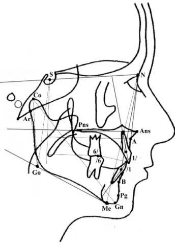
Lateral cephalometric radiographs were taken of all patients in centric occlusion with the head fixed in a cephalostat. The same machine was used for all children and the magnification adjusted to zero. The radiographs obtained were analysed by one opera-tor, following calibration to the senior author, using cephalomet-ric software (Viewbox 4 version 4.0.1.7, dHAL Software, Kifissia, Greece). The cephalometric reference points, lines and angles used in the analysis are shown in Figure 1. The superimposition of the lateral cephalometric radiographs was performed according to the structural method described by Björk and Skieller (25), ensuring that the pre-treatment SN plane was transferred to the subsequent post-treatment cephalometric tracing.
Study casts
Study casts were taken to measure overjet, overbite, and molar rela-tionships. The molar relationship was recorded as a percentage of the Angle Class II relationship, an Angle Class I relationship denoted by zero, and a full cusp Angle Class II relationship denoted by 100 (26). Maximum molar bite force
Maximum voluntary molar bite force was assessed using a digital force gauge with an 8.6mm thick bite element (Occlusal Force-Meter GM 10®; Nagano Keiki Co. Ltd, Tokyo, Japan) (27). The subject was seated upright in a dental chair, and the sensor placed between the first permanent molars of each side separately and the patient was asked to exert a maximum clenching effort but to stop when painful or uncomfortable. In order to obtain as high bite force levels as possible, the subjects were encouraged to ‘do their best’. The recording (measured in Newtons) was taken twice on each side, each recording taking approximately 2–3 seconds, and the high-est value used as the maximum molar bite force for analysis. All
measurements were taken by one operator, who had been calibrated to the more experienced senior author.
Masseter muscle thickness
Masseter muscle thickness was measured by ultrasonography, using a real time ultrasound scanner [FALCO 100, linear array transducer (6–8 MHz), PieMedical, Imaging BV, Maastricht, The Netherlands]. The details of this technique were developed by Kiliaridis and Kalebo (28) and modified by Raadsheer et al. (29). All of the meas-urements were done by one examiner, who had been calibrated to the experienced operator who had developed the method. The par-ticipants were seated in an upright position with no head support. The masseter was scanned bilaterally on a level halfway between the zygomatic arch and the gonial angle. The scan plane was orientated perpendicular to the anterior border of the muscle and perpendicular to the surface of the underlying ramus, so that the reflection of the bone was depicted as a sharp white line. The registrations were made under two conditions, relaxed and contracted. The first was obtained by asking the participants to maintain slight interocclusal contacts, the second by asking them to clench maximally in the intercuspal position. Under all registration conditions, a generous amount of ultrasound contact gel was applied to the probe (Kendall Meditec, Mirandola (MO), Italy) and light pressure was applied so as to avoid compression of the soft tissues and muscle. All registrations were repeated twice, and the final thickness was obtained from the mean
of the repeated measurements. Muscle thickness was registered to the nearest 0.1 mm.
Statistics
All statistical analyses were performed using the Statistical Package for Social Sciences version 15.0 (SPSS Inc., Chicago, Illinois, USA). Data were initially tested for normality using the Shapiro–Wilk test. All data were found to be distributed normally and thus parametric statistics were used throughout.
Maximal molar bite force or masseter muscle thickness changes during treatment or observation (T1–T2) were evaluated, and paired t-tests were used to assess statistical significance within each group. A comparison of changes between the treatment and control groups was also carried out using unpaired t-tests.
For the treatment group, cephalometric and dental changes dur-ing T1–T2 were evaluated, and paired t-tests performed to assess the statistical significance of the changes occurring during the treatment period. Univariate and multivariate linear regression analyses using stepwise regression were carried out to investigate possible correlations between initial maximal molar bite force or masseter muscle thickness and treatment outcomes (dental or cephalometric changes during treatment), including other possible predictor variables in the analysis (pre-treatment age, gender, change in height). Based on the results of a previous study (21), regression analysis was also used to investigate possible correlations between the gonial angle and treatment outcomes. All correlations were considered significant at P < 0.05.
Error of the method
To account for any random error, including possible biologic var-iation, the error of the method for the maximal molar bite force measurements and ultrasound technique was calculated by repeated measurements of 15 patients, on two separate occasions, 2 weeks apart, using Dahlberg’s formula (SE = √Σd2/2n), where n = the number of patients undergoing repeated measurements and d = the difference in measurements (30). For maximal molar bite force measurements, the error was calculated as 61 N, whereas for mas-seter muscle thickness measurements it was found to be 0.4 mm.
The error of the method for the cephalometric variables was calculated by performing duplicate determinations on 15 randomly selected cephalometric radiographs, with a 2-week interval between the measurements, using Dahlberg’s formula. For linear measure-ments, the error of the method did not exceed 0.9 mm, and for angu-lar measurements this did not exceed 1.0 degree.
Results
Sample demographics
The present experimental subjects consisted of 14 boys and 6 girls, between the ages of 9 and 13 (x = 11.4 years; SD = 1.3 years). The con-trol subjects, matched for gender and age to the experimental subjects, also consisted of 14 boys and 6 girls, between the ages of 9 and 13 (x = 11.2 years; SD = 1.9 years). The mean height of the children at T1 was 149.6 cm (SD = 12.0 cm) for the treatment and 146.9 mm (SD = 13.0 cm) for the control group. No significant differences between the treatment and control groups were found concerning T1 age or height.
Pre-treatment cephalometric and dental characteristics and treatment outcomes
The T1 cephalometric and dental characteristics of the 20 children in the treatment group are shown in Table I. The sample had a mean 90 Figure 1. Cephalometric points, lines, and angles used in analysis: SNA;
SNB; ANB; A-reference plane (line through S perpendicular to the maxillary plane (Ans-Pns)); gonial angle (Ar–Go–Me); maxillary incisor (1/) to SN plane; mandibular incisor (/1) to mandibular plane (Me-Go); maxillary first molar (6/) to reference plane through S perpendicular to SN; mandibular first molar (/6) to reference plane through S perpendicular to SN.
per cent Class II molar relationship with a 7.1 mm overjet. The ANB angle was an average of 6.6 degrees. Controls did not have lateral cephalometric radiographs taken.
Following 1 year of active treatment, the dental Class II division 1 relationships improved in all of the experimental children. Molar class shifted towards a Class I relationship by 69.4 per cent and overjet was reduced by 3.4 mm. There was also a mean of 2.2 degrees of mandibular incisor proclination. The ANB angle decreased by an average of 2.3 degrees. The treatment outcomes (dental and cepha-lometric characteristics) are shown in Table I.
Masticatory muscle characteristics
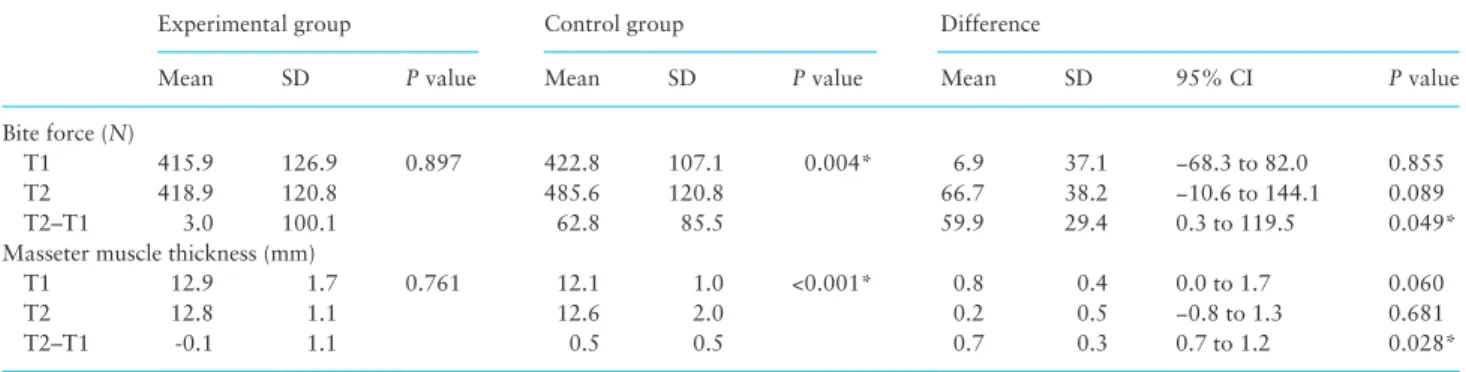
Maximal molar bite force as well as ultrasonographic masse-ter muscle thickness measurements for both experimental and control groups are shown in Table II and Figure 2. There was a direct linear correlation between maximal molar bite force and ultrasonographic masseter muscle thickness in the whole sample (R = 0.390; P = 0.013). There were no statistically significant dif-ferences in initial (T1) masticatory muscle characteristics between the experimental and control groups. Concerning changes however (T2–T1), maximal molar bite force increased significantly in the control group (x = 62.8 N; SD = 85.5 N; P = 0.004) but not the experimental group (x = 3.0N; SD = 100.1 N; P = 0.897). A simi-lar result was seen for masseter muscle thickness whereby masseter muscle thickness increased significantly from T1 to T2 in the con-trol (x = 0.5 mm; SD = 0.5 mm; P < 0.001) but not in the treatment group (x = −0.1 mm; SD = 1.1 mm; P = 0.761).
In the present sample, no associations were found between changes in height and changes in masticatory muscle characteristics (either maximal molar bite force or masseter muscle thickness). Associations
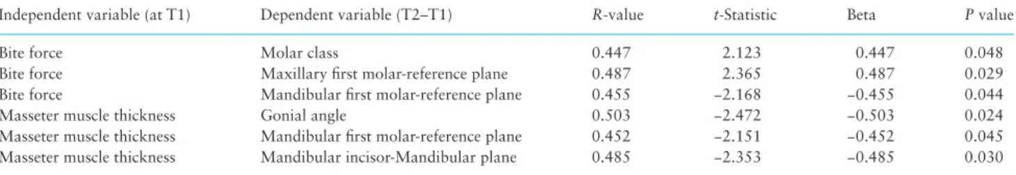
When looking at associations between pre-treatment mastica-tory muscle characteristics, several associations were observed. Children with a lower T1 maximal molar bite force were prone to more mesial movement of mandibular first permanent molars, distal movement of maxillary first permanent molars, and larger change in molar class during treatment (Table III; Figure 3). Children with thinner T1 masseter muscles were more likely to
show more mandibular first molar mesialisation, mandibular inci-sor proclination, and opening of the gonial angle during treatment (Table III). For all of the stepwise multivariate linear regression analyses carried out, the other predictive variables (pre-treatment age, gender, change in height) did not show any significant associa-tion and were thus excluded.
Children with a larger T1 gonial angle showed more maxillary incisor retroclination (R = 0.467; P = 0.038) and mandibular incisor proclination (R = 0.558; P = 0.011; Figure 4) during treatment. In the present sample, no linear correlation was observed between the pre-treatment gonial angle and either maximal molar bite force or masseter muscle thickness.
Discussion
The findings of the present prospective longitudinal study suggest that the functional capacity of the masticatory muscles plays a role in determining treatment outcomes during functional appliance treat-ment in Class II division 1 growing children. The primary null hypoth-esis could thus be rejected. Despite an improvement in the sagittal malocclusion in all children, those with thinner pre‐treatment masse-ter muscles or weaker maximal molar bite force tend to show greamasse-ter dentoalveolar changes than those with thicker masseter muscles or stronger bite forces, contributing to correction of the Class II maloc-clusion and shifting the ocmaloc-clusion with a resulting Class I molar rela-tionship and reduced overjet. These findings corroborate the results of two previous studies (21, 22), providing support from three samples derived from three different populations, strengthening the evidence linking the functional capacity of masticatory muscles to Class II func-tional appliance treatment outcomes. One must keep in mind however that associations were not very strong and thus the predictive power of the model is not to be considered in isolation. The functional capac-ity of the masticatory muscles is perhaps one of multiple predictors of Class II functional appliance treatment outcomes.
Masticatory muscle changes during treatment
Compared to the children who received no treatment, another find-ing of the present study was that treatment of Class II division 1 growing children with functional appliances was found to reduce Table 1. Dental and cephalometirc characteristics
T1 T2 T2–T1
Mean SD Mean SD Mean SD 95% CI P value
Dental characteristics
Molar Class (% Class II) 90.0 22.1 20.6 22.7 −69.4 23.8 −80.5 to −58.2 <0.001*** Overjet (mm) 7.1 1.9 3.7 2.1 −3.4 1.8 −4.2 to −2.5 <0.001*** Cephalometric characteristics Skeletal SNA (°) 80.8 2.8 79.7 3.1 −1.1 1.5 −1.8 to −0.3 0.006** SNB (°) 74.2 2.8 75.4 2.8 1.2 1.4 0.6 to 1.9 0.001** ANB (°) 6.6 1.6 4.3 1.8 −2.3 1.3 −2.9 to −1.7 <0.001*** A-reference plane (mm) 62.3 7.1 63.4 7.7 1.1 1.7 0.3 to 1.9 0.008** Gonial angle (°) 129.7 5.1 129.0 4.2 −0.7 2.5 −1.9 to 0.5 0.221 Dental 1/-SN plane (°) 102.1 9.5 98.6 8.3 −3.5 6.0 −6.3 to −0.7 0.016* /1-Mandibular plane (°) 98.0 7.5 100.2 7.1 2.2 1.8 1.3 to 3.0 <0.001*** 6/-reference plane (mm) 33.2 5.8 34.7 6.2 1.5 2.8 0.1 to 2.8 0.035* /6-reference plane (mm) 32.3 6.0 36.6 7.1 4.3 2.8 3.0 to 5.6 <0.001*** *P < 0.05; **P < 0.01; ***P < 0.001.
both masseter muscle thickness and maximal molar bite force. This is in contrast to the findings seen in growing patients followed up without treatment, where an increase in masseter muscle thickness and maximal molar bite force was observed. The null hypothesis concerning the second aim could thus also be rejected. These findings in untreated individuals are in line with the cross-sectional findings of Raadsheer et al. (18) who showed an increase in masseter muscle thickness in children with increasing age, and with longitudinal stud-ies looking at masseter muscle thickness (21) and bite force (22, 31) changes during functional appliance treatment.
Ideally for the control group, it would have been preferable to include only Class II malocclusion growing individuals. However, there were no statistically significant differences in initial mastica-tory muscle characteristics between the experimental and control groups. Thus, there is no reason to believe that the masticatory mus-cle characteristics of the experimental group would have behaved differently from the control group, had they not received treatment.
The increase in masseter muscle thickness with age seen in untreated growing individuals may be associated with a general increase in muscle force during growth (32), which can also explain the increase in bite force during this period (15, 33). The decrease in masseter muscle thickness and maximal molar bite force observed during Class II functional appliance treatment, when one would expect an increase in growing children of similar ages, could be due to mild atrophy of the masticatory muscles. Our findings suggest that the prolonged use of functional appliances can lead to pro-longed reduction in masticatory muscle activity which may lead to mild atrophy, resulting in a reduction in masseter muscle thickness and consequently maximum molar bite force. This has also been observed in previous electromyographic studies (34–36) but often with a catch up of activity after some time.
A decrease in masticatory muscle activity, at least during the initial period of functional appliance wear, may be due to occlusal instability. A stable occlusion has been shown to be a prerequisite for maximal muscle activity (37, 38). Moreover, functional appliances may induce muscle relaxation similarly to occlusal splints. Previous studies have found a decrease in masseter muscle activity with the use of splints or bite plates (39, 40). The reason for this decrease in muscle activity has been proposed to be that there are less occlusal contacts, leading to an altered tactile sensation by the periodontal receptors and less proprioceptive input, hence decreased muscle activity (40).
Masticatory muscles characteristics and dentoalveolar effects during treatment
Children with a lower pre‐treatment maximal molar bite force were more likely to attain an improvement in molar relationship from Class II to Class I during functional appliance treatment, even when factors such as gender and age were taken into con-sideration. Likewise, the headgear‐like effect of functional appli-ances (41, 42) on the maxillary molars and the maxillary skeleton was more visible in individuals with weaker bite force. Functional appliance treatment in children that can generate weaker vertical intermaxillary forces when shifting the occlusion from Class II to Class I will show less resistance to dentoalveolar effects. Despite their short duration, vertical occlusal forces seem to be impor-tant as regards tooth movement and shifting of the occlusion. It has been observed that if an interarch obstacle is present, tooth movement is partially impeded (43). During functional appliance therapy, this comes into play notably when the child is not wear-ing their appliance. If one can relate maximal molar bite force to masticatory muscle thickness, thick muscles may increase the Figure 2. Box plots showing changes in maximal molar bite force and
masseter muscle thickness measurements (expressed as percentage change) for the control and treatment groups. The lower border of the box represents the lower quartile, the upper border the upper quartile, and the line within the box represents the median. Whiskers represent upper and lower limits. The horizontal line at zero percent represents a line below which the measurement showed a decrease, and above which the measurement showed an increase. Table 2. Masticatory muscle characteristics
Experimental group Control group Difference
Mean SD P value Mean SD P value Mean SD 95% CI P value
Bite force (N)
T1 415.9 126.9 0.897 422.8 107.1 0.004* 6.9 37.1 −68.3 to 82.0 0.855
T2 418.9 120.8 485.6 120.8 66.7 38.2 −10.6 to 144.1 0.089
T2–T1 3.0 100.1 62.8 85.5 59.9 29.4 0.3 to 119.5 0.049*
Masseter muscle thickness (mm)
T1 12.9 1.7 0.761 12.1 1.0 <0.001* 0.8 0.4 0.0 to 1.7 0.060
T2 12.8 1.1 12.6 2.0 0.2 0.5 −0.8 to 1.3 0.681
T2–T1 -0.1 1.1 0.5 0.5 0.7 0.3 0.7 to 1.2 0.028*
anchorage of the maxillary and mandibular dentitions due to the exertion of larger masticatory forces making shifting of the occlusion more difficult. It is implied here that individuals with a higher maximal molar bite force and thicker masseter muscles exert larger masticatory forces.
It cannot be excluded that dentoalveolar changes leading to a change in molar relationship observed in the present study are not partly due to beneficial skeletal effects. The associations seen with changes in the maxillary and mandibular molars, due to the nature
of the cephalometric measurements, may have also had a skeletal component. Even so, it is known that functional appliances improve Class II malocclusions mainly through dentoalveolar effects with minor skeletal influences (44).
Functional appliances have been criticized for their tendency to procline mandibular incisors and retrocline maxillary incisors (41, 45). An increase in mandibular incisor proclination translates to the mesialisation of the whole mandibular dental arch, while maxillary incisor retroclination translates to the distalisation of the entire max-illary arch. A larger dentoalveolar movement may imply a smaller skeletal effect in achieving Class I dental relationships. O’Brien et al. (46), in a multicenter randomized controlled trial, found the average percentage of skeletal change contributing to the reduction in overjet to be 27 per cent, with variation between individuals, the remaining amount being dentoalveolar. They further go on to reason that this variation in apparent skeletal change may be because of other fac-tors, probably reflecting individual growth variation as opposed to growth modification because of appliance wear. A large variation in mandibular incisor proclination is apparent among children treated with functional appliances (47, 48). The results of the present inves-tigation suggest that part of the variation may be explained by the functional capacity of the masticatory muscles. Thin pre‐treatment masseter muscles were observed to correlate with greater proclina-tion of mandibular incisors.
Another factor related to masticatory muscle characteristics and perhaps to treatment effects may be the quality of the mandibular alveolar bone. The mandibular trabecular bone is subject to physi-ological remodeling throughout life, and can be influenced by mas-ticatory demands (49). Jonasson and Kiliaridis (50) have found that masseter muscle thickness is a significant determinant of mandibular alveolar bone mass. In rats, lower bone density has been associated with faster orthodontic tooth movement than in those with signifi-cantly higher bone density (51, 52). If one assumes that children with lower bite forces or thinner masseter muscles exhibit lower bone density, then that is perhaps another reason as to why more dentoal-veolar changes are present during functional appliance treatment in those with weaker or thinner muscles.
Our findings show that individuals with a more obtuse gonial angle tend to show greater retroclination of maxillary incisors and proclination of mandibular incisors during functional appliance treatment, thus a greater compensation of incisor inclination. This association with the gonial angle and incisor compensation actu-ally demonstrates the relationship between the masticatory muscle capacity and the dentoalveolar response. Individuals with a larger gonial angle suggest that the gonial process has not been subject to large mechanical muscular stimulation because of a weaker seter muscle and lower contraction forces. The volume of the mas-seter muscle has been inversely correlated with the gonial angle (53), meaning that those with a more obtuse gonial angle have a smaller masseter muscle volume. Likewise, individuals with a lower bite force have been found to have on average a more obtuse gonial Table 3. Correlations (statistically significant) between pre-treatment masticatory muscle characteristics and treatment changes
Independent variable (at T1) Dependent variable (T2–T1) R-value t-Statistic Beta P value
Bite force Molar class 0.447 2.123 0.447 0.048
Bite force Maxillary first molar-reference plane 0.487 2.365 0.487 0.029 Bite force Mandibular first molar-reference plane 0.455 −2.168 −0.455 0.044 Masseter muscle thickness Gonial angle 0.503 −2.472 −0.503 0.024 Masseter muscle thickness Mandibular first molar-reference plane 0.452 −2.151 −0.452 0.045 Masseter muscle thickness Mandibular incisor-Mandibular plane 0.485 −2.353 −0.485 0.030
Figure 3. Scatter plot showing correlation between T1 maximal molar bite
force and changes in molar class during treatment.
Figure 4. Scatter plot showing correlation between T1 gonial angle and
angle than individuals with a higher bite force (17, 54). It has been put forward that the size and shape of the gonial process being a site of muscle attachment, is dictated by the relative development and organization of the muscles, as they provide a major mechanical stimulus for bone formation (55, 56).
In a study looking into the predictors of mandibular change induced by functional appliances in Class II patients, it was found that the gonial angle could be used as an indicator which dictates whether a treatment will be favorable (increase in total mandibu-lar length), concluding that patients with an obtuse gonial angle are expected to respond less favorably (57). Perhaps in extrapolating their data, one can assume that those with an obtuse gonial angle display weaker maximal molar bite force and masseter muscle thick-ness, and hence less mandibular change would be expected to occur, meaning that in order to achieve a Class I molar occlusion post‐ treatment, more dentoalveolar change would have to take place. This could thus explain part of the variation seen in the response to functional appliance treatment.
Clinical implications of the present results pertain to raising the clinician’s awareness with regard to expected outcomes following removable functional appliance treatment in any given growing indi-vidual with Class II malocclusion. It would be premature to extrapo-late these results and discuss about the possibility of changing an individual’s masticatory muscle characteristics, with training of the muscles, prior to beginning functional appliance treatment, but this warrants exploration in future studies.
Conclusions
The initial condition of the masticatory muscles, represented by masseter muscle thickness and by maximal molar bite force, may be one of the factors that influence treatment outcomes. Children with thinner pre‐treatment masseter muscles or weaker bite force in the present study sample seem to show greater dentoalveolar change. Children with an obtuse gonial angle are also more likely to show greater incisor compensation during treatment. The gonial angle, serving as a site of attachment of the masseter muscle, and hence the mandibular morphology, provides a good indication as to the cross‐sectional thickness and the force of the masseter muscle. In practice, this angle can be measured cephalometrically and used as an indication of expected incisor compensation.
References
1. Tulloch, J.F.C., Phillips, C., Koch, G. and Proffit, W.R. (1997) The effect of early intervention on skeletal pattern in Class II malocclusion: a rand-omized clinical trial. American Journal of Orthodontics and Dentofacial Orthopedics, 111, 391–400.
2. Tulloch, J.F.C., Proffit, W.R. and Phillips, C. (1997) Influences on the out-come of early treatment for Class II malocclusion. American Journal of Orthodontics and Dentofacial Orthopedics, 111, 533–542.
3. Keeling, S.D., Wheeler, T.T., King, G.J., Garvan, C.W., Cohen, D.A., Cabassa, S., McGorray, S.P. and Taylor, M.G. (1998) Anteroposterior skeletal and den-tal changes after early Class II treatment with bionators and headgear. Amer-ican Journal of Orthodontics and Dentofacial Orthopedics, 113, 40–50. 4. Ghafari, J., Shofer, F.S., Jacobsson-Hunt, U., Markowitz, D.L. and Laster,
L.L. (1998) Headgear versus functional regulator in the early treatment of Class II division 1 malocclusion: a randomized clinical trial. American Journal of Orthodontics and Dentofacial Orthopedics, 113, 51–61. 5. Ehmer, U., Tulloch, J.F.C., Proffit, W.R. and Phillips, C. (1999) An
interna-tional comparison of early treatment of Class II/1 cases: skeletal effects of the first phase of a prospective clinical trial. Journal of Orofacial Orthope-dics, 60, 392–408.
6. Wheeler, T.T., McGorray, S.P., Dolce, C., Taylor, M.G. and King, G.J. (2002) Effectiveness of early treatment of Class II malocclusion. American Journal of Orthodontics and Dentofacial Orthopedics, 121, 9–17. 7. O’Brien, K.D., et al.et al. (2003) Effectiveness of treatment for Class II
malocclusion with the Herbst or twin-block appliances: a randomized, controlled trial. American Journal of Orthodontics and Dentofacial Orthopedics, 124, 128–137.
8. Antonarakis, G.S. and Kiliaridis, S. (2007) Short-term anteroposterior treatment effects of functional appliances and extraoral traction on class II malocclusion. A meta-analysis. The Angle Orthodontist, 77, 907–914. 9. Bishara, S.E. and Ziaja, R.R. (1989) Functional appliances: a review.
American Journal of Orthodontics and Dentofacial Orthopedics, 95, 250–258.
10. Tulloch, J.F.C., Medland, W. and Tuncay, O.C. (1990) Methods used to evaluate growth modification in Class II malocclusion. American Journal of Orthodontics and Dentofacial Orthopedics, 98, 340–347.
11. Woodside, D.G. (1998) Do functional appliances have an orthopaedic effect? American Journal of Orthodontics and Dentofacial Orthopedics, 113, 11–14.
12. Hansen, K. and Pancherz, H. (1992) Long-term effects of Herbst treat-ment in relation to normal growth developtreat-ment: a cephalometric study. European Journal of Orthodontics, 14, 285–295.
13. Wieslander, L. (1993) Long-term effect of treatment with the headgear-Herbst appliance in the early mixed dentition. Stability or relapse? Ameri-can Journal of Orthodontics and Dentofacial Orthopedics, 104, 319–329. 14. Manfredi, C., Cimino, R., Trani, A. and Pancherz, H. (2001) Skeletal
changes of Herbst appliance therapy investigated with more conventional cephalometric and European norms. The Angle Orthodontist, 71, 170– 176.
15. Kiliaridis, S., Kjellberg, H., Wenneberg, B. and Engström, C. (1993) The relationship between maximal bite force, bite force endurance, and facial morphology during growth. A cross-sectional study. Acta Odontologica Scandinavica, 51:323–331.
16. Braun, S., Bantleon, H.P., Hnat, W.P., Freudenthaler, J.W., Marcotte, M.R. and Johnson, B.E. (1995) A study of bite force, part 1: relationship to vari-ous physical characteristics. The Angle Orthodontist, 65, 367–372. 17. Ingervall, B. and Minder, C. (1997) Correlation between maximum bite force
and facial morphology in children. The Angle Orthodontist, 67, 415–422. 18. Raadsheer, M.C., Kiliaridis, S., van Eijden, T.M., van Ginkel, F.C. and
Prahl-Andersen, B. (1996) Masseter muscle thickness in growing individu-als and its relation to facial morphology. Archives of Oral Biology, 41, 323–332.
19. Kiliaridis, S. (1998) A step towards the postempirical era of functional dentofacial orthopaedics. In Carels, C. and Willems, G. (eds.), The Future of Orthodontics. Lueven University Press, Lueven, pp. 97–102.
20. Lauweryns, I., Carels, C., Marchal, G., Bellon, E., Hermans, R. and Vli-etinck, R. (1995) Magnetic resonance imaging of the masseter muscle: a preliminary genetic study in monozygotic and dizygotic twins. Journal of Craniofacial Genetics and Developmental Biology, 15, 26–45.
21. Kiliaridis, S., Mills, C.M. and Antonarakis, G.S. (2010) Masseter muscle thickness as a predictive variable in treatment outcome of the twin-block appliance and masseteric thickness changes during treatment. Orthodon-tics and Craniofacial Research, 13, 203–213.
22. Antonarakis, G.S., Kjellberg, H. and Kiliaridis, S. (2012) Predictive value of molar bite force on Class II functional appliance treatment outcomes. European Journal of Orthodontics, 34, 244–249.
23. Pfeiffer, J.P. and Grobéty, D. (1975) The Class II malocclusion: differen-tial diagnosis and clinical application of activators, extraoral traction, and fixed appliances. American Journal of Orthodontics, 68, 499–544. 24. Pfeiffer, J.P. and Grobéty, D. (1982) A philosophy of combined
orthopedic-orthodontic treatment. American Journal of Orthodontics, 81, 185–201. 25. Björk, A. and Skieller, V. (1983) Normal and abnormal growth of the
mandible. A synthesis of longitudinal cephalometric implant studies over a period of 25 years. European Journal of Orthodontics, 5, 1–46. 26. Staudt, C.B. and Kiliaridis, S. (2010) Association between mandibular
asymmetry and occlusal asymmetry in young adult males with Class III malocclusion. Acta Odontologica Scandinavica, 68, 131–140.
27. Nakatsuka, K., Usui, T., Masuda, Y., Rugh, J. and Kurihara, S. (2006) Accuracy and repeatability of the GM10 occlusal force-meter. Nihon Kyo-sei Shika Gakkai Taikai Puroguramu, Shorokushu, 65, 336.
28. Kiliaridis, S. and Kalebo, P. (1991) Masseter muscle thickness measured by ultrasonography and its relation to facial morphology. Journal of Dental Research, 70, 1262–1265.
29. Raadsheer, M.C., Van Eijden, T.M., Van Spronsen, P.H., Van Ginkel, F.C., Kiliaridis, S. and Prahl-Andersen, B. (1994) A comparison of human mas-seter muscle thickness measured by ultrasonography and magnetic reso-nance imaging. Archives of Oral Biology, 39, 1079–1084.
30. Dahlberg, G. (1940) Statistical Methods for Medical and Biological Stu-dents. Interscience Publications, New York.
31. Al-Khateeb, S.N., Abu Alhaija, E.S. and Majzoub, S. (2014) Occlusal bite force change after orthodontic treatment with Andresen functional appli-ance. European Journal of Orthodontics. 10.1093/ejo/cju025.
32. Asmussen, E. (1973) Growth in muscular strength and power. In Rarick, G.L. (ed.), Human Growth and Development. Academic Press, New York, pp. 60–79. 33. Braun, S., Hnat, W.P., Freudenthaler, J.W., Marcotte, M.R., Honigle, K.
and Johnson, B.E. (1996) A study of maximum bite force during growth and development. The Angle Orthodontist, 66, 261–264.
34. Ingervall, B. and Bitsanis, E. (1986) Function of masticatory muscles dur-ing the initial phase of activator treatment. European Journal of Ortho-dontics, 8, 172–184.
35. Pancherz, H. and Anehus-Pancherz, M. (1980) Muscle activity in class II, divi-sion 1 maloccludivi-sion treated by bite jumping with the Herbst appliance. An electromyographic study. American Journal of Orthodontics, 78, 321–329. 36. Pancherz, H. and Anehus-Pancherz, M. (1982) The effect of continuous
bite jumping with the Herbst appliance on the masticatory system: a functional analysis of treated Class II malocclusions. European Journal of Orthodontics, 4, 37–44.
37. Ingervall, B. and Egermark-Eriksson, I. (1979) Function of temporal and masse-ter muscles in individuals with dual bite. The Angle Orthodontist, 49, 131–140. 38. Bakke, M. and Moller, E. (1980) Distortion of maximal elevator activ-ity by unilateral premature tooth contact. Scandinavian Journal of Dental Research, 88, 67–75.
39. Shi, C.S. and Wang, H.Y. (1991) Influence of an occlusal splint on inte-grated electromyography of the masseter muscles. Journal of Oral Reha-bilitation, 18, 253–256.
40. Greco, P.M., Vanarsdall, R.L., Jr, Levrini, M. and Read, R. (1999) An evaluation of anterior temporal and masseter muscle activity in appliance therapy. The Angle Orthodontist, 69, 141–146.
41. Vargervik, K. and Harvold, E.P. (1985) Response to activator treatment in Class II malocclusions. American Journal of Orthodontics, 88, 242–251. 42. Jakobsson, S.O. and Paulin, G. (1990) The influence of activator treatment
on skeletal growth in Angle Class II: 1 cases. A roentgenocephalometric study. European Journal of Orthodontics, 12, 174–184.
43. Dudic, A., Giannopoulou, C. and Kiliaridis, S. (2013) Factors related to the rate of orthodontically induced tooth movement. American Journal of Orthodontics and Dentofacial Orthopedics, 143, 616–621.
44. Koretsi, V., Zymperdikas, V.F., Papageorgiou, S.N. and Papadopoulos, M.A. (2014) Treatment effects of removable functional appliances in patients with Class II malocclusion: a systematic review and meta-analysis. European Journal of Orthodontics. 10.1093/ejo/cju071.
45. Macey-Dare, L.V. and Nixon, F. (1999) Functional appliances: mode of action and clinical use. Dental Update, 26, 240–246.
46. O’Brien, K.D., et al.et al. (2003) Effectiveness of early orthodontic treat-ment with the Twin-block appliance: a multicenter, randomized, controlled trial. Part 1: Dental and skeletal effects. American Journal of Orthodontics and Dentofacial Orthopedics, 124, 234–243.
47. Lund, D.I. and Sandler, P.J. (1998) The effects of twin blocks: a prospec-tive controlled study. American Journal of Orthodontics and Dentofacial Orthopedics, 113, 104–110.
48. Mills, C.M. and McCulloch, K.J. (1998) Treatment effects of the twin block appliance: a cephalometric study. American Journal of Orthodontics and Dentofacial Orthopedics, 114, 15–24.
49. White, S.C. (2002) Oral radiographic predictors of osteoporosis. Den-tomaxillofacial Radiology, 31, 84–92.
50. Jonasson, G. and Kiliaridis, S. (2004) The association between the mas-seter muscle, the mandibular alveolar bone mass and thickness in dentate women. Archives of Oral Biology, 49, 1001–1006.
51. Bridges, T., King, A. and Mohammed, A. (1988) The effect of age on tooth movement and mineral density in the alveolar tissues of the rat. American Journal of Orthodontics and Dentofacial Orthopedics, 93, 245–250. 52. Hashimoto, M., Hotokezaka, H., Sirisoontorn, I., Nakano, T., Arita, K.,
Taneka, M. and Yoshida, N. (2013) The effect of bone morphometric changes on orthodontic tooth movement in an osteoporotic animal model. The Angle Orthodontist, 83:766–773.
53. Benington, P.C., Gardener, J.E. and Hunt, N.P. (1999) Masseter muscle volume measured using ultrasonography and its relationship with facial morphology. European Journal of Orthodontics, 21, 659–670.
54. Ingervall, B. and Helkimo, E. (1978) Masticatory muscle force and facial morphology in man. Archives of Oral Biology, 23, 203–206.
55. Atchley, W.R. and Hall, B.K. (1991) A model for development and evolu-tion of complex morphological structures. Biological Reviews of the Cam-bridge Philosophical Society, 66, 101–157.
56. Mavropoulos, A., Bresin, A. and Kiliaridis, S. (2004) Morphological analy-sis of the mandible in growing rats with different masticatory functional demands: adaptation to an upper posterior bite block. European Journal of Oral Sciences, 112, 259–266.
57. Franchi, L. and Baccetti, T. (2006) Prediction of individual mandibular changes induced by functional jaw orthopedics followed by fixed appli-ances in Class II patients. The Angle Orthodontist, 76, 950–954.