© The Author 2014. Published by Oxford University Press on behalf of the Maryland Psychiatric Research Center. All rights reserved. For permissions, please email: [email protected]
Schizophrenia and Autism as Contrasting Minds: Neural Evidence for the
Hypo-Hyper-Intentionality Hypothesis
Angela Ciaramidaro*,1, Sven Bölte1,2, Sabine Schlitt1, Daniela Hainz1, Fritz Poustka1, Bernhard Weber3,4, Bruno G. Bara5, Christine Freitag1, and Henrik Walter6
1Department of Child and Adolescent Psychiatry, Psychosomatics, and Psychotherapy, Goethe-University, Frankfurt/M, Germany; 2Department of Women’s and Children’s Health, Center of Neurodevelopmental Disorders (KIND), Karolinska Institutet, Stockholm,
Sweden; 3Department of Psychiatry, Psychosomatics and Psychotherapy, Goethe-University, Frankfurt/M, Germany; 4Psychiatric
University Clinics, University of Basel, Basel, Switzerland; 5Center for Cognitive Science, University of Turin, Turin, Italy; 6Department
of Psychiatry and Psychotherapy, Charité Universitätsmedizin, Berlin, Germany.
*To whom correspondence should be addressed; Department of Child and Adolescent Psychiatry, Psychosomatics, and Psychotherapy, Goethe-University, Deutschordenstr. 50, 60528 Frankfurt/M, Germany; tel: 0049-69-630184057, fax 0049-69-63015848,
e-mail: [email protected]
Both schizophrenia (SCZ) and autism spectrum disorder (ASD) are characterized by mentalizing problems and asso-ciated neural dysfunction of the social brain. However, the deficits in mental state attribution are somehow opposed: Whereas patients with SCZ tend to over-attribute inten-tions to agents and physical events (“hyper-intentionality”), patients with autism treat people as devoid of intentions (“hypo-intentionality”). Here we aimed to investigate whether this hypo-hyper-intentionality hypothesis can be supported by neural evidence during a mentalizing task. Using functional magnetic resonance imaging (fMRI), we investigated the neu-ral responses and functional connectivity during reading oth-ers intention. Scanning was performed in 23 individuals with ASD, 18 with paranoid SCZ and 23 gender and IQ matched control subjects. Both clinical groups showed reduced brain activation compared to controls for the contrast intentional vs physical information processing in left posterior superior temporal sulcus (pSTS) and ventral medial prefrontal cortex (vMPFC) for SCZ, and right pSTS in ASD. As predicted, these effects were caused in a group specific way: Relative increased activation for physical information processing in SCZ that was also correlated with positive PANNS score and relative decreased activation for intentional information pro-cessing in ASD. Additionally, we could demonstrate opposed connectivity patterns between the right pSTS and vMPFC in the clinical groups, ie, increased for SCZ, decreased for ASD. These findings represent opposed neural signatures in key regions of the social brain as predicted by the hyper-hypo-intentionality hypothesis.
Key words: autism/schizophrenia/mentalising/intention/ MPFC/pSTS
Introduction
Autism spectrum disorder (ASD) and schizophrenia (SCZ) are 2 distinct neuropsychiatric disorders: ASD is characterized by deficits in social communication along-side stereotyped, repetitive behaviors,1 whereas SCZ is
characterized by a combination of positive (hallucina-tions, delusions, and thought disorder) and negative symptoms (apathy, speech impairment, and flat affect).2
Despite these evident clinical discrepancies, ASD and SCZ both show altered development and function of the social brain,3 a specialized neural network dedicated to
social cognition. Social cognition refers to automatic and controlled psychological processes that support social interaction.4 In particular, a specific cognitive ability,
referred to as “theory of mind” (ToM), mentalizing or mind-reading, allows humans to represent and attribute different types of mental states to others, ie, inferring beliefs and reading intentions. Two specific areas, the medial prefrontal cortex (MPFC) and the right posterior superior temporal sulcus (pSTS) along with the adja-cent temporo-parietal junction (TPJ) are prominently involved in intention reading5–7: The MPFC is activated
in social situations, implying communicative intent and triadic social interaction,5,7 whereas the right pSTS/TPJ
is engaged in processing more basic and simple intentions like recognizing a person’s goal.7
ASD and SCZ share cognitive behavioral and neural dysfunction in mind-reading.8 The over-attribution of
intentions to other people is a key symptom in SCZ, and psychotic symptoms, like delusions, may reflect a failure of affected individuals to monitor and distinguish their own from represented other persons’ mental states and
behavior.9 A behavioral study10 in which moving
geo-metric figures enacted various social situations showed poor performance for SCZ compared to typically devel-oping individuals (TD) implying that mind-reading deficits might lead paranoid patients to misunderstand others’ intentions.11–13 Few fMRI studies in SCZ have
tested intention reading and reported fronto-temporo-parietal altered neural activation, although these findings were not consistently supported by a specific behavioral impairment.14–18
Individuals with ASD appear to be “mind blind,” ie, have difficulties to understand and read others’ mind usu-ally performing poor in tests of mind-reading.19 The
geo-metric stimuli described above were also used for testing social attribution in ASD and reported a lack of spon-taneous attribution of mental states.20 In ASD, reduced
activation during processing of social information has been described in the right pSTS,21–24 as well as decreased
fronto-temporal connectivity during mind-reading.25
Consistent with these studies, it has been hypothesized that ASD and SCZ thus may be located at the extreme ends of a cognitive architecture ranging from a mecha-nistic hypo-intentional (to treat person as objects) to a mentalistic hyper-intentional (to treat objects as persons) mode of cognition, respectively.3,26 These opposed modes
of cognition should be reflected by opposed patterns of brain activation and connectivity patterns between regions of the social brain. It has already been shown that overall brain connectivity is decreased in ASD23,27,28 but
increased in SCZ.29
To date, no fMRI-study has directly compared ASD and SCZ during a mentalizing task. To address the hyper-hypo-intentionality hypothesis, we conducted an fMRI experiment in which participants had to read dif-ferent kinds of intentions, ie, private (1 person acting) and communicative (2 persons interacting) with a physi-cal (non-intentional) condition serving as a control con-dition.6,30,31 It was demonstrated that patients with SCZ
show a decreased differential activation (intentional minus non-intentional) of the MPFC and bilateral pSTS which, and this is relevant here, was driven mainly by increased activation during the (non-intentional) physi-cal control condition.18 This is consistent with a
hyper-intentional mode of cognition. For ASD patients, known for a more mechanistic cognition mode, we predicted also reduced activation, which however should be rather driven by reduced activation during the mentalizing con-dition, consistent with a hypo-intentional mode of cogni-tion (figure 1).
In addition, 2 other predictions were tested. Based on previous results,18 we hypothesized that differences in
neural function would be evident primarily in more com-plex social (communicative) intention reading compared to more simple (private) intention reading. This hypoth-esis is based primarily on our previous study in patients with SCZ who showed no aberrant activation patterns
in simple, but only in complex intention reading.18
Furthermore, we aimed to explore whether the assumed differences between groups would also be reflected in opposed connectivity patterns of the right pSTS, a key region of intention attribution,32–34 with other hubs of the
social brain.
Methods and Materials Participants
We included 23 right-handed patients (2 females) with ASD aged 14–33 years, mean IQ (SD) 105.9 (13.93). We also included 18 right-handed individuals (4 females) with paranoid SCZ, aged 14–32 years, mean IQ (SD) 101.4 (12.37). Twenty-three right-handed TD partici-pants (4 females), aged 15–27 years, mean IQ (SD) 106.8 (10.9), were recruited from the community. The study was approved by the local ethics committee. Written informed consent was obtained from the subjects and their parents (for subjects <18 years). For more details about the 3 groups see supplementary material.
Experimental Design
We used a picture sequencing task similar to18,31 with a
total of 30 comic strips shown via goggles, (each of 15 s duration) with a jittered inter-trial interval of 7–11 s show-ing a fixation-cross. Each comic strip included 2 phases. In Phase 1 (the story-phase) 3 pictures (3 s each) depict-ing an unfolddepict-ing story plot were displayed sequentially. During Phase 2 (the choice-phase) 3 answer pictures were presented simultaneously (6 s). The participants’ task was to choose via button press the picture showing the logical story ending. Comic strips belonged to 1 of the
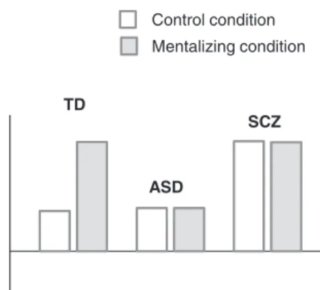
TD
ASD
SCZ Control condition Mentalizing condition
Fig. 1. A schematic illustration of predicted neural signatures for
the hyper-hypo-intentionality hypothesis. The plots represent a priori predicted patterns of activations in mind-reading relevant regions according to the hypo-hyper-intentionality thesis. For TD we predict increased significant activation for the intention condition compared to the control condition. For both patient groups we predict opposed dysfunctional activations: Relatively reduced activation for the intention condition in ASD (hypo-intentional) and relatively increased activation for the control condition in SCZ (hyper-intentional).
following 2 experimental conditions: (1) Private intention (PInt) representing a person’s intention with regard to her/his own action and plans, eg, observing a single per-son changing a broken bulb in order to read a book. (2) Communicative intention (CInt) representing the inten-tion to communicate conveyed by communicative ges-tures, eg, observing a person requiring another person for a glass of water. In the control condition (third condition: physical causality or PhC), the comic strip showed non-intentional causal links among objects, eg, a ball blown by a gust of wind knocking over and breaking a glass of water.
In order to perform the task in the PInt and CInt conditions, it was necessary to read the intention of the characters. In the Ph-C condition (our control condition) the participants viewed scenes showing non-intentional causal links among objects, eg, a rock is rolling down a hill hitting a fence.
The experimental protocol was administered in 1 ses-sion (10 trials per condition) in pseudo-randomized order. Before scanning, each participant was trained outside the scanner with different comic strips for each category. Behavioral Statistics
Descriptive statistics were calculated by χ2 and t-tests as
appropriate. Participants’ reaction times and response accuracy were measured during scanning (for technical reasons, we collected behavioral data for 21 ASD subjects only). Data were analyzed with SPSS Statistics 17.0 by 1-way ANCOVA with age as a covariate (the SCZ group was older than the ASD and TD groups) and subsequent comparisons between means, using Bonferroni’s post hoc test (P < .05).
Imaging Parameters
FMRI data were collected using a 3-Tesla Siemens Allegra scanner. Functional images were acquired using an echoplanar imaging (EPI) sequence. We obtained 384 whole brain scans. One volume consisted of 30 slices (slice thickness 3 mm + 0.75 mm gap, FOV = 192 mm, repetition time = 2 s, echo time = 30 ms, 64 × 64 matrix, flip angle 80°).
Data Analysis
Data Preprocessing. The first 4 volumes were discarded in order to allow for T2-equilibration. Data preprocess-ing and statistical analyses were carried out with SPM8
(http://www.fil.ion.ucl.ac.uk/spm/software/spm8)
follow-ing a standard sequence:
Data were realigned, normalized with a voxel size of 2 mm × 2 mm × 2 mm and smoothed with an 8-mm Gaussian fil-ter. For each trial, the variance of each voxel was estimated according to the general linear model. Intrinsic autocorre-lations were accounted for by the first-order autoregressive
AR(1) process, and low-frequency drifts were removed via a high-pass filtering (128 s).
ROI Analysis. This study based on a region of interest (ROI) analysis. Our ROIs were defined using an indepen-dent localizer “ToM” task35 in order to select 4
inde-pendent ROIs (ventral and dorsal MPFC, right and left pSTS). For more details for the localizer task and ROIs definition, see supplemental material. The rationale for the selection of these ROIs is based on the relevance of these regions considered to be the key brain areas dur-ing mind-readdur-ing.6–8 Furthermore, based on our previous
results, the MPFC and the bilateral pSTS were the unique brain regions showing aberrant activation in SCZ during an intention’s reading.18
The first-level regression model consisted of 3 predic-tors defined for each condition (PhC, PInt, and CInt) modeled as an event (15 s) and convolved with the hemo-dynamic response function and 6 regressors describing residual motion.
In a second-level random effects group analysis, individ-ual regionally specific effects of conditions for each subject were compared using a 3 × 3 full factorial design (ANOVA) with condition and group as factors resulting in a t-statistic for every voxel. We also included an additional covariate “Age.” The significance threshold was set to P < .05 (k > 15) familywise error corrected for multiple comparisons within the a priori-defined regions of interest (right and left pSTS, ventral and dorsal MPFC) provided by the main effect activation of the localizer experiment. In order to compare the mean activations pairwise per condition, indi-vidual peak voxel data within the ROIs were extracted from the respective regressor and region and were subsequently analyzed externally using SPSS Statistics 17.0. Anatomical regions are reported according to standard atlases.36,37
Functional Connectivity
We performed a functional connectivity analysis (fcMRI analysis) in order to explore the extent to which the right pSTS is connected to the rest of the brain during inten-tion reading. A seed-based correlainten-tional analysis was performed using the CONN-fMRI functional connectiv-ity toolbox (http://web.mit.edu/swg/software.htm)38 and
SPM8. After preprocessing, data were band-pass filtered (0.01 Hz < f < inf Hz). Functional connectivity for each subject was examined during each trial. Regressors of no interest included the first-order derivatives of the 6 motion parameters, the 3 main condition effects (PhC, PInt, and CInt) as well as the eigenvectors from a principal com-ponent analysis on the white matter and cerebrospinal fluid voxels. Correlation maps were produced by extract-ing the residual BOLD time course from the seed region and computing Pearson’s correlation coefficients between the time course of the right pSTS and the time courses of all other voxels of the brain. Correlation coefficients were converted to normally distributed scores using Fisher’s
transformation to allow for second-level GLM analyses. The resulting contrast images were entered into a random-effects ANOVA included an additional covariate “Age.”
GLMs were calculated using the contrast images reflect-ing the connectivity change of the CInt and PhC condi-tions for the 3 groups. In fact, due to the functional results with no differences for the PInt>PhC, we performed the fcMRI group analysis for the CInt>PhC contrast only. Results were considered significant using an uncorrected P < .001 (k > 15) corrected for multiple comparisons. Results
Behavioral Performance
For response accuracy we found a group effect [F(2,58) = 8.13; P = .001] (ASD made more errors), but no effect of condition [F(2,58) = 0.79; P = .46] or any interaction [F(4,88) = 0.92, P = .46]. For reaction times we found a group effect [F(2,58) = 3.42; P = .04] (SCZ were slower), but again no effect of condition [F(2,58) = 1.27; P = .29] or any interaction [F(2,58) = 0.86; P = .49]. See also supplementary table 2.
Functional Imaging Results
When comparing each intentional condition (PInt and CInt) with the physical control condition (PhC), differ-ences in activation patterns between groups were found driven by specific aberrant activation within the 4 ROIs in pair-wise comparisons as predicted (table 1).
TD Vs Patients With Paranoid SCZ. For PInt>PhC, no difference was observed between groups. For CInt>PhC, the TD group compared to the SCZ group showed higher activation in the left pSTS and ventral MPFC (table 1
and figure 2A).
In order to test the hypothesis of “hyper-intentionality” in the SCZ group and to replicate our results,18 the mean
activation per condition in both groups in vMPFC was extracted and analysed externally in order to test by which differences the group by condition effects were driven. Note that we do not claim P-values for the interaction contrast itself to be a significant finding, as this would be double dipping—the interaction was found to be sig-nificant already in the ROI analyses on a FWE-corrected level in SPM. The TD group showed increasing activation from PhC < PInt < CInt. In contrast, SCZ showed similar activation for PhC and for both experimental conditions (PInt and CInt) (figure 2A). Comparing directly the con-trol condition (PhC) in concon-trols and SCZ in a post hoc t-test, a strong difference between groups was observed (P = .002). Similar results were found for left pSTS acti-vation (P = .001). Moreover, the SCZ group correlation analysis revealed that PhC activation in vMPFC cor-related positively with the PANSS total score (r = .73, P = .01) (figure 2A). To summarize, the SCZ group revealed dysfunctional activation in vMPFC and left pSTS and these dysfunctional activations were primarily done by an increased activation in the control condition (Ph-C). For the vMPFC this abnormal activation in Ph-C was positively associated with the degree of illness. TD Vs Patients With ASD. No difference was observed between TD and ASD for the contrast PInt>PhC. Instead, decreasing neural activation for CInt>PhC in the right pSTS was observed for ASD (table 1 and figure 2B). In order to test the hypothesis of “hypo-intentionality” in the ASD group mean activation per condition in both groups in the right pSTS was extracted and analyzed externally. Comparing directly the CInt condition by a post-hoc t-test in controls and ASD, CInt showed less activation in ASD (P = .0056).
ASD Vs SCZ. No differential activation for the con-trast PInt>PhC was observed. In the CInt>PhC concon-trast ASD showed more activation in dMPFC compared to SCZ. The inverse contrast (SCZ>ASD) did not reveal any difference (table 1 and figure 2C).
In order to test the hypothesis of “hypo-intentionality” and “hyper-intentionality,” mean activation per condition in the dMPFC ROIs of ASD and SCZ were extracted and analyzed externally. Similar as reported above, the respective plots indicate increasing significant activation in the PhC condition for the SCZ group (P = .0021). fcMRI SEED-Analysis. Functional connectivity analy-ses with the right pSTS (the seed region) was performed
Table 1. Coordinates and Anatomic Localization for the ROI
Analysis (Within and Between Analysis)
PInt vs PhC CInt vs PhC x y z Z x y z Z Control Posterior STS 60 −54 18 5.5 56 −56 20 Inf −58 −62 20 5.15 −58 −58 20 Inf Ventral MPFC 2 48 −18 6.26 Dorsal MPFC 0 56 32 3.97 SCZ Posterior STS 54 −60 10 5.11 52 −60 12 6.49 ASD −58 −52 16 5.62 Posterior STS 56 −60 10 5.11 60 −52 14 Inf −58 −50 18 Inf Ventral MPFC 0 46 −20 6.31 Dorsal MPFC −4 54 34 5.92 CONTROL vs SCZ Posterior STS −46 70 16 3.98 Ventral MPFC 6 48 −20 3.91 CONTROL vs ASD Posterior STS 60 −48 −2 3.95 ASD vs SCZ Dorsal MPFC −4 54 34 4.22
Note: STS, superior temporal sulcus; MPFC, medial prefrontal
Fig. 2. Interaction of mentalizing (CInt>PhC) for TD>SCZ (A), TD>ASD (B), and ASD>SCZ (C). (A) Bar plots indicate effect sizes at
the peak voxel in MPFC and left pSTS for all conditions in TD and SCZ. The amount of MPFC activation for PhC in the SCZ correlated positively with the total positive PANSS score (r = 0.73, P = .001). (B) Bar plots indicate effect sizes at the peak voxel in right pSTS for all conditions in TD and ASD. (C) Bar plots indicate effect sizes at the peak voxel in dMPFC for all conditions in ASD and SCZ. ROI analysis
to identify connected voxels whose activation showed a stronger response during CInt>PhC. Whole brain analy-sis revealed different regions of the social brain connected to the right pSTS that responded more strongly to CInt than PhC (table 2).
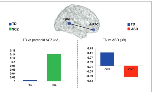
The control group showed increasing connectivity dur-ing CInt compared to PhC with vMPFC and dMPFC, bilateral TPJ, bilateral pSTS, right temporal pole, bilat-eral fusiform gyrus (FG), left supplemental motor area (SMA), and bilateral dorsolateral prefrontal cortex (DLPFC). The SCZ group exhibited functional coupling to fewer areas: left pSTS and temporal pole, right FG, right DLPFC, and left inferior frontal gyrus. In ASD the right pSTS showed increased connectivity with the bilateral TPJ, right FG and left pSTS. In the between-group comparison, between-group differences were found for con-trol group vs SCZ in connectivity strength of the pSTS with vMPFC. During PhC the right pSTS was more strongly coupled with the vMPFC in patients with SCZ
(figure 3A). In contrast, the ASD group showed less
con-nectivity between the right pSTS and vMPFC, not in the physical but during the intentional condition (CInt)
(figure 3B).
To summarize, the right pSTS was dysfunctional con-nected with the vMPFC in both patients group. In par-ticular for the SCZ group the abnormal connection was primarily apparent by increased connectivity strength in the control condition (Ph-C), whereas for the ASD group the anomaly was primarily done by reduced connectivity strength in the communicative intention.
Discussion
This is the first study comparing SCZ and autism with regard to the neural signatures of their mindreading deficits using fMRI. In a task specifically designed for reading different kind of intentions, we observed group specific activation and connectivity patterns in and between key regions of the social brain in line with the hyper-hypo-intentionality hypothesis. These were appar-ent only during complex social interactive but not during simple mentalizing conditions.
At the behavioral level, the ASD group showed over-all reduced accuracy, whereas the SCZ group showed overall increased reaction times. Other studies reported similar findings, namely no specific behavioral impair-ments but specific neural deviances.14,15,18 Since we only
observed a significant main effect, but no interactions, we cannot state that the observed behavioral effects are spe-cific to mind-reading. Rather, they may reflect additional difficulties in both patient groups beyond the ability to read intention. An alternative explanation could be that these results are the consequence of the different way of intention processing: hyper-intentionality leads SCZ to attribute intentions to objects, hypo-intentionality leads ASD to treat persons as objects. In both cases, the stance toward persons and objects could be altered in a group specific way but the result may be the same: to handle persons and objects in a similar way.
At the neural level, the SCZ group showed aberrant activation in vMPFC and left pSTS (figure 2A), core regions of the mentalizing network. Moreover, this was due mainly to increased activation in the control condi-tion. In line with the hyper-intentionality hypothesis, patients with paranoid SCZ seem to process information about physical processes on the neural level in a similar way as information about mental states. Such an “inten-tion detector” may become hyper-active in paranoid thinking as it is characteristic for SCZ.39 These neural
findings replicate our results reported in an indepen-dent patient group,18 and they are also consistent with
behavioral evidence that demonstrated that SCZ attrib-uted intentions to objects.,12 Likewise confirming our
results, in a recent study on action observation40
hyper-activation in the ToM-network (in pSTS and MPFC) was reported in patients with SCZ during a non-social condi-tion. Furthermore, the fact that activation in the MPFC was correlated with the degree of positive symptoms lends some additional support to our interpretation and
Table 2. Seed-Based (Right pSTS) Connection Table for
CInt>PhC (Within and Between Analysis) CInt vs PhC x y z Z Control TPJ 66 −52 24 4.17 −54 −68 20 4.28 Temporal pole 50 −14 −16 4.15 Posterior STS 58 −36 −4 3.72 −54 −40 −4 4.03 Dorsal MPFC 6 52 32 3.9 Ventral MPFC −10 58 −18 3.71 Fusiform gyrus 44 −56 −24 3.54 −36 −46 −30 4.09 Dorso lateral prefrontal cortex 58 22 30 3.87 −50 6 38 3.59 Supplemental motor area −6 2 54 4.22 SCZ
Temporal pole −56 −38 −6 4.11 Posterior STS −56 −60 10 3.72 Fusiform gyrus 46 −46 −32 3.89 Inferior frontal gyrus −56 20 6 4.24 Dorso lateral prefrontal cortex 40 8 28 4.04 ASD
Posterior STS −62 −46 8 3.55 Fusiform gyrus 50 −50 −20 5.43 Anterior temporal pole 50 10 −32 4.22 Temporal pole −64 −34 6 4.08 Control vs SCZ
Ventral MPFC −6 54 −24 3.37 Control vs ASD
Ventral MPFC 10 54 −10 3.81
Note: STS, superior temporal sulcus; MPFC, medial prefrontal
confirms an association between paranoid symptoms and abnormal brain activation during reading of intent and agency.40,41 This neurocognitive bias could be related to an
exaggerated attentional focus on single visual elements in an observed social interaction, resulting in an incorrect reading of the social interaction itself.42 An alternative
explanation is related to the strong decoupling difficul-ties of paranoid patients, which preclude them from dif-ferentiating between mental states belonging to different agents.18 Further evidence comes from connectivity
anal-yses of our data demonstrating that a core region of the mind-reading network, the right pSTS, showed stronger coupling to a second key region of the mind-reading net-work (vMPFC) compared to TD (figure 3A) again during the control condition.
The ASD group also showed group differences in acti-vation within the right pSTS when inferring communi-cative intentions (figure 2B). However, in contrast to the SCZ group, in ASD this difference was driven mainly by less activation during the experimental, ie, CInt condi-tion. This finding is exactly what we predicted for ASD based on the hypo-intentionality hypothesis. Behavioural studies in ASD showed that mental states are often processed similar to physical states of objects.20 Also
eye-tracking data during video-clips viewing showed that individuals with ASD did not focus on the interac-tive actors but on irrelevant physical information pres-ent on the scene.43 Deviant activation of pSTS in ASD
subjects during social cognition is well documented (for review see)44 and abnormal activation in ASD subjects
was reported when they observed geometric forms that elicit mind-reading21 (whereas SCZ reported opposite
results). Its abnormal involvement is described also for a variety of indicators of social information processing
such as eye gaze, language perception, and joint atten-tion.22,45,46 An fMRI study revealed that ASD responded
to faces similarly as to objects.47 Additional evidence for
the hypo-intentional hypotheses comes from our connec-tivity findings with decreased connecconnec-tivity between right pSTS and vMPFC in patients with ASD (figure 3B) in line with the literature supporting hypo-connectivity in
ASD23,25: Hypo-connectivity might result from a lack of
early social experiences due to disturbed early develop-mental mechanisms involved in social cognition.48
Few behavioural studies on social cognition directly compared SCZ and ASD revealing inconsistent results.49–51 One behavioural study using a huge number of
participants, found that ASD showed reduced perception of agency, whereas SCZ showed increasing perception of agency in human but also in other entities (animals and objects).52 Inconsistent results come also from brain
volumetric studies: A meta-analysis study reported simi-lar brain volumes between ASD and SCZ, but also clear distinctions.53 In 2 different EEG-studies using the same
paradigm, ASD and SCZ were separately tested regard-ing their social-emotional abilities54,55 revealing
overlap-ping but opposite results (reduced Mu suppression in ASD and larger Mu suppression in SCZ). To our knowl-edge, only one imaging study on social cognition, namely during processing of trustworthy faces, compared both groups directly: ASD and SCZ reported similar hypo-activation in right amygdala, FG and left ventro-lateral prefrontal cortex.56 The authors argued for “deficit
spe-cific” rather than disorder specific abnormality. In our study, the SCZ group seems to be more affected on the neural level compared to ASD (figure 2C). The available data from the literature make it hard to delineate which psychopathological syndrome should be more affected.
Fig. 3. Group × connectivity interaction. Connectivity was calculated using the right pSTS as a seed region (ANOVA second-level for
CInt>PhC, P = .001 uncorrected, k > 15). (A) Group differences in connectivity strength for TD (blue) versus SCZ (green) for PhC. (B) Group differences in connectivity strength between TD (blue) vs ASD (red) for CInt.
We speculate that the presence of positive symptoms in the SCZ group might elicit more disruption on reading intentions,57 and although a general mind-reading deficit
is described in ASD,19 a specific intention reading deficit
in ASD is not well documented.58
Clearly, there are several limitations to this study. A sub-group of the patients with SCZ and 1 patient with ASD were treated with atypical neuroleptics. Therefore, we can-not exclude that medication effects interfere with the inter-pretation of our findings. Additionally, the age range of our sample was rather large: Participants with SCZ were older than TD and ASD participants. Although age effects were partialled-out in all analyses, confounding influences of age on group comparability cannot definitely be excluded. Taken these limitations into account, we conclude that group differences in neural activation during intention reading in ASD and SCZ depend on the type of intention (communicative or private). Moreover, the group differ-ences in brain activation and the results of our connectiv-ity analyses are consistent with the idea that mind-reading problems in SCZ are due to an overactive intention-detec-tion module, whereas mind-reading problems in ASD are rather due to an underactive intention-detector module. Supplementary Material
Supplementary material is available at http://schizophre
niabulletin.oxfordjournals.org.
Funding
The study was partly supported by the Swedish Research Council (grant nr. 523-2009-7054) and by the LOEWE-Program ‘Neuronal Coordination Research Focus Frankfurt’ (NeFF).
Acknowledgments
The authors acknowledge the contribution of the study participants. The authors have declared that there are no conflicts of interest in relation to the subject of this study. References
1. Lord C, Cook EH, Leventhal BL, Amaral DG. Autism spec-trum disorders. Neuron. 2000;28:355–363.
2. Crow TJ. The two-syndrome concept: origins and current sta-tus. Schizophr Bull. 1985;11:471–486.
3. Crespi B, Badcock C. Psychosis and autism as diametrical dis-orders of the social brain. Behav Brain Sci. 2008;31:241–261 4. Adolphs R. Cognitive neuroscience of human social
behav-iour. Nat Rev Neurosci. 2003;4:165–178.
5. Ciaramidaro A, Adenzato M, Enrici I, et al. The inten-tional network: how the brain reads varieties of intentions.
Neuropsychologia. 2007;45:3105–3113.
6. Amodio DM, Frith CD. Meeting of minds: the medial frontal cortex and social cognition. Nat Rev Neurosci. 2006;7:268–277.
7. Saxe R. Uniquely human social cognition. Curr Opin
Neurobiol. 2006;16:235–239.
8. Chung YS, Barch D, Strube M. A meta-analysis of mental-izing impairments in adults with schizophrenia and autism spectrum disorder. Schizophr Bull. 2014;40:602–616.
9. Frith C. The neural basis of hallucinations and delusions. C
R Biol. 2005;328:169–175.
10. Bell MD, Fiszdon JM, Greig TC, Wexler BE. Social attri-bution test–multiple choice (SAT-MC) in schizophrenia: comparison with community sample and relationship to neurocognitive, social cognitive and symptom measures.
Schizophr Res. 2010;122:164–171.
11. Martin JA, Penn DL. Attributional style in schizophrenia: an investigation in outpatients with and without persecutory delusions. Schizophr Bull. 2002;28:131–141.
12. Bentall RP, Rowse G, Shryane N, et al. The cognitive and affective structure of paranoid delusions: a transdiagnostic investigation of patients with schizophrenia spectrum disor-ders and depression. Arch Gen Psychiatry. 2009;66:236–247. 13. Blakemore SJ, Sarfati Y, Bazin N, Decety J. The detection of
intentional contingencies in simple animations in patients with delusions of persecution. Psychol Med. 2003;33:1433–1441. 14. Brunet E, Sarfati Y, Hardy-Baylé MC, Decety J. Abnormalities
of brain function during a nonverbal theory of mind task in schizophrenia. Neuropsychologia. 2003;41:1574–1582. 15. Brüne M, Lissek S, Fuchs N, et al. An fMRI study of theory
of mind in schizophrenic patients with “passivity” symptoms.
Neuropsychologia. 2008;46:1992–2001.
16. Das P, Lagopoulos J, Coulston CM, Henderson AF, Malhi GS. Mentalizing impairment in schizophrenia: a functional MRI study. Schizophr Res. 2012;134:158–164.
17. Pedersen A, Koelkebeck K, Brandt M, et al. Theory of mind in patients with schizophrenia: is mentalizing delayed?
Schizophr Res. 2012;137:224–229.
18. Walter H, Ciaramidaro A, Adenzato M, et al. Dysfunction of the social brain in schizophrenia is modulated by intention type: an fMRI study. Soc Cogn Affect Neurosci. 2009;4:166–176. 19. Frith U. Mind blindness and the brain in autism. Neuron.
2001;32:969–979.
20. Klin A. Attributing social meaning to ambiguous visual stimuli in higher-functioning autism and Asperger syn-drome: the social attribution task. J Child Psychol Psychiatry. 2000;41:831–846.
21. Castelli F, Frith C, Happé F, Frith U. Autism, Asperger syn-drome and brain mechanisms for the attribution of mental states to animated shapes. Brain. 2002;125:1839–1849. 22. Pelphrey KA, Morris JP, McCarthy G. Neural basis of eye
gaze processing deficits in autism. Brain. 2005;128:1038–1048. 23. Kana RK, Libero LE, Hu CP, Deshpande HD, Colburn JS.
Functional brain networks and white matter underlying the-ory-of-mind in autism. Soc Cogn Affect Neurosci. 2014;9:98– 105.doi:10.1093/scan/nss106.
24. Lombardo MV, Chakrabarti B, Bullmore ET, MRC AIMS Consortium, Baron-CohenS. Specialization of right temporo-parietal junction for mentalizing and its relation to social impairments in autism. Neuroimage. 2011;56:1832–1838. 25. Kana RK, Keller TA, Cherkassky VL, Minshew NJ, Just
MA. Atypical frontal-posterior synchronization of Theory of Mind regions in autism during mental state attribution.
Soc Neurosci. 2009;4:135–152.
26. Abu-Akel A, Bailey AL. The possibility of different forms of theory of mind impairment in psychiatric and developmental disorders. Psychol Med. 2000;30:735–738.
27. Just MA, Cherkassky VL, Keller TA, Minshew NJ. Cortical activation and synchronization during sentence comprehen-sion in high-functioning autism: evidence of underconnectiv-ity. Brain. 2004;127:1811–1821.
28. Assaf M, Jagannathan K, Calhoun VD, et al. Abnormal func-tional connectivity of default mode sub-networks in autism spectrum disorder patients. Neuroimage. 2010;53:247–256. 29. Whitfield-Gabrieli S, Thermenos HW, Milanovic S, et al.
Hyperactivity and hyperconnectivity of the default network in schizophrenia and in first-degree relatives of persons with schizophrenia. Proc Natl Acad Sci U S A. 2009;106:1279–1284. 30. Bara BG, Ciaramidaro A, Walter H, Adenzato M. Intentional
minds: a philosophical analysis of intention tested through fMRI experiments involving people with schizophrenia, peo-ple with autism, and healthy individuals. Front Hum Neurosci. 2011;5:7. doi:10.3389/fnhum.2011.00007.
31. Walter H, Adenzato M, Ciaramidaro A, Enrici I, Pia L, Bara BG. Understanding intentions in social interaction: the role of the anterior paracingulate cortex. J Cogn Neurosci. 2004;16:1854–1863.
32. Saxe R, Xiao DK, Kovacs G, Perrett DI, Kanwisher N. A region of right posterior superior temporal sulcus responds to observed intentional actions. Neuropsychologia. 2004;42:1435–1446. 33. Pelphrey KA, Morris JP, McCarthy G. Grasping the
inten-tions of others: the perceived intentionality of an action influences activity in the superior temporal sulcus during social perception. J Cogn Neurosci. 2004;16:1706–1716. 34. Gao T, Scholl BJ, McCarthy G. Dissociating the detection of
intentionality from animacy in the right posterior superior temporal sulcus. J Neurosci. 2012;32:14276–14280.
35. Walter H, Schnell K, Erk S, et al. Effects of a genome-wide supported psychosis risk variant on neural activation during a theory-of-mind task. Mol Psychiatry. 2011;16:462–470. 36. Duvernoy HM, Bourgouin P. The Human Brain: Surface,
Three-Dimensional Sectional Anatomy With MRI, and Blood Supply. 2nd ed. Wien, NY: Springer; 1999.
37. Talairach J, Tournoux P. Co-planar Stereotaxic Atlas of the
Human Brain. New York, NY: Thieme Medical Publishers;
1998.
38. Whitfield-Gabrieli S, Nieto-Castanon A. Conn: a functional connectivity toolbox for correlated and anticorrelated brain networks. Brain Connect. 2012;2:125–141. doi:10.1089/ brain.2012.0073.
39. Meyer-Lindenberg A. From maps to mechanisms through neuroimaging of schizophrenia. Nature. 2010;468:194–202. 40. Backasch B, Straube B, Pyka M, et al. Hyperintentionality
during automatic perception of naturalistic cooperative behavior in patients with schizophrenia. Soc Neurosci. 2013;8:489–504. doi:10.1080/17470919.2013.820666.
41. Park IH, Ku J, Lee H, et al. Disrupted theory of mind net-work processing in response to idea of reference evocation in schizophrenia. Acta Psychiatr Scand. 2011;123:43–54. 42. Chambon V, Pacherie E, Barbalat G, Jacquet P, Franck N,
Farrer C. Mentalizing under influence: abnormal dependence on prior expectations in patients with schizophrenia. Brain. 2011;134:3728–3741.
43. Klin A, Jones W, Schultz R, Volkmar F. The enactive mind, or from actions to cognition: lessons from autism. Philos
Trans R Soc Lond B Biol Sci. 2003;358:345–360.
44. Zilbovicius M, Meresse I, Chabane N, Brunelle F, Samson Y, Boddaert N. Autism, the superior temporal sulcus and social perception. Trends Neurosci. 2006;29:359–366.
45. Gervais H, Belin P, Boddaert N, et al. Abnormal cortical voice processing in autism. Nat Neurosci. 2004;7:801–802. 46. Redcay E, Dodell-Feder D, Mavros PL, et al. Atypical brain
activation patterns during a face-to-face joint attention game in adults with autism spectrum disorder. Hum Brain Mapp. 2013;34:2511–2523. doi:10.1002/hbm.22086.
47. Schultz RT, Gauthier I, Klin A, et al. Abnormal ventral temporal cortical activity during face discrimination among individuals with autism and Asperger syndrome. Arch Gen
Psychiatry. 2000;57:331–340.
48. Pelphrey KA, Shultz S, Hudac CM, Vander Wyk BC. Research review: Constraining heterogeneity: the social brain and its development in autism spectrum disorder. J Child
Psychol Psychiatry. 2011;52:631–644.
49. Bölte S, Poustka F. The recognition of facial affect in autis-tic and schizophrenic subjects and their first-degree relatives.
Psychol Med. 2003;33:907–915.
50. Couture SM, Penn DL, Losh M, Adolphs R, Hurley R, Piven J. Comparison of social cognitive functioning in schizophre-nia and high functioning autism: more convergence than divergence. Psychol Med. 2010;40:569–579.
51. Craig JS, Hatton C, Craig FB, Bentall RP. Persecutory beliefs, attributions and theory of mind: comparison of patients with paranoid delusions, Asperger’s syndrome and healthy con-trols. Schizophr Res. 2004;69:29–33.
52. Gray K, Jenkins AC, Heberlein AS, Wegner DM. Distortions of mind perception in psychopathology. Proc Natl Acad Sci
U S A. 2011;108:477–479. doi:10.1073/pnas.1015493108.
53. Cheung CH, Wu SY, Lee TR, et al. Cancer cells acquire mitotic drug resistance properties through beta I-tubulin mutations and alterations in the expression of beta-tubulin isotypes. PLoS
ONE. 2010;5:e12564. doi:10.1371/journal.pone.0012233.
54. Oberman LM, Hubbard EM, McCleery JP, Altschuler EL, Ramachandran VS, Pineda JA. EEG evidence for mirror neuron dysfunction in autism spectrum disorders. Brain Res
Cogn Brain Res. 2005;24:190–198.
55. McCormick LM, Brumm MC, Beadle JN, Paradiso S, Yamada T, Andreasen N. Mirror neuron function, psychosis, and empathy in schizophrenia. Psychiatry Res. 2012;201:233– 239. doi: 10.1016/j.pscychresns.2012.01.004.
56. Pinkham AE, Hopfinger JB, Pelphrey KA, Piven J, Penn DL. Neural bases for impaired social cognition in schizophrenia and autism spectrum disorders. Schizophr Res. 2008;99:164–175. 57. Sugranyes G, Kyriakopoulos M, Corrigall R, Taylor E,
Frangou S. Autism spectrum disorders and schizophrenia: meta-analysis of the neural correlates of social cognition.
PLoS ONE. 2011;6:e25322.
58. Hamilton AF. Goals, intentions and mental states: chal-lenges for theories of autism. J Child Psychol Psychiatry. 2009;50:881–892.