A comparative evaluation of intrathoracic latissimus dorsi and
serratus anterior muscle transposition
q
Matthias Kurt Widmer
a, Thorsten Krueger
b,*, Didier Lardinois
a, Andrej Banic
c, Hans Beat Ris
baDepartment of Thoracic and Cardiovascular Surgery, University of Berne, Berne, Switzerland bDepartment of Surgery, University of Lausanne, 1011 Lausanne, Switzerland cDivision of Plastic and Reconstructive Surgery, University of Berne, Berne, Switzerland
Received 11 October 1999; received in revised form 20 June 2000; accepted 12 July 2000
Abstract
Background: Comparison of intrathoracic latissimus dorsi (LD) versus serratus anterior (SA) muscle transposition for treatment of infected spaces, broncho-pleural ®stulae, and for prophylactic reinforcement of the mediastinum after extended resections following induction therapy. Patients and methods: Twenty LD and 17 SA transfers were performed for prophylactic reinforcement (11 LD; nine SA), and treatment of infections (nine LD; eight SA) from 1995 to 1998. Results: The 30-day mortality was 0% following prophylactic reinforcement and 29% following treatment of infections (three LD; two SA). Prophylactic mediastinal reinforcement was successful in 11 of 11 patients with LD and nine of nine with SA transpositions, and treatment of infected spaces in eight of nine patients with LD and two of three with SA transfers. Morbidity requiring re-intervention consisted of ¯ap necrosis (one LD), bleeding (one SA), and skin necrosis over a winged scapula (one SA). Subcutaneous seromas and chest wall complaints were more frequent following LD (45 and 36%, respectively) compared with SA transfers (29 and 27%, respectively), whereas impaired shoulder girdle function was more frequent after SA than after LD transfer (27 vs. 21%). Conclusion: Intrathoracic LD and SA muscle transpositions are both ef®cient for the prevention or control of infections following complex thoracic surgery, and are both associated with similar and acceptable morbidity and long-term sequelae. q 2000 Elsevier Science B.V. All rights reserved.
Keywords: Intrathoracic muscle transposition; Latissimus dorsi muscle; Serratus anterior muscle
1. Introduction
The treatment of chronic empyema, postresectional infected space and broncho-pleural ®stula remains a chal-lenging problem for thoracic surgeons. The incidence of these diseases is likely to increase due to the increasing incidence of major resections in patients suffering from lung cancer, and chronic pleural infections in elderly poly-morbid patients. In addition, resections are being performed more frequently in the setting of multimodality treatment regimens, including preoperative induction therapy with radiation or chemotherapy, or both. Therefore, thoracic surgeons are again confronted with ef®cient prevention and treatment of chronic empyema and infections resulting from previous thoracic interventions.
Various techniques of mediastinal reinforcement have
been used for this purpose, such as ¯aps arising from the diaphragm, pericardium, pleura, or intercostal muscle, as well as intrathoracic transposition of the chest wall muscles and greater omentum [1±6]. Arnold and Pairolero have shown the usefulness of intrathoracic latissimus dorsi (LD) and serratus anterior (SA) muscle transfers in control-ling infection in postresectional spaces and postpneumo-nectomy broncho-pleural ®stulae [7,8]. These muscle ¯aps are well suited for this purpose since they consist of solid tissue with mechanical strength and suf®cient volume to ®ll cavities and reinforce the mediastinum. Moreover, they are easy to dissect at thoracotomy and their procure-ment does not require access to the abdominal cavity. However, there are no comparative studies for LD and SA muscle transfers regarding morbidity and sequelae, such as chest wall complaints and shoulder girdle function, and their ability to prevent or control postresectional infec-tions. This study was performed in order to compare the outcome following intrathoracic LD versus SA transposi-tion in this respect.
1010-7940/00/$ - see front matter q 2000 Elsevier Science B.V. All rights reserved. PII: S1010-7940(00)00538-8
www.elsevier.com/locate/ejcts
qPresented at the 13th Annual Meeting of the European Association for
Cardio-thoracic Surgery, Glasgow, Scotland, UK, September 5±8, 1999. * Corresponding author. Tel.: 141-21-314-2408; fax: 141-21-314-2358.
2. Patients and methods
All patients who underwent intrathoracic LD or SA muscle transposition at our institution between November 1995 and December 1998 were followed prospectively in order to compare the ef®ciency (prevention or control of intrathoracic postresectional infections) and sequelae related to these procedures. The indications for intrathoracic muscle trans-position consisted of prophylactic mediastinal reinforcement in patients undergoing extrapleural pneumonectomy for malignant mesothelioma followed by radio-chemotherapy, and carinal reconstructions following induction radio-chemotherapy for non-small cell lung cancer, and the treat-ment of postpneumonectomy bronchial stump ®stula, post-resectional infected spaces and chronic empyema.
The LD transfer was used in patients with an intact muscle without a previous thoracotomy. The muscle was dissected in the epifascial plane and divided at the thor-aco-lumbal fascia. The dissection was continued, and the muscle was separated from the chest wall while preserving its neurovascular bundle. The muscle was then transposed into the chest cavity through a small additional thoracotomy performed through the bed of the second or third rib, of which 3 cm was resected. The origin of the muscle was not divided in order to avoid undue tension on the vascular pedicle after intrathoracic transposition. This technique allowed complete coverage of the mediastinal surface down to the diaphragm.
The SA muscle was used if the LD muscle had been divided at a previous thoracotomy. The muscle was dissected from the LD muscle, the scapula and the chest wall, while preserving its vascular pedicle. The pedicle to the LD was left intact. The muscle was transposed into the chest cavity through the bed of the second or third rib, after resecting 3 cm of the rib. Coverage of the mediastinum down to the inferior pulmonary vein could thus be obtained.
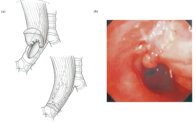
For prophylactic reinforcement of mediastinal structures, the muscle was sutured to the bronchial stump, wrapped around a carinal reconstruction or ®xed to the edges of the mediastinal pleura or pericardium after pericardial resection. Infected spaces were treated in a one stage procedure, including mechanical debridement and irrigation of the chest cavity with diluted povidone±iodine solution in order to remove all debris, ®brin and necrotic tissue followed by muscle transfer. In patients with right-sided postpneumonectomy bronchial stump ®stula and a short bronchial stump the muscle ¯ap was directly sutured to the edges of the debrided bronchial wall without attempting primary closure of the bronchial wall. In one patient with poor cardiac function (ejection fraction of 20%) and broncho-pleural ®stula after upper lobe resection, a similar procedure was chosen in order to preserve the remaining lung and to avoid completion pneumonectomy (Fig. 1a,b).
The 30-day mortality and the procedure-related morbidity were recorded in all patients, as well as the healing of bron-chial stumps and infections. All surviving patients were
Fig. 1. Intrathoracic SA muscle transfer for the treatment of bronchial stump ®stula following upper lobe resection in order to avoid completion pneumo-nectomy in a patient with poor cardiac function. (a) The muscle was sutured to the edges of the debrided bronchial wall of mainstem and intermediate bronchus; (b), endoscopic view 3 months later with well-functioning and healed bronchial wall, with granulation tissue in the intermediate bronchus.
evaluated at 3 and 6 months for healing, chest wall complaints and shoulder girdle function.
The Fisher's exact test was used where appropriate for statistical analysis. Signi®cance was accepted at P , 0:05. 3. Results
From November 1995 to December 1998, 38 intrathor-acic LD or SA muscle transpositions were performed on 37 patients (Table 1).
The follow-up is shown in Table 2. The 30-day mortality of the entire series was 14% (®ve of 37 patients). All deaths occurred after muscle transfer for infections. Three patients died from multi-organ failure 2 (two patients) and 3 (one patient) weeks after the operation; one after decortication and muscle transfer for chronic tuberculous empyema, one after closure of an aorto-pulmonary ®stula, and one after muscle transfer for coverage of an oesophageal perforation. Two patients died 3 weeks after the operation from pulmon-ary embolism which occurred after discharge and an initi-ally uneventful postoperative recovery.
The mean hospital stay for the entire series was 32.5 days, ranging from 6 to 80 days, with no signi®cant difference between the LD and SA groups.
Three patients (8%) required re-operations due to compli-cations related to the muscle transfer. One patient had necrosis of a LD ¯ap, with subsequent re-operation, removal of the necrotic muscle and replacement by a SA muscle ¯ap. The ¯ap necrosis was caused by venous congestion of the ¯ap. In this case, the muscle ¯ap was transposed through an incision made in the second intercostal space without resect-ing of a piece of the rib, which was obviously too narrow for this purpose. One patient had to be re-operated due to bleed-ing of the ¯ap pedicle (SA muscle). In this patient, the rib resection was done too far ventrally, and the vascular
pedi-cle was in contact with the dorsal edge of the resected rib causing bleeding. This complication was corrected by resecting a further piece of the third rib in the dorsal aspect of the wound. Another patient with SA transposition required two additional operations on the dorsal extremity of the thoracotomy wound due to skin necrosis related to an underlying winged scapula. This was corrected by use of a myocutaneous LD muscle ¯ap harvested from the opposite side. All three patients then had an uneventful recovery. A seroma requiring prolonged drainage (more than 15 days) was observed in 45% of patients with a LD and 29% of patients with SA muscle ¯aps (P 0:5).
Uneventful healing of mediastinal structures after prophylactic reinforcement was obtained in 11 from 11 LD ¯aps and nine of nine SA muscle ¯aps. Local control of infected postresectional spaces was obtained in eight out of nine patients who underwent LD muscle transposition. Two of these patients who died from multi-organ failure had no evidence of persistent infection or broncho-pleural ®stula in the involved chest cavity at autopsy. One patient had a persistent infection of the postresectional space despite LD transfer. This patient underwent unilateral lung volume reduction surgery for emphysema and developed postresec-tional space infection with Aspergillus. The LD muscle was too thin to ®ll the cavity and the patient underwent open thoracotomy. Two of three patients with SA transposition for postresectional space infection or destroyed lung showed an uneventful healing. One patient died following SA trans-fer for aorto-bronchial ®stula and lobectomy 2 weeks after surgery from multi-organ failure. Closure of postpneumo-nectomy broncho-pleural ®stula was obtained in ®ve of ®ve patients with SA muscle ¯aps.
Twenty-®ve of 32 surviving patients were available for the 6-month follow-up. Two patients were lost due to their departure and ®ve died during follow-up; four due to tumour
Table 1
Pattern of distribution for LD (n 20) and SA (n 17) muscle transfers
LD SA
Prophylactic mediastinal reinforcement Carinal reconstruction following
induction 3 7
Extended/extrapleural
pneumonectomy with pericardial resection in multimodality setting
8 2
Male/female 2:9 5:4
Mean age (range), years 53.5 (43±71) 57.4 (36±73) Treatment of infections
Postpneumonectomy
broncho-pleural ®stula 0 5
Postresectional space infection 1 2
Chronic empyema 8 1
Male/female 1:8 2:6
Mean age (range), years 47.9 (17±78) 63.3 (45±75)
Table 2
Outcome after LD (n 20) or SA (n 17) muscle transfer
LD SA
Prophylactic mediastinal reinforcement 11 9
30-day mortality (%) 0 0
Re-operation (%) 9 0
Uneventful mediastinal healing (%) 100 100
Seroma (%) 55 22
Treatment of infections 9 8
30-day mortality (%) 33 25
Re-operation (%) 0 25
Control of infection (%) 88a 67 (2/3)
Healing of postpneumonectomy
broncho-pleural ®stula (%) 100 (5/5)
Seroma (%) 33 38
6-month follow-up 14 11
Impaired shoulder girdle function (%) 21 27
Chest wall complaints (%) 36 27
a Two patients died from multi-organ failure within 30 days without
progression and one due to urinary sepsis. No statistically signi®cant differences regarding long-term sequelae were found between patients with SA and LD transposition. Twenty-two of 25 patients with the 6-month follow-up experienced a normal quality of life without restriction in their daily activities (13/14 LD vs. 9/11 SA; P 0:56). Chest wall complaints were observed in eight of 25 patients (5/14 LD vs. 3/11 SA; P 1). The shoulder girdle function was normal and symmetrical in 19 of 25 patients (11/14 LD vs. 8/11 SA; P 1). In patients with impaired shoulder girdle function, a de®cit in elevation and abduction was found only at more than 1208 abduction and elevation of the ipsilateral shoulder girdle.
4. Discussion
At the beginning of the century, Abrashanoff reported thoracic wounds successfully closed by use of a muscle ¯ap [9]; in 1915, Robinson described the healing of a chronic empyema following an intrathoracic muscle trans-position [10]. Pool demonstrated in an experimental setting that a persistent broncho-pleural ®stula could be success-fully healed using a muscle ¯ap [11]. In 1989, Pairolero and Arnold described their experience with intrathoracic muscle transfer for the treatment of life-threatening infections [7], and have demonstrated that either LD, SA or pectoralis major muscles could be used for this purpose [8,12].
Intrathoracic muscle transposition is now a well accepted procedure for the treatment of infected postresectional spaces and postpneumonectomy broncho-pleural ®stula. The advantages of transposed extrathoracic muscle ¯aps over other tissues (omentum, intercostal muscles, or ¯aps derived from pericardium or pleura) are their large size, thickness and mechanical strength, as well as their vascu-larity. The excellent vascularization of the LD and the SA muscle may enhance local wound healing by induction of granulation tissue and antibiotic drug delivery to poorly vascularized infected tissue. Their strength is desirable for mediastinal reinforcement and closure of bronchial stumps since they do resist mechanical shear stress during breathing and coughing. Allen et al. have shown that the muscle can even be sutured directly to the edges of the open bronchial stump without attempting to re-approximate the bronchial wall in order to avoid tension and compromising the vascu-larization of the stump [13]. We have used this technique in ®ve patients with healing in all cases. This technique may also be used in patients with broncho-pleural ®stula follow-ing lobectomy who are not candidates for completion pneu-monectomy. One patient in our series underwent re-operation for this purpose after a right upper lobectomy. The necrotic area of the right mainstem and intermediate bronchus were debrided, and airtight sutures of the muscle to the edge of the oval shaped defect in the bronchial wall of the intermediate bronchus were performed. Approximately one half of the bronchial circumference was replaced by
muscle tissue over a distance of 3 cm. Postoperative follow-up after 6 months revealed a patent bronchus without any tendency to collapse. Intrathoracic muscle transposition was also used to ®ll postresectional infected spaces or chronic empyema cavities not amenable to decortication in elderly fragile patients. In the latter situation, it may be more prudent to remove only the infected calci®ed plaques without attempting to perform a formal decortication, and to ®ll the infected space with a muscle ¯ap. Our results indi-cate that eight out of nine patients revealed uneventful heal-ing with this technique. However, the 30-day mortality rate was high in these patients (33%), although this could not be attributed to the procedure itself, since there was no evidence of residual infection in the chest cavity.
The prophylactic use of intrathoracic muscle transposi-tion is more controversial as there are less aggressive tech-niques available to cover the bronchial stump and other mediastinal structures, such as pericardial, pleural and inter-costal muscle ¯aps [5,14,15]. However, it might be consid-ered in situations at risk of impaired wound healing, such as sleeve pneumonectomy, extended pneumonectomy with a large pericardial defect and carinal reconstruction following induction therapy, or after extrapleural pneumonectomy and pericardial resection in patients suffering from malignant mesothelioma with planned postoperative radio-chemother-apy. We have used prophylactic reinforcement of mediast-inal structures and closure of pericardial defects with intrathoracic muscle transpositions for these indications in 17 patients, with uneventful healing in all patients. Prolonged duration of seroma and impaired shoulder girdle function occurred in 47 and 18% of these patients. The low morbidity of the SA muscle transfer suggests that its use might be justi®ed for prophylactic mediastinal reinforce-ment in dif®cult multimodality settings.
The purpose of this study was to compare the LD and SA muscle transfers regarding ef®ciency and morbidity. Complete mobilization of the LD muscle allows one to reach nearly all parts of the chest cavity. Due to its dominant blood supply (thoracodorsal artery), ¯ap dissection and transposition are safe and easy to perform through the same access as standard thoracotomy. The SA muscle may serve as an alternative, since it is seldom divided during standard posterolateral thoracotomy. It has two principle blood supplies, the lateral thoracic artery and branches of the thoracodorsal artery. However, in contrast to the LD, the SA is not an entirely dispensable muscle which can be replaced by the synergistic function of other muscles of the shoulder girdle. In order to prevent ¯ap devasculariza-tion or bleeding from the pedicle, it is important to resect a suf®cient amount (4 cm) of the second and third ribs for muscle transposition for both types of muscle transfer. Two of three re-operations in this series were related to the neglect of this aspect.
Both techniques have demonstrated equal ef®ciency with respect to the control or prevention of infections in our series. Regarding postoperative morbidity and long-term
sequelae, such as chest wall complaints, discomfort, and shoulder girdle dysfunction, there were no signi®cant differ-ences between the LD and SA transpositions. Subcutaneous seromas and transient chest wall complaints were slightly more frequent in the LD group, and impaired shoulder girdle function was observed in similar proportions after LD and SA transpositions (21 and 27%, respectively). Neither chest wall complaints nor impairment of shoulder girdle function led to an impaired quality of life or changes in daily activ-ities, except in one patient. This patient had a markedly winged scapula with necrosis of the overlying skin follow-ing SA transfer. In this case, heavy irradiation of the ipsi-lateral neck and thoracic inlet preceded thoracotomy for treatment of a head and neck cancer causing atrophy of virtually all the shoulder girdle muscles. All other patients with SA transfer and impaired shoulder girdle function presented a functional de®cit only at abduction and eleva-tion of more than 1208. Our results are in contrast to the common opinion that the use of the whole SA for intrathor-acic transposition will result in all patients having a winged scapula and incapacity to abduct the arm above the horizon-tal. Segmental use of the muscle for intrathoracic transposi-tion has been used to overcome this drawback [12]. However, our results indicate that transfer of the whole SA muscle can be performed if required without impairing the shoulder girdle function to a disabling degree.
We conclude that the intrathoracic transfer of the LD and SA are both ef®cient in preventing or controlling dif®cult intrathoracic infections. The postoperative morbidity and long-term sequelae are similar after both procedures with acceptable functional outcome and quality of life.
References
[1] Iverson LIG, Young JN, Ecker RR, Ennix CL, Lau G, Stallone R, Grimes O, May IA. Closure of bronchopleural ®stulas by an omental pedicle ¯ap. Am J Surg 1986;152:40±42.
[2] Mathisen DJ, Grillo HC, Vlahakes GJ, Dagett WM. The omentum in the management of complicated cardio-thoracic problems. J Thorac Cardiovasc Surg 1988;95:677±684.
[3] Levashev YN, Akopov AL, Mosin IV. The possibilities of greater omentum usage in thoracic surgery. Eur J Cardio-thorac Surg 1999;15:465±468.
[4] Harris SU, Nahai F. Intrathoracic muscle transposition. Surgical anat-omy and techniques of harvest. Chest Surg Clin N Am 1996;6:501± 518.
[5] Klepetko W, Taghavi S, Pereszlenyi A, Birsan T, Groetzner J, Kupilik N, Artemiou O, Wolner E. Impact of different coverage techniques on incidence of postpneumonectomy stump ®stula. Eur J Cardio-thorac Surg 1999;15:758±763.
[6] Hollaus PH, Huber M, Lax F, Wurnig PN, Bohm G, Pridun NS. Closure of bronchopleural ®stula after pneumonectomy with pedicled intercostal muscle ¯ap. Eur J Cardio-thorac Surg 1999;16:181±186.
[7] Arnold PG, Pairolero PV. Intrathoracic muscle ¯aps: a 10 year experi-ence in the management of life-threatening infections. Plast Reconstr Surg 1989;84:92±99.
[8] Pairolero PC, Arnold PG, Trastek VF, Meland NB, Kay PP. Post-pneumonectomy empyema ± the role of intrathoracic muscle transpo-sition. J Thorac Cardiovasc Surg 1990;99:958±968.
[9] Abrashanoff A. Plastische methode der schliessung von ®stelgaÈngen, welche von inneren organen kommen. Zentralbl Chir 1911;38:186± 191.
[10] Robinson S. The treatment of chronic non-tuberculous empyema. Collected Papers Mayo Clin 1915;7:618±644.
[11] Pool EH, Garlock JH. A treatment of persistent bronchial ®stula: an experimental and clinical study. Ann Surg 1929;90:186±187. [12] Arnold PG, Pairolero PV. Intrathoracic muscle ¯aps: an account of
their use in the management of 100 consecutive patients. Ann Surg 1990;211:656±662.
[13] Allen MS. Bronchopleural ®stula. Chest Surg Clin N Am 1992;2:823±837.
[14] Yamamoto R, Inoue K, Hori T, Takehara S, Kaji M, Kinoshita H. Intercostal muscle pedicle ¯ap for prophylaxis against bronchopleural ®stula after pulmonary resection. Osaka City Med J 1994;40:99± 105.
[15] Rendina EA, Venuta F, Ricci P, Fadda GF, Bognolo DA, Ricci C, Rossi P. Protection and revascularization of bronchial anastomoses by the intercostal pedicle ¯ap. J Thorac Cardiovasc Surg 1994;107:1251± 1254.
Appendix A. Conference discussion
Dr T. Grodzki (Szczecin, Poland): I have to ask one doubt. In case of complication, it is de®nitely justi®ed to use large muscles. But I have doubts about prophylaxis in postpneumonectomy patients and so on. In our small experience, those patients present relatively serious shoulder problems and we are trying to avoid using such big muscles as a prophylax. Could you comment on that?
Dr Ris: Our results indicate that morbidity and long-term sequelae after LD and SA transfer were reasonable, and did not lead to impaired quality of life or loss of professional or sports activities. We fully agree that for usual resections, less aggressive stump reinforcement techniques should be used, such as pericard, pleura or intercostal muscle bundles. However, our results indicate that intrathoracic muscle transposition may be considered for prophylactic intentions in dif®cult and selected situations, such as extra-pleural pneumonectomy, extended pneumonectomy or carinal reconstruc-tions in the context of multimodality therapy.
Dr A. Lerut (Leuven, Belgium): In the patients where you use it for prevention, like in induction therapy, I can imagine that a simple intercostal muscle bundle does as well as this operation, which is I think a much bigger operation with some additional morbidity anyway.
Dr Lerut: No, no, I'm saying about the prevention after induction chemotherapy. I assume that you can do as well with an intercostal muscle bundle.
Dr Ris: We fully agree with this. However, in selected patients, as mentioned before, intrathoracic muscle transposition may be considered without decreasing quality of life in those patients.
Dr F. Zonuzi (Istanbul, Turkey): I wonder whether one of these muscles would be suf®cient to ®ll the chronic infectious space?
Dr Widmer: Not the whole space, but the critical area in the thoracic cavity.