• Aucun résultat trouvé

Genetics and mechanisms leading to human cortical malformations

N/A
N/A
Protected

Academic year: 2021

Partager "Genetics and mechanisms leading to human cortical malformations"

Copied!
65
0
0

Texte intégral

(1)

HAL Id: hal-01626222

https://hal.sorbonne-universite.fr/hal-01626222

Submitted on 30 Oct 2017

HAL is a multi-disciplinary open access

archive for the deposit and dissemination of

sci-entific research documents, whether they are

pub-lished or not. The documents may come from

teaching and research institutions in France or

abroad, or from public or private research centers.

L’archive ouverte pluridisciplinaire HAL, est

destinée au dépôt et à la diffusion de documents

scientifiques de niveau recherche, publiés ou non,

émanant des établissements d’enseignement et de

recherche français ou étrangers, des laboratoires

publics ou privés.

Genetics and mechanisms leading to human cortical

malformations

Delfina Romero, Nadia Bahi-Buisson, Fiona Francis

To cite this version:

Delfina Romero, Nadia Bahi-Buisson, Fiona Francis.

Genetics and mechanisms leading to

hu-man cortical malformations.

Seminars in Cell and Developmental Biology, Elsevier, 2017,

�10.1016/j.semcdb.2017.09.031�. �hal-01626222�

(2)

1

Genetics and mechanisms leading to human cortical malformations

Delfina M. Romero

1-3

, Nadia Bahi-Buisson

4-5

, Fiona Francis

1-3 1

INSERM UMR-S 839, 17 rue du Fer à Moulin, Paris 75005, France.

2

Sorbonne Universités, Université Pierre et Marie Curie, 4 Place Jussieu, Paris 75005,

France.

3
Institut du Fer à Moulin, 17 rue du Fer à Moulin, Paris 75005, France.

4

Paris Descartes – Sorbonne Paris Cité University, Imagine Institute, Paris, France.

5

INSERM UMR 1163, Embryology and genetics of congenital malformations.

Corresponding author: Fiona Francis.

Institut du Fer à Moulin, INSERM UMR-S 839, 17 rue du Fer à Moulin, Paris 75005, France.

E-mail address: fiona.francis@inserm.fr

Abstract

Cerebral cortical development involves a complex series of highly regulated steps to generate the

laminated structure of the adult neocortex. Neuronal migration is a key part of this process. We

provide here a detailed review of cortical malformations thought to be linked to abnormal neuronal

migration. We have focused on providing updated views related to perturbed mechanisms based on the

wealth of genetic information currently available, as well as the study of mutant genes in animal

models. We discuss mainly type 1 lissencephaly, periventricular heterotopia, type II lissencephaly and

polymicrogyria. We also discuss functional classifications such as the tubulinopathies, and emphasize

how modern genetics is revealing genes mutated in atypical cases, as well as unexpected genes for

classical cases. A role in neuronal migration is revealed for many mutant genes, although progenitor

abnormalities also predominate, depending on the disorder. We finish by describing the advantages of

human in vitro cell culture models, to examine human-specific cells and transcripts, and further

mention non-genetic mechanisms leading to cortical malformations.

Highlights

Update in cortical development malformations (MCDs) involving single gene mutations.

Comparative description of animal models of the main mutated genes.

Atypical rare mutations leading to neuronal migration phenotypes.

Genetics, mouse models and human cell models as tools to understand mechanisms of MCD.

Non-genetic mechanisms of MCD, the case of Zika virus infection.

Keywords

Neuronal

migration;

cortical

malformations;

lissencephaly;

Tubulinopathies;

heterotopia;

microcephaly; polymicrogyria; atypical rare mutations; exome sequencing; human in vitro cultures;

ZikaV.

1. Cortical development in mammals - a short overview, rodent versus human

1.1 Principal cell neurogenesis

A correctly functioning central nervous system (CNS) relies on the formation of neural

circuits to control activity. For this, newborn neurons migrate, differentiate, form their dendrites and

axons, and establish neuronal connections at the correct time and place in the CNS during

embryogenesis [1,2].

(3)

2

In vertebrates, earliest stages of brain development involve segmentation of the neural tube

into lineage-restricted compartments where a highly elaborated genetic program maintains the

head-to-tail (anteroposterior; AP) and back-to-front (dorsoventral; DV) axes of the CNS [3]. After neural tube

closure at around embryonic day (E) 30 or gestational week (GW4), neuroepithelial cells (NECs)

convert into fate-restricted differentiated radial glial cells (RGCs), a process that in the mouse occurs

at E9-10, and in human embryonic telencephalon at GW5-6. At these stages the ventricular zone (VZ)

is the only proliferative region [4]. RGCs are polarized with an apical-basal orientation and show

hallmarks of both astrocytes and NECs. RGC basal processes constitute the scaffold for migration of

newly born neurons through the intermediate zone (IZ) to the cortical plate (CP). The RGC apical

process allows attachment to the ventricular lining (VL) and also contains key elements of signaling

pathways, such as the primary cilium and the centrosomes. Most neurons of the brain are derived,

either directly or indirectly from RGCs [5]. These cells, expressing the marker Pax6, are either able to

self-renew, generating two RGCs (symmetric cell division), or give rise by asymmetric cell divisions

to basal or intermediate progenitors (BPs or IPs) which move basally to form the subventricular zone

(SVZ). These cells are Tbr2-positive and in rodent give rise to post-mitotic neurons, thus indirectly

from RGCs [6]. A more recently identified Pax6-positive VZ cell is the short neural precursor (SNP),

which is similar to an RGC but has only a short basal process, and these cells generate post-mitotic

neurons directly [7]. SVZ onset occurs at E12 in the mouse and at GW7-8 in humans [4], and

neurogenesis in the SVZ contributes to the generation of upper-layer neurons [8], as well as other

neurons in the deeper cortical layers [9].

In gyrencephalic species, apical RGCs divide asymmetrically to generate a more

fate-restricted type of RG progenitor. Thus, the SVZ is composed of several types of BP: basal or outer

radial glial (bRG) cells, as well as intermediate progenitors (also called bIPs) [10]. This thickened

SVZ and particularly its division into an expanded outer SVZ (OSVZ), is predominantly linked to

neocortex evolution and expansion, as well as formation of folds and fissures [11]. OSVZ progenitors

undergo expansive proliferative divisions which contrasts the rodent SVZ, where IP cells usually

divide only once. These cellular mechanisms are hence associated with the evolutionary expansion of

human neocortex [12].

At GW5 in human there is already evidence of the primordial plexiform layer (PPL) or

preplate (PP), visible also by approximately E11 in the mouse. The earliest born neurons form this

layer which is later divided (around GW7-8, and accomplished by approximately E13 in the mouse)

into the more superficial marginal zone (MZ) and the deeper subplate (SP) by the emerging CP

[13,14]. Later-born neurons arriving in the CP migrate past earlier born neurons, to generate a

multilayered neocortex [8]

.

Post-mitotic cortical neurons migrate radially along RGC processes toward

the pial surface, giving rise to pyramidal neurons [15–17]. After PP splitting, the subsequent waves of

migrating neurons successfully form in an inverse manner layers II–VI in the mouse, with

layer-specific connectivity, morphological and physiological characteristics. Different classes of projection

neuron are thus born in overlapping temporal waves [8, 17].

In humans, the OSVZ forms at GW11, while the neuronal migration peak that in the mouse

cortex takes place from E13-16, occurs from GW12-20 [12, 16]. The IZ, located above the SVZ (see

Fig. 1), contains radially and tangentially migrating interneurons (IN), the latter derived from the

ganglionic eminence (GE). Before beginning radial migration into the CP, principal cells adopt first a

multipolar morphology, basal to the VZ [18]. Then cells become bipolar, facilitated by different kinds

of molecules that allow differentiation and locomotion along the basal process of RGCs towards the

CP [8,16,19]. Extracellular guidance cues also promote the correct positioning of new-born neurons.

The most well-known is the Reelin signaling pathway (see below). Reelin is secreted from

Cajal-Retzius neurons to activate downstream factors within migrating neurons [16,20].

Cajal-Retzius cells

are in place from very early corticogenesis [21].

1.2 Gliogenesis

Neurogenesis is generally followed by gliogenesis in the developing mammalian CNS, with

the same progenitor domains switching the differentiation program to oligodendrocyte or astrocyte

production [22]. Glia may constitute 50-90% of the cells in the human brain, and glial numbers are

thought to be essential for achieving increased brain complexity. This involves the expansion of glial

(4)

3

pool size and increased long range conduction

across white matter tracts [22]. RGCs in the developing

mouse and human cortex hence give rise to two main types of glia, astrocytes and oligodendrocytes,

which are produced both pre- and postnatally. The third major type of glia of different origin is the

microglia population which constitute the macrophages of the CNS, mediators of neuroinflammation

which can induce or modulate a broad spectrum of cellular responses.

Astrocyte dysfunction can lead to developmental and/or psychiatric disorders [23,24].

Mutation of the α-2-delta subunit of the voltage gated calcium channel CACNA2D2 which is highly

expressed in astrocytes and in the neocortex, has been linked with epileptiform activity in the mouse

brain [25]. Mutations in this gene are also associated with polymicrogyria (PMG), global cortical

atrophy, corpus callosum dysgenesis, intellectual disability (ID) and epilepsy in three non-related

patients [26].

Furthermore, the role of glia in epilepsy has recently been reviewed, especially in association

with cortical malformations [27]. These authors emphasize that gliosis is a common factor observed in

histological brain samples from such patients. Glial uncoupling as a consequence of loss of expression

of certain molecules which are normally expressed in glia could contribute to seizures, and targeting

glial cell function in the treatment of epilepsy could be a key mechanism. Finally, these authors

propose the hypothesis that glial dysfunction contributes to pathology through a local proinflammatory

environment driven by abnormal gliovascular interactions. Further research efforts will elucidate glial

contributions in this area.

2. Human Malformations of Cortical Development (MCDs).

ID in children comprises 2-5% of the general population [28,29]. This includes neuronal

migration disorders caused by infectious, toxic or vascular events, as well as genetic disorders with

Mendelian inheritance, reviewed here, caused by mutations in a single gene. Continual advances in

genetic technologies allow a more general understanding of MCDs, and numerous new causative

genes and mechanisms have been identified in MCD patients [30]. Nevertheless, many patients who

present defined clinical features and likely genetic diseases cannot be diagnosed definitively. In

addition, advances in neuroimaging technologies, particularly magnetic resonance imaging (MRI) and

functional MRI (fMRI), allow a delineation of the type of brain malformation, but at the same time

can make their classification complicated [31]. Therefore, the identification of the underlying genetic

cause is also essential in order to diagnose and determine prognosis for the affected patients.

2.1 Genetic methods for diagnosis of MCDs

Clinical diagnosis involves standard practices for the identification of specific phenotypic or

radiographic features, and the analysis of biopsies and metabolites. When there is a clear phenotype

revealed by MRI, targeted Sanger sequencing of selected candidate genes may be performed (e.g.

LIS1, DCX, FLNA, see below). Nevertheless, hypothesis-driven approaches based on the ability to

recognize the most likely disorder associated with defined symptoms, often fails to reach a diagnosis.

Targeted panels are frequently implemented [30]. Other genomic tests include G-band karyotype

analysis, the identification of chromosomal abnormalities using fluorescence in situ hybridization

(FISH) and array comparative genomic hybridization (array-CGH). Array-CGH (also known as

chromosomal microarray analysis, CMA) has been developed as a method to identify and map

sub-microscopic deletions/duplications simultaneously onto the genome sequence [32]. Moreover, during

the last decade, the implementation of Next Generation Sequencing (NGS) protocols has been an

important tool for the diagnosis and classification of patients with rare Mendelian diseases [33]. This

method is also key to understanding the molecular and cellular bases of certain disorders. Individual

patients might be the sole cases showing mutations in a particular gene, although worldwide data

sharing can allow the grouping of several patients, hence better validating these candidate genes.

For NGS, patient DNA samples are broken down into short fragments and amplified using

polymerase chain reaction (PCR) or hybridization-based approaches. The regions that are amplified

could include either a subset of genes (targeted approach) or all the genes in the genome. Sequencing

all protein-coding regions of genes is referred to as whole exome sequencing (WES), whereas if the

target is the entire genome, the method is called whole genome sequencing (WGS) [30, 34]. WES may

(5)

4

also be extended to target functional non-protein coding elements (e.g., micro RNA, long intergenic

noncoding RNA, etc.). While WGS has a greater sensitivity to detect pathogenic variations in a single

individual gene (including outside of the coding region), WES is currently more accessible and

powerful for novel disease gene discovery [35]. Sequences are processed bioinformatically, aligned

and compared using a set of filters to determine the significance of the variants [36].

As different populations and ethnic groups differ in their genomic variations, databases

containing common variants from healthy individuals are essential for determining the significance of

a given variant. Some examples include dbSNP, 1000 Genomes project, Exome Aggregation

Consortium (ExAC), and Exome Sequencing Project (ESP), although certain ethnicities are

underrepresented in these databases [34]. WES has been shown to improve the diagnostic yield to 25%

for patients affected with neurological disorders and/or congenital anomalies with Mendelian

inheritance [33]. Indeed, WES has been shown to be very useful to discover new de novo mutations

associated with epileptic encephalopathy or with severe IDs [37, 38]. However, there are some

limitations on the use of WES, for instance some somatic variants may be missed if read depth or the

alternate-allele read frequency is low [39]. Somatic mutations are post-zygotic events that lead to two

or more populations of cells with distinct genotypes in an organism [40]. The better the sequence

coverage, including the breadth and uniformity of the reads, the better is the probability and accuracy

to find de novo variants. In the case of somatic variants, the use of WES with very high coverage aids

their detection [39]. This is also the case with WGS which also has the advantage to identify structural

and non-coding variants potentially associated with a disorder, although with a higher cost.

2.2. Neuronal migration disorder classification and mutant genes

MCDs can be diagnosed in utero, with the most severe disorders (lissencephaly, PMG and

large heterotopia) detected during pregnancy with fetal ultrasound and fetal MRI [41]. For disorders

detected after birth, children develop early MCD-related symptoms, such as developmental delay,

epilepsy and ID, even during the first year of life depending on the severity of the malformation [42].

In some cases, additional extra-neurological features may be evident including facial dysmorphia and

skin abnormalities. MRI is used as one criteria of classification and diagnosis, as well as karyotypes

and molecular screens when there is a likely genetic origin of the pathology. As mentioned above,

NGS is currently a key tool for the identification of mutated genes in patients, especially those cases

remaining after screening for mutations in the known associated genes. Unraveling the genes involved

in MCD has shown that each cortical disorder is heterogeneous with similar phenotypes explained by

different mutated genes [30,

43].

MRIs from patients that do not respond to drug treatments for epilepsy have revealed a

higher-than-expected prevalence of MCDs (8–14% of cases) [44]. These are indeed often associated with

recurrent seizures, due to the incorrect positioning of cortical neurons, or neuronal defects leading to

alterations in cortical circuitry and a subsequent imbalance between excitatory (glutamatergic) and

inhibitory (γ-aminobutyric acid GABAergic) systems [45]. The latter system is involved in the normal

regulation of electrical discharges, preventing spontaneous abnormal electrical discharges and

seizures.

The severity of clinical manifestations in MCD patients correlates with the extent of the brain

malformation. Neuronal migration disorders of the type I lissencephaly spectrum (including agyria,

pachygyria and subcortical band heterotopia or SBH) are grouped together based on MRI images and

refer to partial or complete failure of newborn neurons to move correctly out of the proliferative zones,

to cross the IZ and enter into the developing CP. It is also possible to distinguish generalized

abnormalities of migration, mainly including lissencephalies (LIS) versus more localized

abnormalities such as regions of heterotopia or cortical dysplasia. There are another set of

abnormalities due to defects in the pial limiting membrane and/or terminal phases of migration (mostly

type II ‘cobblestone’ lissencephaly, COB or polymicrogyria, PMG). These over-migration phenotypes

may involve the detachment of basal processes of RGCs [16, 42] (see below).

MCDs concerning

apical abnormalities of the neuroependyma (related to the position where neurons start migrating),

mainly include periventricular heterotopias (PVH).

We summarize here genetics and mechanisms of the most common neuronal migration MCDs

in human, following the classification proposed by Barkovich and collegues [42]. Disorders are

(6)

5

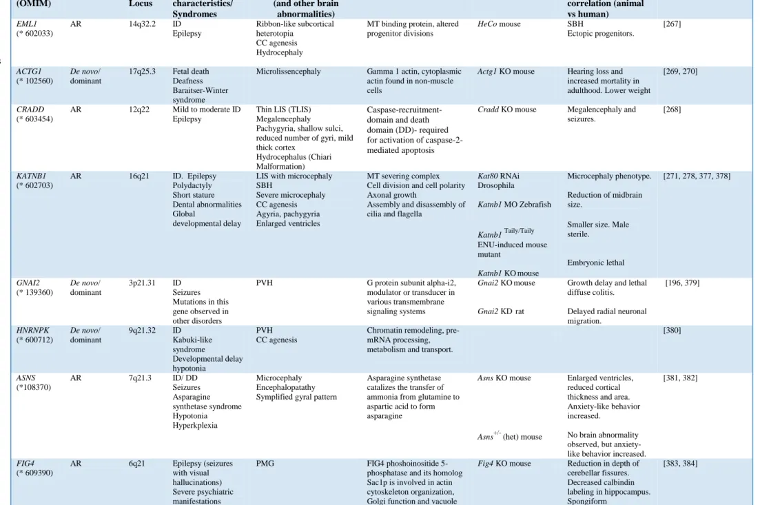
resumed in Fig 2 and corresponding mouse models in Tables 1-6. We mainly focus on single genes

which when mutated have been shown to be involved in neuronal migration abnormalities. Future

more optimal classifications may further involve pathways, linking together multiple mutant genes

[42], and already the transitions between MCDs are being broken down by the identification of gene

mutations spanning multiple linked malformations.

2.2.1. Type 1 lissencephaly spectrum (LIS/SBH) and related disorders

Classical (type 1) lissencephaly (cLIS, see Table 1), refers to a spectrum of related neuronal

migration disorders which include a smooth, thickened and disorganized cortex without gyri (agyria or

‘smooth brain’); simplified and abnormal gyri (pachygyria) and misplaced or ectopic neurons which

form SBH (or double cortex) [42, 46]. Generally, agyria is characterized by the absence of deep sulci

in more than one lobe, and a cortex thickness of 10–20 mm, while in pachygyria, gyri are simplified

and wider than in the normal cortex, and the thickness of the cortex is 4–9 mm [31]. In SBH, a

superficial cortex which may be grossly normal or show aberrant gyration, is separated by a thin layer

of white matter from a band of grey matter.

It has been estimated that 40 to 75% of patients with cLIS including also Miller Dieker

Syndrome (MDS, see below) have heterozygous mutations involving LIS1 (approximately to 60%) or

hemizygous DCX mutations (12%), meaning that many of lissencephaly patients have other mutations.

Of these, rare cases of cLIS are for example, caused by mutations in TUBA1A [47, 48], and also in

DYNC1H1 and KIF2A [49].

Irrespective of the causal gene, patients with cLIS typically demonstrate severe to profound

hypotonia, intractable epilepsy and feeding problems [50]. The overall incidence of lissencephaly is

rare and estimated around 1.2/100,000 births. Clinical presentation of cLIS may vary depending on the

severity. The most severe cases, prenatally detected using ultrasound performed during the second

trimester of the pregnancy, show an absence of primary sulci, confirmed by fetal cerebral MRI

performed from 30 GW. Most patients present with hypotonia or severe neurological distress, or

develop seizures during the first year of life [51].

Imaging studies for patients with cLIS also reveal a smooth surface brain with diminished

white matter and a shallow vertically oriented Sylvian fissure. Remarkably, when the lissencephaly is

not complete, the defects (agyria, or severe forms of pachygyria or SBH) are typically more severe in

either anterior or posterior regions of the brain, e.g. parieto-occipital regions for LIS1-associated, and

fronto-temporal regions for DCX-associated lissencephaly [52]. Thus, the neuroradiological

appearance is often graded using a six-point grading system based on the severity and

anterior-posterior gradient of the abnormalities. Only grade I actually deserves the name of lissencephaly;

grades 2–4 are cases of pachygyria [42] and at the end of the spectrum, grades 5 and 6 apply to SBH.

According to Friocourt and colleagues [53], the first detailed neuropathological description of

lissencephaly was reported in 1956 by Crome who introduced the notion of a “four-layered” cortex.

On gross examination, brain weight is either normal or reduced. When the hemispheres are smooth,

there are poorly defined central and Sylvian fissures. When present, gyri are broad, small in number

and coarse, with a failure or a delayed operculization of the Sylvian fissure. The cortical ribbon is

thicker than in a normal brain (10–20 mm vs. the normal 4 mm) and poorly delineated from the white

matter which is markedly reduced, leading to an inverted proportion between the white matter and the

cortical ribbon [53]. On microscopic examination, the CP exhibits a characteristic four-layered pattern.

The molecular layer I contains Cajal–Retzius cells located close to the pia. Layer II is composed of

densely packed pyramidal neurons which are mainly observed in its upper part, and underlined by an

irregular sheet of granular cells. Layer III consists of scattered fusiform, rounded or multipolar

neuronal elements. Layer IV is particularly poorly delineated from the underlying white matter and

composed of pleiomorphic neuronal cells, with misoriented pyramidal neurons. The reduced white

matter contains multiple arrested post-mitotic neurons. The remaining subependymal cell layer is also

poorly delineated from the deep white matter and sometimes contains PVHs. Moreover, anomalies of

infratentorial structures and cortico-spinal tracts are also frequently observed, but these are typically

less severe than found in cases of lissencephaly with cerebellar hypoplasia [54].

(7)

6

2.2.1.1a LIS1 (Platelet Activating Factor Acetylhydrolase 1b Regulatory Subunit 1,

PAFAH1B1)

The first gene identified to be causative of lissencephaly was LIS1, localized on chromosome

17p13.3. LIS1 belongs to the non-catalytic alpha subunit of the intracellular Ib isoform of

platelet-activating factor acteylhydrolase, which is a heterotrimeric enzyme that specifically catalyzes the

removal of the acetyl group at the SN-2 position of platelet-activating factor (identified as

1-O-alkyl-2-acetyl-sn-glyceryl-3-phosphorylcholine) [55]. LIS1 also associates with microtubules (MTs) [56]

(see below). This gene was found deleted in MDS patients [57], a contiguous gene deletion resulting

from deletions of 17p13.3 including LIS1 [58– 60]. cLIS can also occur as isolated forms due to other

LIS1 mutations (isolated lissencephaly sequence or ILS, isolated SBH). MDS is more severe deleting

LIS1 and adjacent genes, in particular 14-3-3ε (YWHAE, tyrosine 3- monooxygenase/tryptophan

5-monooxygenase activation protein, epsilon) [57]. The 14-3-3ε protein binds to phospho-serine

containing proteins and mediates intracellular signal transduction. Approximately 80 % of individuals

with MDS have a de novo deletion and approximately 20 % have inherited a deletion from one of the

parents who presents a balanced chromosome rearrangement [61]. Heterozygous LIS1 mutations show

an equal frequency in males and females, as expected from an autosomal disorder. Gene dosage is

clearly very important, with these mutations leading to haploinsufficiency [56]. As well as MDS, it has

also been proposed that 60% of the remaining mutations that cause the disorder involve the disruption

of splicing of LIS1 variants [62]. Moreover, it has recently been shown that a recurrent LIS1 intronic

mutation disrupts LIS1 transcript splicing and is responsible for cLIS of variable severity [63]. Other

types of point mutations are observed e.g. [62] including cases of somatic mosaicism [39].

2.2.1.1b

Animal models and functional studies

Mouse models of Lis1 (Pafah1b1) revealed that homozygous knockout (KO) mice died in

embryogenesis soon after implantation suggesting an essential role of this gene in early embryonic

development [64]. Brain abnormalities displayed by heterozygous (+/-) mutant mice mainly disrupt

the hippocampus. However, 5% of Lis1

+/−

mutant mice die from seizures at 3–5 weeks of age. More

severe defects are observed by lowering the amounts of Lis1 further, by crossing with a hypomorphic

allele [64]. In this case, the phenotype involves cortical, hippocampal and olfactory bulb

disorganization resulting from delayed neuronal migration, by a cell-autonomous neuronal pathway.

The Lis1 phenotype is hence dosage-sensitive, and a critical role for Lis1 in neuronal migration

throughout the brain is suggested. The heterozygous phenotype in mice however, appears milder than

the phenotype observed in patients with heterozygous LIS1 mutations.

Another Lis1 mouse model gave rise to a shorter Lis1 protein that initiates from the second

methionine (M63) and elicits a different phenotype. In heterozygote mutant embryos (Lis1/sLis1),

athough the CP does form in occipital regions, the process is slowed or delayed and as a consequence,

the width of the CP is reduced at E14.5 [65]. The authors showed that these Lis1 mutants show no

progenitor abnormalities in either cell cycle length or interkinetic nuclear migration (INM), despite the

fact that the overall organization of RGCs seems affected. In both mouse models (Lis1

+/−

and

Lis1/sLis1), adult brain cortices are normal, but hippocampal abnormalities were observed only in

Lis1

+/−

mice [64,65]. Altogether, dose-dependent disorganization of cortical layers, hippocampus,

cerebellum and olfactory bulb due to cell autonomous neuronal migration defects is observed in mice

with severely decreased levels of Lis1 [64].

(8)

7

In utero electroporation studies performed in rat embryos showed that down regulation of Lis1

in neuronal progenitors induced the accumulation of multipolar progenitor cells within the SVZ of the

neocortex. Moreover, a complete failure in progression from the multipolar to migratory bipolar state

was observed. RGC proliferation failed and INM was abolished [66]. This is hence a severe phenotype

generated by acute inactivation. In other studies, LIS1 was also shown to be important for cell

proliferation during neurogenesis, as well as for neuronal migration and survival [67]. Furthermore,

loss of Lis1 showed perturbed mitotic progression resulting in a prolonged cell cycle that induced

anaphase onset delay in cKO mouse embryonic fibroblasts (MEFs) [68].

At the protein level, LIS1 has been shown to form a homodimer through N terminal

associations [69], while it interacts through its C-terminal β-propeller with cytoplasmic dynein

(DYNC1H1) and its regulatory partner dynactin (DCTN) [70-73]. DYNC1H1 belongs to a group of

MT-activated ATPases that function as molecular motors, being the primary minus-end-directed MT

motor protein. DCTN is involved in dynein-mediated retrograde transport of vesicles and organelles

along MTs by recruiting and tethering dynein to them. Thus, DCTN binds to both dynein and MTs

providing a link between specific cargos, assisting cargo loading and increasing processivity. In vitro

studies in mammalian cells showed that dynein is important for LIS1’s role in cell division [70], and

for INM in mammalian neural progenitor cells. Indeed, dynein regulated by LIS1, is the force

generator for nuclear movement in RGCs [74]. Additionally, experiments performed in migrating

neural cells demonstrated that Lis1 and dynein RNAi inhibit centrosomal and nuclear movement,

whereas myosin II inhibition blocks only nuclear translocation [74].

LIS1 binds other proteins such as NDE1 [75] and its paralogue NDEL1 [76]. NDE1 is a

spindle-associated gene and mutations in humans cause a severe microlissencephaly syndrome that

reflects both morphological and quantitative defects in RGCs [76-79]. Blocking NDE1 self-association

induced defective centrosomal duplication, and this defect was at least partially responsible for spindle

mis-assembly [74]. Pawlisz and col. [80] showed that Nde1 null and Lis1 heterozygous double mutant

mice have an 80% thinner and severely disorganized cortex, presenting reduced numbers of radial

neuronal units, due to the loss of progenitors and a consequence of failures in mitotic spindle function.

A dramatic increase in neuronal differentiation was shown at the onset of corticogenesis which in the

mutant led to overproduction and abnormal development of early-born PP neurons and Cajal–Retzius

cells, at the expense of progenitors.

Ndel1 loss of function, like Lis1 or dynein, impairs neuronal positioning and induces the

uncoupling of the centrosome and nucleus. Shu and col. [81] showed that overexpression of Lis1

partially rescued the positioning defect caused by Ndel1 RNAi but not dynein RNAi, whereas

overexpression of Ndel1 was not able to rescue the phenotype induced by Lis1 down-regulation,

strongly suggesting that Ndel1 interaction with Lis1 is essential for dynein function. Lis1 and dynein

colocalize at mitotic kinetochores, and microinjection of an anti-Lis1 antibody in mammalian cells

was shown to interfere with the attachment of chromosomes to the metaphase plate, leading to

chromosome loss and a delayed mitotic progression [70]. Ndel1 and Nde1 also colocalize with Lis1 at

the MTOC in mitotic embryonic neuroblasts or fibroblasts [69] and were shown to interact directly

with dynein [71,81,83]. Nde1 is hence required for targeting Lis1 to the cytoplasmic dynein complex

to generate persistent motor forces [72], while Ndel1 has been implicated in the process of Lis1

recruitment to dynein, to act as a scaffold protein [81,84]. Lis1 also appears essential for precisely

controlling mitotic spindle orientation via Lis1/Ndel1/dynein-mediated cortical MT capture [70,77].

This can help regulate symmetric versus asymmetric divisions of neural progenitor cells.

LIS1 and NDEL orthologs in Saccharomyces cerevisiae (Pac1 and Ndl1, respectively) were

shown to be important for concentration of dynein at the plus ends of MTs [84,85]. This favors dynein

at the cell membrane, where it positions the nucleus during cell division. Moreover, Lis1 is required

there for the proper distribution of three distinct dynein cargoes such as endosomes, peroxisomes and

nuclei [56,86].

Thus there are multiple roles for LIS1, intensively studied since its identification in 1993.

Heterozygote LIS1 mutations in human reveal likely neuronal migration defects underlying

disorganized cortical layers and gyral abnormalities. Abnormal neuronal migration can also be

observed in mouse mutants. Varying Lis1 amounts in the mouse and in in vitro systems, reveals as

well effects in dividing cells via multiple mechanisms. The human LIS phenotype might be due to a

(9)

8

combination of LIS1 functions in nuclear positioning and organelle transport, as well as in mitotic

spindle function and cell cycle progression. It should be noted that a proportion of LIS1 cases also

exhibit microcephaly, and so it seems likely that certain mutations combined with particular genetic

backgrounds will have a detrimental effect on progenitors, perhaps as well as during migration. It

becomes important to dissect out these possible effects, potentially with the use of temporally and

spatially controlled knockouts.

2.2.1.2a

Doublecortin (DCX)

The DCX gene is located on chromosome Xq22.3–q23, and mutations cause lissencephaly

mostly in males (X-linked LIS or XLIS), whereas DCX mutations in females frequently give rise to

SBH [87,88]. A minority of males have SBH [52], often due to mosaic DCX mutations. The product of

DCX is a cytosolic microtubule-associated protein (MAP) with two doublecortin domains (DC) which

bind MTs and several potential regions for regulation by phosphorylation and protein interaction, such

as proline and serine rich regions [89–91]. DCX nucleates and stabilizes MTs and is highly expressed

in migrating and differentiating neurons, but appears to be little expressed in progenitors. This protein

is potentially the best characterized of the neuronal migration classical MAPs [89,90,92-94].

In XLIS, neuronal migration in the cortex is severely disrupted, leading to the formation of an

abnormal four-layered cortex. In hemizygous DCX-mutated males, the brain surface is agyric or

pachygyric, the anterior areas being more severely affected, with hypoplasia of the frontal lobes

(anterior-posterior gradient, [95]. The corpus callosum is variably affected, described as thick, thin or

absent. The cortical ribbon is thick and poorly delimited from the white matter and generally

recognized as 4-layered. However, with further investigation some DCX brains have been described as

having an ill-defined but six-layered cortex [53]. With a relatively normal layer I, layer II consists of a

thin band containing an admixture of pyramidal and granular neurons. Layer III is poorly delimited

and paucicellular and made up of granule and immature neurons. Within the superficial white matter,

layer IV forms a thick ribbon of scattered, sometimes clustered pyramidal cells and immature neurons.

The extra abnormal layers are adjacent in the white matter and composed of large nests of arrested

migrating neurons, arranged in a radially and columnar pattern [96]. The cerebellum and the brainstem

are macroscopically normal.

Most of missense mutations causing cLIS are located in the evolutionary conserved DC

domains of DCX, suggesting the importance of MT binding for DCX function during brain

development [91,97,98]. Mutations in DCX have been found in families, with females presenting SBH

and males presenting LIS. Also 80-90% of sporadic DCX mutations are found in females, versus 25%

in males with SBH [31,99]. De novo mutations show a higher proportion of nonsense and frameshift

changes, and are likely therefore to lead to a more severely perturbed protein [98]. Indeed, maternal

germline or somatic mosaicism may explain the difference between sporadic SBH and XLIS in males

[39,95,100]. The 10% of SBH patients showing no DCX mutations are likely to have mutations in

other genes such as TUBA1A or occasionally LIS1 [101,102].

2.2.1.2b

Animal models and functional studies

DCX is predominantly expressed in post-mitotic neurons and shows developmental regulation.

In developing neurons Dcx has been shown to have a specific subcellular localization, enriched at the

ends of neuritic and leading processes [89,103]. It has been shown to play a role limiting the number

of leading processes during migration [92,94]. Mutations in MAPs which stabilize MTs, such as Dcx,

Dclk, Tau, Map2 and Map1b, can impair cell polarization, formation of the leading process, neurite

outgrowth and/or lead to excessive branching that hinders migration [16].

Like Lis1 mutants, Dcx KO mutations in the mouse induce no major neocortical malformation

although hippocampal lamination defects are observed [104,105]. Dcx

−/Y

mice show abnormally

laminated CA3 hippocampal pyramidal cells, a dual or dispersed layer of pyramidal cell somata with

potentially aberrant connectivity associated with mossy fibers [106]. Pyramidal cells displayed

morphological abnormalities, as they were smaller with a reduced total dendritic length, and were

more excitable than WT cells. In vitro hyperexcitability and spontaneous epilepsy are characteristic of

this mouse model [107].

(10)

9

Lack of severe neocortical abnormalities suggests a functional redundancy with Dcx-related

genes, involving genetically redundant pathways [93,108]. However, acute down-regulation of Dcx in

the developing rat cortex by RNAi led to disruption of radial migration [109]. In addition, Pramparo

and col. [110] demonstrated by time-lapse video-microscopy in embryonic neocortical slices that Dcx

-/Y

neurons do have defective migration velocities, similar to Lis1

+/-

neurons, as well as multidirectional

movements, abnormal morphology and increased branching [110]. This is similar to previous data

showing the same defects in IN populations [92,94]. Pramparo and col. [110] also detected RGCs with

abnormal spindle orientations in Dcx

-/Y

mutant mice, similar to Lis1

+/-

cells, leading to mild

proliferation defects in vivo and in vitro. Moreover, a genetic interaction in double mutant males

(Lis1

+/-

; Dcx

-/Y

) was shown, resulting in severe defects of proliferation of neuronal precursors,

neurogenesis and neuronal migration.

Dclk (Doublecortin-Like Kinase 1) is a protein kinase that belongs to the DCX family. Gene

mutations cause no obvious migration abnormalities, although mice mutant for both Dcx and Dclk

show perinatal lethality, disorganized neocortical layering and hippocampi [93,108]. Dcx

-/y

; Dclk

-/-mutants have axonal defects in the corpus callosum, anterior commissure, subcortical fiber tracts and

the internal capsule. Lamination defects of the hippocampus observed in these animals were more

severe than previously reported in Dcx KO mice [104] but appeared similar to hippocampal defects

seen in Cdk5 KO mice [108,111]. In Dcx

-/Y

; Dclk

-/-

animals, as in Cdk5 KO mice, hippocampal cells

appeared to be less packed in the dentate gyrus, with a dispersion of neuronal cell bodies. The stratum

pyramidale (SP) in fields CA3, CA2 and CA1 showed multiple, disorganized layers with an irregular

influx of white matter in no consistent pattern. The cingulate cortex was also affected in contrast to

Dcx and Dclk single KO mice, which show no apparent neocortical defects [108]. Double mutants of

Dcx and Dclk2 have also been generated, however in this case the hippocampus is the most affected

structure [112].

Dcx mutations in mice have been shown to lead to defects in IN migration [92,94]. This was

shown both during embryogenesis [92] and in the rostral migratory stream of the adult [94].

Videomicroscopy revealed morphological defects (excessive branching) during migration, expected

due to the loss of a MAP. These defects lead to slowed neuronal migration. The down-regulation of

Dcx in rat embryos also showed slowed IN migration during development [113], also observed with

the down-regulation of Dclk, although branching defects were not observed in Dclk-mutant migrating

INs [113]. Further information about Dcx and branching was obtained by the study of the stromal

cell-derived factor-1 (Sdf11), a modulator of IN migration speed and leading process branching in the

mouse [114]. Sdf1 reduces IN branching frequency while promoting stream migration. The Sdf1

signaling pathway leads to the activation of the calpain protease which cleaves cortactin, reducing

Arp2/3 activity, consolidating the actin network and leading to a reduced branch lifetime. Sdf1

signaling was however also shown to activate Dcx, favorising binding and bundling of MTs,

stabilizing the MT array and reducing neuronal branches [115]. In addition to bundling MTs in

processes, Dcx influences nuclear movement and also promotes MT elongation [89,90,94,97,116].

Transcriptome studies have also been performed in Dcx

-/Y

mice [110,117]. In the

Khalaf-Nazzal and col. study [117], an analysis of the abnormal double layer in the hippocampal CA3 region

was performed, showing that one layer is likely to be due to migration of early born pyramidal neurons

to abnormal superficial regions [117]. This is interesting since although all hippocampal neurons are

deleted for Dcx, clearly they can be distinguished in two different populations, potentially related to

different modes (Dcx dependent/ independent) of migration.

The Dcx mutant phenotypes showing abnormal neuronal migration can be associated with the

required regulation of MTs by Dcx. An extensive clinical study also made the association of patient

mutations with predicted effects on MTs [98]. In a separate study, the analysis of different DCX

patient mutations in vitro showed either loss-of-function or the activation of different cell pathways

including increased autophagy [118]. Transcriptional studies also reveal increased cell stress [117].

Thus, DCX alleles may cause dysfunction by different mechanisms [118]. This may help to explain

variable severities in patients, including the small percentage (∼15%) which also show microcephaly

[98].

(11)

10

The ARX (aristaless-related homeobox) gene encodes a homeodomain transcription factor

which is essential for brain development and patterning [119]. This gene is located in Xp22.3 and

loss-of-function mutations are associated with infantile spasms and X-linked lissencephaly with abnormal

genitalia (XLAG) characterized by an unusual three-layered cortex and agenesis of corpus callosum,

or alternatively X-linked non-syndromic ID.

Patients with XLAG present occipital-predominant lissencephaly, particularly anterior

pachygyria and posterior agyria or a simplified gyral pattern, as well as agenesis of the corpus

callosum, and abnormal basal ganglia [120]. In the most severe form of XLAG, patients show

hydranencephaly with a large occipital cavity. Female carriers of ARX mutations causing XLAG have

a risk of agenesis of the corpus callosum with no cortical defects. In boys, abnormalities of external

genitalia range from a hypoplastic penis or undescended testes to complete female appearance, while

the karyotype is 46 XY. Patients with XLAG show intractable seizures soon after birth, suggesting

major perturbations between excitatory projection neurons and inhibitory INs.

Approximately, 5-10% of X-linked ID cases are associated with ARX mutations [119].

Mutations in non-coding regions close to the ARX gene have also been associated with ID and/or

autism spectrum disorder (ASD) and infantile spasms [121]. Polyalanine expansions account for over

half of all mutations in ARX. Missense and polyalanine expansions hence generate an extremely broad

spectrum of phenotypes, associating malformation and non-syndromic cases, and showing a strong

correlation between genotype and phenotype [120]. De novo mutations can give rise to the previously

mentioned conditions, but in addition, hydraencephaly with abnormal/ambiguous genitalia, Proud

syndrome (agenesis of corpus callosum with abnormal genitalia), West syndrome, X-linked myoclonic

epilepsy with generalized spasticity and developmental delay, Partington syndrome, ASD,

developmental delay with severe feeding problems, intracranial hemorrhage and seizures [122]. ARX

has also been found mutated in atypical cases e.g. a boy presenting infantile spasms, PMG, PVH and

cleft lip/ palate [123]. In particular, X-linked West syndrome, X-linked myoclonic epilepsy with

spasticity and ID, Partington syndrome (ID, ataxia, and dystonia), and non-syndromic forms of ID

have not been reported to present brain imaging abnormalities [124]. Poirier and col. [125] have also

reported a patient presenting eye rolling combined with atypical hypsarrhythmia, severe ID and

non-ictal dyskinetic movements. Thus a huge variety of syndromic and non-syndromic phenotypes are

possible.

Neuropathological reports for XLAG syndrome are rare [54,126–128]. In all reported cases,

the cortical ribbon is of intermediate thickness (less than 10 mm) and the corpus callosum is absent.

Several diencephalic structures are not identified. With histological analysis, XLAG is characterized

by a three-layered cortex with a global decrease in neuron number affecting massively INs. The

molecular layer contains a few Cajal–Retzius cells lying under the pia. Layer II is composed of

numerous densely packed pyramidal neurons. Layer III, whose thickness increases in the posterior

lobes, is irregular in width and less cellular, and contains small-to-medium-sized neurons, with among

them randomly dispersed pyramidal cells of various sizes. In the diencephalon, striatal and thalamic

structures are disorganized. The caudate nuclei are markedly reduced in size and cellularity, and so are

the putamen, the pallidum and the thalami. Most hypothalamic nuclei are not identified and their

absence has been correlated with some clinical hallmarks of the disease.

Moey and col. [129] and Fullston and col. [121] have shown that certain missense variants

early on in the ARX gene induce a premature termination of the protein (amino acid residue 12 or 27)

in patients with early-onset infantile spasms. In these cases they show that a reinitiation of translation

is possible (e.g. at methionine residue 41) producing lower levels of an N-terminal truncated protein.

The authors conclude that low levels of the truncated protein are enough to improve the patients’

phenotypes compared with severe loss-of-function XLAG patients. This is hence another way to

account for certain variable phenotypes. Of note, heterozygous female carriers can show a variety of

psychiatric conditions such as anxiety, depression and schizophrenia jointly with learning disability

[130]. Genomic variations in ARX have also been reported in some unaffected individuals [131].

Nevertheless, in general, different types of ARX mutations lead to severe phenotypes.

A high proportion of GABAergic neocortical INs are generated in the medial ganglionic

eminence (MGE) of the ventral telencephalon and reach the cortex by tangential migration, also

(12)

11

termed non-radial cell migration. They migrate from the MGE to the cortex during embryonic

development, guided by a combination of chemoattractive and repulsive cues [132]. IN dysfunction

and perturbations of their migration have been associated with severe neurological and psychiatric

illnesses, including epilepsy, ASD and schizophrenia [133]. Indeed, “interneuronopathies” is a term

which refers to aberrant migration and differentiation of GABAergic INs which do not reach their

proper destination in the neocortex [134]. This phenomenon also occurs mildly in human type-I cLIS,

as well as relevant mouse models, although this is usually to a lesser extent, compared to XLAG

[16,46,96,134,135]. Particularly, in LIS1 and DCX samples, defective migration was suggested by the

presence of heterotopic calretinin- and calbindin-positive cells in the VZ/ SVZ or IZ. In addition, few

GABAergic INs were detected in the CP. In comparison, an ARX case in this study showed an almost

complete absence of INs in the CP, as well as a paucity of Cajal-Retzius cells in layer I, suggesting

that ARX is important for correct migration of several neuronal types [96]. Mutations in TUBA1A (see

below) also can include GE abnormalities [136]. Thus, IN migration defects of different severities are

frequent occurrences, also associated with MCDs.

2.2.1.3b Animal models and functional studies

Arx is expressed in several areas of the nervous system, and in the floorplate Arx together

with FoxA2, directly induces Sonic hedgehog (Shh) expression through binding to an Shh floor plate

enhancer (SFPE2) [137]. Nkx2.2, induced by Shh, then down-regulates Arx expression, showing

hence a regulatory loop during vertebrate development. These data suggest that Arx plays a role as a

context-dependent transcriptional activator, which may help explain how mutations in this gene are

associated with a wide variety of phenotypes in affected patients. Different types of mutation also

contribute to variability. Missense mutations found in patients in either the N- or C-terminal nuclear

localization signal (NLS) regions lead to aggregates of ARX both in the peri-nuclear region of the

cytoplasm and inside the nucleus [138]. These mutations did not prevent the interaction of ARX with

Importin 13, a mediator of the nuclear import process, however they lead to inadequate accumulation

and incorrect distribution of ARX [138].

Arx mutant mice die perinatally as a consequence of hypoglycemia related to pancreatic

defects [139]. However, neuropathological analyses and study of Arx in the mouse show perturbed

INs, confirming that this gene has an important role in their migration from the subpallium during

brain development [138,140]. Mouse mutants also showed ectopic accumulation of neurons in ventral

proliferative zones close to the ventricles [141], mimicking hence a ventral PVH phenotype. Arx KO

mice also show hypoplasia of the olfactory bulbs, suggesting that IN migration routes are similarly

perturbed in the adult [140].

During development Arx is also expressed in pallial VZ progenitors. Loss of Arx specifically

from these cells (Arx

F/Y

; Emx1

Cre/Cre

mice) resulted in less anxious, less social, hyperactive mice,

resembling mildly affected human ARX patients [142]. In addition, mice presented reduced cortical

thickness, a hypoplastic corpus callosum and anterior commissure, consistent with perturbations in

cortical projection neuron production and connectivity. Mutant mice were shown to have reduced size

of brains and proliferation defects, especially affecting IPs [143]. This had an overall effect on upper

layer neurons. Thus, Arx plays a role regulating pallial progenitors as well as being involved in

subpallial mechanisms.

Conditional loss of Arx from developing INs in the neocortex and hippocampus induces a shift

of these precursors to more ventral locations, which persists in the adult nervous system, leading to

reduced numbers of INs in the cortex at early and late postnatal periods [144]. Conditional

Arx

−/y

;Dlx5/6

CIG

male mice and half of the Arx

-/+

;Dlx5/6

CIG

female mice exhibit different seizure types

in early-life, including those that resemble infantile spasms which evolve throughout development. No

specific anatomical defects were detected in this mouse model, although both male and female mutants

showed a reduction of calbindin INs in the neocortex, as well as a more subtly reduced calretinin INs

[145]. This study also found that 8/25 (32%) human females heterozygous for ARX mutations, had

seizures, neurocognitive deficits, and/or agenesis of corpus callosum, suggesting that disruption of

these IN subpopulations contributes to the pathogenesis of developmental epilepsy in both males and

females.

(13)

12

Reln codes for a large extracellular glycoprotein, a serine protease, abundantly synthesized and

secreted by Cajal-Retzius cells in the MZ, and involved in the positioning of post-mitotic neurons

during migration in the developing cortex [146-148]. Cajal-Retzius cells are also localized in the

hippocampus during embryonic development and disappear soon after birth (1-2 postnatal weeks), as

soon as neuronal migration and layer formation are completed. Reln has multiple functions in the brain

and also plays a role in neuronal morphology and synapse formation, e.g. in hippocampal connections

during forebrain development, promoting the branching of axonal projections from the

entorhinal-hippocampal pathway [149].

Autosomal recessive mutations in Reelin (RELN) were identified in patients with a form of

lissencephaly and cerebellar hypoplasia [150,151]. The association of cortical and cerebellar defects

are also observed in patients with TUBA1A mutations and less commonly in patients presenting

TUBB2B and CDK5 mutations [101,136,152–154]. The rare mutations in RELN or its receptor VLDLR

(Very Low Density Lipoprotein Receptor) lead to frontal predominant mild lissencephaly (diffuse

pachygyria) with severe hippocampal and cerebellar hypoplasia [150,155]. In rare cases, patients with

RELN or VLDLR mutations may show pontocerebellar hypoplasia (PCH), a neurodegenerative

disorder with prenatal onset mainly affecting infratentorial structures, leading to combined

pontocerebellar hypoplasia and atrophy at birth [156,157].

2.2.1.4b Animal models and functional studies

The spontaneous mouse mutant for Reln, reeler, is a well-studied model mimicking the human

phenotype, with a disorganized, inverted lamination pattern in the cortex and hypoplastic cerebellum

featuring a decreased number of Purkinje cells [148]. The reeler phenotype was observed in several

original mouse mutants displaying either a complete loss of transcription of the gene [199] or Reln

protein that lacks a C-terminal portion [64,158]. These mouse strains present neurological phenotypes

including tremors, ataxia, cerebellar hypoplasia and malformation of cellular layers throughout the

brain.

Another more recent mutant of Reln, a truncated variant containing a deletion of the

C-terminal region (CTR), shows different cortical defects with no cerebellar hypoplasia [159]. This

mutant (Reln

CTRdel

), carries a chemically induced splice-site mutation that truncates the Reln protein.

This induces the intracellular accumulation of the mutant protein causing signaling defects.

Interestingly, brains of Reln

CTRdel

mice do not present an inversion of the cortical layers. However,

some cortical neurons (Cux1-positive cells), over-migrate and are observed in the MZ, whereas other

Cux1-positive cells migrate less and forms small ectopic clusters within the deep layers of the mouse

cortex [159]. The differences in this mutant are likely to be related to the different signaling pathways

initiated by Reln, involving either Vldlr or ApoER2. This particular mutation is likely to only affect

binding to Vldlr.

Indeed, Reln binds both cell-surface receptors Vldlr and ApoER2 (apolipoprotein E receptor

type 2) on migrating neurons, and induces clustering of the receptors and activation of the intracellular

adaptor phosphoprotein Disabled-1 (Dab1). This is phosphorylated by Src family kinases (SFK) at

different sites, which initiates a signaling cascade to induce neuronal migration [160], including

Crk/Rap1 signaling affecting cell adhesion, and phosphatidylinositol-3 kinase PI3K/Akt and mTOR

signaling [161,162]. Dab1 is then ubiquitinated leading to proteasome degradation, suggesting that this

sequence of activation and shutoff may be crucial for the execution of multiple steps in neuronal

migration [162]. During radial migration, neurons require Dab1 for glia-independent somal

translocation, but not for glia-guided locomotion. During somal translocation, Dab1 function allows

neuron translocation stabilizing their leading processes in a Rap1-dependent manner [162]. After

binding to Vldlr on the cell surface, Reln has also been shown to be internalized into vesicles. ApoE

has also been shown to reduce the level of Reln-induced intracellular tyrosine phosphorylation of

Dab1 in mouse cortical neurons in vitro [163]. Vldlr and ApoER2 double mouse mutants have a

reeler-like phenotype, involving inversed cortical lamination, hippocampal and cerebellar defects. Hence

disruption of both pathways is required to lead to this severe cortical phenotype [159]. Dab1 mutants

(one of which is known as scrambler), also have similar defects.

Reln canonical pathways, involving the activation of the ApoER2/Vldlr/Dab1 signaling,

appear to mediate many Reln-dependent functions during pre- and postnatal brain development and

(14)

13

function [160]. However, SFKs that are central in Reln signal transduction allow the activation of two

distinct branches: PI3K-Akt- and MEK-Erk1/2- dependent signaling cascades, which converge on

downstream effectors, as was reported in cortical neurons in vitro [161]. A non-canonical pathway is

hence known, involving the induction of Erk1/2, p90RSK, and immediate early genes and was

suggested to mediate at least some postnatal functions of Reln signaling [161] which may also be

related to neuropsychiatric disorders.

Reln also binds to the extracellular domain of ephrin Bs, which interact at the membrane with

Vldlr and ApoER2 in neurons [164]. Clustering of ephrin Bs leads to the recruitment and

phosphorylation of Dab1, which is necessary for Reelin signaling. Compound mouse mutants (Reln

+/−

;

Efnb3

−/−

or Reln

+/−

; Efnb2

−/−

) and triple ephrin B1, B2, B3 KOs show neuronal migration defects that

recapitulate the ones observed in the neocortex, hippocampus and cerebellum of the reeler mouse

[165]. Loss of function of ephrin Bs severely impairs Reelin-induced Dab1 phosphorylation. Dab1

activates additionally other signaling pathways, eg involving PI3K. Interestingly, the activation of

ephrin Bs can rescue the phenotype of neuronal migration defects in reeler slices, allowing neurons to

reach layers II-III [165]. Ephrin B induces the formation of a macromolecular complex required for

Src recruitment/activation and Reln signaling. Sentürk and col. [165] concluded that ephrin Bs are

essential components of the Reln receptor/signaling pathway controlling neuronal migration during

nervous system development.

Interestingly, He and col. [166] demonstrated that during cortical development, sister

excitatory neurons progressively and selectively develop gap junctions with each other while they

migrate. This accurate electrical synapse formation depends on inside-out radial migration. Indeed,

inactivation of Reln or knockdown of Dab1 disrupted clonal inside-out neuronal migration and

impaired electrical synapse formation between sister neurons. Moreover, elevated levels of

EfnA/EphA-mediated signaling, which was shown to laterally disperse clonally related excitatory

neurons, also prevented electrical coupling. Mutations in the Reln pathway are hence key for the

elucidation of such mechanisms.

The Rho family of small GTPases, RhoA, Rac and Cdc42, are significant contributors for

orchestrating neuronal development including many cellular processes, such as cytoskeleton stability

during neurite outgrowth and branching, cell polarity and vesicle trafficking [167-169]. Leemhuis and

col. [169] showed that Reln-treated neurons have increased growth cone motility and these are

enlarged with abundant filopodia. This stimulated motility occurred via ApoeR2, activating Cdc42

through the PI3K cascade, which resulted in axonal filopodia formation [169]. More recently, Clasp2

(cytoplasmic linker associated proteins), an MT plus-end tracking protein that specifically accumulates

in the growth cone, was also shown to be a key cytoskeletal effector downstream of the Reln

activation pathway, orchestrating cytoskeletal dynamics during neurite extension and motility during

brain development [170].

Reelin activation of the PI3K/Akt/mTOR pathway may not normally play a crucial role in the

early events leading to layer formation and corticogenesis. However, the activation of a specific AKT

isoform, AKT3, in focal MCDs was shown to be responsible for local, non-cell autonomous cortical

migration defects, leading to drug-resistant epilepsy [171]. In this study it was shown that restricted

cells exhibiting postzygotic brain-specific somatic mosaic AKT3 mutations showed activation of the

PI3K-AKT-mTOR pathway. Importantly, mutation-expressing neural progenitors in the mouse brain

showed misexpression of Reln, due to a derepression of reelin transcription, involving Foxg1. These

mechanisms are hence involved in perturbed migration of restricted mutated and surrounding cells,

leading to this focal malformation. De novo heterozygous mutations in FOXG1 can also lead to

cortical malformations associated with Rett-like symptoms, postnatal microcephaly and abnormalities

of the corpus callosum [172].

Reln is clearly a potent, multi-functional protein with different receptors and signaling

pathways, expressed in several cell types and even in the adult brain. Signaling pathways involving

this protein revealed mutations in patients for several components, representing one of the first

examples of an assembled biochemical ‘pathway’ for MCDs, identified via ‘phenocopy’ mouse

mutants and biochemical studies [146,148,163]. Putting together pathways is now being made easier

with the use of NGS technologies in patients with similar disorders.

Figure

Fig4 KO mouse  Reduction in depth of  cerebellar fissures.

Références

Documents relatifs

[r]

Technical advances, which mostly consist of stereotactic procedures, have already led to practical modifications of the surgical management of malformations of

L’archive ouverte pluridisciplinaire HAL, est destinée au dépôt et à la diffusion de documents scientifiques de niveau recherche, publiés ou non, émanant des

L’archive ouverte pluridisciplinaire HAL, est destinée au dépôt et à la diffusion de documents scientifiques de niveau recherche, publiés ou non, émanant des

The mutations in KIF5C, KIF2A and DYNC1H1 drastically affect ATP hydrolysis, productive protein folding or microtubule binding, while suppression of Tubg1 expression in

While the previ- ous work focused on the stellar halo, which has revealed interac- tion history with a smaller companion that left streams and loops around NGC 891, in the following

Incidence angle dependence of the reflected and transmitted amplitude ratios for an incident SV wave of frequency 10 Hz in water-saturated sand impinging on an interface with

progenitor cell diversity found in gyrencephalic species (ferret, primate). My PhD work has been focused on studying a mouse model for a severe human cortical malformation. It is thus