Computational Processing and Modeling of Intravascular Images
Precisely Couple Arterial Morphology and Biomechanics
by
Max Louis Olender M.S.E. Biomedical Engineering
University of Michigan, 2016 B.S.E. Mechanical Engineering
University of Michigan, 2015
SUBMITTED TO THE DEPARTMENT OF MECHANICAL ENGINEERING IN PARTIAL FULFILLMENT OF THE REQUIREMENTS FOR THE DEGREE OF
DOCTOR OF PHILOSOPHY AT THE
MASSACHUSETTS INSTITUTE OF TECHNOLOGY FEBRUARY 2021
© 2021 Massachusetts Institute of Technology. All rights reserved.
Signature of Author: ………. Department of Mechanical Engineering January 15, 2021 Certified by……… Elazer R. Edelman Edward J. Poitras Professor in Medical Engineering and Science Thesis Supervisor Certified by……… Peter T. C. So Professor of Mechanical and Biological Engineering Thesis Committee Chair Accepted by………... Nicolas G. Hadjiconstantinou Professor of Mechanical Engineering Chairman, Committee on Graduate Students
Computational Processing and Modeling of Intravascular Images
Precisely Couple Arterial Morphology and Biomechanics
by
Max Louis Olender
Submitted to the Department of Mechanical Engineering on January 15, 2021 in Partial Fulfillment of the Requirements for the Degree of Doctor of Philosophy in
Mechanical Engineering ABSTRACT
Cardiovascular diseases, and coronary artery disease in particular, remain a persistent devastating and prevalent menace to health and wellbeing globally despite great strides in vascular biology and medicine. While biomechanical forces are known to play a driving role in the natural history of atherosclerosis, the nuanced yet profound impact of patient- and lesion-specific biomechanics in disease presentation, course, and treatment are not fully appreciated or accounted for in clinical practice. The incredible strides in melding image processing with artificial intelligence, computational modeling, and numerical methods is increasingly filling gaps in knowledge, especially at the intersection of pathological anatomy and biomechanical structural behavior. We derived geometric and morphological structure, as well as constitutive material properties, from invasive intravascular image sequences to quantitatively assess and characterize the state of atherosclerotic arteries. Overcoming the challenge of limited penetration depth in the presence of signal-attenuating plaque, contextual information and spatial continuity was leveraged by a novel surface fitting method to fully delineate the mural conformation of the diseased vessel wall. Neural networks enriched with domain knowledge of vascular geometry and imaging classified pathological regions of interest within heterogeneous lesions. Construction of in silico computational models and in vitro phantom models facilitated the execution and validation of inverse methods to determine material constitutive mechanical properties non-destructively and in clinically amenable fashion. Strategic simplifying assumptions freed the approach from data acquisition limitations which inhibited previous methods of in situ mechanical characterization. Finally, to bridge the chasm between virtual and physical medicine and facilitate integration of these new capabilities into clinical practice, synthetic images were generated by an adversarial network trained in the familiar visual vernacular of vascular imaging.
Through the insights described in this thesis, greater information can be extracted, augmented, and made accessible from clinically-available imaging data. Approaches to more quantitatively and reliably assess, model, and convey biomechanical disease states may offer mechanistic insight into disease development, progression, and treatment response, ultimately leading to improved personalized patient care in an emerging era of computational cardiology.
Thesis Supervisor: Elazer R. Edelman, M.D., Ph.D.
Title: Director, Institute for Medical Engineering and Science & Edward J. Poitras Professor in Medical Engineering and Science
Acknowledgments
The work of this dissertation, and the range of experiences which permeate and extend beyond its pages, have been made possible and enriched beyond measure by the involvement of countless individuals. To each, not limited to those named here, I am eternally grateful.
I could not have been more fortunate than to have my advisor, Professor Elazer Edelman, to guide and join me through the journey of my PhD studies. His genuine and sincere care for all under his tutelage pervades every interaction and supersedes all professional considerations. I have benefited greatly not only from his magnanimity, but also his invaluable technical insight, advice, unrelenting support, and positive encouragement.
I am also incredibly grateful for the support and contributions of my thesis committee, comprised of a remarkable and inspiring collection of individuals. Professor Peter So, my committee chair, has asked insightful, challenging, and illuminating questions at each step, from my qualifying examination through the formalization and completion of my dissertation research. Professor Ellen Roche’s practical and pragmatic perspective, informed by an incredible constellation of educational and professional experiences, shone through in her creative and innovative recommendations that elevated the quality of my work. Professor Guillermo (Gary) Tearney’s expertise as a founding figure in intravascular imaging could not have been more relevant, and his deep familiarity with the field, historical insight, and strong vision as an active researcher were invaluable in determining the direction of my own research. His singular focus on translation and patient impact also helped to shape my perspective and direction. Finally, Professor Dimitrios Fotiadis, who graciously volunteered to serve as an external reviewer and examiner of my thesis, offered expertise and insight as a leader in the fields of biomedical and health informatics, co-author, colleague, and mentor that has made me a better researcher.
I have been indelibly impacted by the lasting friendships I have been privileged to form with those whose time in the Edelman Lab has intersected with my own. An account of even a fraction of these could match the length of this voluminous dissertation. Their dedication, commitment, and initiative was inspiring and motivating, but more importantly their companionship within and beyond the lab brought me immeasurable happiness. Fellow graduate students, both long-time comrades and visiting, have filled my mind with new perspectives and fond memories, and shared in the daily struggles, triumphs, and grind of research. From simple coffee and lunch breaks, to concerts and camping trips, the slog of graduate school was constantly studded with the joys of uplifting interactions.
There are a few individuals from the Lab who made enormous contributions to my development as a researcher and to this dissertation. Drs. Farhad Rikhtegar Nezami and Lambros Athanasiou, who initially convinced me to join the research group (for which I am indebted in perpetuity), continued to be central figures throughout my graduate experience. I feel incredibly fortunate to have had the opportunity to work and grown alongside both of them on a nearly daily basis, and to have benefited so much from their personal investment in my success. As technical experts and personal mentors, but foremost as trusted friends, they have guided me through and indelibly shaped my past 4+ years. Drs. José (Chema) de la Torre Hernández and Eyal Ben-Assa contributed
similarly in the most formative, early years of my graduate studies. Offering not just their expert medical expertise, clinical perspective, and patient tutoring, but also their warm, open-armed friendship, I am grateful for these deep relationships that have persisted over time and across oceans.
Beyond the laboratory, several communities have added dimension to my experience. My cohorts in the Mechanical Engineering, Health Sciences and Technology, and Graduate Education in Medical Sciences programs, as well as supporting administrators and faculty, have provided an accommodating and supportive academic home. The MIT Science Policy Initiative, Outing Club, MakerWorkshop, GradHillel, and Graduate Student Leadership Institute communities have offered opportunities and outlets to learn, grow, and find fulfillment.
Above all, though, I thank my family. Even when I lacked the patience to explain the details of my research, they were always convinced that my work was important and valuable, and offered their unwavering support. With absolute confidence and interest, they have listened with patience and offered encouragement, sharing in my successes and disappointments throughout my graduate studies, just as they have for all endeavors throughout my life.
Finally, I’d also like to acknowledge that I’ve been fortunate to receive generous support from various funding sources that have enabled me to pursue this work and enriching experiences throughout my graduate studies. I am grateful to have been supported by the MathWorks Engineering Fellowship, William Asbjornsen Albert Memorial Fellowship, and Whitaker Health Sciences Fund Fellowship, and to have received funding from the U.S. National Institutes of Health through a research grant to Dr. Edelman.
Table of Contents
Acknowledgments ... 3
Table of Contents ... 5
Introduction: Biomechanical Modeling & Assessment of Diseased Coronary Arteries ... 7
1.1. Disease of the Cardiovascular System ... 8
1.2. Imaging of Diseased Vessels: A Pillar of Clinical Practice... 11
1.3. Arterial Modeling & Simulation: Promise & Challenges ... 14
1.4. Artificial Intelligence in Cardiology ... 19
1.5. Thesis Contributions & Organization ... 21
Extracting Vessel Geometry: Overcoming Current Limitations of Intravascular Imaging 25 2.1. Diseased Vessel Geometry: Needs, Progress, & Challenges ... 26
2.2. Detecting Visible Segments of the Outer Border ... 36
2.3. Identifying the Complete Border from Visible Segments ... 44
2.4. Extracting or Tracking Geometry from Disparate Datasets ... 91
2.5. Implications & Future Directions ... 97
2.6. Conclusion ... 101
Determining & Characterizing Vessel Morphology ... 103
3.1. Diseased Vessel Morphology: Needs, Progress, & Challenges ... 104
3.2. A Flexible Approach for Intravascular Image Characterization: Fundamental Methodological Framework ... 112
3.3. Characterizing Tissue in Grayscale IVUS ... 119
3.4. Characterizing Tissue in Intravascular OCT ... 138
3.5. Segmenting Indwelling Devices in Intravascular OCT ... 144
3.6. Variations on the Framework ... 154
3.7. Importance of Domain Enrichment... 167
3.8. Importance of Training Data ... 173
3.9. Implications & Future Directions ... 183
Computational Modeling & Simulation of Patient-Specific Vessels ... 189
4.1. Diseased Vessel Modeling: Needs, Progress, & Challenges ... 190
4.2. Constructing Detailed Image-Based Volumetric Models ... 199
4.3. Determining Patient-Specific Constitutive Parameters ... 206
4.4. Implications & Future Directions ... 212
4.5. Conclusion ... 215
Bridging the Chasm between Virtual & Physical Medicine with Synthetic Imaging ... 217
5.1. Synthetic Medical Imaging: Needs, Progress, & Challenges ... 218
5.2. Synthetic Image Generation ... 225
5.3. Applications in Cardiology ... 254
5.4. Implications & Future Directions ... 265
5.5. Conclusion ... 267
Conclusion: Biomechanical Assessment & Modeling of Diseased Coronary Arteries ... 269
6.1. Summary of Work & Contributions ... 270
6.2. A Vision and Roadmap for the Future of Cardiovascular Medicine ... 273
6.3. In Closing ... 278
Chapter 1
Introduction: Biomechanical Modeling & Assessment of
Diseased Coronary Arteries
“The heart of living things is the foundation of their life, the sovereign
of everything within them, the sun of their microcosm, that upon which
all growth depends, from which all power proceeds.”
1– William Harvey (1628)
Cardiovascular disease (CVD) is a monumental burden to society, accounting for over 30% of deaths globally [1]. In particular, atherosclerosis and coronary artery disease (CAD), characterized by plaque buildup in and hardening of artery walls [2], presents a significant and ubiquitous concern. Although tremendous progress has been made in combating and diminishing mortality of the disease [2], many questions regarding the mechanical progression of disease and patient response to intervention remain unanswered; mathematical and computational modeling are key to answering these biomechanical questions [3]. This thesis therefore melds novel quantitative analysis tools and models representing diseased coronary arteries to assess and characterize the state of atherosclerotic vessels, contributing insight at the intersection of pathological anatomy, imaging, and biomechanical structural behavior to enhance the diagnosis, prognosis, and treatment of CAD. In this chapter, major themes of this work, and scientific fundamentals of the fields it advances, are introduced, providing an overview of the disease, vascular imaging, and arterial modeling.
1 “Cor animalium, fundamentum eſt vitæ, princeps omnium, Microcoſmi Sol, à quo omnis vegetatio dependet, vigor
omnis & robur emanat.”
AI ... Artificial Intelligence CAD .... Coronary Artery Disease CHD .... Coronary Heart Disease CT ... Computed Tomography CVD .... Cardiovascular Disease CFD .... Computational Fluid
Dynamics
ESS ... Endothelial Shear Stress FEA... Finite Element Analysis FSI ... Fluid-Structure/Solid
Interaction
IVUS ... Intravascular Ultrasound ML ... Machine Learning OFDI... Optical Frequency
Domain Imaging
OCT .... Optical Coherence
Tomography
PCS ... Peak Cap Stress
PSS ... Plaque Structural Stress RF ... Radiofrequency
TCFA .. Thin-Cap Fibroatheroma WSS .... Wall Shear Stress 3D ... Three-Dimensional
1.1. Disease of the Cardiovascular System
The cardiovascular, or circulatory, system is the intricate and exquisitely complex network by which the body nourishes the immense mass of cells which constitute the living body [4]. The constituent vessels serve as a transportation system, delivering oxygen and nutrients while removing carbon dioxide and other waste products, as well as immune cells and antibodies, signaling factors, and other essential circulating molecules. Far from a passive system of pipes, the circulatory system is remarkably dynamic, responding to mechanical and chemical stimuli in order to actively regulate blood pressure, shunt blood to meet physical demands, maintain body temperature, and preserve core functions as needed, and even modulate vessel wall permeability to control transmural exchange [5], [6]. Circulating blood through this system—from prenatal development to death without respite—is the heart [4]. Like the vessels through which it drives blood, the heart is no monotonous pump, and in consort with the vessels, the heart controls the rate and volume of blood flow in response to the demands of the body by actively varying the strength and frequency of its beating [5], [6]. They dynamism, range, and responsiveness of this system, central to the sustenance of human life, is truly profound.
1.1.1. Impact & Pathology of Heart Disease
Given the complexity and importance of the system, it is no surprise that CVD, disease of the cardiovascular system, is both prevalent and dangerous. Indeed, CVD is the leading cause of death and responsible for 50% of all noncommunicable disease deaths—over 30% of all mortality worldwide [1]. Of those deaths, at least 57% can be conservatively and directly attributed to atherosclerosis in the form of coronary heart disease (CHD; 42.6%) or ischemic stroke (14.8%). In more concrete terms, CHD causes around 1 in every 7 deaths in the United States—an American has a heart attack (myocardial infarction) every 40 seconds [1]. Beyond this startling statistic, prevalence of subclinical atherosclerosis belies the true extent and impact of this pervasive menace [1], which is further compounded by the toll in diminished quality of life, financial burden, and emotional strain.
Atherosclerosis2, named for the characteristic pathological presentation of arterial
hardening, is a widespread chronic disease which can suddenly escalate in a devastating acute
2 The terms “atherosclerosis” and “arteriosclerosis,” of similar etymology, are sometimes used interchangeably in
health event. The development of atherosclerosis systemically affects arteries, the blood vessels which carry oxygenated blood away from the heart. These vessels, like nearly every tubular structure in the body, are comprised of three layers. The innermost layer, the (tunica) intima, is comprised of endothelial cells which lines the blood-filled lumen; it acts as an active barrier and serves important signaling functions. The (tunica) media, separated from the intima by the membranous internal elastic lamina, is comprised of smooth muscle and extracellular matrix, components which allow it to control vasomotion (and thereby regulate blood pressure and flow) and store potential energy during oscillatory periods of high pressure. Finally, the (tunica) adventitia or externa, separated from the media by the membranous external elastic lamina, supports the vessel with nerves, lymphatics, and blood vessels [5]–[7]. While the entire vessel structure can be modified and disrupted in atherosclerosis, the disease is considered “a disease of the intima” [8]. It is in this layer that endothelial dysfunction leads down a cascade of fatty deposition, inflammation, and dysmorphic remodeling, driven in large part by mechanical forces [5], [7], [9]. Composition of the resulting diseased intima can include calcifications, fatty lipid deposits or necrotic cores, fibrotic tissue, or numerous variations thereof. These depositions of so-called “plaque” result in a highly variable and heterogeneous mechanical structure.
The consequences of plaque deposition and accumulation are variable, and evolve with the plaque morphology—composition, structure, and form—distribution, and ever-changing milieu— the microenvironment. While sub-clinical atherosclerosis is present to some extent in nearly every adult, the disease presents clinically when blood flow is inhibited and oxygen demand of downstream tissue exceeds the supply delivered by the blood, a state known as ischemia. This may be caused either by the protrusion of intima-bound plaque into the luminal space or rupture or erosion of the cap separating highly-thrombogenic plaque from the bloodstream, resulting in immediate clotting and luminal blockage [5]–[7]. Blockages starve distal (downstream) tissue of necessary oxygen; if flow of oxygen is not restored quickly, deprived tissue begins to undergo non-reversible injury and subsequently dies. When a blockage is located in the carotid artery or one of its distal branches, which supply blood to the brain, such a catastrophic event results in an ischemic stroke. When atherosclerosis is localized in the coronary artery, which supplies blood to
infiltration. This thesis primarily focuses on the former, of which intramural lipid accumulation is a hallmark, though is generally applicable in the latter pathology as well.
the heart itself, the disease is classified as coronary artery disease (CAD)3, and consequences of
ischemia range from angina (ischemic chest pain) to deadly myocardial infarction (heart attack) [5], [6]. Atherosclerotic plaque can form elsewhere in the arterial vasculature (such as the renal, mesenteric, and peripheral arteries). These lesions can be devastating in their own right, though the consequences are typically less acute and severe as those caused by plaques in vessels supplying the brain and heart due to the indispensability and high oxygen requirements of their never-ceasing functions.
1.1.2. Diagnosis & Treatment of Atherosclerosis
The ischemic symptoms of atherosclerosis, and to some degree their cause, have been recognized for centuries. In the early 1500’s, Leonardo da Vinci performed an autopsy and concluded that the cause of death was a “lack of sustenance…brought about by the continuous narrowing of the passage of the vessels…by thickening of the coats of these vessels” [10]. While da Vinci erred in some critical details—he, like his contemporaries, did not even grasp the circulatory nature of blood flow—his autopsy observations and hypotheses are forebears to modern understanding of disease that evolved over the following half-millennium [10]. By the late 1700’s and early 1800’s, medical experts were beginning to describe a dangerous disease that manifested as chest pain, particularly during physical exertion, which often concluded with the patient’s death [11], [12]. However, no treatment was available; one doctor remarked at this time, “With respect to the treatment of this complaint, I have little or nothing to advance,” recommending, quiet, warmth, alcohol consumption, and opium [12]. Even into the 1900’s, while understanding was much improved, it was incomplete. Regarding “marked sclerosis of the arteries,” one prominent doctor dismissively noted that “it is important not to over-estimate their seriousness. The outlook may depend much less upon the existence of these factors than on the sort of man in control of them,” likening diseased arteries to a ship and the patient to a captain [13]. True diagnosis could be made only at postmortem autopsy, and prognosis was also unavailable; the same doctor noted that there was “no method by which we can reasonably tell whether a sufferer will be cut off the next minute or survive many years.” While the importance
3 The terms “coronary artery disease” (CAD) and “coronary heart disease” (CHD) are often used interchangeably.
Technically, however, CHD is the result of CAD, and occurs when disease of the blood vessels servicing the heart (coronary arteries) progresses to cause disease of the heart. No strict distinction is made between the two in this thesis.
of lifestyle changes were recognized, with doctors admonishing to “Go slowly” and “Eat less,” medical treatment was limited and clinicians sometimes resorted to palliative administration of chloroform [13]. The disease was still described at this time as “meditatio mortis,” translated as “contemplation of death” [13].
Comprehension and treatment of CAD has advanced rapidly in the age of modern medicine: facilitated by improving mechanistic understanding of the disease, overall mortality has dropped around five-fold in the past five decades, with in-hospital mortality declining even more so [1], [2]. Medical innovations propelling this trend, starting around the 1950’s, have included interventional devices (such as stents, scaffolds, and balloons), pharmaceutical agents and delivery methods, increasingly effective and decreasingly invasive (e.g. percutaneous) surgical procedures, and large coordinated risk-identification and education campaigns. Together, these advances have tapered the rising incidence and increased survivability of the disease.
1.2. Imaging of Diseased Vessels: A Pillar of Clinical Practice
Imaging has played an important direct and supporting role in lowering CVD mortality and growing knowledge of vascular physiology and pathology [2], [14]. While autopsy and histology studies helped to determine the cause of acute myocardial infarction, measure and quantify population distributions of normal and pathological vessel features, and classify plaque types, such approaches cannot, by their nature, be used to diagnose, assess, or guide treatment of current patients, or to monitor disease progression and response to treatment. For these purposes, in vivo imaging is required. In vivo imaging plays a role in all stages of disease diagnosis, prognosis, treatment, and monitoring [15], [16]. It is widely used to determine vessel geometry; such imaging can visualize macroscopic vessel and blood distribution patterns, identify and monitor stenoses (aberrant narrowings of blood vessels), measure vessel eccentricity, determine extent and direction of remodeling, and quantify plaque prevalence (i.e. plaque burden) [17]–[19]. Beyond geometry, plaque morphology can be assessed by determining plaque composition [18], [19] and biological features such as areas of inflammation can be identified and located [20]. The constellation of data and insights available from the imaging is beneficially used to identify plaques demonstrating high-risk features [14], [17], [21]–[24] and to guide, optimize, and assess outcomes of intervention (angioplasty and stent deployment), monitor patient reaction to, and recovery from, these interventions, and identify instances of subsequent device (e.g. stent or scaffold) failure [25]–[30].
An expanding arsenal of in vivo imaging modalities has been developed to service these clinical and research needs [31]. Noninvasive imaging techniques include multidetector computed tomography (CT), magnetic resonance imaging, nuclear imaging (including single-photon emission computed tomography and positron emission tomography), and contrast-enhanced ultrasound imaging. Invasive imaging techniques for arteries include angiography, intravascular ultrasound (IVUS), IVUS-radiofrequency (RF) analysis, palpography (intravascular elastography), optical coherence tomography (OCT), near-infrared spectroscopy, intravascular magnetic resonance imaging, angioscopy, and thermography [21]. Each imaging modality offers different benefits and strengths at different scales, and suffers from distinct shortcomings (Figure 1.1) [21], [26], [32]. For this reason, hybrid and multimodal technologies (combining modalities) are being explored to take advantage of distinct strengths and negate shortcomings [21], [26], [32]; such technologies, however, are not yet widely available. In fact, none of the non-invasive modalities are robustly validated and widely used, while among the invasive modalities only angiography, IVUS(-RF), and OCT are widely used in clinical routine; others are used only in limited and research contexts [21].
Intravascular OCT is a relatively new modality which offers exciting opportunities for clinical use and fresh challenges. Developed by researchers at MIT and Harvard Medical School [33]–[36], OCT is an invasive, catheter-based technique that utilizes near infrared light to construct cross-sectional slices of the artery and its internal (micro)structure [37]. The use of light instead of mechanical ultrasonic waves—used in the more established IVUS [19]—leads to remarkable resolution, particularly in frequency domain OCT/optical frequency domain imaging (OFDI), which is superior to the displaced time domain OCT [38]. Indeed, OCT resolution is typically 10-20 μm in the axial direction and 10-20-40 μm in the lateral direction [18], [39], [40], compared to 100-200 μm axial resolution and 200-300 μm lateral resolution in IVUS [39]. This difference in resolution is critical, as it uniquely allows in vivo measurement of particularly small features and identification of life-threatening thin-cap fibroatheromas (TCFAs), which are characteristically less than 65 μm in thickness [41], [42]. Furthermore, the light signal can better penetrate calcified tissue, which is opaque to IVUS’ ultrasound signal and consequently obscures abluminal tissue (Figure 1.1C).
However, OCT has failed to overtake IVUS in global popularity due to several of its own shortcomings and challenges. Most importantly, the use of light instead of mechanical waves, while improving resolution, results in substantially lower penetration depth (0.1-2.5 mm in OCT, compared to 10 mm in IVUS [18], [39]). Though better able penetrate calcified tissue, light is
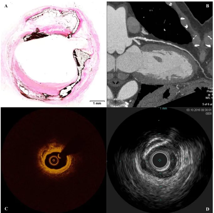
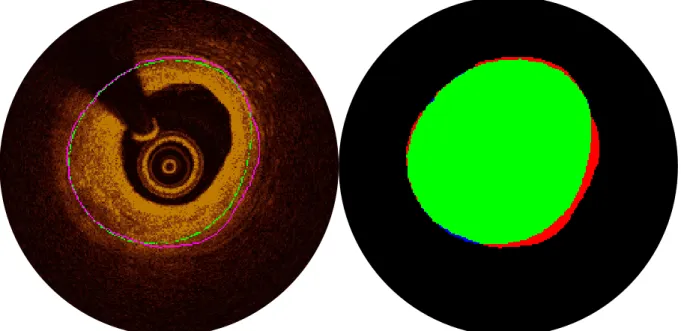
Figure 1.1. Various imaging modalities are associated with distinct benefits and limitations. The cross sectional
profile of diseased coronary arteries can be visualized through (A) histology (von Kossa stain), (B) CT, (C) OCT/OFDI, and (D) IVUS. Each of the above illustrates a highly calcified lesion in a coronary artery at different scales and resolutions. While CT visualizes the cross-section of a large region, providing context and spatial information, and readily highlights calcified tissue, resolution is low and distinction of soft tissues is poor. OCT and IVUS provide highly detailed information with moderate tissue differentiation in smaller regions; the former has greater resolution but inferior penetration depth relative to the latter. Histology, with its arrays of specialized stains, provides the greatest resolution and tissue differentiation, but at sparse, discrete locations, and requires that the tissue be excised from the body—a critical restriction. (Histology image provided by CBSET.)
D C
B
quickly attenuated by fatty tissue and lipid-rich lesions, obscuring abluminal tissue. The light signal also cannot penetrate the metal guidewire used to place the intravascular imaging catheters, resulting in a substantial reflection and shadow artifact (Figure 1.1B). Other artifacts sometimes also occur [43]. Furthermore, blood is a highly-attenuating medium for light, and so must be cleared from the field of view within the vessel. While older, slower forms of OCT (time domain) required proximal balloon occlusion, modern versions use non-occlusive flush of an optically transparent media (e.g. Lactated Ringer’s or crystalloid/radiocontrast solution) alone; contrast may be administered through the guide catheter either by manual injection using a syringe or automatically with a power injector [18], [44]. While shown to be safe in typical quantities for those without contraindicative complications (e.g. renal insufficiency) [45], [46], this flush imposes additional challenges. Insufficient flush can result in obscured vessel wall tissue and artifacts, which significantly deteriorates image quality [18], [39]. Repeated acquisitions may be precluded due to the concomitant safety risks of additional media [47]. And flush administration can also impact the observed and measured geometry of the vessel by altering luminal pressure and inducing hypervolemic conditions [44], [48], [49].
Despite these shortcomings, OCT has been shown repeatedly to be not just reliable and safe for clinical use [45], [46], [50]–[54], but has demonstrated the capacity to offer insights and detailed information not offered by IVUS or other imaging modalities [50], [55]. OCT is regularly used in catheterization labs around the world to diagnose and prognosticate CAD [18], assess lesions and characterize plaque with excellent reproducibility [40], [56]–[58], guide coronary interventions such as stent deployment [45], [59], [60], and assess stents after deployment [37], [56], [61]. Ongoing work continues to demonstrate and improve the clinical utility of the modality through validation studies [62], [63], updated signal analysis and processing [31], [64]–[66], hardware modifications, and introduction of exogenous agents and fluorescence [66]. For a more extensive expert review of the intravascular applications of OCT, with comparisons to IVUS, see Prati et al. [37], [39].
1.3. Arterial Modeling & Simulation: Promise & Challenges
While imaging has led to greater understanding of basic pathology development and improved treatment in the clinic, it has also empowered researchers with the ability to develop patient-specific computational models to glean further insights. One vision for a bourgeoning
paradigm in computational cardiology describes the mutual supplementation of imaging, modeling, and informatics in advancing understanding of atherosclerosis, novel treatment, and decision support systems [67]; this trend has already commenced. By generating virtual representations of physical biological systems—informed by imaging and patient data and adhering to fundamental governing principles—the system can be non-invasively interrogated in complex, comprehensive, and systematic ways that would be dangerous, expensive, or impossible in a patient.
In pursuit of the three ultimate goals—fundamental disease insight, novel and improved treatment options, and individualized patient care guidance—in silico (i.e. computer-based) models of three primary classes have become popular [68].The first category of such models addresses fluid dynamics, primarily using computational fluid dynamics (CFD) [69], [70]; by simulating blood flow through a model vessel, researchers can approximate spatiotemporal pressure, speed, exerted forces (i.e. shear stress, including wall or endothelial shear stress), and gradient patterns in the vessel. The second category considers solid mechanics, primarily through finite element analysis (FEA) [71], [72]. By simulating vessels with applied blood pressure, heart wall deformation, balloon inflation (with or without stent deployment), or other loading conditions, researchers can approximate spatiotemporal stress distribution (circumferential, von Mises, principal, or other measure) throughout the vessel wall and device, if applicable. Finally, the third category addresses mass transfer—generally that of drug from a drug-eluting stent or balloon through a vessel wall or infiltration of molecules (such as lipoproteins) and/or cells (such as macrophages) from the blood stream into the vessel wall. Active and passive diffusion models are used to numerically predict the temporal and spatial distribution of the substance of interest. Advanced models intersect multiple categories: fluid-structure (or solid) interaction (FSI) models numerically compute the interplay between fluid dynamics and solid mechanics, allowing blood to deform vessel walls and walls to likewise exert pressure on the contained blood; multi-level (or multi-scale) modeling iteratively calculates mass transport, fluid dynamics, and/or solid mechanical state to numerically simulate the coupled system. Such multi-level approaches have utilized image, experimental, and blood exam data as inputs to reconstruct three-dimensional (3D) arteries, model blood flow and mass transport, simulate plaque growth and progression, and subsequently calculate clinical metrics (e.g. fractional flow reserve) and perform virtual treatment (e.g. stent implantation) [73].
Computational models arise from constitutive equations describing material and interface properties, initial and boundary conditions enforcing known or estimated states, driving forces applied to the system, and system geometry. By implementing the fundamental equations expressing conservation of mass and momentum within this context, a system of differential equations can be constructed and solved iteratively to calculate state parameters at various times and locations. For all of these model systems (Figure 1.2), geometries, constitution, constitutive models, initial and boundary conditions, and loading conditions can be, in whole or part, idealized, extracted from imaging data, determined experimentally (i.e. ex vivo), or derived by some combination of imaging or experimentation and simulation (e.g. inverse FEA). Various metrics may be calculated by these computational models. With CFD analyses, researchers can indirectly determine regions of disrupted flow, identify locations of elevated or depressed pressure, and calculate wall shear stress (WSS), which is referred to as endothelial shear stress (ESS) when occurring at the inner endothelial lining of a vessel. Associated metrics distilling spatial and temporal relationships are also commonly computed, including time-averaged WSS, area-weighted average WSS, axial WSS, WSS gradient, time-averaged WSS gradient, oscillatory shear index, modified oscillatory shear index, relative residence time, flow separation parameter, and other derived parameters. With FEA, researchers can locate points of elevated plaque structural stress (PSS) and calculate peak cap stress (PCS)—a value of interest because catastrophic mechanical failure of the cap results in immediate clotting and luminal blockage of the vessel. The metrics calculated by these models can provide insights in themselves, or may be coupled with clinical and/or animal model data to determine causal or mechanistic relationships.
Ongoing research in the arterial modeling field can be broadly divided into three partly-intersecting aims: improving modeling techniques, evaluating interventions, and seeking
Figure 1.2. The validity of model outputs and the actionable outcomes derived therefrom ultimately relies on the
validity of the input data. Input data is required to specify various model and system parameters, and can be derived from various sources.
Input Data
Clinical & patient data Imaging
Benchtop testing
Model & System Inputs
Geometry Material properties Diffusion & reaction kinetics Biological response Boundary conditions Loads Interfaces Model Systems Vessel Device Actionable Outcomes
Develop and evaluate new interventions Optimize or evaluate existing interventions Identify optimal intervention (minimize risk-benefit ratio) Establish need for intervention (assess risk)
Aims & Results
Fundamental insights General guidelines for standard practice Individual patient care guidance
Improve Patient Care & Clinical
(patho)physiological insights. Facilitated by improvements in computational capacity, modeling techniques have in some ways advanced substantially since some of the earliest, trailblazing work on vessel stress analysis [74], [75]. However, in many respects techniques are largely reminiscent of those from several decades ago, and much is still not understood regarding the selection of various parameters, inclusion of assorted features, and level of detail and resolution. Work is still ongoing to determine how to best model these complex systems, but findings continue to recommend implementation of ever-increasing complexity and specificity. Recent work has illustrated the importance of intima heterogeneity in accurately assessing stress (particularly PCS) [76], the high sensitivity of stent angioplasty simulation results to tissue constitutive, damage, and regrowth representation [77], [78], the need for multimodal deformations to accurately assess and predict stent performance [79], and the impact of anatomic and physiologic uncertainties on blood flow simulations [80]. Findings have also highlighted the need to incorporate wall compliance and displacement (through FSI, as opposed to CFD) to accurately compute WSS distribution and profile [81], the immense stress concentration factor that can be produced by micro-calcifications (and the dependence of calcification spacing, aspect ratio, and relative alignment) [82], and the importance of incorporating residual and initial stress/strain into vessel models for accurate stress calculation [72], [83]–[85], among other findings that seem to warrant models of escalating intricacy.
Meanwhile, more applied work has focused on intervention evaluation. Virtual execution of interventions in silico allows non-invasive assessment and/or prediction of both qualitative and quantitative procedure outcomes (such as lumen deformation and final shape of a stent following deployment and recoil, as well as stress and strain distribution in the vessel wall and stent or scaffold). Recent areas of investigation in this regard have included often-complex bifurcation stenting techniques [86], [87], the impact of plaque type, bifurcation angle, and procedure strategy on side branch compromise [88], the effect of predilation on stenting outcome (final lumen configuration and resulting tissue damage) [77], and approaches to optimize and select balloon and stent designs, materials, and deployment strategies [89]–[92]. Other work in this field seeks to project fatigue behavior and likely points of failure for stents and scaffolds, and determine the impact of calcifications on stent fatigue life [93].
Additional work has looked beyond devices and intervention to pursue pathophysiological insights. The most influential work in this field has been the use of CFD models to demonstrate
co-localization of low ESS and both plaque progression and post-implantation neointimal hyperplasia (often evaluated using OCT), suggesting new mechanistic pathways [94]–[97]. Less prevalent, but equally intriguing, is work dissecting the role of structural stress on plaque evolution; computational research has unveiled associations between elevated plaque structural stress (PSS) and increased atheroma volume and incidence of TCFA [98], as well as neointimal thickening after stenting [99]. More common work regarding structural stress doesn’t assess plaque evolution, but rather seeks to identify areas susceptible to rupture or damage at a given time and the relative impact of various features (such as micro-calcifications, necrotic core, and overall plaque composition and geometry) on PCS to identify features of “vulnerable” plaques [100]– [102].
Idealized and image-based models of diseased coronary arteries are already used to better understand the pathological mechanisms of lesion progression and vulnerability [103]. There has long been interest in using such approaches to quantitatively segment patient populations, evaluate patient- and lesion-specific risk (or “vulnerability”) [104], and guide complex interventions [87]. As such, translation of patient-specific computational modeling, integrated with imaging and informatics, into the clinic has gained increasing attention and traction among researchers, clinicians, and regulators [67], [105]. Indeed, there have already been some limited successes in the field: a tool which uses computational fluid dynamics simulations—based on non-invasive imaging of coronary arteries—to calculate a critical hemodynamic measurement which drives clinical decision-making has been approved by the U.S. Food and Drug Administration [106]. However, fully realized concepts of “computational cardiology” remain encumbered by barriers to integration with physical medicine, stranding years of work in modeling and image analysis across a chasm from the clinical bedside. New approaches are needed to bridge the gap.
While all of the aforementioned models offer exciting prospects and opportunities to advance understanding and treatment of atherosclerosis, the vast majority of current models aren’t reliable and lack the detailed input data necessary to robustly characterize and represent the systems and confidently address important questions. Despite the progress that has been made in refining and improving fidelity of models, uncertainty remains and the level of model complexity demanded to inspire confidence in results is ever-increasing and often prohibitive. Furthermore, since features such as micro-calcifications—which can induce stress concentration factors of at least 2 to 5 [82], [107]—are not visible in any routinely used clinical imaging modality, it is unclear
whether all relevant features needed to reliably estimate absolute (as opposed to relative) values of a given metric are actually represented in any generated model system. Implementing a model with sufficient but manageable complexity is indeed a challenge, in the determination of feature relevance, the attainment of input information needed to accurately represent these features, and the technical implementation of these features in a representative model.
Additional uncertainties arise from implementation of generic parameters in patient-specific models. While gross geometry—and to an extent constitution—can be replicated for individual patients, many parameters needed for modeling and simulation cannot be readily or practically determined in vivo on a per-patient basis. These parameters include boundary conditions (such as pressure and flow waveforms), initial and residual stress/strain, material mechanical properties, diffusion and reaction kinetics, and biological response profile. Many of these properties are determined ex vivo for a small number of samples and generalized to all patient models. However, elastic moduli and ultimate stresses of pseudo-homogeneous plaque components measured between—and even within—different studies have varied by multiple orders of magnitude; the various proposed constitutive models also predict drastically varying stress-strain behavior of the hyperelastic and highly anisotropic materials [101], [108], [109]. While some approaches to in vivo mechanical characterization have been proposed [110]–[112], none have been adopted for clinical use and a reliable method for identifying appropriate constitutive parameters remains an outstanding challenge. Coupled with a dearth of model validation, these challenges leave many in silico models unsubstantiated and do not allow reconciliation of their conclusions, diminishing prospects of delivering the oft-cited goal of providing real-time patient-specific modeling and clinical decision-making guidance [103], [113].
1.4. Artificial Intelligence in Cardiology
In addition to computational and image-based modeling, artificial intelligence (AI) is blossoming within the field of cardiology, buoyed by tentative optimism and high expectations among cardiologists and engineers alike [67], [114]–[120].
AI is the capacity of machines or computers to make decisions, perform higher-order analytical processes, or mimic other behavior typically attributed to natural human (or non-human animal) mental capacities. The concept is incredibly broad, and can range from a simple explicitly-encoded decision tree to a highly-complex self-trained network which can perform intricate tasks
like playing a game, driving a vehicle, or translating text into a different language. These latter examples, in which abilities are learned directly from a set of data without explicit programming— often because the tasks are too difficult for a programmer to provide a concrete, governing set of instructions or rules addressing all possible scenarios—fall within a subset of AI called machine learning (ML). ML methods often use a software structure modeled after the brain, called a neural network, to extract patterns from the data. Connections between sets of interconnected nodes, through which input data is transformed to an output value (or set of values), are optimized based on a set of training data to achieve the desired output results. When ML approaches implement this network structure, expanded to deeper levels, the process is called deep learning [121]. One specific type of deep learning neural network is effectively structured to handle data arrays, such as images and videos: convolutional neural networks. By leveraging prior structural knowledge of spatial locality and translation invariance—meaning that relevant information is typically located near each other and does not depend on its relative position in the array—the complexity of the network (and consequently the required volume of training data and the risk of over-fitting the model to the data) can be substantially reduced [121].
Though many fundamental concepts in AI are decades old, their potential has only begun to be realized. It was not until sufficient computational power recently became widely available to meet the needs of these demanding tools that the most powerful AI methods, including deep learning, became feasible. They are now widely applied to classification, prediction (or recommendation), and action selection (or planning) tasks in a wide array of fields and applications.
AI in healthcare, as in other fields, has experienced a resurgence in recent years. As for the wider field of medicine [122], the growing reliance on, and proliferation of, digitized data, supported by ever-increasing computational power, conjures an eagerness to harness and fully utilize this immense resource. Garnering particular interest are applications to collaboratively support clinicians in the amelioration of routine tasks [123]–[125] and processing of incomprehensibly vast troves of medical data [124]–[129], as well as translational research seeking fundamental insights from patterns in massive datasets [130]–[132]. Moving forward, AI to augment human intelligence and offer decision support offers perhaps the most challenging and beneficial future applications [133]–[135]. However, some have cautioned that the hype and tantalizing promise of AI has overshadowed typical scrutiny and vigilance of the field; ML-based
decision support systems are often not subject to sufficient critical assessment and may lead to unintended consequences through inappropriate use or application [136]–[138]. Furthermore, disconnects between AI specialists, clinicians, and medical infrastructure have impeded successful development and effective deployment of AI systems in clinical practice [139]–[141], and a lack of interpretability, explainability, and uncertainty-awareness among AI systems impedes establishment of trust necessary in such a high-stakes environment as healthcare [142], [143]. Still, however, with proper oversight and critical assessment, and collaboration between subject matter experts and stakeholders, such systems offer great promise in improving the delivery of healthcare, particularly in cardiovascular medicine.
Residing at the intersection of routine task automation, big data processing, and augmented decision support, deep learning for medical image analysis is a booming, promising specialty [144]–[147]. In cardiology, where imaging is a staple of clinical practice, opportunities to support clinicians in image interpretation are expected to have a particularly profound impact [148]–[152]. A niche corollary in medical image generation and synthesis has attracted some interest in the research community as well, but is yet to find a foothold in any clinical applications [153], [154]. However, recent exciting advances in ML addressing image processing and computer vision offer new possibilities to enable the continuum between physical and virtual medicine, the absence of which currently stifles translation of technical innovation into medical practice.
1.5. Thesis Contributions & Organization
Despite great strides in understanding and treating atherosclerosis, mortality and disease burden remains unacceptably high [1], [2]. While the diagnosis is fortunately no longer a sentence of meditation mortis, there remains limited actionable understanding or ability to differentiate those likely to experience an acute event, ineffectual treatment outcomes, or responsiveness to non-invasive interventions—a disappointing echo of the past century [13]. Tools of intravascular imaging, computational modeling, and AI promise to usher in a new era of computational cardiology [67] in which the field adopts more quantitative approaches leveraging the best available data to deliver patient-specific, personalized care. As noted, however, several challenges must first be overcome before this vision can be realized. The contribution of this thesis makes significant strides in overcoming several of these major obstacles. Using and advancing the aforementioned tools, the enclosed work develops new methods to determine arterial geometry,
extract vessel and plaque morphology and material properties, model and simulate patient-specific diseased vessels, and integrate this rich set of information into clinical practice and visual workflows (Figure 1.3), as described in the following chapters. The results are improved quantitative assessment and characterization of the state of atherosclerotic arteries to fill gaps in knowledge at the intersection of pathological anatomy, vascular imaging, and biomechanical structural behavior.
Figure 1.3. This thesis advances the confluence of imaging, modeling, and artificial intelligence. Processing
intravascular images of diseased arteries provides information on lesion geometry and morphology, which can inform computational models and simulations. Models and simulations can conversely be supplemented with information extracted from imaging to elucidate lesion morphology and/or geometry. These intrinsically coupled systems can be integrated into clinical practice workflows through the generation of synthetic images. This thesis address and advances each of these synergistic elements.
Clinical Imaging (Chapter 5) Computational Simulation (Chapter 4) Modeling & Mechanical Characterization (Chapter 4) Morphological Segmentation (Chapter 3) Geometry Extraction (Chapter 2)
Chapter 2 discusses a breakthrough method of extracting vessel geometry which overcomes current limitations of intravascular imaging. By employing a priori knowledge of arterial geometry and image acquisition procedure, and progressing from two-dimensional feature detection to 3D integration of adjacent and contextual information, existing barriers arising from limited penetration depth is largely surmounted to determine the geometry of diseased vessel wall geometry in high-resolution OCT [155]–[158]. Insights on the application and limitation of lumen-based image co-registration are also presented [159].
Chapter 3 describes the development and critical assessment of methods to determine and characterize vessel wall morphology. Aided by constraints imposed by vessel geometry and effective AI tools, plaque components of diseased vessels are labeled in OCT and IVUS images [160], [161]. The approach is extended as well to labeling intravascular devices (bioresorbable vascular scaffolds). The importance of enriching the method with domain knowledge and information on vessel geometry is quantitatively assessed, as is the impact of training data on overall performance [162].
Chapter 4 details the computational modeling and simulation of patient-specific vessels. Enabled by greater geometric and constitutive information extracted from clinically available intravascular imaging, improved patient-specific models of individual arteries are developed, offering the promise of more comprehensive assessment to guide clinical decision-making. The tradeoffs between two implemented modeling strategies are considered. An inverse approach utilizing the new models is also demonstrated and validated as a proof-of-concept for determining lesion-specific material properties.
Chapter 5 addresses a new way to bridge the chasm between virtual and physical medicine with synthetic imaging. Uniquely-applied methods of synthetic image generation are described and compared, and clinical applications of such images in cardiology are presented.
Chapter 6 concludes this thesis, discussing implications and future directions of the work to achieve a vision for a continuum of physical and virtual medicine enabling computational cardiology.
The work presented here has immediate impact through the insights revealed on geometric relationships, plaque visualization and appearance, impact domain knowledge enrichment, consequences of model structure, training, and evaluation metric decisions, and more. With further
refinement and validation, the achievements of this thesis will empower clinicians and researchers with enhanced information on diseased vessel constitution and biomechanical properties. With this information, which could previously only be derived through in vitro testing, coronary artery modeling and clinical assessment will allow clinicians to make more informed choices regarding their course of treatment and researchers to pursue new insights into the pathobiology of atherosclerosis and the impact of interventions.
Chapter 2
Extracting Vessel Geometry: Overcoming Current
Limitations of Intravascular Imaging
The physical form of a blood vessel not only elucidates the progressive state of cardiovascular disease (CVD), but drives vessel functional capacity and, consequently, functional impact of the disease. Coronary artery disease (CAD) arises from insufficient delivery of blood to tissue of the heart due to atherosclerosis, which hardens and narrows the arteries [2]. The amount of plaque and extent of narrowing are, therefore, key metrics considered in the diagnosis, prognosis, and treatment of CAD—metrics inherent to the geometry of the vessel. Indeed, quantitative metrics of vessel geometry figure prominently in the expert consensus documents and clinical guidelines for intravascular imaging and coronary interventions [17]–[19], [25], [163]. However, such measures are typically made manually, presenting a host of challenges, and can be difficult in some modalities due to limited
penetration depth. Such shortcomings severely restrict large-scale determination of vessel geometry, necessary not only for clinical assessment and clinical studies, but development of computational models of diseased arteries. In this chapter, a breakthrough method of extracting vessel geometry which overcomes current limitations of intravascular imaging [155]–[158] is presented, as well as a method and illuminating analysis of disparate geometric dataset alignment [159].
CAD .... Coronary Artery Disease CSA... Cross-Sectional Area CT ... Computed Tomography CVD .... Cardiovascular Disease EEM ... External Elastic
Membrane
FPC ... First Point of
Correspondence
IEM... Internal Elastic
Membrane
IVUS ... Intravascular Ultrasound OCT .... Optical Coherence
Tomography
SD ... Standard Deviation VH ... Virtual Histology 2D ... Two-Dimensional 3D ... Three-Dimensional
2.1. Diseased Vessel Geometry: Needs, Progress, & Challenges
The geometry of a vessel can be distinctly altered by disease and profoundly impacts its function. It is therefore a critical consideration in clinical diagnosis, prognosis, and treatment-planning, as well as research. This importance has prompted efforts to offload the tedious work of annotation and measurement from human experts, but complications arising from the inherent limitations of imaging modalities have hindered progress in doing so.
2.1.1. The Need for Accurate Geometry
Long before vessels could be observed in vivo, the geometry of an artery has been known to vary with disease. Indeed, even before the circulatory system was known to be that— circulatory—Leonardo da Vinci made the astute pathological deduction on the basis of autopsy observations that certain CVD “is brought about by the continuous narrowing of the passage of…vessels by thickening of the coats of these vessels” [10]. (Here, “coats” refers to the layers of the vessel wall, which themselves have been imaginatively named “tunicae” due to their rough semblance to the tunic garment.) More recent detailed autopsy and histology studies have quantified the range of normal and diseased vessel geometry [8], [164]–[167]. Findings have shown that the plaque focally develops in the intima layer, driving most geometric changes, but also that the media layer (comprised of smooth muscle cells and connective tissue) is markedly thinner in diseased segments of atherosclerotic vessels than in healthy ones [164]. However, only measurement of the intima has been widely adopted as a strong signal of disease burden; standard quantitative metrics of the media alone are not noted in the standards for either [17]–[19], [25], [163].
To characterize a patient’s vessel and atherosclerotic lesion, if present, the dimensions of the vessel wall and plaque must be determined. With the advent of in vivo imaging techniques, including optical coherence tomography (OCT) and intravascular ultrasound (IVUS), recognition of the importance of vessel geometry led to several quantitative metrics being defined in the expert consensus documents and clinical guidelines for intravascular imaging and coronary interventions; these include, among others, (cross-sectional) area, diameter, and eccentricity of the lumen, plaque, or indwelling stent (when present), and additional metrics derived therefrom [18], [19]. Measurements are typically made in relation to the lumen, internal elastic membrane (IEM; equivalently the internal elastic lamina), and external elastic membrane (EEM; equivalently the
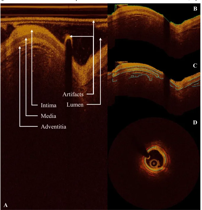
external elastic lamina). The IEM and EEM are fenestrated laminae separating the media layer from the intima and adventitia layers, respectively. Though too thin to be directly observed in currently-available clinical imaging modalities, these physical features are considered for practical purposes to reside at the visible border between layers. Of note, the media itself is differentiable from the adjacent layers in OCT but not IVUS—in which only the trailing edge of the media (EEM; adjacent to the adventitial layer) is visible [18], [19]. However, due to the limited penetration depth of OCT, the intima-media and media-adventitia borders (IEM and EEM, respectively) can be difficult to discern in diseased vessels, though studies have shown that experts can still derive accurate measurements of vessel area in arteries with highly-attenuating lipid-rich plaque [62], especially when the images are attenuation compensated and contrast enhanced [65]. Still, clinical measurements can be time-consuming and suffer from inter- and intra-observer variability, and are typically made at distributed, discrete points, limiting the attainable insights of holistic consideration. Furthermore, physicians without sufficient training, experience, or expertise are often unable to identify the complete outer border of vessels in OCT images, inhibiting important clinical measurement and limiting application in treatment decisions.
Aside from clinical practice, scientists and engineers seeking to computationally model arteries, particularly through structural mechanics or fluid dynamics analyses, are required to extract not just singular metrics of vessel geometry, but digitally recapitulate full two-dimensional (2D) or three-dimensional (3D) geometry as a cornerstone of the model. Furthermore, such geometric boundaries are needed to delineate the region of interest in images for any subsequent tissue characterization processes. Delineating vessel borders and characterizing healthy and diseased (plaque) wall constituents allows for the construction of models of patient-specific vessel geometry for research (and future clinical applications), as highlighted in Chapter 1. However, previous coronary artery anatomic reconstructions have been subject to limitations in application and/or fidelity arising from methodological approaches. Many modeling approaches have utilized either idealized geometries or relatively low-resolution image sources, such as intravascular ultrasound (IVUS), angiography, magnetic resonance imaging, or computed tomography (CT), resulting in necessarily simplified models. Others have relied on histology of excised tissue, which precludes predictive studies, serial monitoring, or application to current patients. Modeling approaches to date utilizing high-resolution OCT have been limited by assumptions or simplifications made during the reconstruction of the arterial wall necessitated by the absence of
geometric information; some use only the inner (lumen) border obtained from OCT (e.g. for computational fluid dynamics simulation without fluid-structure coupling) [70], [168]–[172], others have extrapolated radially from the inner (lumen) border by a fixed distance to approximate the outer (IEM or EEM) border [173], [174], and some have lofted circumferential cross-sections of indirectly-calculated diameters, defined only in plaque-free regions, to create external arterial wall surfaces [172].
The ability to reliably identify the full IEM and/or EEM in OCT images of diseased coronary arteries would provide great benefit by allowing high-resolution and holistic investigation of plaque and wall distribution and composition, along with macroscopic, continuous plaque burden measurements. Because automatic lumen segmentation and plaque characterization methods have been previously developed and validated for this modality [175], the major unresolved challenge is automatically identifying the outer border the vessel wall, which is often obscured by highly-attenuating plaque constituents which limit the penetration depth of OCT. Unfortunately, it is often difficult or impossible to identify the full IEM in a given frame. To generate the best possible estimate of the IEM within a frame, it is therefore necessary to use information from adjacent frames and a priori knowledge of vessel shape—strategies regularly employed by expert clinicians.
2.1.2. Progress in Automatic Geometry Extraction
In response to the clinical need for fast and consistent geometric quantification of vessels, several automated methods have been developed. In addition to progress in imaging methods and technology, methods for post-processing of IVUS and OCT images have brought about tools and capabilities that provide valuable insight to researchers and clinicians [175], [176]; algorithms now exist which can identify many key vascular structural and morphologic features with increasing accuracy, allowing for their measurement and quantification. With sufficient data, advanced algorithms can accurately detect lumen contours and identify stent struts [175], [177]–[179], and even rudimentary image processing algorithms can provide rough approximations of these features. The outer border can be readily segmented automatically in IVUS by various approaches [175], [180]. However, automatic segmentation of the outer border in OCT has been more elusive and challenging.
Automated methods for identifying the outer wall of a vessel in OCT are sparse, despite their potential value. Some approaches have sought to circumvent—and indeed leverage—the penetration depth limitations of OCT by using plaque free wall angle (i.e. visible arc) as a proxy for plaque burden, which is typically derived from the area delineated by the inner and outer borders of the vessel wall [181]. Others have directly delineated the outer wall, but only in non- or minimally-diseased vessels in which the entire outer border was clearly visible within the penetration depth-mediated field of view [182], [183]. Zahnd et al. were the first to extract multiple surfaces simultaneously in intravascular OCT, segmenting intima, media, and adventitia layers through a front propagation scheme [182]. However, the approach was only applied successfully in healthy regions of the vessel in which the layers were clearly visible; these regions neither present the most challenging features characteristic of intravascular OCT, nor do they represent segments of clinical relevance. A later graph-based approach introduced by Chen et al. was used to segment the lumen, intima, and media in cardiac allografts [183]. While applied to an interesting and clinically-relevant application of OCT image segmentation, the method analyzed vessel segments which represent relatively ideal conditions with clear visibility, and is unlikely to translate to more challenging cases such as advanced atherosclerosis. Curiously, these approaches were strikingly similar to OCT segmentation methods developed nearly a decade earlier for an entirely different application.
Unexpectedly, success in extracting geometry from OCT can be found in a different medical field: ophthalmology. Layer segmentation in OCT is not itself novel, though doing so in intravascular OCT is still a fledgling endeavor. When the revolutionary modality was first introduced, OCT was demonstrated in both coronary arteries and the peripapillary area of the retina [33]. Retinal OCT, which is non-invasive, has been widely adopted in ophthalmology; in this use case, penetration depth is not a significant issue, as the retina is generally less than 0.23 mm thick in its thickest region [184]. As such, layer segmentation has matured significantly in retinal OCT, and multiple methods and approaches have been developed to segment retinal layers in OCT images. Grayscale edge detection (with substantial pre-processing including specialized smoothing and de-noising filters) [185]–[187], active contours (energy-minimizing splines) [188], multi-step region growing [189], layered graph minimum-cost closed set determination (including feasibility constraints and regional information) [190], and coarse grained diffusion mapping using texture analysis to identify regional boundaries [191] have all been employed successfully to
segment layers in retinal OCT. Kafieh et al. reviewed and classified a number of other previous approaches that achieved varying success [191]. As engineers and scientists tackle the challenge of identifying and segmenting layers in intravascular OCT, it is prudent to learn from the preceding work in retinal OCT layer segmentation. Penetration depth aside, many complications are shared by both applications. Both retinal and intravascular OCT experience speckle noise, discontinuities in layer boundaries, intensity inhomogeneity, low image contrast, poor layer-to-layer image gradients, and feature shape irregularities. Unfortunately, these issues are more extreme in intravascular OCT. For example, layer boundary discontinuities in retinal OCT are caused by small intraretinal blood vessels, whereas large discontinuities in intravascular OCT are caused by shadows cast widely by a metal catheter guidewire, obscuring up to around 45o (one eighth) of the
vessel wall. Nevertheless, challenge similarities suggest there may be much to be gleaned from retinal OCT layer segmentation approaches.
Despite all of the work to date, more progress must be made to enhance the information gleaned from intravascular images and to make this information more accessible to scientists and clinicians. Critically, current methods can only extract spatially superficial information from OCT images, providing incomplete information necessary for clinical assessment and biomechanical study.
2.1.3. Prior Interpolation and Surface Fitting Methods & Spring Model Precursors
Surface fitting and interpolation are common techniques for filling gaps in 3D information—approximating data in 3D when information is only available in certain areas of the solution space. It ultimately served a critical function in my solution to extract geometric information on diseased vessel walls from OCT images. Unfortunately, while many variations of interpolation and surface fitting exist, most are limited in flexibility, intuitiveness, ability to incorporate secondary information, and computational efficiency. Though existing approaches proved insufficient, a brief introduction to common approaches to interpolation and surface fitting is informative in understanding the shortcomings of prior approaches and the tremendous benefits of the method I developed for this purpose.
Interpolation methods are widely applied to construct a continuous function from irregularly spaced data, including in medical imaging [192]. In these schemes, functions are constructed that intersect known data points or positions, and utilize various methods to