Bulletin of Mathematical Biology (1998) 60, 1017-1037 Article No. bu980061
Modeling
Actin Filament
Reorganization
in Endothelial
Cells Subjected
to Cyclic Stretch
G. CIVELEKOGLU,
Y. TARDY
AND
J.-J. MEISTERT
Biomedical Engineering Laboratory, Swiss Federal Institute of Technology, PSE-Ecublens,
1015 Lausanne,
Switzerland
Hemodynamic forces affect endothelial cell morphology and function. In particular, circumferential cyclic stretch of blood vessels, due to pressure changes during the cardiac cycle, is known to affect the endothelial cell shape, mediating the alignment of the cells in the direction perpendicular to stretch. This change in cell shape pro- ceeds a drastic reorganization at the internal level. The cellular scaffolding, mainly composed of actin filaments, reorganize in the direction which later becomes the cell’s long axis. How this external mechanical stimulus is ‘sensed’ and transduced into the cell is still unknown. Here, we develop a mathematical model depicting the dynamics of actin filaments, and the influence of the cyclic stretch of the substratum based on the experimental evidence that external stimuli may be transduced inside the cell via transmembrane proteins which are coupled with actin filaments on the cytoplasmic side. Based on this view, we investigate two approaches describing the formulation of the transduction mechanisms involving the coupling between fila- ments and the membrane proteins. As a result, we find that the mechanical stimulus could cause the experimentally observed reorganization of the entire cytoskeleton simply by altering the dynamics of the filaments connected with the integral mem-
brane proteins, as described in our model. Comparison of our results with previous studies of cytoskeletal dynamics reveals that the cytoskeleton, which, in the absence of the effect of stretch would maintain its isotropic distribution, slowly aligns with the precise direction set by the external stimulus. It is found that even a feeble stim- ulus, coupled with a strong internal dynamics, is sufficient to align actin filaments perpendicular to the direction of stretch.
@ 1998 Society for Mathematical Biology
1.
INTRODUCTIONVascular endothelium is a monolayer of cells lining the inner wall of blood vessels,
and serves as the interface between the flowing blood and the vessel wall. As blood
flows through an artery, it imparts a force on the endothelial cells and the vessel wall.
This force can be decomposed into two components: one tangent to the surface of the
artery, in the direction of blood flow, and the other normal to the arterial surface. The
~TCI whom correspondence should be addressed.
1018 G. Civelekoglu et al.
tangential component, fluid shear stress, results from the flow of blood and is exerted
on the apical surface of endothelial cells. The normal component results from the
blood pressure causing a radial expansion of the artery wall during the cardiac
cycle, and therefore subjecting the endothelial cells to a cyclic circumferential
hoop stretch as the artery diameter increases during systole and decreases during
diastole, as a result of changes in blood pressure. Thus, this latter mechanical
stimulus is exerted on the basal surfdce of endothelial cells by cyclic stretching of
the extracellular matrix. Under the effect of these dynamic forces, the endothelial
cells undergo a number of morphological and structural changes, both in their
physiological environment and in vitro experiments (Fry, 1968; Dewey et al., 1981;
Levesque et al., 1986; Kim &al., 1989; Davies, 1989). The cells respond to changes
in their physical environment, such as flow patterns and pressure variations, by active
adaptation. Today, the importance of hemodynamic forces in the development of
various cardiovascular diseases, such as atherosclerosis, is well recognized (Zarins
et al., 1983; Ku et aE., 1985). However, the detailed mechanism(s) by which the
endothelial cells sense the changes in their environment and respond to them is still
poorly understood.;
In vitro studies indicate that endothelial cells elongate and align in the direction
offlow when they are subjected to shear stress [see, Zhao et al., 1995, Fig. l(b)]
and in the direction perpendicular to stretch when they are subjected to uniaxial
substrate stretch [see, Zhao et al., 1995, Fig. l(c)]. These changes in cell mor-
phology reflects a profound reorganization at the intracellular level. Changes in
intracellular ionic fluxes, gene regulation and a drastic alteration of the cytoskeletal
structure accompany and/or produce this metamorphosis (Shen et al., 1992; Resnick
et al., 1993; Davies and Tripathi, 1993; Girard and Nerem, 1995). The endothelial
cytoskeleton mainly consists of two different types of dynamic actin structures: the
bundles of actin filaments, referred to as stress$bers, grouped together by actin
binding proteins, and the distributed isotropic network of filaments that fills the
cytoplasm (Satcher, 1993). There are many attachment points between the F-actin
cytoskeleton and the cell membrane, and, in the endothelial cells, this structural
backbone is directly attached to the luminal and basal membranes without an addi-
tional distinct submembraneous network (Satcher and Dewey, 1996; Satcher, 1993).
Under the effect of these hemodynamic forces the two components of the actin cy-
toskeleton undergo a drastic change: the bundles of filaments become longer and
orient in the flow direction, and the distributed network of filaments reorient pref-
erentially in the direction offlow converting to more acute angles, when subjected
to shear stress, and similarly, the filament bundles orient in the direction perpendic-
ular to stretch when subjected to uniaxial substrate stretch [see, Zhao et al., 1995,
Fig. 3(b,c); Satcher and Dewey, 1996; Moore et al., 1994; Dewey et al., 19811.
Despite the strong relationship that exists between the mechanical stimuli and
the shape change and internal reorganization of the cell, the mechanism(s) of force
transduction still remains unknown. Mechanosensitivity of membranes through
Modeling Actin Filament Reorganization 1019
or mechanically induced gene regulation, are potential force transduction mecha-
nisms which are currently under investigation (Harrigan, 1990; Sachs, 1988; Resnick
et al., 1993). Intuitively, the cytoskeleton connected to the membrane and extend-
ing throughout the cell seems the most likely candidate for force transmission to
the interior of the cell (Davies and Barbee, 1994; Davies and Tripathi, 1993; 0~01,
1995; Satcher and Dewey, 1996).
The effect of shear stress on cell morphology and structure has been extensively
investigated experimentally (Dewey et al., 198 1; Davies, 1989; Girard and Nerem,
1995) and to lesser extent in a theoretical framework (Suciu et al., 1997). However,
the effect of substrate stretch has received attention only recently, and has not yet
been studied from a theoretical point of view (Ives et aE., 1986; Tba and Sumpio,
1991; Moore et al., 1994; Zhao et al., 1995). Although the shear stress and the
substrate stretch are sensedthrough different parts of the cell (the apical and the basal
surface, respectively), the resemblance of their effects on the internal reorganization
[compare Zhao et al., 1995, Fig. 3(b) and (c)j suggests that the mechanism(s) by
which these physical stimuli are transduced throughout the cell may be similar.
In particular, transmembrane proteins, such as integrins, which spread throughout
the plasma membrane, both at the apical and the basal surfaces, may play a key
role in these mechano-transduction mechanisms (Wang et al., 1993; Ingber, 199 1).
These proteins provide the link between the actin cytoskeleton and the external
environment of the cell, directly or via a chain of a&in-associated proteins, and
their dynamics is coupled with cytoskeletal dynamics (Schmidt et al., 1994). In this
paper, we investigate the reorganization of the endothelial actin cytoskeleton as a
response to uniaxial substrate stretch in a theoretical framework.
The model presented here should be viewed as a sequel of two previous theoreti-
cal studies of actin filament organization, described in Civelekoglu and Edelstein-
Keshet (1994) and in Suciu et al. (1997). In Civelekoglu and Edelstein-Keshet
(1994), the spontaneous organization of actin filaments into bundles and/or orthog-
onal networks, and the transition between these structures was studied. It was shown
that, under certain conditions, a minute change in the parameters representing the
details of biochemical interactions, such as the binding affinities or the rate of asso-
ciation or dissociation, is sufficient to break the stability of the isotropic distribution,
leading to a rapid formation of bundles or orthogonal networks. Furthermore, the
transition between these two structures was found to be highly sensitive to certain
parameters. In Suciu et al. (1997), the above model was modified to account for
the effect of an external force, the fluid shear stress, in endothelial cytoskeleton. It
was found that the effect of shear stress, altering the dynamics of a small fraction
of the cytoskeletal filaments, namely those attached to transmembrane proteins, is
sufficient to break the stability of the isotropic network and to favor the formation
of bundles of filaments.
This paper concerns the effect of a different mechanical stimulus, namely the
substrate stretch, on the dynamics and organization of the cell’s actin cytoskeleton.
1020 G. Civelekoglu et al.
is not clear, we propose two different scenarios by which a uniaxial stretch of the
substratum may alter the dynamics and the organization of endothelial actin fila-
ments. The model presented in this paper involves a description of actin dynamics
closely resembling the ones in Civelekoglu and Edelstein-Keshet (1994) and Suciu
et al. (1997). In addition to the internal dynamics of the cytoskeleton described
in these papers, we consider two different approaches accounting for the effect of
substrate stretch on the actin filament organization. The linear analysis and nu-
merical simulations of the model equations confirm that the proposed mechanisms
are sufficient for the initiation and the development of the observed response of en-
dothelial cytoskeleton to stretch. We further compare the results of the two different
hypotheses proposed to describe the transduction of the external stimulus.
In the following section, we describe the features and the structure of the model,
and present the two mechano-transduction hypotheses which lead to two different
formulations of the effect of substrate stretch. Then, we proceed with the formu-
lation of the model equations, corresponding to the two hypotheses given in the
previous section. The results include linear analysis of the equations and numerical
simulations. The final section comprises an overall discussion and comparison of
the results obtained from the two different approaches, and with previous work.
2.
THEMODELINGSTRATEGYANDHYPOTHESES
As in previous theoretical considerations of actin filament organizations, here too
we focus on the orientation of actin filaments on a plane representing the quasi two-
dimensional cytoskeletal network (Civelekoglu and Edelstein-Keshet, 1994; Suciu
et al., 1997). Therefore, we assume that all the variables represented in the present
model are spatially homogeneously distributed. The angle 0 represents an angle on
the plane, with respect to a fixed orientation and the plane is the projection of the
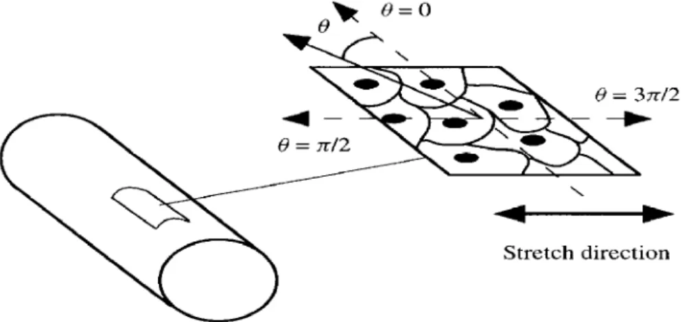
cell(s) on the substratum (see Fig. 1).
In incorporating the effect of substrate stretch on the cytoskeletal reorganization
into the model, we base our hypothesis on the experimental evidence that transmem-
brane proteins play a key role in the transduction of this external mechanical stimulus
into the cell (Ingber, 1991; Wang et aE., 1993; Petrov and Usher-wood, 1994). The
radial expansion of the artery causes a uniaxial stretch {in the circumferential direc-
tion) of the extracellular matrix which is in the order of 10% in a normal aorta. Along
with this stretch/relaxation cycle, the integral membrane proteins embedded in the
plasma membrane, traversing the bilayer and anchoring the cell to the extracellular
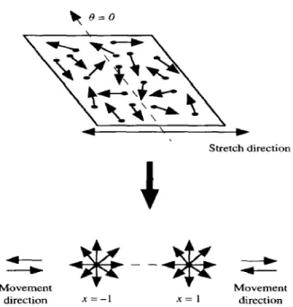
matrix, move away from or towards each other, as shown in Fig. 2, along the rim of
each circular cross section of the cylindrical surface. Note that this displacement,
being a uniaxial one, does not affect the relative position of these proteins along the
longitudinal axis of the cylinder (the vertical axis of the cylinder), that is the axis
perpendicular to stretch. The actin filaments which are attached to these integral
Modeling Actin Filament Reorganization 1021
Stretch direction
Figure 1. The quasi-two-dimensional flat piece cut from the artery wall, the plane of projection of the cells and their cytoskeleton, the angle Q on the plane, and the direction of stretch.
Direction perpendicular to stretch % (longitudinal axis of the artery)
Transmembrane proteins
Figure 2. The uniaxial stretch of the substratum and the displacement of the integral membrane proteins away from each other along the circumference, and the direction per- pendicular to stretch.
G. Civeiekoglu et al.
are in turn influenced by this displacement. We assume that this effect has either
one of the following implications on the cytoskeletal dynamics.
HYPOTHESIS I. The displacement of the proteins away from each other influences
the dynamics of actinfilaments attached to them by breaking the bonds between the filaments (via actin binding proteins), an~or the connection between the filaments
and the transmembraneproteins. We base theformulation of this efSect on the follow- ing experimental results. Numerous studies indicate that actinJilaments are$exible to bending yet extremely rigid to extension (Kishino and Yanagida, I988; Ookawa
et al., 1993; Kojima et al., 1994; Ben-Avraham and Tirion, 1995). That is, there
is substantial difference in the flexibility of jil aments to bending and extensibility. When the anchoring points of the filaments, the transmembrane proteins, are dis- placed under the effect of substrate stretch, whether the Jilaments are exposed to
bending or extension is determined by their angle of orientation with respect to the stretch direction In particular, those which are parallel to the stretch direction are exposed to extension, whereas those perpendicular are exposed to bending. Therefore, in the view of the experimental$ndings onfilamentflexibility, the bonds
between filaments which are perpendicular to stretch direction, or simply the fila-
ments perpendicular to that direction, are less likely to break than those oriented in
the direction of stretch. This means that the dissociation coeficients of the binding
proteins anchoring the filaments to the membrane, or holding the filaments together assume an angle-dependent form when the substratum is stretched.
HYPOTHESIS II. The cyclic translocation of the proteins influences the dynamics
of actin$laments by inducing a cyclic turn, a rotational motion, of the microfilaments attached to them. Due to the viscous nature of the intracellular milieu, the amplitude of this motion depends on the angle of orientation of the$lament with respect to the direction of stretch. In particular; the filaments which are perpendicular to stretch direction undergo the widest turn, whereas the filaments along the direction of stretch remain still. That is, the angular flux velocity is a function of the angle of orientation. It is also a function of time since the stretch is cyclic and fully elastic, hence the filaments swing, sweeping an angular section de$ned by their angle of orientation. Therefore, in this view, filaments which are perpendicular
to stretch direction are more motile than those which are parallel to it, and hence
are more likely to meet other filaments and to form bonds. Here, note that the
physiological stretch cycle is 1 s (assuming 60 heart beats per min), whereas the chemical interactions involved in binding offilaments is in the millisecond range. This permits numerous binding between filaments within each cycle.
The dynamics of the actin cytoskeleton, as described in Suciu et al. (1997),
together with the formulation of the effect of the substrate stretch, based on the
hypotheses above, result in two different descriptions of the cytoskeletal dynamics
1023 Modeling Actin Filament Reorganization
Figure 3. Schematic diagram showing the four populations of filaments: free filaments, in gray; bound filaments, hatched; free filaments anchored to the membrane, in black; bound filaments anchored to the membrane, dotted. The gray circles denote the transmembrane proteins through which the filaments are anchored to the membrane, and the solid heart- shaped figures denote the actin bundling proteins. The exchange rates between different populations of filaments are indicated with arrows.
3.
MODELEQUATIONS
The dynamics of actin filaments described in this paper is in close resemblance
with the model considered in Suciu et al. (1997). We first focus on the internal
dynamics of the actin filaments and introduce the necessary variables and parame-
ters. In order to describe the interactions between actin filaments via actin binding
proteins and their coupling with the transmembrane proteins we have four states of
actin filaments. That is, a given filament belongs to either one of the following four
populations of filaments at a given time. The ‘free’ filaments, L, are those which
are neither attached to other filaments nor anchored to the membrane. The ‘bound’
filaments, B, are those which are attached to other filaments via actin bundling
proteins but not anchored to the membrane. The ‘membrane-free’ filaments, ML,
are those directly anchored to the membrane but not attached to other filaments. Fi-
nally, the ‘membrane-bound’ filaments, M B, are those which are directly anchored
to the membrane and attached to other filaments via acting bundling proteins (see
Fig. 3). In our model equations, the free filaments are the analogue of the dis-
tributed isotropic cytoskeletal network and the bound ones are the analogue of the
stress$fibres. The repertoire of each four type of filaments include interactions with
all other types of filaments, binding and dissociation via actin binding proteins,
and coupling and uncoupling with integral membrane proteins. These interactions
1024 G. Civelekoglu et al.
ospecific characteristics, and result in exchange of filaments between populations.
The ratio of G and F-actin is assumed to be constant throughout this reorganization
process (Satcher, 1993). However, the randomizing effect of the filament turnover,
breaking, annealing and such, is accounted for by a slow diffusion term for the
filaments L and ML, which represent the distributed isotropic actin network (Suciu
et al., 1997). For a more detailed description of the terms concerning the actin
dynamics the reader is referred to Civelekoglu and Edelstein-Keshet (1994), and/or
Suciu et al. (1997).
Both systems of equations include the following variables and parameters which
represent the cytoskeletal dynamics:
L, B, ML, Me four types of filament populations, distinguished by their connec-
tions with other filaments and transmembrane proteins
KC@) the kemal representing the angular dependence of the rate constant
for binding of filaments via parallel actin binding proteins
B the binding affinity of filaments via actin binding proteins
P the concentration of free actin binding protein
6 the dissociation rate of acting binding proteins
PL the rotational diffusion constant for free filaments connected
with the transmembrane proteins
PML the rotational diffusion constant for free filaments connected to the
transmembrane proteins
e the dissociation rate of filament connections with the transmem-
brane proteins
w the rate of attachment of filaments to transmembrane proteins.
3.1.
System1.
Under Hypothesis I, the effect of translocation of the integralmembrane proteins directly influences the dynamics of the crosslinks between fila-
ments as well as the dynamics of filaments anchorage points to the membrane. That
is, the external effect can be accounted for directly in the dissociation constants 6
and @. We proceed with a description of the four populations of actin filaments
as a function of their angle of orientation, &J and t. This leads to four state vari-
ables L(B, t), B(0, t), ML (0, t), MB (f3, t)- All other variables and parameters are
as described in the beginning of this section. We only modify the constants 6 and
@, accounting for the rate of dissociation of the bonds between filaments and the
transmembrane proteins, respectively, to be functions of 0. We choose the following
functions to reflect the angular dependence of these dissociation rates:
6 =61 +&tsin81
$f = *I + 34 sin01 (1)
where 61 and @r are the constant dissociation rates, independent of stretch, and 62
and +2 are the maximal contribution rates to the dissociation due to the effect of
Modeiing Actin Filament Reorganization 1025
results of the model, any other function f bearing the following main characteristics
leads to similar conclusions: f must attain its maximum at 6 = n/2 and 8 = 31-r/2;
fmustbeOat8 =Oandn; f 2 Ofor f [0,2n].
These properties are based on the variability of the resistance of bonds between fil-
aments oriented at various directions, due to the bending and extensibility properties
of the microfilaments, as discussed in Section 2.
The following system of equations describe the dynamics of actin filaments and
the influence of stretch on the dissociation of bonds between crosslinked filaments
and the filaments’ anchorage points to the membrane.
-gB(B, t) = ppL(K*L)
+ ppB(K*L)
+ p/mfLw*L)
+ PmfEw*Ll
--6(8)B + p+(e)n/r, - fwB
;L(t’,
t) = --p&L(K*B)
- ,@L(K*L)
- p/%(K*f&)
-
Pj=(K*MB)~M~(~.~)=~PL(K*~L)+~PB(K*~L)+~PML(K'ML)+~PM~(K*ML)
-~(~)MB
- +(6’>Ms
+ uB
The terms such as pfi ML (K * B) include a convolution integral where:
0
K*B =
s 2rr K(Q - 0’)B(O’, r) de’,
and describe, in this case, the rate at which the bound filaments, Z3, in any orientation
interact, bind and align with membrane-free filaments, ML, in direction 19 in the
presence of actin binding proteins, p [for a more detailed explanation of such
terms the reader is referred to Civelekoglu and Edelstein-Keshet (199411. Terms
such as S(G) B denote, for example, the rate at which bound filaments become
free as the bonds between crosslinked filaments dissociate and similarly $ (0) ML
denotes the rate at which the membrane-free filaments become free filaments as
filaments detach from the membrane. For the terms 6 (0) B in the first and second
equations, and +(f3>ML in the second and fourth equations, the rate constants S(0)
and I++(@) are considered to be constant. That is, 82 = I& = 0, since the effect
of stretch is not exerted on these types of filaments directly. For the membrane-
1026
G. Civelekaglu et al.stretch. The terms such as WL or wB denote the rate at which the free or bound
filaments anchor to the membrane, and become membrane-free or membrane-bound
filaments, respectively. Finally the PL $$, ,L.LML % terms describe the random
reorientation of filaments of type L and M L, respectively, due to the randomizing
effect of breaking and annealing of filaments and the filament turnover rate [for a
more detailed explanation of these teps the reader is referred to Suciu et al. (1997)].
All the above terms describe a dynamic exchange between four populations of
filaments and/or alteration in their orientation. For example, as a result of a filament-
filament type interaction, say p/3 ML (K * B), the bound filaments, B, bind and align
with membrane-free filaments and remain as bound filaments as they are still only
attached to other filaments and not directly anchored to the membrane; however,
the corresponding membrane-free filaments, ML, become membrane-bound types.
Thus there is a corresponding loss term, the second term, in the rate equation for
ML, and a corresponding increase in the equation for MB, the second term.
The mathematical form of these equations is similar to the ones given in
Suciu et al. (1997), however, here, there are some additional terms and all func-
tions are periodic; in 8, with period 2~. The additional terms appear in the last
two equations and are all interaction terms allowing the binding of membrane at-
tached filaments with all other populations. The period of all state variables being
2n, as opposed to n in Suciu et al. (1997), results from the plane of projection
being the top view of the cell rather than a cross section as in Suciu et al. (1997).
Therefore, here, all four types of filaments can be oriented from 0 to 2~. These
differences render the equations in a suitable form for linear analysis and allow us
to compare the analytical results with numerical simulations and to draw important
conclusions.
3.2. System
ZZ.
In order to describe the cylic rotational motion of the filamentson the two-dimensional surface in consideration, it is most desirable to describe
the distribution of all four populations of actin filaments, including their position,
I, and orientation, 8. However, a model based on such a description is far from
being amenable to analytical or numerical treatment. Therefore, we proceed with
the following arguments leading to a description of a simplified geometry, fully
capturing the desired dynamics.
Due to the cyclic circumferential stretch of the substratum, the anchorage points
of the filaments, the transmembrane proteins, move only along the direction of
stretch, and not in the direction perpendicular to stretch. Moreover, all points
along the line of the circumference move away from or towards each other with
the effect of stretch or relaxation (see Fig. 2). Therefore, lumping the contin-
uum of filament anchorage points simply in two discrete points in the stretch di-
rection, moving away from or towards each other captures the essential aspects
of the dynamics (see Fig. 4). Hence, to describe the geometry for the reori-
entation of filaments due to the translocation of the transmembrane proteins on
Modeling Actin Filament Reorganization 1027
Stretch direction
direction x=-l .X=1 direction
Figure 4. The surface in consideration and the simplified geometry. The angle 0 and the variable x = - 1, 1 indicate the orientation and the two anchorage positions of the filaments moving away from each other.
and x = - 1 and 1, indicating the positions of the filaments’ anchorage points
along the stretch direction. Note that the introduction of the variable x at two
discrete points does not mean we study a fully two-dimensional problem. We
consider a one-dimensional problem as before; however, at two discrete points
in this case. This allows us to capture the movement of the anchorage points
of the filaments on the membrane away from or towards each other as a result
of the stretching of the membrane caused by the swelling of a cylindrical sur-
face.
In the present case, all variables, L, B, A4 L, MB are functions of 13, x and t .
8 varies from 0 to 2n, and represents an angle of orientation, with respect to the
axis perpendicular to the stretch direction. x = - 1, 1 are the two positions of
the transmembrane proteins, moving away from or towards each other, and the
distance between these two points represents the maximal distance within which
two filaments anchored to the membrane canfeel each other, and bind to each other,
during a stretch cycle.
The rate of change of the density of the membrane-bound filaments due to the
rotational motion of the filaments, caused by the displacement of the transmembrane
proteins away from or towards each other, can then be described by an angular flux
term, similar to the one in Suciu et aE. (1997) formulated for shear stress:
1028 G. Civelekoglu et al.
In this case, the flux velocities,
V
ML,VMS,
depend on both 8 and x, and are of theform:
VML(O,x) = uMLcose
sgn(x)sinhtvMB(e, X) = v MB cos 0 sgn(x) sin ht (4)
where VML and V,jJB are the maxim&l reorientation rates for the free and bound
filaments attached to integral membrane proteins, and the constant h defines the
period of oscillations. We assume different maximal turning rates for the free and
bound types of filament, WMB -C uML, due to restriction of motion of large molecules
in the viscous cytoplasm. Note that both of these angular velocity terms depend
on the variable x through sgn(x) which captures the reorientation movement of
filaments away from or towards each other. For a detailed description and derivation
of the values of these velocities the reader is referred to Suciu et al. (1997).
As discussed in the previous subsection, here too, the exact form of these functions
are unimportant for the results, and any other function g, satisfying the following
properties, results ln similar conclusions: g must attain its maximum at 8 = 0 and
n;gmustbeOat8 = n/2 and 3x/2; g (e, I) must be positive in the first and second
quadrants, and negative in the third and fourth quandrants; g must be odd in X, that
is g(- 1) = -g( 1). These properties are based on the physical description of the
angular displacement of filaments due to translocation of their anchorage points.
The following system of equations describe the dynamics of actin filaments and
the effect of the translocation of the transmembrane proteins due to cyclic stretch
of the substratum.
a2L
+m+wL-OL+CLL~
Modeling Actin Filament Reorganization 1029
The mathematical form of these equations is, as in System I, similar to the ones
given in Suciu et al. (1997); however, here there is an additional variable x. Also,
as in System I, here too all functions are periodic in 19 with period 2~7 and the flux
velocity function has a different form than the one given in Suciu et al. (1997).
Note that in the systems of equations (2) and (5), the total density of actin filaments:
h4=
s JT
-?7
{L(@,
t)
+
B(B,
t)
+
ML(~,
t)
+
MB(Q,
t)}
db’ (6)
remains constant over time. [For system (5), the actual total mass is indeed M =
M-i + Mi, where M-1 is the mass at x = -1, and MI is the mass at x = 1, each
calculated as in equation (6)f. That is, there is no change in the total amount of
actin filaments in the system. This represents the treadmilling case where there is
no net increase or decrease in total F-actin in the cytoskeleton.
4.
RESULTS4.1. Analysis. First, note that in the absence of the external stimulus, the actin
dynamics is similar in the systems of equations (2) and (5). That is, equations (2)
with constant dissociation rates, 6 and I,?, and equations (5) with null angular flux
velocities,
VmL
andVMB,
are identical except an additional discrete positionalrepresentation in equations (5). For the linear analysis of the systems (2) and (5),
we therefore use the common description of the actin dynamics alone, where the
effect of the external stimulus is neglected. Thus, the following analysis concerns
the ‘truncated’ equations where 6 and $ are constant in case of equations (2) and
the angular flux velocities,
VML
andVMB,
are null in case of equations (5).We begin by discussing the properties of a uniform steady state of the ‘truncated’
systems (2) and (5), L, B, i%$ L, MB, that is systems (2) and (5) in the absence of the
external effect. That is, a time independent steady state in which every orientation
(and position, in the case of System II) is equally probable. Setting the time, angle
and positional derivatives to zero, we obtain the following equations in terms of the
homogeneous steady states (L , fi, ML, MB) :
These are the ratio of the sum of filaments of type L, and ML, the analogue of the
distributed isotropic network of filaments, to the sum of filaments of type
B
and MB,the analogue of stress fibers, and the ratio of the cytoskeletal filaments anchored to
the membrane to those which are not, in terms of the binding and dissociation rate
1030 G. CiveLekoglu et al.
explicit descriptions of how the cytoskeleton is divided into its different components
in a homogenous equilibrium state, in the absence of external stimulus.
We further wish to test the stability of the homogeneous steady state of these trun-
cated equations to small perturbations, as was done in Civelekoglu and Edelstein-
Keshet (1994). Defining new variables, B’ and L’ as:
and assuming ,uL g ~ML, our ‘truncated’ equations reduce to the system of equa-
tions (1) in Civelekoglu and Edelstein-Keshet (1994). Hence, in the absence of
the external stimulus, the results of the linear stability analysis presented in their
paper apply to our equations, assuming that the diffusion coefficients pML and fiL
are the same. The coefficient C, given in the dispersion relation in Civelekoglu
and Edelstein-Keshet (1994), is a combination of the parameters in the equations,
and its value determines the stability properties of the homogeneous steady state.
Namely, if C < 0.04 [see Civelekoglu and Edelstein-Keshet, 1994, Fig. 3(b)] the
linear analysis predicts that there will be a spontaneous self-organization of actin
filaments in parallel bundles. The results of their numerical simulations of the
nonlinear system confirm these predictions. Based on their results, we conlcude
that our system is at a stable equilibrium in the absence of the external stimulus,
provided that the parameters do not satisfy the dispersion relation (Civelekoglu and
Edelstein-Keshet, 1994, equation (4)). And, we wish to analyze the effect of the
external stimulus on this stable system, and study its dynamics.
Table 1. The range of values for the parameters used in the numerical simulations. Most parameters were found in raw form in the literature and were adapted to correspond to the geometry and their representation in the model.
Symbol Standard value References
ff 20-30“ Civelekoglu and Edelstein-Keshet (1994)
B 0.1-5 ,uM-’ s-l Burridge et aE. (1990), Dufort and Lumsden (1993)
P 0.1-Z PM Pollard and Cooper (1986)
s 0.1-2 s-1 Burridge et al. (1990), Dufort and Lumsden (1993) E*L 0.01-l rad2 s-f Civelekoglu and Edelstein-Keshet (1994)
PAIL 0.001-0.2 rad* s-’ adapted from the value of pi
4 0.5-2 s-l Meyer and Aebi (1990), Dufort and Lumsden (1993), coupling rate in Schmidt et al. (1994)
w 0.01-0.5 PM-’ s-f Meyer and Aebi (1990), Dufort and Lumsden (1993), uncouphng rate in Schmidt et al. (1994)
4.2. Numerical simuZutions. Equations (2) and (5) were simulated with meth-
ods identical to the ones described in Suciu et al. (1997) and Civelekoglu and
Edelstein-Keshet (1994). The parameters used for the computations were gathered
from biological literature and/or from previous theoretical models where similar
rate constants were used. A list of the parameters and the corresponding references
Modeling Actin Filament Reorganization 1031
iG$T--& i -p]
0 90 180 270 360 0 90 180 270 360 0.65 @ (deg) 0 90 180 270 360 @(de@ - ho”mge”cous sol”rl”nr --- non- homogeneoui WI”I10”’0.18
0 90 180 270 360 0 (deg)
Figure 5. The homogeneous and non-homogeneous solutions of the system of equations (2) in the absence (solid lines), and in the presence (dashed lines) of the external stimulus, respectively. The plots (a)-(d) correspond to the final stationary densities of the bound
B(8, t), free L (0, t), membrane-bound MB (0, r), and the membrane-free ML (0, r) filament types, respectively. In the case where the external force is absent, the rate of detachment of the filaments from the membrane and the dissociation rate between filaments is constant for all populations S = St and + = et. It is seen that the initial homogeneous state is stable in this case and all filaments remain equally distributed over all orientations. In the case where the external stimulus is present (dashed lines) the rate of detachment of the filaments from the membrane and the dissociation rate between filaments are angle dependent for the filaments coupled with the membrane S2, + f 0. The solution is non- homogeneous in this case, and two peaks, centered at 19 = 0 and 180”, are seen in the distribution of all four types of filaments. For the solutions shown a discretization with A8 = 36, At = 0.001 was used and the total number of iterations was 12 x 1 O6 in the case where the external stimulus is absent, and 40 x lo6 in its presence. The parameters used were: p = 0.5, fi = 0.3,cr = 21°, 61 = os,w = O.l,~l = 1.2, /.L‘+fL = 0.6, pL = 0.6, and in the absence and the presence of the external stimulus the following values of 62 and $2 were used, respectively 62 = 0, $9 = 0 and 62 = 0.1, $2 = 0.2.
At = 0.001, and Courant-Friedrichs-Lewy stability conditions were verified for
the stability of the numerical scheme. The time step At corresponds to a minimal
time required for interaction between molecules, in the order of milliseconds. In
the figures shown here, the initial densities were small random deviations, of mag-
nitude lo%, superposed on a uniform distribution. The resuls of all simulations
were in total agreement with the linear analysis results, that is, in the absence of
the external stimulus, the system remained at a stable homogeneous state for the
parameters which did not satisfy the dispersion relation adapted from Civelekoglu
and Edelstein-Keshet (1994).
Figures 5(a)-(d) shows the numerical solutions of the system of equations (2) in
the absence of the external stimulus (solid lines), and including the effect of stretch as
1032 G. Civelekoglu et al.
(b)
60
Figure 6. The homogeneous solution of equations (5) in the absence of the external stimulus. That is, the rate of angular flux rate of filaments coupled with the membrane (UML, UMB) = 0. Fe plots (a)-(d) correspond to the stationary homogeneous densities of the bound B(8, n, t), free L (0, n, t), membrane-bound MB (f3, x , t), and the membrane-free ML (0, X, t) filament types, respectively. The discretization was as in Fig. 5, the total num- ber of iterations was 6 x 10⁶. The parameters used were p = 0.2, #3 = 0. I, a = 40°, 6 = 0.5, w = 0.025, ?,k = 0.1, ,.&ML = 0.2, PL = 0.5, and the values of UMB = VML = 0. It is seen that the initial homogeneous state is stable in this case and all filaments remain equally distributed over all orientations.
solution of the system (2) where the functions S(0) and 1+4(e) are constant, thus do
not depend on 8. This corresponds to a system where the external stimulus is absent.
We wish to compare the solution of this system with one where the external effect
is taken into account while all other conditions remain the same. These results are
shown with dashed lines, thus, the dashed lines indicate the solution of the system (2)
with parameters and initial conditions identical to the ones employed to obtain the
solid line solutions, except that the functions S(Q) and I++(O) here depend on 8 as
described in Section 3.1. Each set of four plots correspond to angular densities of
the four types of filaments, L , B, MB and ML in the equations. In the absence of
the external force, the densities of all four populations remain homogeneous, at the
levels predicted by the linear stability analysis [Fig. 5(a-d), solid lines]. When the
effect of stretch is added to the system (meaning that the functions S(0) and $ (0)
are made to be 8 dependent), two small peaks in the distributions of the filaments
anchored to the membrane appear in early stages, then are transmitted into the
other two types of filaments, and become amplified with time. Only the final state
of the system, obtained through numerical simulations, is shown is Fig. 5(a)-(d)
(dashed lines). Note that the peaks in all populations are centered at 8 = 0, 180”, the
direction perpendicular to the stretch direction, indicating the alignment of filaments
along the direction perpendicular to stretch, as observed experimentally. Also the
Modeling Actin Filament Reorganization 1033 (b)
Figure 7. The non-homogeneous solution of equations (5) in the presence of the external
stimulus. That is, the rate of angular flux rate of filaments coupled with the membrane (UML, UMB) $: 0. The plots (a)-(d) correspond to the final stationary densities of the bound B(8, x, t), free L(8, n, t), membrane-bound MB (0, x, t), and membrane-free ML (0. x, t) filament types, respectively. The discretization and the parameters given in Fig. 6 were used, only the total number of iterations was 12 x lo6 in order to reach a stable solution, and the values of VML = 0.1 and UMB = 0.1. Two peaks, centered at x = - 1, 1 and 8 = 0 and 1 80°, are seen in the distribution of all four types of filaments.
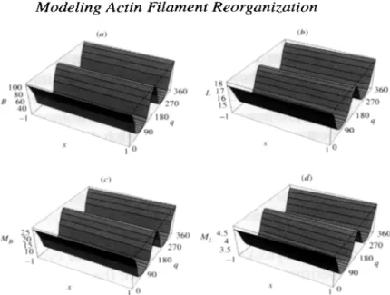
Figure 6(a)-(d) shows the numerical simulations of the system of equations (5)
in the absence of the external stimulus, and the Fig. 7(a)-(d) shows the solution
of the system, with the same parameters and initial conditions, but including the
effect of stretch as described in Section 3.2. Each set of four plots correspond to
angular densities of four types of filaments L , B, MB and ML, at positions x = - 1
and 1. For an easier visualization, the plots at x = - 1 and 1 were connected to
form a smooth surface. In Fig. 6(a)-(d), all four densities remain homogeneous in
orientation and position, whereas with the addition of the effect of stretch (Fig. 7(a)-
(d)) two peaks at 8 = 0, 180” appear at both x = - 1, 1. Again, the orientations
which become accentuated are those perpendicular to stretch direction, as observed
experimentally. As in the previous case, the peaks which first appear in the densities
of the filaments coupled with the membrane, are then transmitted into the other two
densities, and are magnified with time.
In all simulations of the systems of equations (2) and (5), two peaks in the filament
densities in the direction perpendicular to stretch were observed in the presence of
the effect of this external stimulus. These results are in total agreement with the
experimental observations. For the parameter set for which spontaneous parallel
alignment was predicted in Civelekoglu and Edelstein-Keshet (1994), the two peaks
in the filament densities formed rapidly, in the presence, as well as in the absence
1034 G. Civelekoglu et al.
two peaks that formed were always in the direction perpendicular to stretch, but in
its absence, the position of the 180” apart two peaks were arbitrary and depended
on the initial conditions. This shows that this mechanical stimulus may have the
following effect on the cytoskeletal dynamics. Stretching directly alters the internal
cytoskeletal dynamics by promoting the formation, or the survival, and/or turning
and alignment of filaments perpendicular to the stretch direction, and hence sets the
direction of alignment regardless of the initial polarity of the cytoskeleton.
5.
DISCUSSION
Alignment of endothelial cytoskeleton under the effect of cyclic stretch has been
previously studied experimentally (Ives et al., 1986; Iba and Sumpio, 1991; Moore
et al., 1994; Zhao et al., 1995). To our knowledge, this is the first attempt to de-
scribe this phenomenon in a theoretical framework. Previous theoretical studies of
cytoskeletal remodeling are based on either a mechanical approach, where the dy-
namics at the molecular level is not included (Sherratt and Lewis, 1993; Satcher and
Dewey, 1996; Dembo, 1989; Ah, 1987; Oster and Odell, 1984), or a biochemical
approach where the cytoskeleton is viewed as a pool of interacting molecules, sep-
arated from its mechanical environment (Civelekoglu and Edelstein-Keshet, 1994;
Dufort and Lumsden, 1993). In Suciu et al. (1997) the effect of an external me-
chanical force, namely that of shear stress, was incorporated into a model based on
biochemical interactions.
In this paper, we present a theoretical model of the endothelial cytoskeletal re-
modeling under the effect of cyclic stretch. Our model includes a description of the
cytoskeletal dynamics, and a direct effect of the external stimulus, in a simplified
geometry. In System I, the uniaxial stretch of the substrate induces a directional
preference on the breaking of bonds between filaments and their coupling with the
membrane. In System II, the stretch causes a cyclic swing of filaments which are
connected with the membrane. Both of the hypothetical mechanisms considered
in this paper are based on the experimental evidence that the cytoskeleton directly
detects the external stimulus exerted on the membrane, through coupling with in-
egral transmembrane proteins (Schmidt et al., 1993; Kusumi et al., 1993; Wang
et al., 1993). With this work, we do not imply that other force transduction systems
based on stretch sensitive ion channels or second messenger systems are unimpor-
tant, rather, we suggest a complementary mechanism which can explain the precise
choice in the direction of cytoskeletal reorientation. Intuitively, with other mecha-
nisms based soley on biochemical events, it is indeed difficult to explain this precise
direction of reorganization.
Our results indicate that the time course of the cytoskeletal reorganization based
on the effect of an external mechanical stimulus, is orders of magnitude slower
than a cytoskeletal reorganization based on the intrinsic biochemical mechanisms.
Modeling Actin Filament Reorganization 1035
reorganization as described in (Civelekoglu and Edelstein-Keshet, 1994; Dufort
and Lumsden, 1993). However, the cytoskeletal polarity is firmly dictated by the external stimulus, already in early stages, whereas it is arbitrary in a model based
solely on activation/inactivation of molecules.
Actin depolymerization has been reported as a fast initial response to mechanical
stimuli such as shear stress Morita et aE. (1994). On a longer time scale (24 h),
exposure to fluid shear stress shows no significant change in the F/G-actin ratio in
endothelial cells (Satcher, 1993). Incorporating an initial transient decrease in F/G-
actin ratio, or other force transduction mechanisms into our model which already
comprises a microscopic description of the cytoskeleton may be achieved quite
easily once the details of the biochemical pathways underlying these mechanisms
become clear.
REFERENCES
Alt, W. (1987). Mathematical models in actin-myosin interaction, in Nature and Function of Cytoskeletal Proteins in Motility and Transport, K. E. Wohlfarth-Bottermann (Ed.), Gustav Fisher: Stuttgart.
Ben-Avraham, D. and M. Tirion (1995). Dynamic and elastic properties of F-actin: A normal modes analysis. Biophys. J. 68, 123 1-1245.
Burridge, K., G Nuckolls, C. Otey, F. Pavalko, K. Simon and C. Turner (1990). Actin- membrane interaction in focal adhesions. CeEZ OiJg: Dev. 32, 337-342.
Civelekoglu, G. and L. Edelstein-Keshet (1994). Modeling the dynamics of F-actin in the cell. Bull. Math. Biol. 56,587-616.
Davies, P. F. (1989). How do vascular endothelial cells respond to flow? NIPS 4, 22-25.
Davies, P. F., and K. A. Barbee (1994). Endothelial cell surface imaging: insights into
hemodynamic force transduction. News Physiol. Sci. 9, 153-157.
Davies, P. F., and S. C. Tripathi (1993). Mechanical stress mechanisms of the cell, an en- dothelial paradigm. Circ. Res. 72, 239-245.
Dembo, M. (1989). Field theory of the cytoplasm. Corn. Theor. Biol.
1-3,
159-177.Dewey, C. F. Jr., S. R. Bussolari, and M. A. Gimbrone (1981). The dynamic response of vascular endotheliun cells to fluid shear stress. J. Biomech. Eng.
103,
177-185.Dufort, P. A. and C. J. Lumsden (1993). Cellular automaton model of the actin cytoskeleton. Cell Mot. Cytoskel. 25, 87-104.
Fry, D. L. ( 1968). Acute vascular endothelial changes associated with increased blood velocity gradients. Circ. Res. 22, 165-197.
Girard, P. G. and R. M. Nerem (1995). Shear stress modulates endothelial cell morphology and F-actin organization through the regulation of focal adhesion-associated proteins. J. Cell Physiol. 163, 179-193.
Harrigan, T. P (1990). Transduction of stress to cellular signals, in First World Congress in
Biomech. University of C. San Diego. Vol. 2, p. 5 1.
Iba, T. and B. E. Sumpio (199 1). Morphological response of human endothelial cells subjected to cyclic strain in vitro. Microvasc. Res. 42,245-254.
1036 G. Civelekoglu et al.
Ingber, D. (1991). Integrins as mechanochemical transducers. Cure Op. Cell Biol. 3, 841-
848.
Ives, C. L., S. G. E&in and L. V. McLntire (1986). Mechanical effects on endothelial cell morphology: in vitro assessment. In viva CeZZ Dev. BioZ. 22, 50&507.
Kim, D. W. A. I. Gotlieb and B. L. Langille (1989). In vivo modulation of endothelial F-actin microfilaments by experimental alterations in shear stress. Arteriosclerosis 9,439445. Kishino, A. and T. Yanagida (1988). Force,measurements by micromanipulation of a single
actin filament by glass needles. Nature 334,74-76.
Kojima, H., A. Ishijima and T. Yanagida (1994). Direct measurements of stiffness of single actin filaments with and without tropomyosin by in vitro nanomanipulation. Proc. NatZ Acad. Sci. 91, 12962-12966.
Ku, D. N., C. K. Giddens, C. K. Zarins and S. Glagov (1985). Pulsative flow and atheroscle- rosis in the human carotid bifurcation. Arteriosclerosis. 5, 293-302.
Kusumi, A., S. Yasushi and Y. Mutsuya (1993). Confined lateral diffusion of membrane receptors as studied by single particle tracking. Effects of calcium-induced differentiation in cultured epithelial cells. Biophys. J. 65,2021-2040.
Levesque, M. J., D. Liepsch, S. Moravec and R. M. Nerem (1986). Correlation of endothelial cell shape and wall shear stress in a stenosed dog aorta. Arteriosclerosis 6, 220-229. Meyer, R. K. and U. Aebi (1990). Bundling of actin filaments by alpha-actin depends on its
molecular length. .J. Cell BioZ. 110, 2013-2024.
Moore, J. E. Jr., E. Brki, A. Suciu, S. Zhao, M. Bumier, H. R. Brunner and J. J. Meister (1994). A device for subjecting vascular endothelial cells to both fluid shear stress and circumferential cyclic stretch. Ann. Biomed. Eng. 22, 41-22.
Morita, T., H. Kurihara, K. Maemura, M. Yoshizumi, R. Nagai and Y. Yazaki (1994). Role of Ca2+ and protein kinase C in shear stress-induced actin depolymerization and endothelin
1 gene expression. Circ. Res. 75, 630-636.
Ookawa, K., M. Sato and N. Ohshima (1993). Time course changes in cytoskeletal structures of cultured endothelial cells. Front. Med. BioZ. Eng. 5, 121-125.
0~01, G. (1995). Mechanotransduction by vascular smooth muscle. J. Vast. Res. 32,275-292. Oster, G. F. and G. M. Odell. (1984). Mechanics of cytogels I: oscillations in physarum. Cell
Motil. 4, 464-503.
Petrov, A. G. and P. N. R. Usherwood (1994). Mechanosensitivity of cell membranes. ELM Biophys. .I. 23, 1-19.
Pollard, T. D. and J. A. Cooper (1986). Actin and actin-binding proteins. A critical evaluation of mechanisms and functions. Ann. Rev. Biochem. 55,987-1035.
Resnick, N., T., Collins, W., Atkinson, D. T. Bonthron, C. F. Jr. Dewey and M. A. Gimbrone (1993). Platelet-derived growth factor B chain promoter contains a cis-acting fluid shear- stress-responsive element. Proc. NatZ Acad. Sci. USA 90, 45914595.
Sachs, F. (1988). Mechanical transduction in biological systems. Crit. Rev. Biomed. Eng. 6, 141-169.
Satcher, R. L. Jr. (1993). A mechanical model of vascular endothelium, PhD Thesis, MIT, Cambridge, U.S.A.
Satcher, R. L. Jr and F. Dewey, Jr (1996). Theoretical estimates of mechanical properties of the endothelial cell cytoskeleton. Biophys. .l 71, 109-l 18.
Schmidt, C. E., T. Chen and D. A. Lauffenburger (1994). Simulation of integrin-cytoskeletal interactions in migrating fibroblasts. Biophys. J. 67,461-474.
Schmidt, C. E., A. F. Horwitz, D. A. Lauffenburger and M. P Sheetz (1993). Integrin- cytoskeletal interactions in migrating fibroblasts are dynamic, asymmetric, and regulated.
Modeling Actin Filament Reorganization 1037 .I. Cell Biol. 123,977-991.
Shen, J., F. W. Luscinskas, A. Connolly, C. F. Dewey Jr and M. A. Gimbrone Jr (1992). Fluid shear stress modulates cytosolic free calcium in vascular endothelial cells. Am. J. Physiol. 262, C384-C390.
Sherratt, J. A. and J. Lewis (1993). Stress-induced alignment of actin filaments and the mechanics of cytogel. Bull. Math. Biol. 55, 637-654.
Suciu, A., G. Civelekoglu, Y. Tardy and J. J. Meister (1997). A model for the alignment of actin filaments in endothelial cells subjected to fluid shear stress. Bull. Math. Biol. in press.
Wang, N., J. P. Butler and D. E. Ingber (1993). Mechanotransduction across the cell surface and through the cytoskeleton. Science 260, 1124-l 127.
&t-ins C. K., D. P. Giddens, B. K. Bharadvaj, V. S. Sottiurai, R. F. Mabon and S. Glagov (1983). Carotid bifurcation atherosclerosis: quantitative correlation of plaque localization with flow velocity profiles and wall shear stress. Circ. Res. 53, 502-514.
Zhao, S., A. Suciu, T. Ziegler, J. E. Jr. Moore, E. Brki, J. J. Meister and H. R. Brunner (1995). Effects of combined fluid shear stress and cyclic circumferential stretch on the morphology and cytoskeleton of vascular endothelial cells. Arterio. Thromb. Vast. Biol. 15, 1781-1786.