ORIGINAL ARTICLE
New species, reports, observations and taxonomical changes
of southern African rust fungi (Uredinales)
Reinhard Berndt&Elisabeth Uhlmann
Received: 10 February 2006 / Revised: 29 June 2006 / Accepted: 30 June 2006 / Published online: 15 August 2006
#German Mycological Society and Springer 2006
Abstract This work presents research on the diversity of the southern African rust mycobiota (Uredinales). It describes new species, lists new reports and adds new information on several rust fungi. Puccinia cornurediata, Puccinia dioscoreae-mundtii, Puccinia horti-kirstenboschi, Puccinia othonnoides, Puccinia rapipes, Puccinia subindu-mentana, Uredo otholobii and Uromyces lotononidicola are described as new; Puccinia verwoerdiana is assigned to Puccinia lycii as a synonym, and Uredo lotononi to U. lotononidicola. Comprehensive accounts and keys are presented for Puccinia species on Lycium (Solanaceae), Helichrysum and Othonna (Asteraceae). Puccinia butleri and Uromyces bidenticola are new reports for South Africa, and Puccinia spinulosa is new for Namibia. So far, the latter species has only been known from Madagascar, and P. butleri from the Indian subcontinent. Taxonomical novelties are P. cornurediata R. Berndt; P.
dioscoreae-mundtii R. Berndt, A.R. Wood & E. Uhlmann; P. horti-kirstenboschi R. Berndt & E. Uhlmann; P. othonnoides R. Berndt, A.R. Wood & E. Uhlmann; P. rapipes R. Berndt & E. Uhlmann; P. subindumentana R. Berndt; U. otholobii R. Berndt, A.R. Wood & E. Uhlmann and U. lotononidicola R. Berndt
Introduction
Our knowledge of the taxonomy and diversity of South African rust fungi (Basidiomycota, Uredinales) is still mainly based on a series of papers (compare Doidge 1950) published by the eminent South African mycologist Ethel M. Doidge (1887–1965). Since her time, new species and observations have been reported only erratically, although a commendable checklist of South African phytopathogenic fungi was compiled (Crous et al. 2000 onwards). Additionally, important contributions were made to the understanding of the biology of certain rust fungi to use them as biocontrol agents against plants indigenous to South Africa but which have become invasive weeds elsewhere (e.g. Kleinjan et al. 2004; Morris 1982; Wood 2002; Wood et al.2004). Recently, a research project was initiated to investigate the composition of the rust myco-biota of southwestern Africa (Berndt et al. 2002). So far, this has led to the publication of findings of several new species and observations on South African rust fungi (e.g. Mennicken et al. 2003,2005). With the present paper we aim to contribute to the knowledge of the southern African rust mycobiota in several respects: (1) By describing new species and reporting new findings from South Africa, we emphasise the importance and necessity of continuing to survey the area mycologically. (2) We present “micro-monographs” of groups of rust species on selected host taxa DOI 10.1007/s11557-006-0510-0
R. Berndt (*)
Herbarium turicense, Institute of Integrative Biology (IBZ), ETH Zurich, CHN,
Universitätsstr. 16,
CH-8092 Zurich, Switzerland e-mail: reinhard.berndt@env.ethz.ch E. Uhlmann
Systematic Botany and Mycology, University of Tübingen, Botanical Institute,
Auf der Morgenstelle 1, D-72076 Tübingen, Germany
e-mail: elisabeth.uhlmann@uni-tuebingen.de e-mail: elisabeth.uhlmann@ufz.de
E. Uhlmann
Department Soil Ecology (BOOEK),
UFZ Centre for Environmental Research Leipzig-Halle, Theodor-Lieser-Strasse 4,
D-06120 Halle, Germany Present address:
with detailed species descriptions, keys and illustrations. These treatments are intended to contribute to the future project of a rust flora of the region.
Materials and methods
Spores and hand sections of herbarium material were mounted in lactophenol and gently heated to boiling. The preparations were examined with a C. Zeiss “Axiophot” light microscope and photographs were taken with a C. Zeiss MC-80 camera on Kodak Ektachrome 64 Profession-al slide film. All micrographs were taken using differentiProfession-al interference contrast optics. At least 30 spores were measured for each spore stage; exceptions are mentioned in the descriptions. The arithmetic means are given after the ranges of measurements (in brackets). Names of herbaria are abbreviated by their acronyms according to Index herbariorum (Stafleu et al. 1981). The rust species are listed under their respective host families, which are ordered alphabetically.
Results
On Acanthaceae
Puccinia species with loculate telia on Acanthaceae Several Puccinia spp. on members of Acanthaceae are characterised by loculate telia: Puccinia makenensis Cumm. (including Puccinia boerhaviaefoliae Thirum.), Puccinia multiloculata Cumm., Puccinia namibiana Mennicken et al., Puccinia semiloculata Laundon and Puccinia thunbergiae Cooke. These species are very similar in the telial stage but reveal characters in the aecial stage that may help to distinguish them.
The aecidia of P. multiloculata (holotype and PUR 16117 et 16116) and P. thunbergiae (IMI 61368a) were virtually identical. The aecidiospores revealed numerous “light refractile bodies” (or “pore plugs”) which were approximately muffin-shaped and detached easily from the spore surface (Fig. 1a). Both species are also very similar in the telial stage and may not be specifically different. As we could not examine type material of P. thunbergiae, we prefer to keep them separate, however. Light refractile bodies were not present in P. namibiana or P. makenensis. In Puccinia blepharidis P. Henn., a species on Blepharis with non-loculate telia, we found aeciospores with isolated coarse granules which did not assume the size of the described globules and only occasionally separated from the spore surface. Light refractile bodies have been described in unrelated groups
of Puccinia/Uromyces, and their value for systematic purposes is probably restricted (Berndt2004; Holm1966; Sato and Sato 1982).
Fig. 1 a Puccinia multiloculata (type), aeciospores with numerous, more or less muffin-shaped light refractile bodies. Scale bar=10μm. b Puccinia horti-kirstenboschi (type), aeciospores. Scale bar=10μm. c Puccinia horti-kirstenboschi (type), peridial cells; note the finely granular surface. Scale bar=10μm
Puccinia namibiana was described for the reason that the peridial cells of the aecia had very thick (ca. 9–13 μm) outer walls (Mennicken et al. 2005). Aecidium acanthop-sidis Syd. & P. Syd. was listed as a synonym. We measured only 5.5–8 μm for the thickness of the external periclinal walls of the peridial cells in the type specimen. Addition-ally, we found them to be verruculose rather than finely striate as described. These peridial characters overlap or coincide with those of other Puccinia species on Acantha-ceae (Laundon1963) and do not seem to suffice to delimit P. namibiana as a separate species. One should note here that the thickness of the walls of the aecial peridial cells can be variable, as Mayus (1904) observed in the field and Iwanoff (1907) demonstrated experimentally that the thickness of peridial walls varied considerably within in a single rust species according to the light exposition of the infected plants.
We found that P. namibiana differs from the other species with loculate telia by certain teliospore characters (compare key). The differences are subtle, however, and it appears that the species are closely related. We did not investigate whether Ae. acanthopsidis really is a synonym of P. namibiana.
Key to the Puccinia species with loculate telia on Acanthaceae:
1 Telia distinctly loculate (teliospores produced in “cham-bers” bounded by densely aggregated, slenderly cylin-drical paraphyses), more or less compact, spores on average broader than 15 μm
2 Aeciospores with conspicuous,“muffin-shaped” globules 3 Teliospore pedicels generally darker than the spore
wall—P. multiloculata
3* Teliospore pedicels coloured like spores or paler— P. thunbergiae
2* Aeciospores without such globules
4 Teliospore wall light chestnut brown, 1.5–2 μm thick at sides—P. makenensis
4* Teliospores paler, 1–1.5 μm thick at sides—P. namibiana 1* Telia loculate, but not distinctly so, and thus appearing “velvety”, teliospores 10–15 μm broad, with a pale spore wall—P. semiloculata
Material examined: Puccinia blepharidis: Africa, Angola (?), am Knebe bei Manonge, on Blepharis buchneri, leg. H. Baum (no. 835 et 855), 21/23 April 1900 (type, Z+ZT). Puccinia makenensis: Africa, Sierra Leone, Makene, on Blepharis maderaspatensis Heyne ex Roth, leg F.C. Deighton (no. 1741), 28 January 1939 (holotype, PUR 9558). Puccinia multiloculata: Africa, Sierra Leone, Segbwama, on Thunbergia cynanchifolia Benth., leg. F.C. Deighton (no. 1460), 11 December 1937 (holotype, PUR
9568). Gold Coast (=Ghana), Kumasi, on Justicia? insu-laris, leg. L. Piening (no. 2216 et 2319), 17 January 1956 et 4 April 1956 (PUR 16117 et 16116 ex IMI 62148a et 63544a). Puccinia namibiana: Africa, Namibia, between Okahandja and Wilhelmstal, on Blepharis obmitrata C.B. Clarke, leg. M. Mennicken, 6 April 2002 (type, to be deposited in PREM). Puccinia thunbergiae: Africa, Gold Coast (=Ghana), on Justicia sp., leg. L. Piening, 1955 (IMI 61368a). Uganda, Kiboga, on Asystasia schimperi T. Anders., leg. G. Hakiza, 19 August 1988 (IMI 327948). On Asteraceae
Puccinia species on Helichrysum (Gnaphalieae) in Africa Recent collections of Puccinia rust on Helichrysum spp. from South Africa could not be assigned to known species readily. A study of the relevant rusts led to the recognition of three new species, Puccinia horti-kirstenboschi, Puccinia subindumentana and P. cornurediata.
Puccinia horti-kirstenboschi R. Berndt & E. Uhlmann, sp. nov. (Figs.1b,c and2).
Etymology: Named after the collection site close to Kirstenbosch Botanical Garden.
Spermogonia typi 4 adsunt. Aecia aecidiomorpha, sparsa vel laxe aggregata in maculis atro-brunneis, non vel paulum hypertrophis paginae abaxialis foliorum; peridio albo, anguste cylindrico vel inaperte subconico, ca. 0.5–1 mm longo et 0.2–0.3 mm lato, apicaliter aperenti et laceranti, interdum longitudinaliter inciso; cellulis peridii hyalinis, intus verrucis subtilissimis verruculosis vel fere subreticu-latis, ca. 4–5 μm crassis, extus verrucis inconspicuis, humilibus praeditis et ca. 3–5 μm crassis; aeciosporae subangulariter subglobosae, globosae vel late ellipsoideae, 22–26.5×19.5–22.5 μm (medium 24.1×21.0 μm), pariete hyalino, ca. 1.5μm crasso (including ornamentum), verrucis moderate grossis, ca. 1 μm diam. dense obsito; poris germinationis non visis. Telia abaxialia in foliis, sub tomento foliorum occulta, minuta, rotundata, pulvinata, laxe fibroso-carnosa; teliosporae ellipsoideae, oblongae vel, rariore, late ellipsoideae vel subclavatae, secundum septum leniter vel moderate constrictae, 40–52 (55)×(17.5) 20–23 μm (medi-um 45.4×21.0μm), pariete levi, dilute ochraceo ad strami-neo, ca. 0.5–1 μm crasso, in apice usque ad 1.5 μm, poris germinationis apicaliter in cellula distali, septum juxta in cellula proximali, apapillatis; teliosporae post maturitatem basidiis germinantes, pedicellis brevibus, hyalinis, tenue tunicatis praeditae. Mesosporae interdum adsunt.
In foliis Helichrysi sp. (Asteraceae).
Spermogonia of type 4 present. Aecia Aecidium-like, abaxially on leaves, singly or in loose groups on blackish-brown spots, causing no or only slight hypertrophy of leaf tissue; peridium white, slenderly cylindrical or subconical when still closed, ca. 0.5–1 mm long and 0.2–0.3 mm wide, opening and lacerating irregularly at the apex, sometimes longitudinally incised; peridial cells delicately verruculose to subreticulate on the inner surface and finely verrucose on the outer surface by low, inconspicuous warts, inner cell wall ca. 4–5, outer wall ca. 3–5 μm thick; aeciospores subangularly subglobose, globose or broadly ellipsoidal, 22–26.5×19.5–22.5 μm (mean 24.1×21.0 μm), spore wall hyaline, ca. 1.5 μm thick (including ornament), densely covered by moderately coarse, flat-topped warts with an irregular, subpolygonal outline and about 1μm in diameter, germ pores not seen. Telia present abaxially on leaves, hidden under the wooly indument, scattered, tiny, rounded and pulvinate, ferrugineous, soft and somewhat sticky; teliospores ellipsoidal, oblong, more rarely broadly ellip-soidal or subclavate, slightly to moderately constricted at the septum, 40–52 (55)×(17.5) 20–23 μm (mean 45.4×21.0 μm), spore wall smooth, light ochraceous or straw-coloured, ca. 0.5–1 μm thick, to 1.5 μm at the apex, germ pores apical in distal cells and close to septum in
proximal cells, without papillae, spores germinating with basidia upon maturity, pedicels short, reaching up to spore length, hyaline, thin-walled. One-celled mesospores occurred occasionally.
On leaves of Helichrysum sp. (Asteraceae).
Holotype (PREM): South Africa, Western Cape Prov-ince, Cape Peninsula, at Klaasen Road adjacent to Kirstenbosch Botanic Garden, on Helichrysum sp. (Aster-aceae), leg. R. Berndt and E. Uhlmann, 2 November 2004 (isotype Z+ZT).
Puccinia horti-kirstenboschi differs from P. cornurediata R. Berndt, Puccinia kalchbrenneri De Toni vars., Puccinia macowani Winter and Puccinia rocherpaniana Mennicken & Oberw. and P. subindumentana R. Berndt in teliospores (Fig. 2), which are not apically thickened and from P. pienarii Pole Evans in the smooth and thin teliospore wall. Uredinia were not observed in the present rust and are probably lacking. The species was collected at several other sites in the Western Cape Province and may be quite common.
Puccinia subindumentana R. Berndt, sp. nov. (Figs. 3 and4a,b).
Etymology: Indicating that the sori are hidden under the indumentum of the leaves.
Aecia aecidioidea, in maculis violaceis ad brunneis paginae abaxialis foliorum aliquot aggregata vel singulatim sparsa,
peridio cylindrico, eburneo ad ochraceo, longitudinaliter inciso, copia sporarum ochroleuco; aeciosporae subglobosae vel globosae, subangulares, 27–33×25–30 μm (medium 29.1×27.4μm), pariete hyalino, ca. 2–3 μm crasso, verrucis irregulariter rotundatis vel subpolygonalibus, ca. 0.8–1.5 μm latis et ca. 0.8–1.5 μm altis dense vel densissime obsito; Fig. 4 a Puccinia
subindumen-tana (type), aeciospores. Scale bar=10μm. b Puccinia subin-dumentana (type), peridial cells; note the finely granular surface. Scale bar=10μm. c Puccinia cornurediata (type), aecio-spores. Scale bar=10μm. d Puccinia cornurediata (type), distal part of uredinial peridium with enclosed urediniospores. Scale bar=10μm
cellulae peridii extus subtilissime subreticulatae ad subfoveo-latae verrucis delicatissimis, basaliter inter se coniunctis, intus ornamento simili vel leniter grossiori ornatae. Telia in pagina abaxiali foliorum aliquot aggregata vel singulatim sparsa, rotundata et pulvinata, 0.3–0.5 mm diam., primum armeniaca, deinde ferruginea et post germinationem teliosporae basidiis pruinosa, sub indumento foliorum subocculta; teliosporae fusiformes vel ellipsoideae, secundum septum non vel leniter constrictae, basaliter vel leniter oblique pedicellatae pedicellis hyalinis, delicatis, tenue tunicatis, usque ad 55μm longis, (36) 41–68 (71)×16–22.5 μm (medium 53.7×18.7 μm), pariete levi, stramineo ad dilute aureo, ca. 1μm crasso, incrassato secundum septum, apicaliter (sub)lateraliter usque ad 10μm incrassato, poris germinationis (sub)apicalibus et septum juxta.
In foliis Helichrysi chrysophori (Asteraceae).
Aecia Aecidium-like, scattered on violet to light brown spots of abaxial leaf surface, in small groups or singly on thickened areas, with a cylindrical, whitish to ochraceous, longitudinally incised peridium, spore mass cream-col-oured; aeciospores subglobose or globose, subangular by mutual pressure, 27–33×25–30 μm (mean 29.1×27.4 μm), spore wall hyaline, ca. 2–3 μm thick including the irregularly rounded or subpolygonal, densely situated warts which are ca. 0.8–1.5 μm high and 0.8–1.5 μm in diameter, warts sometimes confluent and then forming almost smooth areas; outer surface of peridial cells very finely subreticu-late to almost foveosubreticu-late by basally coalescing very fine warts, inner surface with similar, but slightly coarser, ornament. Telia scattered or in small groups on the abaxial leaf surface, first apricot to orange, later ferrugineous and pruinose after germination of teliospores, pulvinate, round-ed, 0.3–0.5 mm in diameter, subcompact, almost hidden under the indumentum of the leaves; teliospores fusiform to ellipsoidal, not or slightly constricted at the septum, basally stalked by a delicate, thin-walled, hyaline, collapsing pedicel up to 55μm long, sometimes the pedicel is slightly offset, (36) 41–68 (71)×16–22.5 μm (mean 53.7×18.7 μm), spore wall smooth, straw-coloured to light golden, ca. 1μm thick, thickened around septum and to 10μm in the most often laterally situated thickening of the spore apex, germ pores (sub)apical and at septum.
On leaves of Helichrysum chrysophorum (Asteraceae). Holotype: Africa, Nyasaland (=Malawi), Mlanje Mt., on H. chrysophorum S. Moore, leg. P.O. Wiehe (as“Niehe”), 17 November 1949 (IMI 45352, sub Aecidium helichrysi). The present species is similar to P. horti-kirstenboschi by the pulvinate, apricot to ferrugineous telia and the pallid teliospores. The teliospores are longer and apically thick-ened (Fig.3), however, and the larger aeciospores (Fig.4a) have thicker walls and coarser warts. The aecial stage is also different from Ae. helichrysi by larger aeciospores with coarser warts.
Puccinia cornurediata R. Berndt, sp. nov. (Figs. 4c,d and 5).
Etymology: Named after the corniculate peridium of the uredinia.
Spermogonia typi 4 adsunt, adaxialia, aeciis opposita. Aecia aecidioidea, maculo pallescenti insidentia, peridio albo, cylindrico, laceranti praedita; cellulae peridii extus labyrinthoide delicateque striatae, intus verrucis moderate grossis, coalescentibus et superficiem irregulariter labyrin-thicam ad foveolatam efficientibus; aeciosporae irregular-iter subglobosae, vel late ellipsoideae, cum ornamento 29– 36 (41)×23–31 μm (medium 32.3×27.9 μm), pariete hyalino, cum ornamento ca. 2–2.5 μm crasso, verrucis cylindricis, ca. 0.5–1 μm altis, usque ad 1.5 μm latis moderate dense obsito. Uredinia in maculis pallescentibus paginae abaxialis foliorum singulatim laxeque sparsa, ampullacea, peridio tenui, aurantio-flavo, ca. 0.7–1 mm longo et ca. 0.08–0.12 mm lato, hyphis arctissime coalitis, linearibus vel fusiformibus, longitudinalibus, aureis, cras-sule tunicatis, in apice subacutis composito; urediniosporae pedicellatae, late ellipsoideae, late obovoideae ad subglo-Fig. 5 Puccinia cornurediata (type), teliospores. Scale bar=20μm
bosae, 28–34.5×24.5–29.5 μm (medium 31.2×27.3 μm), pariete hyalino, ca. 2 μm crasso, aequaliter et moderate sparse echinulatae spinis ca. 1μm longis et 2–4 μm inter se distantibus, poris germinationis non visis. Telia in gregibus parvis maculis pallescentibus paginae abaxialis foliorum insidentia, nitide plumbea quamdiu epidermide tecta, postea ferruginea, mollia ad pulverulenta; teliosporae ellipsoideae vel subclavatae, saepe leniter incurvatae vel deformes, cellulis distalibus apicaliter subacutis vel rotundatis, cellulis proximalibus basim versus attenuatis vel plus minusve rotundatis, secundum septum leniter, rariore paullulum vel distincte constrictae, (48) 50–69 (80)×20.5–25.5 (27) μm (medium 58.3×23.5 μm), pariete levi, aureo, vel dilute aureo in cellula proximali et poro germinationis apicali, ca. 1 μm crasso, usque ad 4–8 μm incrassato in apice, poris germinationis apicalibus et juxta septum.
In foliis Helichrysi petiolati (Asteraceae).
Spermogonia of type 4 present, adaxial, opposite aecia. Aecia Aecidium-like, in small groups (always?) on round, bleached leaf spot, peridiate with a white, cylindrical, lacerating peridium, peridial cells with a delicate, labyrin-thine pattern of striae on outside and moderately coarse warts on the inner side which generally coalesce to form an irregular labyrinthine or almost foveolate surface; aecio-spores irregularly subglobose or broadly ellipsoidal, includ-ing ornament 29–36 (41)×23–31 μm (mean 32.3×27.9 μm), spore wall hyaline, ca. 2–2.5 μm thick (including orna-ment), rather densely or moderately densely covered by cylindrical warts ca. 0.5–1 μm high and up to 1.5 μm in diameter. Uredinia singly and sparsely scattered on small bleached spots on abaxial side of leaves, flask-shaped, with a slender, tubular and slightly tapering, orange-yellow peridium ca. 0.7–1 mm long and ca. 0.08–0.12 mm wide, composed of firmly adherent linear or spindle-shaped, longitudinal, slightly thick- and ochraceous-walled hyphae ending in subacute apices; urediniospores pedicellate, broadly ellipsoidal, broadly obovoidal to subglobose, 28– 34.5×24.5–29.5 μm (mean 31.2×27.3 μm), spore wall hyaline, about 2μm thick, evenly and moderately sparsely echinulate, spines ca. 1 μm long and ca. 2–4 μm apart, germ pores not seen. Telia found on bleached leaf spots abaxially on leaves, pulvinate, ferruginous, or with plumb-eous lustre as long as they are covered by epidermis, soft to pulverulent; teliospores ellipsoidal or subclavate, often slightly bent or deformed by mutual pressure within sori, distal cells apically subacute to rounded, lower cells tapering into the pedicel or more or less rounded, slightly constricted at septum, sometimes hardly or more deeply constricted, (48) 50–69 (80)×20.5–25.5 (27) μm (mean 58.3×23.5μm), spore wall smooth, golden brown or lighter pigmented in proximal cells and at spore apex, ca. 1 μm thick in proximal cells, thickening to 4–8 μm towards the apex in distal cells, germ pores apical and at the septum.
On leaves of Helichrysum petiolatum (Asteraceae). Holotype: Africa, Nyasaland (=Malawi), Zomba Mt., on H. petiolatum D. Don. (as Helichrysum petrolatum), leg. P. O. Wiehe, 17 September 1950 (IMI 44327, sub P. kalchbrenneri).
This is a remarkable member of the genus Puccinia, belonging to a group of species sometimes classified in the separate genus Miyagia and characterised by peridiate uredinia and telia with or without peridia. It is well distinguished from the other members of Miyagia, Miyagia anaphalidis Syd. & P. Syd. and Miyagia macrospora Hirats. f., by its very long and slender, corniculate uredinial peridium (Fig. 4d). Miyagia anaphalidis and M. macro-spora occur in Asia on Anaphalis, which belongs to tribe Gnaphalieae, as does Helichrysum. Miyagia pseudosphae-ria (Mont.) Jørstad differs morphologically and grows on Sonchus (Lactuceae). In some specimens of M. pseudos-phaeria, one-celled teliospores predominate (Wilson and Henderson1966). Corbulopsora is a related genus differing from Miyagia by consistently one-celled teliospores. It is interesting to note that Corbulopsora cumminsii Thirum. occurs on Lactuca, like Sonchus, a member of Lactuceae, and is morphologically quite similar to M. pseudosphaeria (Jørstad1956b).
Because of similar morphological traits and host rela-tionships, we speculate that the species on Helichrysum and Anaphalis are more closely related and that M. pseudos-phaeria and C. cumminsii on Lactuceae may be linked. A closer affinity with the remaining two Corbulopsora species on Olearia (tribe Astereae) is uncertain. As Miyagia and Corbulopsora may not circumscribe natural relation-ships, we prefer to retain the present rust in Puccinia.
Assuming a closer relationship of the Miyagia-like rust species on Gnaphalieae and not knowing additional locations between Malawi and Asia, these rusts may represent an astonishing southern African–Asian disjunction. Puccinia kalchbrenneri De Toni 1888. Sacc. Syll. Fung. vol. VII:645 (Figs.6a–d and7a)
The aecial stage is probably represented by Ae. heli-chrysi Doidge (compare below). Uredinia predominantly abaxial on leaves, semi-immersed in host tissue, opening irregularly at apex and liberating the pulverulent, pallid cinnamon spore mass; urediniospores obovoidal, subglo-bose or broadly ellipsoidal, 24–32 (38)×20–27 μm [mean 25.7×23.2 μm in PREM 29862, 28.8×22.8 μm in PREM 26021 (n=19), 30.6×23.5μm in PREM 23440], spore wall ca. 1–1.5 μm thick, subhyaline to straw-coloured, finely echinulate with slender, sharp spines about 2–3 μm apart, germ pores obscure, scattered. Telia are abaxial on leaves, loosely scattered on bleached areas, blackish brown, most often tiny and crust-like (almost phakopsoroid), surrounded by a layer of tangled, thick-walled, light brown hyphae,
permanently (?) covered by the epidermis, more rarely bullate, with a compact texture and naked; teliospores 37– 65 (76)×19–27 μm (mean 43.6×22.8 μm in PREM 29862, 50.6×23.7 μm in PREM 26021, 50.9×23.1 μm in Z+ZT, 58.3×24.2 in PREM 23440), ellipsoidal, broadly ellipsoidal or subclavate but often bent or deformed by mutual pressure within the densely packed sori, slightly to strongly constricted at septum, distal cells rounded to subapiculate,
proximal cells tapering towards the hilum, pedicels short, thin-walled, subhyaline to ochraceous, collapsing, spore wall light brown to light chestnut (generally more lightly pigmented at the apex and where the spore wall is thinner), smooth, 1–1.5 μm thick laterally, thickening to 8 μm in the apex, germ pores apical and close to the septum, usually with a shallow, conical pit. Mesospores rare or scattered.
Fig. 6 a Puccinia kalchbren-neri (type), teliospores. Scale bar=20μm. b Puccinia cf. kalchbrenneri (Cape peninsula specimen), teliospores; note the knotty wall thickenings. Scale bar=20μm. c Puccinia kalch-brenneri var. valida (PREM 12823), teliospores. Scale bar=20μm. d Puccinia kalchbrenneri var. valida (PREM 23440), teliospores. Scale bar=20μm
Material examined: South Africa, Transvaal, Zoutpansberg Distr., Piesanghoek, on Helichrysum nudifolium var. quinquenerve (Thunb.) Moes. (=Helichrysum quinquenerve Less.), leg. P. Watson, 1 June 1929 (PREM 29862). South Africa, Transvaal, Barberton Distr., on H. quinquenerve, leg. L.C.C. Liebenberg, June 1931 (PREM 26021). South Africa, Transvaal, Haenertsburg, on H. nudifolium var.
quinquenerve, leg. K. Putterill, 13 November 1938 (PREM 32752, only aecidia present, det. E.M. Doidge). South Africa, Natal, Port Shepstone Distr., Oribi Gorge, on H. nudifolium var. quinquenerve, leg. H. Schuepp, 16 May 1959 (Z+ZT). South Africa, Pretoria Distr., Silverton Ridge, on H. nudifolium var. leiopodium (DC.) Moes., leg. E.M. Doidge, 12 June (?) 1928 (PREM 23440). South Fig. 7 a Puccinia
kalchbren-neri (PREM 32752), aecio-spores. Scale bar=10μm. b Aecidium helichrysi (PREM 10128), aeciospores. Scale bar=10μm. c Puccinia macowani (type), aeciospores. Scale bar=10μm. d Puccinia macowani (type), peridial cells of aecial peridium. Scale bar=10μm
Africa, Transvaal, Olifantsfontein, on Helichrysum coria-ceum, leg. Pienaar, 21 February 1920 (?) (PREM 12823, var. valida). Uganda, Kigezi, Mpalo, on H. nudifolium Less. (as H. nudiflorum), leg. C.G. Hansford, August 1937 (IMI 55385).
Puccinia kalchbrenneri may be a variable species. Doidge (1927) distinguished a variety valida for specimens with longer teliospores and a more strongly thickened teliospore apex. In a specimen assigned to the latter variety (PREM 12823), a few telia were discovered among many uredinia. The teliospores measured 48–63×22–27 (29) μm (mean 57.2×24.7μm) and were apically thickened to 7 μm (Fig. 6c). This is within the observed range of var. kalchbrenneri. It is possible that the telium morphology (crustose or erumpent) could influence the shape and the size of the teliospores. If this is true, the separation of vars. valida and kalchbrenneri would be artificial. No type specimen was designated by Doidge (1927) in the proto-logue of P. kalchbrenneri var. valida, and a lectotype needs to be selected. This is not done here as none of the syntypes could be studied. A uredinial specimen from Uganda (IMI 55385) assigned to P. kalchbrenneri differed from the other specimens by considerably smaller urediniospores with thinner walls [22–28 (30.5)×17–22.5 μm (mean 25.0×19.3μm), spore wall ca. 1 μm thick]. It is doubtful whether it belongs to the present species.
We collected two specimens of Puccinia on Helichrysum spp. on the Cape Peninsula. One was telial, the other revealed uredinia and telia, as well as some aecia almost hidden in the dense tomentum of the lower leaf surface. The teliospores were virtually indistinguishable, and both specimens most probably belong to the same species. They were similar to the studied P. kalchbrenneri but showed some differences in all present spore stages: telia occurred on leaves or stems and were chestnut brown to blackish brown, pulvinate and subcompact. Teliospores measured 52–65×20–27 μm (mean 59.0×24.0 μm) and (44) 51–73 (76)×21–27 μm (mean 61.1×23.9 μm). They were distinguished by a prominent apical thickening (8–13 μm) of the cell wall and additional knotty thicken-ings (Fig. 6b). The urediniospores were very similar to those of P. kalchbrenneri [28–31 (33)×23–27 μm, mean 29.6×25.7μm] but were extremely fine and rather densely echinulate (ca. 1–2 μm between spines). The aeciospores were much more delicately verruculose than spores of Ae. helichrysi and had thinner spore walls [1.5–2 μm instead of 2–2.5 (3) μm]. Doidge (1927) suggested that Ae. helichrysi (Fig. 7b) belonged to the life cycle of P. kalchbrenneri; however, this remains unproven. The only evidence is a specimen of Ae. helichrysi, which occurred together with the uredium stage of P. kalchbren-neri on the same leaves. Despite the differences observed in the Puccinia from Cape Peninsula, we assign it to
P. kalchbrenneri. More material needs to be studied to evaluate the constancy or variability of the relevant characters.
Puccinia kalchbrenneri has also been reported from India based on a uredinial collection on Helichrysum buddleioides DC. (Ragunathan and Ramakrishnan 1972). If the determination is correct, this would be an example for a South African–Indian pattern of rust distribution.
Puccinia macowani Winter, Puccinia pienaarii Pole Evans and P. rocherpaniana Mennicken & Oberw. are the other Puccinia species occurring on Helichrysum in South Africa. Puccinia pienaarii is unmistakable in the telial stage by very irregularly thickened, bulging teliospore walls and more or less equatorial germ pores (Fig. 9). Puccinia rocherpaniana has thicker urediniospore walls than P. kalchbrenneri and broader teliospores. The teliospores of P. macowani are quite similar to those of P. kalchbren-neri while the aeciospores are entirely different (Fig.7a,c). The teliospore apex is thickened and does not show a pit before germination but a subglobose to broadly ellipsoidal thickening (Fig. 8). The aeciospores are characteristically Fig. 8 Puccinia macowani (type), teliospores. Scale bar=20μm
ornamented by flat and broad, button-like warts (Fig.7c). Aeciospores of P. horti-kirstenboschi have similar warts that are smaller, however. The peridial cells of the aecial peridium are very finely warty to granular in P. macowani (Fig.7d) and resemble those of P. subindumentana.
Key to the Puccinia species on Helichrysum:
1 Teliospores with very irregularly thickened, knotty or bulged cell wall, germ pores more or less equatorial; uredinia unknown—P. pienarii
1* Teliospores with other characters, germ pores apical and close to septum
2 Teliospores uniformly thin-walled, straw-coloured to ochraceous, aeciospores with flat, button-like warts, uredinia unknown—P. horti-kirstenboschi
2* Teliospores with apically thickened walls, straw-coloured to light brown (to chestnut)
3 Uredinia with a slenderly cylindrical to subconical peridium up to 1 mm long, composed of linear, golden, thick-walled hyphae—P. cornurediata
3* Uredinia not peridiate or unknown
4 Telia ferrugineous, soft to subcompact; teliospores ellipsoidal to fusiform, with a subglobose or broadly
ellipsoidal thickening at germ pores; aeciospores with broad, flat, button-like warts, uredinia unknown— P. macowani
4* Teliospores apically thickened, with or without conical pits at the germ pores but not biconvexly thickened; aeciospores not with button-like warts, verruculose to verrucose or aecial stage absent
5 Telia apricot to ferrugineous, erumpent; teliospores fusiform to ellipsoidal with straw-coloured wall and an oblique thickening at the apex, often conspicu-ously thickened in the angles of the septum— P. subindumentana
5* Telia dark brown to blackish, erumpent or crustose; teliospore wall darker coloured, apically thickened but normally not obliquely
6 Urediniospore wall 2–3 μm thick, yellow-brown, teliospores relatively broad (21–37 μm), aecia un-known—P. rocherpaniana
6* Urediniospore wall 1–2 μm thick, straw-coloured, teliospores narrower on average, aecial stage probably present (Ae. helichrysi)—P. kalchbrenneri (vars.) Rust on Othonna (Senecioneae)
The genus Othonna comprises ca. 120 species and is centred in South Africa. It is host to Uredo othonnae Jørstad and Puccinia othonnae Doidge of which only the telial stage is known. We collected several Puccinia specimens on Othonna spp. in the Western Cape Province that revealed both uredinia and telia. To elucidate the identity of these specimens and to check a possible connection with U. othonnae, we made a comparison of the rusts. This resulted in the recognition of a new species, Puccinia othonnoides, and a detailed evaluation of mor-phological characters particular to these rusts.
Puccinia othonnae Doidge 1927. Bothalia 2:203 (Fig. 10a–c)
Puccinia othonnae is known from Othonna natalensis Sch. Bip. in Transvaal (=Gauteng Province, NE South Africa). We studied the type and the paratype and present a description to supplement the diagnosis published by Doidge (1927).
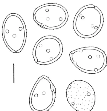
Telia amphigenous on leaves, though predominant on the abaxial side, dark brown, rounded or irregularly shaped by confluence, ca. 0.3–2.4 mm in diameter, slightly pulvinate, pulverulent but with a somewhat fibrous texture as the pedicels appear to stabilise the spore mass; teliospores broadly ellipsoidal, rounded at both ends, not or very slightly constricted at the septum, 53–65 (68)×38– 46μm (mean 59.8×42.2 μm), pedicels stout, thick-walled, hyaline, up to 120μm long, inserted basally but generally Fig. 9 Puccinia pienarii (type), teliospores. Scale bar=20μm
shifted sidewards or, more rarely, spores stalked laterally, spore wall 7–9 μm thick, at germ pores up to 12 μm thick, laminate, with an outer, ochraceous layer ca. 4–5 μm thick and an inner, light chestnut brown, ca. 3–4-μm-thick layer (layers are not sharply delimited but tend to blend), outer wall layer more or less evenly ornamented by flat, broad warts (ca. 1μm in diameter) about 2.5–3.5 μm apart and by an inconspicuous ripple mark-like pattern that seems to
underly the surface, germ pores apical in distal cells and subequatorial to equatorial in the proximal cell, without papillae but with a thickening of the outer wall layer and a pit in the inner layer. In the telia, very few, mostly old, urediniospores were found with a bilaminate, echinulate wall and numerous, scattered, papillate germ pores.
Material examined: Puccinia othonnae. South Africa, Transvaal, Olifantsfontein, on O. natalensis Sch. Bip., leg. Fig. 10 a Puccinia othonnae (type), teliospores. Scale bar=10μm.
b Puccinia othonnae (type), teliospores; note the delicately verrucose spore surface. Scale bar=10 μm. c Puccinia othonnae (type), teliospores; note the fine ripple mark pattern underlying the verrucose ornament. Scale bar=10 μm. d Puccinia othonnoides (type),
telio-spores. Scale bar=10 μm. e Puccinia othonnoides (type), uredinio-spores; note the ripple mark pattern of the spore wall. Scale bar=10 μm. f Puccinia othonnoides (type), urediniospores; note the striate pattern of the spore wall in the optical section. Scale bar=10μm
Pienaar (no. 260), 14 April 1920 (holotype, PREM 13052). South Africa, Transvaal, Roodepoort, on O. natalensis, leg. I.B. Pole Evans, 4 June 1919 (paratype, PREM 14184).
Puccinia othonnoides R. Berndt, A.R. Wood & E. Uhlmann, sp. nov. (Fig.10d–f).
Etymology: The epithet designates morphological simi-larity to P. othonnae.
Spermogonia et aecia ignota. Uredinia in foliis amphi-gena, subepidermalia, erumpentia, rotundata, ca. 0.5–1 mm diam., ferruginea, pulverulenta, nonnunquam telios evol-ventia; urediniosporae subglobosae ad late ellipsoideae, 27– 45×25–36 μm, pariete 2.5–3.5 μm crasso, in hilo et poris germinationis crassiori (interdum usque ad 8μm), bilami-nato, lamina exteriori tenui, straminea vel subhyalina, sate dense moderate delicateque echinulata, lamina inferiori crassiori, aurea ad dilute castanea, striis finissimis, impres-sioni digitalis similibus crebre praedita, poris germinationis sparsis, 9–13, infra incrassatis, extra papillis latis humil-ibusque. Telia urediniis similia, atro-brunnea ad nitide atra, pulvinata, textura subfibrosa, 0.5–1.2 mm diam.; teliosporae late ellipsoideae, late obovoideae vel rariter -subglobosae, secundum septum non vel leniter constrictae, (39) 42–64×32–48 μm, pariete (5) 6–8 (10) μm crasso, usque ad 12 μm in poris germinationis, indistincte bilaminato lamina exteriori tenui, usque ad 2 μm crasso, ochracea ad aurantio-brunnea, levi vel inconspicue verru-cosa verrucis humilibus, lamina interiori crassiori, aurantio-brunnea ad castanea, poris germinationis apicalibus in cellula distali, aequatorialibus vel subaequatorialibus in cellula proximali, papillatis; pedicello basaliter inserto vel obliquo, rariter laterali, usque ad 150μm longo, hyalino vel subhyalino, crasse tunicato et persistenti. Mesosporae absunt, vel rarae ad sparsae adsunt.
In foliis Othonnae specierum (Asteraceae).
Uredinia amphigenous on leaves, originally subepider-mal, erumpent, rounded, ca. 0.5–1 mm in diameter, ferrugineous, pulverulent, sometimes giving rise to telia; urediniospores subglobose to broadly ellipsoidal, 27– 45×25–36 μm, spore wall 2.5–3.5 μm thick, thicker at germ pores and generally at hilum (there occasionally up to 8μm), two-layered with a thin, straw-coloured or subhya-line outer layer and a thicker, golden to light chestnut brown inner layer, germ pores numerous, scattered, mostly 9–13, with an internal thickening and a broad flat papilla, outer wall layer evenly moderately fine and rather densely echinulate, inner layer with an inconspicuous to conspicu-ous ripple mark or fingerprint-like pattern around and between the germ pores. Telia similar to uredinia, blackish brown to shiny black, pulvinate with subfibrous texture, ca. 0.5–1.2 mm in diameter; teliospores broadly ellipsoidal, broadly ovoidal or rarely subglobose, not or very slightly constricted at the septum, (39) 42–64×32–48 μm, spore
wall (5) 6–8 (10) μm thick, at germ pores to 12 μm, two-layered, with an indistinctly delimited thin, ochraceous, golden or orange brown outer layer up to 2μm thick and a much thicker, orange brown to chestnut brown inner layer, entirely smooth to (very) inconspicuously verrucose by flat warts, sometimes only visible on young spores, germ pores apical in the distal cell and equatorial to subequatorial in the proximal cell, with a pit in the inner wall layer and a thickening of the outer one, pedicels inserted basally or slightly shifted sidewards, rarely almost lateral, persistent and up to 150μm long, hyaline to subhyaline, thick-walled. One-celled mesospores absent, rare or scattered.
On leaves of Othonna species (Asteraceae)
Holotype (PREM): South Africa, Western Cape Prov-ince, at the side of the road to Little Boy Kraal ca. 500 m from road between Citrusdal and Algeria, on Othonna cf. coronopifolia L., leg. E. Uhlmann and R. Berndt (no. RSA 26), 21 October 2004 (isotype Z+ZT).
Paratypes: South Africa, Western Cape Province, Cederberg Mts., at Wolfberg, on Othonna sp., leg. R. Berndt (no. RSA 7), 13 October 2004 (PREM, Z+ZT). South Africa, Western Cape Province, road from Citrusdal to Ceres (R 303), towards Middelberg Pass, on Othonna cf. parviflora L., leg. E. Uhlmann and R. Berndt (no. RSA 38), 22 October 2004 (PREM, Z+ZT). South Africa, Western Cape Province, on side road to Touws Rivier, ca. 1 km after turn-off from Ceres to Calvinia road, on Othonna sp., leg. E. Uhlmann and R. Berndt (no. RSA 46), 23 October 2004 (PREM, Z+ZT). South Africa, Western Cape Province, on road from Barrydale to Heidelberg, towards Tradouw Pass, on O. cf. parviflora, leg. E. Uhlmann and R. Berndt (no. RSA 56), 24 October 2004 (PREM, Z+ZT). South Africa, Western Cape Province, Cape Peninsula, Miller’s Point S Simon’s Town, on Othonna arborescens L., leg. A.R. Wood, 18 November 2000 and 28 September 2002 (no. 253 and 398).
Puccinia othonnoides appears to be a variable species. The investigated collections differ mainly with regard to teliospore size and ornament and to urediniospore size and shape. The differences are listed in Table1. All collections share essential characters, however, and we consider it best to regard them as pertaining to a single species as the characters intergrade and sizes overlap. It is unclear at the moment whether the observed differences correlate with different Othonna hosts or just represent small sections from a morphological continuum.
Comparing the characters listed in Table 1, one can see that P. othonnoides is very similar to P. othonnae and U. othonnae: the urediniospores have many scattered germ pores and a thick, inconspicuously to distinctly bilaminate wall whose inner layer generally shows a fine striate or ripple mark pattern (Fig.10e,f). The teliospores are thick-walled and two-layered with germ pores apical and more or less equatorial (Fig. 10d). Puccinia othonnae differs from
P. othonnoides, however, by larger teliospores with a thick, distinctly verrucose outer wall layer and an underlying ripple mark pattern (Fig.10c). The latter may not always be visible. Uredo othonnae [South Africa, Western Cape Province, Koude Bokkeveld, Wagedrift, altitude of 5,500 ft, on Othonna multicaulis Harv. var., leg. Schlechter, 21 January 1897 (type, S F32636)] could not be assigned to either species with confidence. Its urediniospores (Fig.11) have fewer germ pores (6–10, mostly 8) than those of P. othonnoides, otherwise it is very similar. Our study showed that P. othonnae has urediniospores, too, but they were too scanty to make a proper comparison.
Puccinia butleri Syd. & P. Syd. 1906. Annales Myco-logici 4:431. New for South Africa (Fig.12).
Rust collected on Launaea dregeana DC. (Eastern Cape Province, Kayser’s Beach S of East London, 20 August 2002, leg. A.R. Wood) was determined as P. butleri, known so far from India and Pakistan. The South African specimen (Fig.12) differs in certain aspects from the type of P. butleri (Z+ZT): it has teliospores with thinner walls (ca. 1.5–2 μm versus ca. 2.5 μm) with a more prominently verrucose
ornament and urediniospores that are larger [22.5–28 (30)×18.5–22.5 μm (mean 24.9×20.9 μm) versus 18– 21.5×17.5–21 μm (mean 20.7×19.4 μm)]. These differ-ences may lie within the variability of P. butleri, but it will be interesting to collect more material from southern Africa to evaluate whether they occur constantly.
As the original description of P. butleri is short, we present here additional observations obtained from the type (Z+ZT): Urediniospores obovoidal to subglobose, 18– 21.5×17.5–21 μm (mean of 20 spores 20.7×19.4 μm), spore wall ochraceous to light brown, ca. 1.5 μm thick, moderately fine and closely echinulate, spines mostly 1.5– 2.5 μm apart, germ pores inconspicuous, about five, scattered. Teliospores broadly ellipsoidal to elongate broad-ly ellipsoidal, rounded at both ends and slightbroad-ly constricted at the septum, 36–42.5×24–29 μm (mean 39.2×25.6 μm), pedicels subhyaline, thin-walled, up to as long as the spores but most often breaking shorter, spore wall light chestnut brown, about 2.5μm thick, not thickened at the apex, rather densely verrucose by flat warts, germ pores apical or subapical in the distal cell, equatorial to subequatorial in the proximal cell, with flat, rather broad, ochraceous papillae. Table 1 Characters of telio- and urediniospores of rust fungi on Othonna
Specimen Teliospores Urediniospores
P. othonnoides, (Wood no. 398, [II] and no. 253 [III]), on O. arborescens
(39) 44–58 (65)×33–44 μm (mean 51.1×37.3), wall 6–7 μm thick, at germ pores 8–12 μm, bilaminate, smooth
32–36×28–36 μm (mean 34.7×31.6), wall
indistinctly bilaminate, 3–3.5 μm thick, inner layer with ripple mark pattern, germ pores 9–12 (14?) P. othonnoides (RSA 7),
on Othonna sp.
42–52×34–40 μm (mean 45.9×36.9), wall about 6 μm thick, at germ pores ca. 8μm, bilaminate, smooth (but indistinct warts visible in young spores)
32–39.5×29–33 μm (mean 36.2×30.8), wall bilaminate, 3–4 μm thick, with ripple mark pattern, germ pores (10) 11–13
P. othonnoides, (RSA 26, holotype), on O. cf. coronopifolia
45–57×31.5–38 (41) μm (mean 51.2×35.3), wall 5–7 μm thick, apically to 12 μm, indistinctly bilaminate, smooth
27–33×26–29 μm (mean 29.6×27.9), wall 2.5–3 μm thick, thicker at hilum, uniform, with ripple mark pattern, germ pores (8) 9–11
P. othonnoides, (RSA 38), on O. cf. parviflora
44–58 (60)×36–42.5 μm (mean 49.1×39.7), wall 7–10 μm thick, apically to 12 μm, appearing smooth to very inconspicuously verrucose with flat warts
31–37.5×27–33.5 μm (mean 34.3×29.8), wall 2.5–3 μm thick, bilaminate, inner layer with ripple mark pattern, germ pores 9–12 P. othonnoides, (RSA 46),
on Othonna sp.
53.5–64×40–48.5 μm (mean 59.1×45.0), wall 7–8 μm thick, apically to 12μm, indistinctly bilaminate, indistinctly verrucose with small flat warts
33.5–45×25.5–31 μm (mean 36.9×28.1), wall about 2.5μm thick, much thicker at hilum and slightly thicker apically, indistinctly bilaminate, inner layer with ripple mark pattern, germ pores 12–14 P. othonnoides, (RSA 56),
on Othonna sp.
42.5–48×33.5–38 (40) μm (mean 45.0×36.1), wall 5.5–7 μm thick, apically to 9 μm, indistinctly bilaminate, almost smooth but with inconspicuous flat warts
28–35×28–32 μm (mean 31.2×29.6), wall about 2.5μm thick, much thicker at hilum, very
indistinctly bilaminate, wall with ripple mark pattern, germ pores mostly 11–13
P. othonnae 53–65 (68)×38–46 μm (mean 59.8×42.2), wall 7–9 μm thick, at germ pores to 12μm, distinctly bilaminate with thick outer layer, verrucose and with underlying “ripple mark-pattern” (Doidge: 43–50×33–40 μm, wall 6.5–8 μm thick, not thickened at pores or apically)
Very scarce: with numerous, scattered, papillate germ pores and a two-layered, echinulate wall, ripple mark pattern not observed
Uredo othonnae (holotype) Not present (31) 33–39×(29) 31–35 μm (mean 35.9×32.7),
wall ca. 3–3.5 μm thick, bilaminate with ripple mark pattern of inner layer, germ pores 6–10. (Jørstad: 30–42×25–32 μm, wall 3–5 μm thick, bilaminate, germ pores ca. 8, scattered)
Uromyces bidenticola Arthur 1917. Mycologia 9:71. New for South Africa, formerly reported as Uromyces bidentis Lagh.
Doidge (1927) reported U. bidentis from Natal and the Cape Province. She stated that only the urediniospores had been found so far in South Africa. Uromyces bidentis is a micro-cyclic species, however, producing only teliospores (Arthur 1917). Doidge’s reports refer to U. bidenticola, therefore, and U. bidentis must be deleted from the South African rust list.
Material examined: South Africa, Western Cape Prov-ince, Cape Peninsula, Kirstenbosch Botanical Garden, on weed Bidens cf. pilosa L., 4 November 2004, leg. E. Uhlmann and R. Berndt.
On Dioscoreaceae
Puccinia dioscoreae-mundtii R. Berndt, A.R. Wood, & E. Uhlmann sp. nov. (Figs.13and14).
Etymology: Named after the host species, Dioscorea mundtii, a climber in forest and thicket vegetation in South Africa.
Uredinia in pagina abaxiali foliorum dense sparsa, subepidermalia, minuta, 0.2–0.4 mm diam. vel majores si coalescent, primo pallide ochracea, deinde aurantio-brun-nea, pulverulenta; urediniosporae obovoideae vel pyri-formes, 23.5–36×18–20.5 μm (mean 28.3×19.2 μm), pariete subhyalino ad pallide ochraceo, ca. 1 μm crasso, moderate dense et aliquantum delicate echinulato, spinis 1.5–2 μm inter se distantibus, aegre echinulato hilum versus, poris germinationis obscuris, 4–5 sparsis vel cum duobus poris basalibus et duobus subapicalibus. Telia non visa; teliosporae inter urediniosporas, ellipsoideae, clavatae, rariter late ellipsoideae vel oblonge ellipsoideae, paulum constrictae secundum septum, cellula distali ellipsoidea, late ellipsoidea vel subglobosa, quam cellula proximali, cuneato, breviori est, 39–65×14.5–20 μm (mean 49.4×17.9μm), pariete levi, ca. 0.5–1 μm crasso, apicaliter 1–2 μm, subhyalino usque ad pallide ochraceo, poris germinationis obscuris, pedicellis tenue tunicatis, plus minusve hyalino, brevibus; sporae tricellulares rariter adsunt.
Fig. 11 Uredo othonnae (type), urediniospores; a small section of the ripple mark pattern observable within the spore wall has been delineated (not to scale). Scale bar=20μm
In foliis Dioscorea mundtii Baker (Dioscoreaceae) Uredinia densely scattered on abaxial side of leaves that are marbled by blurred discoloured to ochraceous or pallid green areas, subepidermal, tiny, 0.2–0.4 mm in diameter, sometimes larger when confluent, pallid ochraceous when young to orange brown when older, pulverulent; uredinio-spores obovoidal or pyriform, 23.5–36×18–20.5 μm (mean 28.3×19.2μm), spore wall subhyaline to light ochraceous, ca. 1 μm thick, moderately dense and rather finely echinulate (spines ca. 1.5–2 μm apart), echinulation vanishing towards the hilum; thus, more or less bald around hilum, germ pores difficult to discern, mostly by a thickening of the wall, four to five, scattered or, quite often, two pores basal and two subapical. Telia not seen, uredinia in which teliospores were found appeared slightly wax-like; teliospores ellipsoidal, clavate, rarely broadly ellipsoidal or oblong ellipsoidal, slightly constricted at septum, the distal cell ellipsoidal, broadly ellipsoidal or subglobose most often shorter than the proximal, cuneate one, 39–65×14.5– 20μm (mean 49.4×17.9 μm), wall smooth, ca. 0.5–1 μm thick, at the apex 1–2 μm, subhyaline to light ochraceous, germ pores not seen, pedicels thin-walled, more or less hyaline and breaking shortly from the hilum; three-celled spores occur rarely.
On Dioscorea mundtii Baker (Dioscoreaceae)
Holotype (PREM): South Africa, Western Cape Prov-ince, at road from Knysna to George shortly after Homtini pass, in the afromontane forest of a river valley, on Dioscorea mundtii Baker (Dioscoreaceae), 27 October 2004, leg. E. Uhlmann and R. Berndt. Isotype in Z+ZT.
Additional material studied: South Africa, Western Cape Province, Victoria Bay E of George, on D. mundtii, 25 July 2001, leg. A.R. Wood (no. 337, only II present).
The present species differs from known Puccinia spp. on Dioscorea, namely Puccinia dioscoreae Kom. and Puccinia valida Arthur, by uniformly thin-walled teliospores (Fig.13). Its uredinial stage appears to be different from that of the described Uredo spp. on Dioscorea, though it is quite similar to Uredo dioscoreae-filiformis, which differs, however, by deep-seated uredinia, which liberate thicker-walled uredinio-spores through a central aperture (Ono1982).
On Fabaceae
Uredo otholobii R. Berndt, A.R. Wood & E. Uhlmann, sp. nov. (Fig. 15a,b).
Etymology: Named after the host genus Otholobium of Fabaceae
Fig. 13 Puccinia dioscoreae-mundtii (type), teliospores. Scale bar=20μm
Fig. 14 Puccinia dioscoreae-mundtii (type), urediniospores. Scale bar=20μm
Uredinia foliicola, amphigena, parva, pulverulenta, cas-tanea; urediniosporae obovoideae, late ellipsoideae vel (rariter) subglobosae, 21–27×18–22 μm (medium 23.4×15.0 μm), pariete brunneo, ca. 1.5–2 μm crasso (in poris germinationis parum crassiore), delicate moderate denseque echinulato, spinis inter se 2–3 μm distantibus, poris germinationis plerumque 2–3, aequatorialibus et aequidistantibus, papillis subhyalinis, humilibus vel mod-erate altis et tumore debili interno praeditae.
In foliis Otholobii cf. candicantis (Fabaceae, Psoraleae) Uredinia amphigenous on leaves, small, chestnut brown, pulverulent; urediniospores obovoidal, broadly ellipsoidal or rarely subglobose, 21–27×18–22 μm (mean 23.4×15 μm), spore wall brown, ca. 1.5–2 μm thick, slightly thicker at the germ pores, evenly fine and moderately dense echinulate, spines mostly 2–3 μm apart, two to three germ pores, in most cases approximately equatorial and equidistant with a flat to moderately high subhyaline papilla and a slight internal swelling.
On leaves of Otholobium cf. candicans (Fabaceae, Psoraleae)
Holotype (PREM): South Africa, Western Cape Province, Barrydale, at the southern border of the village, on Oth-olobium cf. candicans (Eckl. & Zeyh.) C.H. Stirt., 23 October 2004, leg. E. Uhlmann and R. Berndt (isotype Z+ZT).
Additional material investigated: South Africa, Western Cape Province, Stellenbosch, Jan Marais Nature Reserve, on Otholobium hirtum (L.) C.H. Stirton, 16 September 2002, leg. A.R. Wood (no. 394). South Africa, Western Cape Province, E Bredasdorp, De Hoop Nature Reserve, on
Otholobium fruticans (L.) C.H. Stirton, 24 June 1998, leg. A.R. Wood (no. 53).
Other rust fungi on Otholobium are Phakopsora meibo-miae (Arthur) Arthur with paraphysate uredinia and Uromyces psoraleae Peck, a demicyclic rust. Uredo otholobii is also different from U. psoraleae-polystictae Doidge and Uromyces abbreviatus Arthur, which occur on other members of the tribe Psoraleae.
Rust on Lotononis (Fabaceae, Crotalarieae)
Rust collected on Lotononis falcata (E. Mey.) Benth. revealed the telial stage of Uromyces sp. together with the uredinial stage. According to the authors’ knowledge, the only known rust on Lotononis spp. is Uredo lotononi Doidge. A comparison of both rust fungi did not reveal significant differences. Teliospores were found in the uredinia of the type specimen of U. lotononi, and therefore, a new species, Uromyces lotononidicola, is proposed for the holomorph:
Uromyces lotononidicola R. Berndt sp. nov. (Figs. 16 and 17)
Syn. U. lotononi Doidge 1927. Bothalia 2:213.
Uredinia in foliis amphigena, sparsa, minuta (0.3–0.5 mm diam.), subepidermalia, ferruginea ad dilute badia, pulver-ulenta; urediniosporae obovoideae, late ellipsoideae vel, rariter, subglobosae, 25–33×20–24.5 μm (medium 28.1×22.5 μm), pariete dilute brunneo, ca. 2 μm crasso, delicatissime echinulato, 1.5–2.5 μm inter spinas, poris Fig. 15 a Uredo otholobii
(type), urediniospores; optical section. Scale bar=10μm. b Uredo otholobii (type), urediniospores; echinulate spore surface. Scale bar=10μm
germinationis 3–5 (praecipue 3–4), aequatorialibus vel sparsis cum papillis latis, hyalinis et echinulatis. Telia non visa; teliosporae urediniosporis inmixtae, deciduae, late ellipsoi-deae, obovoideae (ad subglobosae), (23) 25–32×20–23 μm (medium 27.7×21.7μm), pariete dilute brunneo, ca. 2–3 μm crasso, grosse denseque verrucoso, cum verrucis ca. 1.5– 2.5μm latis, conicis vel subacutis, poro germinationis apicali, papilla lata, ca. 1.5–2.5 μm alta, subhyalina, rugosa vel rugulosa praedito; pedicello hyalino fragili.
In foliis Lotononidis cytisoidis Benth. (Fabaceae) Uredinia amphigenous on leaves, scattered, tiny (0.3– 0.5 mm in diameter), subepidermal, ferrugineous to light chestnut brown, pulverulent; urediniospores obovoidal, broadly ellipsoidal or, more rarely, subglobose, 25– 33×20–24.5 μm (mean 28.1×22.5 μm), wall light brown, ca. 2 μm thick, very finely echinulate, ca. 1.5–2.5 μm between spines, germ pores 3–5 (predominantly 3–4), almost equatorial to scattered, with a moderate internal thickening of the wall and a broad, hyaline, echinulate papilla. Telia not seen; teliospores among the uredinio-spores, broadly ellipsoidal, obovoidal (to subglobose), (23) 25–32×20–23 μm (mean 27.7×21.7 μm), spore wall light brown, ca. 2–3 μm thick, coarsely verrucose, warts ca. 1.5–
2.5 μm broad, conical to subacute, densely situated, germ pores apical with a broad, subhyaline rugose to rugulose papilla ca. 1.5–2.5 μm high, spores deciduous with fragile, hyaline and delicate pedicels that normally break off shortly below the hilum.
On leaves of Lotononis cytisoides Benth. (Fabaceae) Holotype: South Africa, Natal, Mont-aux-Sources, 20 April 1919, leg. Mogg (holotype of U. lotononi, PREM 12955).
Additional material studied: Paratype (PREM): South Africa, Northern Cape Province, at road no. 7 north of Gariep, on Lotononis falcata Benth., 14 October 2004, leg. E. Uhlmann and R. Berndt (isoparatype in Z+ZT).
Uredo lotononi: South Africa, Pretoria Distr., Irene, on Lotononis hirsuta Schinz, 14 March 1917, leg. Pole Evans (paratype of U. lotononi, PREM 10986).
The urediniospores of the specimen from the Northern Cape Province were very similar to those of the type but slightly smaller: Urediniospores broadly ellipsoidal, obo-voidal or subglobose, 22–30 (31.5)×18–22 μm (mean 24.7×20.3 μm), spore wall ochraceous to light brown, about 1.5 (2) μm thick, evenly rather sparsely to moder-ately dense and finely echinulate, three to four germ pores, mostly three, more or less equatorial and equidistant, sometimes almost scattered, with broad, flat, subhyaline papillae. Telia amphigenous on leaves, more rarely on pedicels, subepidermal, first singly then confluent, rounded or elongated, about 0.4–2 mm in diameter, up to 3 mm long, pulvinate, dark chestnut brown, pulverulent; telio-spores broadly ellipsoidal, subglobose or subpyriform, Fig. 16 Uromyces lotononidicola (type), teliospores. Scale bar=15μm
sometimes slightly irregularly deformed, with short remnants of the fragile, thin-walled, hyaline pedicels, 25–32×20.5–25 μm (mean 27.6×22.8 μm), spore wall 2.5–3 μm thick, orange brown to light chestnut brown, densely covered by rather coarse, broadly conical or hemispherical warts, germ pore apical, without or with inconspicuous to conspicuous broad, ochraceous and rugose papilla.
In a slide prepared from PREM 10986 two coarsely verrucose teliospores with apical papillae were present. The urediniospores measured 21.5–29×18–22.5 μm (mean 24.2×19.8 μm) and the specimen is assigned to U. lotononidicola as well. Other Uromyces spp. on members of Crotalarieae, namely Uromyces africanus (Gjærum) Ono, Uromyces bolusii Massee, Uromyces crotalariae (Arth.) Baxter, Uromyces decoratus Syd. & P. Syd., Uromyces harmsianus (Henn.) Doidge, and Uromyces occidentalis Dietel, are clearly different from the present species.
On Orchidaceae
Puccinia aurea Winter 1884. Flora 67:260
Syn.: Puccinia satyrii P. Syd. & Syd. 1903. Monogr. Uredin. Vol. I:594 (Fig.18).
Puccinia rust collected on a sterile orchid in Fynbos vegetation at Bain’s Kloof above Paarl resembled P. aurea and P. satyrii. A comparison was made, therefore, with original material.
Material examined: Puccinia aff. aurea: South Africa, Western Cape Province, Paarl, Witte Rivier at Bain’s Kloof, on sterile orchid (cf. Ceratandra), 11 December 2004, leg. E. Uhlmann and R. Berndt. Puccinia satyrii: South Africa, Western Cape Province, Cape flats near False Bay, on Satyrium carneum R. Br., September 1884, leg. P. Mac Owan (holotype, Berlin) and Rabh.-Winter Fungi Europ. no. 3614 (isotype, Berlin, sub P. aurea).
Puccinia satyrii is very similar to P. aurea and, according to Sydow and Sydow (1904), differs essentially by slightly smaller teliospores and the presence of ure-diniospores. Jørstad (1956a) considered P. satyrii to be conspecific with P. aurea and listed six different host genera. We studied the type of P. satyrii and another specimen issued under P. aurea in Rabh.-Winter Fungi Europ. no. 3614. Our measurements (Table2) show that the teliospores of P. satyrii are not smaller than those of P. aurea, and we follow the opinion of Jørstad (1956a) that both species are the same.
The specimen recently collected at Bain’s Kloof is distinct from P. aurea by smaller teliospores (Fig. 19), which are less thickened at the apex and have a lighter-coloured spore wall. Despite these differences, we prefer to assign this specimen to P. aurea at the moment as it shows the same set of characters in principal (leptosporic teliospores with a much thickened apex and tiny, puncti-form, subcompact telia). Puccinia aurea may therefore be regarded as a variable species able to infect a rather broad spectrum of orchids.
On Solanaceae
Puccinia on Lycium species in Africa and the Near and Middle East
Five species of Puccinia have been described on Lycium spp. in Africa, the Near and Middle East and southern Europe: Puccinia afra Winter (South Africa, Spain), Puccinia lycii Kalchbr. [South Africa, Yemen (Island of Abd-al-Kuri)], Puccinia spinulosa Jørstad (Madagascar), Puccinia turgida P. Syd. & Syd. (Egypt, Israel, Pakistan) and Puccinia verwoerdiana Van der Byl (South Africa). Specimens of Puccinia collected recently on Lycium in Namibia and in South Africa could not be determined by Fig. 18 Puccinia aurea (type of P. satyrii), teliospores. Scale
comparison with available descriptions. To evaluate their status, they were compared to original material of the listed Puccinia species.
The specimen from South Africa was different from the known species and is described as new:
Puccinia rapipes R. Berndt & E. Uhlmann, sp. nov. (Figs.20a,b, and21a)
Etymology: Named after the shape of the swollen teliospore pedicel.
Spermogonia et aecia ignota. Uredinia in foliis amphi-gena, sparsa, parva, subepidermalia, ferruginea, pulveru-lentia, 0.3–1 mm diam.; urediniosporae fusiformes ad ellipsoideae, apicaliter subacutae, basim versus attenu-atae vel truncattenu-atae, 38–60×16.5–22 μm (medium 50.8×18.5 μm), pariete ochraceo ad dilute brunneo, ca. 1.5 μm crasso, apicaliter leniter incrassato usque ad 2– 3 μm, delicate et moderate dense echinulato spinis brevibus, inter se ca. 2.5–3.5 μm distantibus, hilum versus decrescentibus, deinde levi, poris germinationis 3–4, praecipue 4, plusminusve subaequatorialibus et aequidis-tantibus, papillis parvis, humilibus, subhyalinis praeditis. Telia urediniis similia, atrobrunnea vel atra; teliosporae ellipsoideae, late ellipsoideae vel subfusiformes, non vel paululum constrictae ad septum, apicaliter saepe apiculo usque ad 6 μm alto praeditae, 41.5–61×21.5–27 μm (medium 48.8×24.2 μm), pariete castaneo, 1.5–2.5 μm crasso, crebre verruculoso verrucis parvis, humilibus, non-nihil longitudinaliter dispositis, areas fere leves includenti-bus, poro germinationis cellulae distalis subapicali ad aequatoriali, plusminusve aequatoriali in cellula proximali, cum vel sine papillis verruculosis et inconspicuis, pedicello basaliter inserto, levi, subhyalino, dilute brunneo hilum versus, crasse tunicato, ca. 25–30 μm ab hilo vesiculoso inflato vel obovoideo-caudato.
In foliis Lycii cf. ferocissimi Miers (Solanaceae) Spermogonia and aecia not present. Uredinia amphige-nous on leaves, scattered, subepidermal, small, more or less rounded, 0.3–1 mm in diameter, ferrugineous, pulverulent, often developing to telia later; urediniospores fusiform to ellipsoidal, apically subacute, tapering towards the hilum or truncate basally, 38–60×16.5–22 μm (mean 50.8×18.5 μm), spore wall ochraceous to light brown, ca. 1.5 μm thick, apically slightly thickened to 2–3 μm, rather finely and moderately densely echinulate with short spines, spines spaced about 2.5–3.5 μm, or slightly closer apically, becoming smaller towards the hilum and fading, smooth Table 2 Spore measurements of Puccinia aurea (including P. satyrii)
Taxon Teliospores Average Urediniospores Average
P. aurea [after Sydow and Sydow (1904)]
35–65×13–20 μm, apex thickened to 16μm
– Not described
P. satyrii, holotype 36–56×15–20 μm, apex thickened to 14μm [after Sydow and Sydow (1904): 32–52×13–19 μm, apex thickened to 13–19 μm]
45.7×17.6μm Not seen in available fragment of type [after Sydow and Sydow (1904): globose or subglobose, 16–24 μm in diameter]
–
P satyrii (sub P. aurea), isotype issued in Rabh. Fg. Europ. no. 3614
(37.5) 44–61 (64)×16–20 μm, apex thickened to 8–17 μm [after Sydow and Sydow (1904): 35–65×13–20 μm]
50.9×17.9μm Only a few collapsed spores present in specimen from B [after Jørstad (1956a): 19–26×15–19 μm, germ pores obscure]
–
Puccinia aff. aurea, (Bain’s Kloof)
30–47×13.5–18 μm, apex thickened to 6–11 μm
38.6×15.8μm 18–23.5×14.5–18 μm, germ pores not seen
20.8×16.4μm
Fig. 19 Puccinia aff. aurea (specimen from Bain’s Kloof), telio-spores. Scale bar=20μm
around the hilum, three to four germ pores, predominantly four, more or less subequatorial and equidistant, with small and flat, subhyaline papillae. Telia like uredinia, blackish-brown to black; teliospores ellipsoidal, broadly ellipsoidal or subfusiform, not or hardly constricted at the septum, apically often with an inconspicuous to conspicuous yellow-brown apiculus up to 6 μm high, 41.5–61×21.5– 27μm (mean 48.8×24.2 μm), spore wall chestnut brown, 1.5–2.5 μm thick, densely verrucose with small, flat,
irregular warts that tend to be arranged in broad longitudi-nal stripes including almost smooth areas, germ pores subapical to equatorial in the distal cell and more or less equatorial in the proximal cell, without or with inconspic-uous verrucose papillae, pedicel broadly (8–10 μm) at-tached at base of spore, smooth, light brown below the hilum, then subhyaline, slightly thick-walled, swelling conspicuously ca. 25–30 μm from the hilum and then obovoid-caudate to almost globose.
Fig. 20 a Puccinia rapipes (type), teliospores; in one spore, the fine warty spore surface has been delineated. Scale bar=20μm. b Puccinia rapipes (type), urediniospores. Scale bar=20μm. c Puccinia lycii (type), teliospores; in one spore, a section of the surface has been delineated with the verrucose ornament. Scale bar=20μm. d Puccinia lycii (type), urediniospores. Scale bar=20 μm
On leaves of Lycium cf. ferocissimum Miers (Solanaceae) Holotype (PREM): South Africa, Western Cape Prov-ince, Simon’s Town, on the shore above the penguin sanctuary at Boulders, on Lycium cf. ferocissimum, leg. E. Uhlmann and R. Berndt, 1 November 2004 (isotype Z+ZT). The present species is morphologically intermediate between P. lycii and P. afra. It differs from P. afra by more slender and thinner-walled teliospores that are finely
verrucose and by smaller, thinner-walled urediniospores that are more finely echinulate and have inconspicuously papillate germ pores. It is very similar to P. lycii but has more slender and longer, thinner-walled, often apiculate teliospores and smaller urediniospores, which are more finely echinulate and hardly thickened apically.
Puccinia lycii Kalchbr. 1882. Grevillea 11:21 (Figs.20c,d and21b).
Syn.: P. verwoerdiana Van der Byl 1927. S. Afr. J. Science 24:226.
The diagnosis of P. verwoerdiana is very short and does not allow positioning the species among the other Puccinias on Solanum. A study of the type (PREM 46255, Fig.21b) did not reveal significant differences to P. lycii (type, Z+ZT, Fig. 20c,d). Puccinia verwoerdiana is therefore reduced to a synonym of the latter.
Puccinia spinulosa Jørstad 1957. Arkiv för Botanik 3 (17):592. New report for Namibia (Fig. 21c).
Syn.: P. turgida auct., non P. Syd. & Syd.: Mennicken, Maier and Oberw. 2005. Mycol. Progr. 4:69.
Rust collected in Israel on Lycium europaeum L. by Bornmüller was first assigned to P. lycii by Magnus (1898), but was later regarded as a separate species, P. turgida (Sydow and Sydow1904). It has been known hitherto only from the Near and Middle East but was reported recently from Namibia by Mennicken et al. (2005). We studied the Namibian material and found that the specimens are different from P. turgida but not distinguishable from P. spinulosa described by Jørstad (1956a) from Madagascar. This is the first report of the latter rust from outside Madagascar and a new report for Namibia.
A specimen of P. lycii reported from the Island of Abd-al-Kuri (Yemen) by Gjærum (1987) may belong to P. turgida as the location is close to the reported area of this rust.
The Puccinias on Lycium considered here share a set of characteristic morphological traits: the proximal or central parts of their teliospore pedicels swell conspicuously in aqueous fluids, their teliospores are verrucose and have about equatorial germ pores and the urediniospores are slender with more or less equatorial pores and an even echinulation that becomes very fine or fades towards the hilum. This similarity clearly indicates that these rusts are closely related and belong to a natural group. It may also suggest that speciation within this group took place relatively recently. Fukuda et al. (2001) studied the phylogeny and biogeographical aspects of Lycium with a cpDNA sequence analysis. Their results indicate that Lycium originated in the New World from where it dispersed to South Africa, and that the Eurasian Lycium spp. probably derive from South African ancestors. With Fig. 21 a Puccinia rapipes (type), teliospores. Scale bar=10μm.
b Puccinia lycii (type of P. verwoerdiana, PREM 46255), teliospores. Scale bar=10 μm. c Puccinia spinulosa (Namibian specimen), teliospores. Scale bar=10μm