HAL Id: hal-02655515
https://hal.inrae.fr/hal-02655515
Submitted on 29 May 2020
HAL is a multi-disciplinary open access
archive for the deposit and dissemination of
sci-entific research documents, whether they are
pub-lished or not. The documents may come from
teaching and research institutions in France or
abroad, or from public or private research centers.
L’archive ouverte pluridisciplinaire HAL, est
destinée au dépôt et à la diffusion de documents
scientifiques de niveau recherche, publiés ou non,
émanant des établissements d’enseignement et de
recherche français ou étrangers, des laboratoires
publics ou privés.
plastidial type A methionine sulfoxide reductases
Nicolas Rouhier, Brice Kauffmann, Frédérique Tete-Favier, Pasquale
Palladino, Pierre Gans, Guy Branlant, Jean-Pierre Jacquot, Sandrine
Boschi-Muller
To cite this version:
Nicolas Rouhier, Brice Kauffmann, Frédérique Tete-Favier, Pasquale Palladino, Pierre Gans, et al..
Functional and structural aspects of poplar cytosolic and plastidial type A methionine sulfoxide
re-ductases. Journal of Biological Chemistry, American Society for Biochemistry and Molecular Biology,
2007, 282 (5), pp.3367-3378. �10.1074/jbc.M605007200�. �hal-02655515�
Functional and Structural Aspects of Poplar Cytosolic and
Plastidial Type A Methionine Sulfoxide Reductases
*
Received for publication, May 24, 2006, and in revised form, November 15, 2006 Published, JBC Papers in Press, November 29, 2006, DOI 10.1074/jbc.M605007200
Nicolas Rouhier‡1,2, Brice Kauffmann§1, Fre´de´rique Tete-Favier§, Pasquale Palladino§, Pierre Gans¶, Guy Branlant储,
Jean-Pierre Jacquot‡, and Sandrine Boschi-Muller储
From the‡UMR 1136 INRA-UHP, Nancy Universite´s, Interactions Arbres Microorganismes, IFR 110, Faculte´ des Sciences et Techniques, BP 239, 54506 Vandoeuvre Cedex,§UMR 7036 CNRS-UHP, Nancy Universite´s, LCM3B, Groupe de Biocristallographie, Faculte´ des Sciences et Techniques, BP 239, 54506 Vandoeuvre Cedex,¶Laboratoire de Re´sonance Magne´tique Nucle´aire, Institut de Biologie Structurale, CEA-CNRS-UJF “Jean-Pierre Ebel,” 38027 Grenoble Cedex 1, and储UMR 7567 CNRS-UHP, Nancy Universite´s, Laboratoire MAEM, Groupe Enzymologie Mole´culaire, Faculte´ des Sciences et Techniques, BP 239, 54506 Vandoeuvre Cedex, France
The genome of Populus trichocarpa contains five methionine sulfoxide reductase A genes. Here, both cytosolic (cMsrA) and plastidial (pMsrA) poplar MsrAs were analyzed. The two recom-binant enzymes are active in the reduction of methionine sulf-oxide with either dithiothreitol or poplar thioredoxin as a reductant. In both enzymes, five cysteines, at positions 46, 81, 100, 196, and 202, are conserved. Biochemical and enzymatic analyses of the cysteine-mutated MsrAs support a catalytic mechanism involving three cysteines at positions 46, 196, and 202. Cys46is the catalytic cysteine, and the two C-terminal cys-teines, Cys196and Cys202, are implicated in the thioredoxin-de-pendent recycling mechanism. Inspection of the pMsrA x-ray three-dimensional structure, which has been determined in this study, strongly suggests that contrary to bacterial and Bos tau-rus MsrAs, which also contain three essential Cys, the last C-ter-minal Cys202, but not Cys196, is the first recycling cysteine that forms a disulfide bond with the catalytic Cys46. Then Cys202 forms a disulfide bond with the second recycling cysteine Cys196 that is preferentially reduced by thioredoxin. In agreement with this assumption, Cys202is located closer to Cys46compared with Cys196and is included in a202CYG204signature specific for most plant MsrAs. The tyrosine residue corresponds to the one described to be involved in substrate binding in bacterial and B. taurus MsrAs. In these MsrAs, the tyrosine residue belongs to a similar signature as found in plant MsrAs but with the first C-terminal cysteine instead of the last C-terminal cysteine.
The production and accumulation of reactive oxygen and nitrogen intermediates, inherent to metabolic processes such as respiration or photosynthesis or to stress conditions, initiate oxidative reactions that affect the biochemical constituents of the cells (1, 2). Living organisms use different strategies to
pre-vent oxidative damage and lethal effects that would result from these compounds. Reactive species are trapped and degraded, or modifications that occur anyway are reversed by repair sys-tems, and finally nonrepaired macromolecules can be degraded and removed. Methionine residues of proteins were shown to be one of the preferred targets of oxidation with the formation of methionine sulfoxide (MetSO)3(3). Enzymes named
methi-onine sulfoxide reductases were found to catalyze the reduc-tion of MetSO back to methionine residues (4, 5). The conse-quences of this side-chain modification are variable and can be partial to protein unfolding (6, 7) and modification of biological functions (8 –10). Sometimes surface methionine residues can undergo oxidation without much impact on the protein prop-erties, and this modification can be seen as a mechanism to scavenge oxidative species in a detoxification process based on methionine sulfoxide reductase activity (11). Because of its asymmetric sulfur atom, MetSO exists as two stereoisomeric forms, Met-(S)-SO and Met-(R)-SO. Their reduction back to methionine is catalyzed by two structurally unrelated classes of Msr, MsrAs are specific for Met-(S)-SO, whereas Met-(R)-SO is the substrate of MsrBs.
MsrAs and MsrBs display no significant sequence identity and have different three-dimensional structures. Only three MsrA x-ray structures from Escherichia coli, Bos taurus, and
Mycobacterium tuberculosis and two MsrB structures from
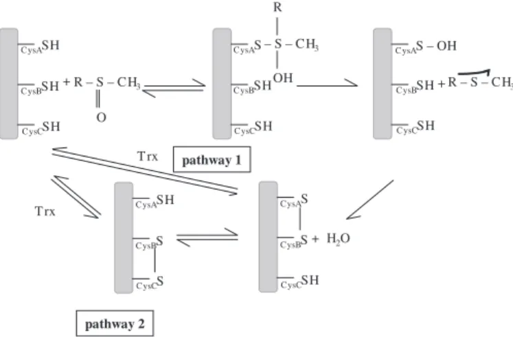
Neisseria species have been described so far (12–16). Both classes of Msrs share, for most of them, a similar three-step chemical mechanism, including the following: 1) a nucleophilic attack of the catalytic CysA residue on the sulfur atom of the sulfoxide substrate leading to the formation of a sulfenic acid intermediate and to the release of 1 mol of Met per mol of enzyme; 2) a formation of an intramonomeric disulfide bond between the catalytic CysA and the recycling CysB with a con-comitant release of 1 mol of water; and 3) a reduction of the CysA–CysB methionine sulfoxide reductase disulfide bond by thioredoxin (Trx) (Fig. 1) (17–19). Nevertheless, for MsrAs, at least three subclasses, based on the number and the position of the recycling Cys residues, have been proposed (20). The
Neis-seria meningitidisand M. tuberculosis MsrA represent the first *The costs of publication of this article were defrayed in part by the payment
of page charges. This article must therefore be hereby marked
“advertise-ment” in accordance with 18 U.S.C. Section 1734 solely to indicate this fact. The atomic coordinates and structure factors (code 2J89) have been deposited in the Protein Data Bank, Research Collaboratory for Structural Bioinformatics, Rutgers University, New Brunswick, NJ (http://www.rcsb.org/).
The nucleotide sequence(s) reported in this paper has been submitted to the Gen-BankTM/EBI Data Bank with accession number(s) AAS46231 and AAS46232.
1Both authors contributed equally to this work.
2To whom correspondence should be addressed. Tel.: 33-3-83684225; E-mail:
nrouhier@scbiol.uhp-nancy.fr.
3The abbreviations used are: MetSO, methionine sulfoxide; Msr,
methio-nine sulfoxide reductase; DTT, dithiothreitol; Trx, thioredoxin; TNB⫺, thionitrobenzoate.
at INRA Institut National de la Recherche Agronomique, on November 9, 2010
www.jbc.org
subclass, characterized by the presence of the recycling CysB in the C-terminal end, and the Bacillus subtilis enzyme represents the second one with the CysB located three amino acids behind CysA. The third subclass, represented by E. coli and B. taurus MsrAs, contains two recycling Cys residues in the C-terminal end and requires the formation not of one but of two successive disulfide bonds. The first one is formed between the catalytic CysA and the recycling CysB. The second one, formed between CysB and the second recycling cysteine CysC, is the one pref-erentially reduced by Trx in the last step (18).4The
denomina-tion of the catalytic cysteines as CysA, -B, and -C is based on the primary structure order.
Most of the MsrAs studied so far are bacterial or mammalian enzymes. In comparison, little has been done concerning plants. Five MsrA-like genes were identified in Arabidopsis
thaliana; one encodes chloroplastic isoforms, and another one is predicted to be targeted to the secretory pathway, and three are cytosolic enzymes (21, 22). The expression of the chloro-plastic isoform, found mainly in photosynthetic tissues, is strongly induced by illumination of etiolated seedlings and is responsive to various oxidative stress conditions (21–23). Moreover, this plastidial MsrA was also shown to maintain chaperonin activity of a small heat-shock protein Hsp21 by pre-venting its denaturation and consequently inactivation after methionine oxidation (24). Finally, the expression of cytosolic MsrAs was also shown to respond to various changing condi-tions as follows: (i) in the dark period of A. thaliana plants growing in short-day conditions (25), (ii) during a pathogen infection by the cauliflower mosaic virus (21), or (iii) during softening of cold-hardened leaves (26). The previous reports
about the plant MsrAs have focused essentially on their expres-sion patterns, but the catalytic mechanism, in particular that related to Trx-dependent recycling process, and the three-di-mensional structure of a plant MsrA have not yet been addressed. One of the first methionine sulfoxide reductase activities that was evidenced for a plant enzyme was established for a chloroplast-targeted MsrA from Brassica napus (28).
In this study, the biochemical and catalytic properties of poplar MsrA are presented, in particular those related to the Trx-depend-ent recycling process. The crystal structure of a poplar MsrA in complex with a mercaptoethanol molecule bound to the catalytic CysA is also reported. Altogether, the data support a Trx-recycling process with formation of a disulfide bond first between the cata-lytic Cys46(CysA) and Cys202(CysC) and then between Cys202and
Cys196(CysB). This latter disulfide bond was reduced by Trx.
MATERIALS AND METHODS
Cloning and Site-directed Mutagenesis
The open reading frame sequences encoding a cytosolic MsrA (cMsrA) and a plastidial MsrA (pMsrA) (respective Gen-BankTM accession numbers AAS46231 and AAS46232) were
cloned by PCR into the expression plasmid pET-3d using as templates a root cDNA library of Populus⫻ interamericana (clone Beaupre´) and a leaf cDNA library of Populus tremula⫻
tremuloides, respectively. Both reactions also contained Pfu DNA polymerase (Promega) and the forward and reverse MsrA oligonucleotides described in Table 1. In the pMsrA cloning, a codon for alanine was inserted downstream from the methio-nine closest to the putative cleavage site and the corresponding N-terminal amino acid sequence starts thus with MANIL. The five cysteines of cMsrA were substituted into serine one by one using either two complementary mutagenic primers per muta-tion (C46S, C81S, C100S, C196S cMsrA and C46S, C196S pMsrA) (Table 1), using a two-step procedure described previ-ously (28), or a one-step procedure when the mutation is directly inserted in the reverse primers (C202S cMsrA and C202S pMsrA). In addition, various combinations of cysteine substitutions by serine were also introduced in cMsrA (C81S/ C100S; C81S/C100S/C196S; C81S/C100S/C202S; and C81S/ C100S/C196S/C202S cMsrAs). The introduction of the muta-tion in the cDNA sequence was verified by DNA sequencing.
Expression and Purification of the Recombinant Proteins
The recombinant plasmids were used to transform the BL21(DE3) E. coli strain, which also contains the helper plas-mid pSBET (29). Cultures of 5 liters of a kanamycin-resistant (50 g/ml) and ampicillin-resistant (50 g/ml) colony were grown at 37 °C and induced by 100Misopropyl 1-thio--D -galactopyranoside in the exponential phase. Bacteria were har-vested by centrifugation, resuspended in buffer A (30 mM Tris-HCl, 1 mMEDTA, 200 mMNaCl) containing 20 mMDTT, and lysed by sonication. The soluble and insoluble fractions were separated by centrifugation (16,000⫻ g, 30 min). The recom-binant wild-type pMsrA was in the soluble fraction and precip-itated between 0 and 50% of ammonium sulfate. All the other recombinant proteins were produced essentially as inclusion bodies with only a small soluble part when cultures were grown at 30 °C without induction. When needed, the insoluble frac-4S. Boschi-Muller and G. Branlant, unpublished results.
FIGURE 1. Catalytic mechanism of the different subclasses of MsrAs. The mechanism implicates two or three cysteinyl residues, depending on the sub-class (CysA, CysB, and possibly CysC) (17). The first step consists of the attack of CysA onto the sulfur of the methionine sulfoxide, leading to a sulfenic acid on CysA and the concomitant release of the methionine. CysB attacks the sulfenic acid intermediate to form a disulfide bond CysA–CysB. Return of the active site to a fully reduced state proceeds either by direct reduction of the CysA–CysB disulfide bond by Trx (pathway 1) or by two thiol-disulfide exchanges via CysC and Trx (pathway 2), depending on the presence or not of a CysC. For MsrAs possessing a CysC, in the absence of reductant, the first turnover leads to the formation of 1 mol of Met per mol of enzyme that accumulates under CysB–CysC disulfide bond form. Thus, CysA is free and is able to reduce another MetSO molecule, explaining why the stoichiometry of the reaction is 2 in the absence of reductant.
at INRA Institut National de la Recherche Agronomique, on November 9, 2010
www.jbc.org
tion was thus resuspended in buffer A in the presence of 20 mM DTT and 8Murea, centrifuged, and then dialyzed against 1 liter of buffer A containing 500 mMurea for at least 5 h at 5 °C (all subsequent steps were realized at that temperature). The extract was centrifuged, and the soluble fraction was dialyzed against 1 liter of buffer A for 5 h and finally centrifuged again. The resulting soluble fraction was purified by exclusion size chromatography onto an ACA 44 column equilibrated in buffer A. The fractions of interest were pooled, dialyzed to remove salts, and separated by DEAE-Sephacel chromatography. The recombinant proteins were eluted around 100 mMNaCl using a linear gradient from 0 to 400 mMNaCl. The purity of the pro-teins was assessed using 15% SDS-PAGE. The protein concen-trations were estimated spectrophotometrically using a molar extinction coefficient of 25,700 M⫺1 cm⫺1 for cMsrA and pMsrA. The proteins were stored at⫺30 °C in buffer A either in the presence of 14 mM-mercaptoethanol or 25 mMDTT.
Crystallization, X-ray Data Collection, Structure Determination, and Refinement
Crystallization of pMsrA was achieved using the hanging-drop vapor-diffusion method in Linbro multiwell tissue culture plates at room temperature. Many crystallization conditions resulted in very thin needles that were not usable for data col-lection and were impossible to improve. Only one condition described here gave suitable crystals for x-ray crystallography. The purified enzyme was concentrated to 40 mg/ml in a solu-tion containing 30 mMTris-HCl, pH 7.0, 14 mM -mercapto-ethanol, and 1 mMEDTA. The crystals were grown from 4-l droplets composed of equal volumes of the protein solution and of the precipitant solution (10% w/v polyethyleneglycol 6000, 2 MNaCl) and equilibrated against 700-l reservoirs. Long nee-dles (1 mm) with a thin triangular cross-section (0.03 mm) appeared after 6 weeks. Crystals were briefly soaked in a cryo-protectant solution (10% v/v methylpentanediol mixed with the precipitant solution) and flash-frozen by fast immersion in nitrogen gas stream at 100 K, maintained during the x-ray dif-fraction experiments performed on beamline BM30A (FIP) at the ESRF.
Crystals belong to space group P31with unit cell parameters a⫽ b ⫽ 68.6 Å, c ⫽ 40.7 Å and contain one monomer per
asymmetric unit. Using a wavelength of 1.009 Å, one native data set was collected up to a resolution of 1.7 Å and processed using DENZO (30). Further details are given in Table 2.
The structure was solved using the molecular replacement method implemented in Molrep (31) of the CCP4 program suite. The initial model used in Molrep consisted of the core (41Gly–Pro194) of the E. coli MsrA structure (Protein Data Bank
entry 1FF3). The molecular replacement solution was submit-ted to the Molrep mode and then to the warpNtrace mode of the Arp/wArp5.1 automatic model building and refinement program (32). It produced a model that contained four polypep-tide chains representing 164 amino acids, with R and Rfree fac-tors of 20.6 and 25.8%, respectively. Manual corrections (in par-ticular, building of the missing residues) and automatic CNS refinement (33) of the model were then performed in an itera-tive procedure, until the model fulfilled satisfactory criteria. The final structure corresponds to 183 amino acids among 204 (residues22Pro–Gly204), 183 water molecules, with R⫽ 19.5%, Rfree⫽ 20.1%. Further details are given in Table 3.
Thiol Content Titration
Known concentrations (generally around 25M) of recom-binant proteins were reduced with 50 mMDTT, extensively dialyzed, and then treated or not with 100 mM L-MetSO for 1 h at room temperature. The proteins were then precipitated on ice by addition of 1 volume of 20% trichloroacetic acid for 30 min. The proteins were pelleted by centrifugation and washed twice with 2% trichloroacetic acid. The pellets were resus-pended in 30 mMTris-HCl, pH 8.0, 1 mMEDTA, and 2% SDS. The concentrations of the proteins were determined spectro-photometrically at this stage, and then 5,5
⬘-dithiobis(nitroben-TABLE 1
Cloning and mutagenic oligonucleotides
The NcoI and BamHI restriction sites are underlined and the mutagenic bases are in boldface characters.
cMsrA forward 5⬘-CCCCCCATGGCAACCAGCACCACCAAT-3⬘
cMsrA reverse 5⬘-CCCCGGATCCTTAACCATAGCATCTAATAGG-3⬘
cMsrA C46S forward 5⬘-GCTCAATTCGGAGCTGGAAGTTTCTGGGGGGTT-3⬘
cMsrA C46S reverse 5⬘-AACCCCCCAGAAACTTCCAGCTCCGAATTGAGC-3⬘
cMsrA C81S forward 5⬘-ACTTACAAGCTGGTATCCACCAACACCACCAAC-3⬘
cMsrA C81S reverse 5⬘-GTTGGTGGTGTTGGTGGATACCAGCTTGTAAGT-3⬘
cMsrA C100S forward 5⬘-TTTGACCCGGAAGTTTCCCCATATACCAACCTC-3⬘
cMsrA C100S reverse 5⬘-GAGGTTGGTATATGGGGAAACTTCCGGGTCAAA-3⬘
cMsrA C196S forward 5⬘-TCTGCTGAAAAAGGTTCCAATGACCCTATTAGA-3⬘
cMsrA C196S reverse 5⬘-TCTAATAGGGTCATTGGAACCTTTTTCAGCAGA-3⬘
cMsrA C202S reverse 5⬘-CCCCGGATCCTTAACCATAGCTTCTAATAGGGTCATTGCA-3⬘
pMsrA forward 5⬘-CCCCCCATGGCTAACATCCTTAGCAAACTAGGC-3⬘
pMsrA reverse 5⬘-CCCCGGATCCTTAGCCATAGCATCGGATTGGATC-3⬘
pMsrA C46S forward 5⬘-TTTGGAGCTGGTTCTTTTTGGGGTGTT-3⬘
pMsrA C46S reverse 5⬘-AACACCCCAAAAAGAACCAGCTCCAAA-3⬘
pMsrA C196S forward 5⬘-GCTGAGAAAGGATCCAATGATCCAATC-3⬘
pMsrA C196S reverse 5⬘-GATTGGATCATTGGATCCTTTCTCAGC-3⬘
pMsrA C202S reverse 5⬘-CCCCGGATCCTTAGCCATAGGATCGGATTGG-3⬘
TABLE 2
Statistics of X-ray diffraction data collection for the pMsrA crystals Values in parentheses refer to data in the highest resolution shell.
Wavelength 1.009 Å (ESRF, BM30)
Temperature 100 K
Resolution 25.0-1.7 (1.74-1.70) Å
No. of measured reflections 114,471 No. of independent reflections 23,121
Completeness 98.2 (83.4)%
Rsym 4.8 (21.9)%
具I典/具I典 8.6 (2.5)
at INRA Institut National de la Recherche Agronomique, on November 9, 2010
www.jbc.org
zoic acid) was added to a final concentration of 100M, and the absorbance was read at 412 nm 1 h later. The thiol content was determined using a molar extinction coefficient of 13,600M⫺1 cm⫺1for thionitrobenzoate (TNB⫺).
Characterization of the Sulfenic Acid Intermediate
The sulfenic acid intermediate was characterized spectro-photometrically by using TNB⫺under nondenaturing condi-tions, as described previously (18). Briefly, progress curves of TNB⫺disappearance were recorded at 412 nm in 50 mM Tris-HCl, pH 8.0, 1 mMEDTA buffer. Enzyme concentrations were 7.35 and 14.7M, and the TNB⫺concentration was 60M. The amount of TNB⫺consumed was calculated using an extinction coefficient at 412 nm of 13,600M⫺1cm⫺1.
Enzymatic Assays
NMR Determination of Activity in the Presence of DTT—The catalytic activity was determined by monitoring the reduction of MetSO to Met using DTT as the reducing agent. The con-centrations of the substrate and product of the reaction were obtained from the intensity of the resonance signals at 2.65 and 2.15 ppm corresponding to the MetSO methyl resonance and the Met methyl resonance, respectively. The assay conditions were 100 mMphosphate buffer, 50 mMDTT, 20 mM L -Met-(RS)-SO at pH 8.5 in 90/10% H2O/D2O.L-Alanine (10 mM) was used for internal concentration calibration. The enzyme was added directly in the NMR cell, and careful homogenization of the sample was performed just before recording. NMR spectra were recorded with eight scans at 27 °C every 79 s on Varian Inova 400 MHz spectrometer equipped with a triple resonance (1H,13C, and15N) probe including shielded z-gradients. Data
were processed using FELIX 97 (Accelrys).
Thioredoxin-dependent Methionine Sulfoxide Reductase Activity—The activity of cMsrA and pMsrA was also measured by following the NADPH oxidation at 340 nm in the presence of Trx and NADPH Trx reductase system. A 500-l cuvette
con-tained 30 mMTris-HCl, pH 8.0, 1 mMEDTA, 200MNADPH, 2MA. thalianaNADPH thioredoxin reductase (purified as in Ref. 34), various concentrations of a cytosolic poplar Trx h1, and 100 mM L-Met-(RS)-SO. After 1 min of incubation, MsrA was added to the reaction mixture. Poplar Trx h1 was purified as described previously (35). The reaction was carried out at 30 °C with a Cary 50 spectrophotometer. The catalytic param-eters for Trx and MetSO were determined at saturating con-centrations of the other substrate and adjusted using GraFit.
Stoichiometry of Methionine Formation in the Absence of Reductants—The different proteins were reduced by 50 mM DTT and dialyzed twice against 1 liter of 30 mMTris-HCl, pH 8.0, 1 mMEDTA. A typical 200-l reaction mixture containing 100 – 400 M of recombinant proteins and 100 mM L -Met-(RS)-SO was incubated at room temperature for 10 min. After adding 2% trifluoroacetic acid to stop the reaction, 100l were injected onto a Sephasil C18 column to quantify the concentra-tion of Met formed as described previously (18).
RESULTS AND DISCUSSION
Genome and Sequence Analysis—Among the five isoforms found in the released genome of Populus trichocarpa, two very close genes (86% identity) are predicted to be located in plastids and two other (93% identity) to be cytosolic. Except for the presence of an N-terminal targeting sequence, the four genes are very similar. It is likely that these genes have been actually duplicated two by two. The fifth isoform (EST accession num-ber DT503157) is quite divergent (28 –32% identity) compared with the four other sequences, although it displays the canoni-cal GCFW active site sequence that allows us to classify it as an MsrA, but it does not possess the two C-terminal cysteines (see below). The cDNA sequences of a chloroplastic and a cytosolic isoform, which we call here conveniently pMsrA (plastidial MsrA) and cMsrA (cytosolic MsrA), were isolated by PCR from poplar leaf and root cDNA libraries, respectively. Based on transit peptide prediction programs and amino acid compari-sons with homologous proteins from A. thaliana, pMsrA (260 amino acids for the precursor) is predicted to present a 57-amino acid-long N-terminal chloroplastic transit peptide. The size of the mature recombinant pMsrA devoid of the transit peptide produced here (see “Materials and Methods”) is 204 amino acids (including the initial methionine and an alanine added for cloning facility). The cmsra open reading frame encodes a protein of 190 amino acids. The additional 14 amino acids of the plastidial form are all located in the N-terminal part of the sequence. The two mature enzymes possess 62% strict identity at the amino acid level. Fig. 2 displays an amino acid sequence comparison of various plant MsrAs with enzymes from other kingdoms with known catalytic mechanisms or structures.
For the comprehensive analysis of this work, we used the numbering of the recombinant pMsrA both for pMsrA and cMsrA cysteines, although they are not exactly at the same posi-tion because of the N-terminal extension in pMsrA. Only the first cysteine at position 46, the catalytic CysA, is conserved among all the sequences presented here. In plants, there are three other strictly conserved cysteines at positions 81, 196, and 202, whereas a fourth at position 100 is present in all sequences
TABLE 3
Refinement and model statistics Resolution rangea
(Å) 25.0 to 1.7 (1.76 to 1.70)
No. of reflections used for R calculationsa
21,710 (1812) No. of reflections used for Rfreecalculations
a 1103 (84) Data cutoff F/s(F) 0.0 Rvaluea (%) 19.5 (23.4) Rfreevalue a (%) 20.1 (23.4)
No. of non-hydrogen protein atoms 1478
No. of water molecules 183
Mean B-factor, protein main-chain atoms (Å2
) 23.6 Mean B-factor, protein side-chain atoms (Å2
) 25.2
Mean B-factor, solvent atoms (Å2
) 28.5
B-factor from the Wilson plot (Å2
) 27.6
Ramachandran plot
Residues in most favored regions (%) 92.7 Residues in additionally allowed regions (%) 7.3 Residues in generously allowed regions (%) 0 Residues in disallowed regions (%) 0 Root mean square deviation from ideal geometry
Bond length (Å) 0.006
Bond angle (°) 1.31
Root mean square deviation for isotropic thermal factor restraints (Å2
) Main-chain bond 0.98 Main-chain angle 1.53 Side-chain bond 1.82 Side-chain angle 2.65 a
Values in parentheses correspond to statistics in the outer resolution shell.
at INRA Institut National de la Recherche Agronomique, on November 9, 2010
www.jbc.org
but B. napus. The Cys at position 81 is equivalent to Cys86of E. coliMsrA, which has been shown to play no role in the cata-lytic mechanism (18). The C-terminal part of plant MsrAs also contains two cysteines, located in a consensus sequence K(G/ V)C(I/N/K)DPI(R/K)CYG, which is clearly different from those of the E. coli and B. taurus MsrAs. Indeed, in the two latter cases, the C-terminal part is less conserved, with many glycinyl residues around the two recycling cysteines.
Another feature of E. coli and B. taurus MsrAs is the presence of a conserved GYC motif around CysB. In few cases, as in M.
tuberculosisMsrA in which only the CysB is present, an addi-tional residue is inserted before CysB, leading to a GYXC motif. Based on the three-dimensional structure of M. tuberculosis MsrA, the tyrosine residue was proposed to participate in the binding of the substrate (14). Interestingly, in plant MsrAs, the GYC motif is neither present near Cys196nor Cys202, but the
reversed sequence (CYG) is present after the last C-terminal cysteine (Cys202).
Thus, based on these C-terminal sequences comparisons, the plant MsrAs could represent a new subclass of MsrA in terms of Trx-recycling process with CysC intervening first to form a disulfide bond with CysA, then followed by formation of a disulfide bond between CysC and CysB. To validate this hypothesis, the catalytic mechanism of poplar cMsrA and the three-dimensional structure of poplar pMsrA have been investigated.
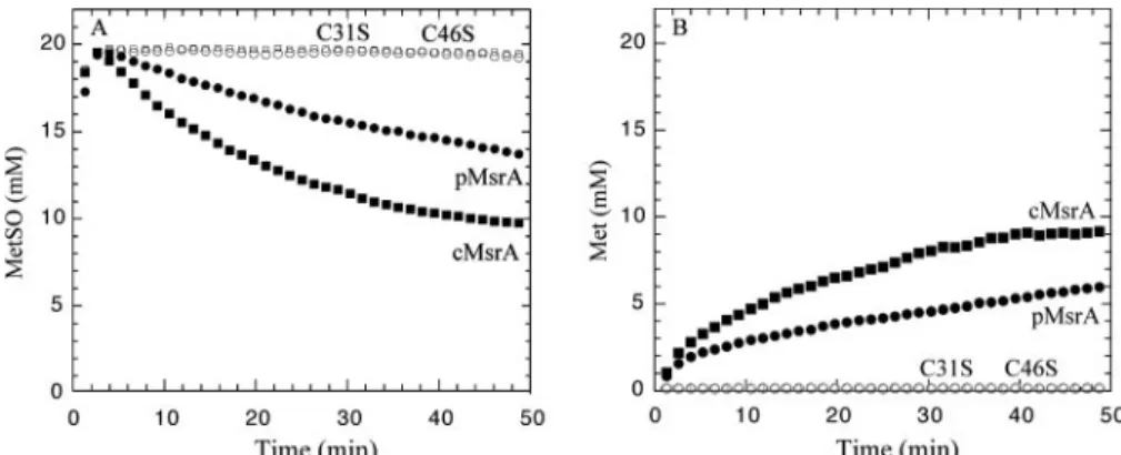
Methionine Sulfoxide Reductase Activity and Mechanism of the Wild-type Enzymes—The two poplar MsrA isoforms were produced as recombinant proteins and purified to homogene-ity. The methionine sulfoxide reductase activity of these two MsrAs was first measured by NMR using DTT as a reductant. As illustrated in Fig. 3, addition of either cMsrA or pMsrA resulted in a rapid decrease of theL-Met-(RS)-SO concentra-tion (Fig. 3A) concomitant with the appariconcentra-tion of Met (Fig. 3B). We have shown previously that pMsrA only reducedL -Met-(S)-SO (23). In agreement with the known stereoselectivity of MsrA species, only one-half of the initialL-Met-(RS)-SO race-mic mixture was reduced after completion of the reaction.
To evaluate further the methionine sulfoxide reductase activity of cMsrA and pMsrA, we used a spectrophotometric test, which coupled the thioredoxin (Trx) system (NADPH/ NADPH thioredoxin reductase/Trx) to NADPH oxidation. Table 4 presents the kinetic parameters of both wild-type enzymes obtained in the presence of a cytosolic poplar Trx called Trx h1. For both MsrAs, the apparent KMvalues, deter-mined under steady-state conditions for Trx h1 andL -Met-(RS)-SO, are around 15 and 300M, respectively. Compared with other biochemically characterized MsrAs, the apparent affinity constant for MetSO is slightly lower (300Mcompared with 600 or 1900Mfor N. meningitidis MsrA (36) or E. coli MsrA respectively (18)). Nevertheless, because the rate-limit-ing step is likely associated with the Trx-dependent recyclrate-limit-ing process as shown for MsrA from N. meningitidis (37) and not to the reductase step, the KMvalues for MetSO cannot be directly
interpreted as representative of a better substrate affinity. It is, however, important to note that the catalytic efficiency, expressed as kcat/KMetSO, is in the same order for the two poplar
MsrAs and for E. coli MsrA.
With the aim to characterize which cysteines are involved in the Trx-recycling mechanism for poplar MsrAs, the stoichiom-etry of the reductase reaction was measured using high pressure liquid chromatography by following the quantity of methionine formed at a known concentration of reduced MsrA without any other reductant. In parallel, the thiol content of the wild-type MsrAs before and after reduction ofL-Met-(RS)-SO was esti-mated (Table 5). The cMsrA and pMsrA exhibit a stoichiome-try of nearly 2 mol of Met by mol of enzyme with a concomitant disappearance of three free thiol groups per monomer (Table 5). These data indicate that three cysteines are involved in the catalytic mechanism, in particular that two of them are impli-cated in the Trx-dependent regeneration with formation of two successive disulfide bonds. Indeed, based on the scheme in Fig. 1, if three cysteines are involved, at the end of the reduction of the first MetSO, the catalytic cysteine is free to reduce another MetSO, whereas the recycling two others are under disulfide state. Such a result is similar to that of E. coli MsrA (18).
Role of the Different Cys Residues in the Catalytic Mechanism— To investigate the role of the cysteine residues in plant MsrAs, each of the five cysteinyl residues of cMsrA was replaced inde-pendently into serine. Among the five monocysteinic mutated MsrAs, only C81S and C100S cMsrAs retained an activity sim-ilar to the wild type with DTT or Trx as reductants, indicating that these two cysteines, as expected from their position, are involved neither in MetSO reduction nor in Trx-dependent recycling process. The three other single substitutions affected cMsrA activity whatever the reductant used (data not shown). The C46S cMsrA and C46S pMsrA were both found to be totally inactive with DTT or Trx (Fig. 3 and Table 5), confirm-ing, as expected from its position in the primary sequence, that this Cys residue is the catalytic one. The absence of thiol decrease after MetSO treatment for these two enzymes is also consistent with these results (Table 5).
To further characterize the catalytic mechanism, the cys-teines at positions 81 and 100 have been systematically replaced by serine in other mutated MsrAs, leading to triple- and qua-druple-substituted cMsrAs called C81S/C100S/C196S, C81S/ C100S/C202S, and C81S/C100S/C196S/C202S cMsrAs. The double-substituted C81S/C100S cMsrA is equivalent to cMsrA in terms of catalytic parameters and stoichiometry of Met formed in the absence of reductant (Tables 4 and 5). The C81S/ C100S/C196S/C202S cMsrA, in which only the catalytic Cys46
remains, still displays a MetSO reductase activity (Table 5). The resulting enzyme is not regenerated by Trx h1, and its Cys46is
oxidized under sulfenic acid form. Indeed, the decrease in thiol is close to 1 mol per mol of enzyme and 0.5 mol of sulfenic acid per mol of enzyme is titrated by TNB⫺, which is a specific rea-gent of sulfenic acid derivative.
The stoichiometry of the MetSO reduction by C81S/C100S/ C196S and C81S/C100S/C202S cMsrAs, in the absence of reductant, is 0.43 and 0.82, respectively (Table 5). These results are expected if one of the two cysteines involved in the regen-eration of the catalytic Cys46is removed. This indicates thus
that both Cys196and Cys202are involved in the regeneration
process but does not permit us to conclude the order in which Cys196and Cys202are involved. Both C81S/C100S/C196S and
C81S/C100S/C202S cMsrAs were found to be active in the
at INRA Institut National de la Recherche Agronomique, on November 9, 2010
www.jbc.org
at INRA Institut National de la Recherche Agronomique, on November 9, 2010
www.jbc.org
presence of Trx (Table 4), and no sulfenic acid was titrated in both triple-mutated cMsrAs with TNB⫺after MetSO reduc-tion in the absence of reductant. These data show that under the experimental conditions used, i.e. 10 min of incubation, a disulfide bond is formed whatever the nature of the substituted recycling Cys. Based on the catalytic mechanism of the E. coli and of B. taurus MsrAs, which both support the successive involvement of the two recycling Cys, these results are rather unexpected (see Fig. 1). Indeed, a titration of one sulfenic acid was expected for the substitution of the first recycling cysteine, whereas no sulfenic titration was expected for the substitution of the second recycling cysteine because of formation of a disul-fide bond in this latter case. Other unexplained data come from the thiol titrations. Indeed, titration of free thiol indicates loss of 0.7 and 0.8 thiols after MetSO reduction in the absence of reductant for the C81S/C100S/C196S and C81S/C100S/C202S cMsrAs, respectively (Table 5), whereas a loss of either 1 or 2 was expected. Increasing the incubation time or concentrations of the substrate or enzyme did not change significantly the data on thiol titration. These results remain to be explained and could be related to the observed instability of these substituted cMsrAs.
When looking at the Trx-dependent activity, the C81S/ C100S/C196S and C81S/C100S/C202S cMsrAs displayed modified catalytic parameters compared with those of C81S/ C100S cMsrA (Table 4). First, the KMvalues of C81S/C100S/
C196S and C81S/C100S/C202S cMsrAs for Trx h1 are increased around 12- and 6-fold compared with that of C81S/ C100S cMsrA and cMsrA, respectively. On the other hand, the
kcat value of the two C-terminal cysteine-mutated MsrAs is
decreased by 3- and 9-fold, respectively, with C81S/C100S/ C202S cMsrA being the more affected. In terms of catalytic
efficiency (kcat/KTrx), a decrease of
36-fold for C81S/C100S/C196S cMsrA and of 107-fold for C81S/ C100S/C202S is observed com-pared with the C81S/C100S cMsrA. Together, these results indicate that the disulfide bond between Cys46
and Cys202 is the more efficiently
reduced by Trx in terms of kcat. The
fact that, because of a lower KM value for Trx, the kcat/KTrx values
are significantly higher for cMsrA and C81S/C100S cMsrA compared with that of C81S/C100S/C196S and C81S/C100S/C202S cMsrAs supports the intervention of Trx on the disulfide Cys196/Cys202 bond
and not on Cys46/Cys196or Cys46/
Cys202 bonds. Indeed, a lower K
M
value for Trx necessitates less Trx in vivo to regenerate the catalytic CysA. In terms of evolution, this is certainly an advan-tage that has already been shown for E. coli MsrA.4
Thus, the results presented here clearly show the following: (i) Cys46 is the catalytic CysA, and (ii) the recycling process
implicates Cys196and Cys202but does not discriminate from
the order in which Cys196and Cys202are involved. To answer
this question, it would be necessary to determine the rate of the disulfide bond formation in each triple-substituted cMsrA in which only one recycling cysteine is present. These rates are likely very different depending on the recycling cysteine and its position relative to the catalytic one. Unfortunately, the nonre-liable thiol titration on both mutated cMsrAs cannot permit us to attain these rates experimentally. Thus, to get further insights into the Trx-dependent recycling process of plant MsrA, the determination of the three-dimensional structure of pMsrA has been undertaken.
Overall Structure of pMsrA—Crystallization trials were per-formed for both chloroplastic and cytosolic MsrA in the reduced and oxidized forms, but crystals suitable for x-ray anal-ysis were obtained only for pMsrA in the reduced form. The trigonal crystals (space group P31) of pMsrA contain one mol-ecule per asymmetric unit and 45% of solvent (Tables 2 and 3). The structure was solved by molecular replacement and refined up to a resolution of 1.7 Å with final R and Rfreefactors of 19.5
and 20.1%, respectively. No electron density was observed upstream from Pro22in the N-terminal part. The missing
resi-dues1Met–Asp21are very disordered and probably situated in
the large solvent cavity observed in the crystal packing inter-face. The catalytic Cys46(CysA) and the recycling Cys196(CysB)
and Cys202(CysC) but also Cys81clearly show extra densities on
FIGURE 2. Amino acid comparison of the two poplar MsrAs with plant MsrA sequences and biochemically or structurally characterized MsrAs. The sequences were compared using ClustalW. The accession numbers (GenBankTMor MATDB) are as follow: P. tremula⫻ tremuloides pMsrA (Pt pMsrA), AY530805;
A. thaliana cMsrA (At cMsrA), At5g61640; A. thaliana pMsrA (At pMsrA), At4g25130; B. napus pMsrA (Bn pMsrA), P54151; P.⫻ interamericana cMsrA (Pi cMsrA),
AY530804; Lactuca sativa (Ls pMsrA), Q9SEC2; F. ananassa (Fa cMsrA), P54153; Lycopersicum esculentum (Le cMsrA), P54153; E. coli MsrA (Ec MsrA), NP_418640;
B. taurus MsrA (Bt MsrA), P54149; M. tuberculosis (Mt MsrA), NP_334555; and N. meningitidis (Nm PilB MsrA), E82024. The cysteine numbering of cMsrA and pMsrA
is indicated, with CysA corresponding to Cys46, CysB to Cys196, and CysC to Cys202. The black arrow indicates the putative peptide cleavage site. In white on black
are indicated the cysteinyl residues and the GYC, GYXC, or CYG signatures. In black on gray are presented the other very conserved or fully conserved residues. FIGURE 3. MetSO reductive activity of wild-type and mutated cytosolic (cMsrA) and plastidial (pMsrA)
poplar sulfoxide methionine reductases. A, MetSO concentration evolution as a function of time. B, Met
concentration evolution as a function of time. pMsrA (filled circles), cMsrA (filled l squares), C46S cMsrA (open
squares), C46S pMsrA (open circles). Reactions were recorded at 25 °C in the presence of 100 mMphosphate buffer with 50 mMDTT, 20 mM L-Met-(RS)-SO at pH 8.5. The enzyme concentrations for cMsrA, pMsrA, C46S cMsrA, and C46S pMsrA were, respectively 4.7, 6.1, 4.7, and 4.9M. MetSO and Met concentrations were determined from the methyl peak intensity of the MetSO at 2.65 ppm and the Met at 2.15 ppm.
at INRA Institut National de la Recherche Agronomique, on November 9, 2010
www.jbc.org
their sulfur atoms that were unambiguously modeled as bonded -mercaptoethanol, present in the crystallization solution. Sur-prisingly, Cys100, the most solvent-exposed cysteine, was
observed in its reduced form.
As expected from the sequence identity, the poplar pMsrA model displays the same overall fold than the E. coli, B. taurus, and M. tuberculosis MsrAs (respective Protein Data Bank entries 1FF3 (12), 1FVA, 1FVG (13), and 1NWA (14)). C-␣ atoms corresponding to the cores of these enzymes can be superimposed with root mean square distances of about 0.8 Å.
On the contrary, major differences concern the N- and C-terminal ends of the poplar enzyme. Because the C-terminal end contains two of the three cysteines involved in the catalytic mechanism (Cys196 and
Cys202), it is of high interest to
com-pare it with other MsrAs.
In E. coli, B. taurus, and M.
tuber-culosisMsrAs, the C-terminal ends, starting from structurally equiva-lent positions 192, 212, and 147, respectively, are observed as long extended coils without tertiary organization, which lean against the core of the domain and run roughly in the same direction, at the border of the active site. On the contrary, in the pMsrA model, the 182Lys–
Gly204 C-terminal end is observed
for the first time in a totally new sta-bilized conformation (Fig. 4). The polypeptide chain turns by 90° with respect to the previously described C-terminal ends, so that it starts to run parallel to the first␣-helix (named H1) of the central core of the structure and then, after a turn, antiparallel to it. It results in a packed conformation, as opposed to the extended conformation previously observed for the C-terminal ends of other MsrA structures. Numerous hydrogen bonds are observed within this C-terminal part and between the C-termi-nal part and the core of the enzyme. Three amino acid residues, only and strictly conserved in plant MsrA sequences, seem to play a key role in the global architecture of the C-terminal part
FIGURE 4. Stereoview of the pMsrA monomer structure. The core enzyme is shown in gray and the C-terminal part in blue. The C-terminal ends of the E. coli, B. taurus, and M. tuberculosis MsrA superimposed to the poplar model are shown in red, green, and yellow, respectively. The five cysteine residues present in the pMsrA structure are drawn in
ball-and-stick. This figure was generated using Pymol (The PyMOL Molecular Graphics System is available on line).
TABLE 4
Catalytic parameters of wild-type pMsrA and cMsrA and mutated cMsrAs
These parameters were determined under steady-state conditions by following NADPH oxidation in the presence of 100 mM L-Met-(RS)-SO, 0 –250Mpoplar cytosolic
Trx h1 as an electron donor, and 0.5McMsrA, 3MpMsrA, 1MC81S/C100S cMsrA, 3.7McMsrA C81S/C100S/C196S, and 5.7MC81S/C100S/C202S cMsrA. KM
values for Trx h1 and MetSO were measured at saturating concentrations of the other substrate. ND indicates not determined.
KTrx KMetSO kcat kcat/KMetSO kcat/KTrx
M M s⫺1 ⫻103 M⫺1s⫺1 ⫻103 M⫺1s⫺1 EcMsrAa 10⫾ 2 1900 3.7 2.0.⫾ 0.5 370⫾ 70 pMsrA 18⫾ 8 240⫾ 30 0.30⫾ 0.03 1.2⫾ 0.3 17⫾ 6 cMsrA 15⫾ 3 380⫾ 100 1.20⫾ 0.10 3.1⫾ 0.6 80⫾ 19 C81S/C100S cMsrA 8⫾ 1 ND 0.60⫾ 0.04 ND 75⫾ 9 C81S/C100S/C196S cMsrA 94⫾ 15 ND 0.20⫾ 0.02 ND 2.1⫾ 0.5 C81S/C100S/C202S cMsrA 103⫾ 17 ND 0.07⫾ 0.01 ND 0.7⫾ 0.1 a
Results were obtained with E. coli MsrA, from Boschi Muller et al. (18).
TABLE 5
Stoichiometry of Met formed in the absence of reductant and decrease in thiol content after treatment or not withL-Met-(RS)-SO for
wild-type pMsrA and cMsrA and mutated cMsrAs
The results of stoichiometry are expressed in mol of Met formed per mol of enzyme. In parentheses is indicated the theoretical content (or decrease) in thiols, i.e. the expected values based on the hypothesis of a mechanism involving Cys46as the catalytic cysteine, and Cys196and Cys202as the recycling cysteines (both orders are
considered).
Stoichiometry Number of free thiols withoutL-Met-(RS)-SO
Number of free thiols
withL-Met-(RS)-SO Decrease in free thiols
pMsrA 1.8 4.3⫾ 0.4 (5) 1.3⫾ 0.1 (2) 3 (3) cMsrA 1.7 4.4⫾ 0.4 (5) 1.1⫾ 0.1 (2) 3.3 (3) C46S cMsrA 0 3.9⫾ 0.4 (4) 3.8⫾ 0.3 (4) 0.1 (0) C81S/C100S cMsrA 1.5 2.7⫾ 0.3 (3) 0.10⫾ 0.02 (0) 2.6 (3) C81S/C100S/C196S cMsrA 0.43 1.5⫾ 0.1 (2) 0.8⫾ 0.1 (0 or 1) 0.7 (1 or 2) C81S/C100S/C202S cMsrA 0.82 1.6⫾ 0.1 (2) 0.8⫾ 0.1 (0 or 1) 0.8 (1 or 2) C81S/C100S/C196S/C202S cMsrA 0.80 1.0⫾ 0.1 (1) 0.10⫾ 0.02 (0) 0.9 (1)
at INRA Institut National de la Recherche Agronomique, on November 9, 2010
www.jbc.org
(Fig. 5). First, the N-1 and N-2 atoms of Arg56, located at the
end of the␣-helix H1, interact with the carbonyl groups of Asp198and Ser191, respectively, whereas its carbonyl group is
hydrogen-bonded with the NH group of Gly195. Second, the
N- atom of Lys194 is hydrogen-bonded with the carbonyl
groups of Gln55, Val57, and Val60situated in the loop
connect-ing the␣-helix H1 with the following -strand. Finally, Gln190
contributes to the stabilization of the C-terminal end via an interaction between its elongated side chain and the main chain of Arg201. Furthermore, a water molecule that interacts with
Gln190, Asp198, Pro199, and Arg56further stabilizes the packed
conformation of the C-terminal end. When superimposing the structures of E. coli, B. taurus, M. tuberculosis, and poplar pMsrA, the main chain of the latter enzyme starts to deviate from the others at a position that can be considered as a hinge region. It consists of the sequence182KGG184in poplar MsrA,
whereas it is192KNP194in the E. coli MsrA,212KDP214in the B. taurussequence, and147RYP149in the M. tuberculosis enzyme.
Interestingly, the KGG sequence is also conserved in other plant MsrAs. The lack of the proline and the presence of two glycine residues in pMsrA strongly modify the tor-sion constraints in this region.
The conformation of the C-ter-minal end observed in the poplar pMsrA structure does not seem to arise from the crystal form studied in this x-ray analysis. Indeed, even if this part of the structure faces two monomers in the crystal lattice, the few observed interactions with the neighboring partners could not account for a misfolding of the 23 C-terminal residues, especially because this new conformation of the C-terminal end features several intramolecular interactions that involve residues conserved exclu-sively in plant sequences. Analysis of the active site, developed below, reinforces this assumption.
Modification of the Catalytic Cys-teine and Architecture of the Active Site—The side chain of the catalytic cysteine Cys46(CysA) conserves the
same position in the poplar pMsrA structure with respect to the other known MsrA x-ray models. The -mercaptoethanol molecule bonded to Cys46occupies a site
sit-uated within the active site. The modification of CysA has been observed in other MsrA structures. Indeed, in E. coli MsrA, a dimethyl-arsenate group resulting from the crystallization medium is bonded to Cys51(CysA) (12). In the B. taurus
structure, a bound dithiothreitol molecule is observed on Cys72
(CysA) (13). Furthermore, in the M. tuberculosis MsrA struc-ture, a crystal contact fortuitously places the methionine side chain of a neighboring monomer in the active site of the enzyme (14). These different complexes have been proposed to mimic either a Michaelis complex or a transient intermediate.
The position of the-mercaptoethanol molecule observed on CysA in the pMsrA structure supports the proposals made for the recognition and stabilization of the substrate (13, 14). Indeed, the sulfur atom of the-mercaptoethanol molecule bonded to Cys46occupies the same position as the arsenic atom
that mimic the sulfur atom of methionine sulfoxide in E. coli MsrA. The entire-mercaptoethanol molecule elongates in the same direction as the methionine side chain in the M.
tubercu-losisactive site (14) and as the dithiothreitol molecule present in the B. taurus MsrA (13). Consequently, Asp124, which is
con-served in most MsrA sequences, forms a hydrogen bond with the hydroxyl group of the-mercaptoethanol molecule, similar
FIGURE 5. Stereoviews of the pMsrA C-terminal end. A, the C-terminal end of pMsrA is shown in blue, and the core of the enzyme is displayed in gray. The hydrogen bonds shared between the C-terminal domain and the core of pMsrA are shown as dashed lines. The red sphere corresponds to a water molecule. B, the 2Fo⫺ Fc
electronic density (contour level: 1.2) superimposed on the pMsrA C-terminal part.
at INRA Institut National de la Recherche Agronomique, on November 9, 2010
www.jbc.org
to that observed in M. tuberculosis MsrA between the side chain of the equivalent aspartate residue (Asp87) and the amide
group of the methionine main chain (14) (Fig. 6). Modeling of a peptidic methionine sulfoxide bound in the active site of pMsrA, based on the M. tuberculosis model, shows the follow-ing: 1) the invariant Phe47and Trp48of the GCFW active-site
consensus sequence form a hydrophobic pocket in which the ⑀-methyl group of MetSO can bind as already observed in all the three-dimensional structures of MsrAs determined so far; 2) the oxygen of the sulfoxide is situated at hydrogen bonding distances of Tyr77, Glu89, and Tyr129(Fig. 6); and 3) the location
of Asp124is suitable for its involvement in the stabilization of
the main chain of the substrate. In M. tuberculosis MsrA, the side chain of the equivalent aspartate residue (Asp87) points
toward the side chain of Tyr152, which belongs to the GYTC
sequence (the ending cysteine residue being CysB). The aro-matic ring of this tyrosine residue is proposed to stack the pep-tidic bond between the methionine sulfoxide of the substrate and its upstream residue, through a- interaction (14). In the
B. taurus MsrA structure, a similar interaction is observed between structurally equivalent aspartate and tyrosine
dues. In this case, the tyrosine resi-due (Tyr217) belongs to the more
frequent GYC sequence (see under “Genome and Sequence Analysis”). As mentioned earlier, the GYC or GYXC string is absent from the plant MsrA sequences. However, in the latter, a stabilizing tyrosine resi-due is still observed, because Tyr203
of pMsrA is structurally equivalent to Tyr217of the B. taurus structure
or to Tyr152of the M. tuberculosis
enzyme. The new conformation of the C-terminal end observed in the plant structure allows the poplar main chain of only three residues,
202CYG204, to run at roughly the
same position than the main chains of the other two structures, but in an antiparallel way. The reverse direc-tion of the superposed polypeptidic chains thus remarkably places the C-␣ atoms of the CYG string of res-idues in the poplar enzyme at roughly the same location as the C-␣ atoms of the 218CYG216
resi-dues of B. taurus MsrA or as the C-␣ atoms of the153TYG151residues in
the M. tuberculosis MsrA structure. In each case, it allows the central tyrosine residue to exactly maintain the same position and orientation of its side chain. The side chain of Tyr202, conserved in plants through
a C-terminal CYG sequence, is thus proposed to act as Tyr152 in M. tuberculosis MsrA, conserved in a GYXC or GYC sequence in a large subset of MsrAs.
The conformation of the C-terminal end observed in the pMsrA structure also suggests that the extended side chain of Arg185participates in substrate binding through a hydrogen
bond with the main chain of the methionine sulfoxide. The substrate could then be maintained within the active site by interactions on both sides of its main chain. However, the posi-tion of this arginine residue is shifted by one amino acid in some plant sequences, and a substitution for an asparagine residue is observed in Fragaria anannassa MsrA. Whether these differ-ences are compatible with the proposed role of Arg185remains
to be assessed.
Position of the Cysteine Residues—Among the five conserved cysteines, Cys46(CysA) lies at the bottom of the active site. A
comparative analysis with other MsrA structures shows that its bonding to the-mercaptoethanol molecule reasonably mim-ics part of the transient tetrahedral intermediate formed after the CysA attack onto the substrate (see Fig. 6). Cys196(CysB) is
found far from the active site, near the C-terminal end of ␣-he-lix H1, in the portion of the C-terminal coil that runs antipar-allel to this␣-helix. This location of Cys196places its sulfur
FIGURE 6. Stereoviews showing a superimposition of the pMsrA (blue) and M. tuberculosis MsrA (yellow)
models in the active site region. A, the C-␣ trace of the pMsrA structure is shown in coil mode. The methionine
residue observed in the M. tuberculosis active site is shown in stick mode. B, detailed view that emphasizes the comparable stabilization of the-mercaptoethanol molecule bound to the catalytic cysteine in the pMsrA model and the methionine residue observed in the catalytic site of the M. tuberculosis enzyme.
at INRA Institut National de la Recherche Agronomique, on November 9, 2010
www.jbc.org
atom at a distance of 18 Å from the S-␥ atom of CysA. Cys202
(CysC), as part of the C-terminal CYG sequence of plant MsrAs, is found near the active site, with its C-␣ atom being in a position close to the C-␣ atom of the B. taurus MsrA CysB (Cys218). However, the torsion angles of the polypeptide chain
direct the CysC side chain of the poplar enzyme opposite to that of B. taurus MsrA. Cys202makes hydrophobic contacts with the
aromatic side chain of Tyr203and elongates its bonded
-mer-captoethanol parallel to the Asp124–Tyr203pair of residues. The
sulfur-sulfur distance between Cys202 and Cys196 is 15.8 Å,
whereas it is only 7.1 Å with Cys46. Rotations of the Cys46and
Cys202side chains bring their sulfur atoms to a minimum
dis-tance of 3.3 Å that could allow the formation of the disulfide bond. However, the required movement of Cys202 would be
slightly hindered by the Tyr203side chain, maintained in
posi-tion through an interacposi-tion with Asp124. It thus necessitates
that small conformational changes occur during the first step of the regeneration process of the enzyme. On the contrary, large movements are necessary to approach Cys196 toward Cys46.
Such a requirement seems to be a common property of all MsrAs that involve three cysteine residues in their catalytic mechanism. Indeed, in the B. taurus MsrA structure (13), the C-␣ atom of CysB (Cys218) is found at 7.1 Å of the C-␣ atom of
CysA (Cys72), but the CysB side chain points outside the active
site and cannot be reoriented toward CysA by simply modifying its side chain torsion angle. Furthermore, CysC (Cys227) is
sit-uated at 21 Å from CysB. In the E. coli MsrA structure (12), the C␣–C␣ atom distance for CysA (Cys51) and CysB (Cys198) is 11
Å, whereas it is 20 Å between CysB and CysC (Cys206).
How-ever, the disulfide bond is supposed to form once the product,
i.e.the reduced methionine, is released, concomitantly with the formation of the sulfenic acid on CysA. These events might modify the enzyme conformation, especially at the C-terminal end, in order to approach CysB or CysC (for the poplar enzyme) closer to the sulfenic acid form on CysA. Only three-dimen-sional structures of an oxidized state of MsrA would bring an answer that cannot be deduced from the current structures that represent either a Michaelis-like complex or a transition state preceding formation of the sulfenic intermediate. Concerning poplar pMsrA, the structure determination of the oxidized forms of the enzyme would help to understand the nature of the conformational changes that occur at the different steps of the catalytic mechanism. However, to date, attempts to crystallize such forms of the enzyme have failed.
Conclusion—The biochemical studies indicate, as expected, that the enzyme is able to reduce the S enantiomer of MetSO using Trx as a reductant. Out of a total of five cysteines, three are involved in the catalytic mechanism, the catalytic Cys46, on
which the sulfenic intermediate is formed, and two recycling cysteines (Cys196and Cys202), which are involved in the
Trx-de-pendent recycling process and are located in a highly conserved motif specific to plant enzymes. Inspection of the three-dimen-sional structure of the poplar pMsrA suggests a Trx-dependent recycling process for this plant MsrA different from the one described for the E. coli and B. taurus enzymes. The sulfenic acid formed on Cys46after MetSO reduction would be attacked
by the more C-terminal Cys202(CysC), leading to formation of
a Cys46–Cys202 disulfide bond. Then, Cys196 (CysB) would
attack the disulfide bond to form a Cys202–Cys196bond, which
is finally reduced by Trx. The conformational changes needed to place Cys202close to the active site and especially close to
Cys46are small. In addition, Cys202is included in a CYG motif,
which is conserved in all plant MsrAs. The tyrosine residue corresponds to the one described to be involved in substrate binding in bacterial and B. taurus MsrAs. In these MsrAs, the tyrosine residue belongs to a similar motif as found for pMsrA but with the first C-terminal cysteine instead of the last C-ter-minal cysteine.
Acknowledgments—We are very grateful to Jean-Luc Ferrer and Pierre Legrand at the ESRF (beamline BM30A) for giving us the opportunity to perform data collection and for their kind help during this experiment.
REFERENCES
1. Imlay, J. A., and Linn, S. (1986) Science 240, 1302–1309
2. Berlett, B., and Stadman, E. (1997) J. Biol. Chem. 272, 20313–20316 3. Vogt, W. (1995) Free Radic. Biol. Med. 18, 93–105
4. Brot, N., Weissbach, L., Werth, J., and Weissbach, H. (1981) Biochemistry
78,2155–2158
5. Grimaud, R., Ezraty, B., Mitchell, J. K., Lafitte, D., Briand, C., Derrick, P. J., and Barras, F. (2001) J. Biol. Chem. 276, 48915– 48920
6. Gao, J., Yin, D. H., Yao, Y., Sun, H., Qin, Z., Schoneich, C., Williams, T. D., and Squier, T. C. (1998) Biophys. J. 74, 1115–1134
7. Harndahl, U., Kokke, B. P., Gustavsson, N., Linse, S., Berggren, K., Tjer-neld, F., Boelens, W. C., and Sundby, C. (2001) Biochim. Biophys. Acta
1545,227–237
8. Taggart, C., Cervantes-Laurean, D., Kim, G., McElvaney, N. G., Wehr, N., Moss, J., and Levine, R. L. (2000) J. Biol. Chem. 275, 27258 –27265 9. Khor, H. K., Fisher, M., and Scho¨neich, C. (2004) J. Biol. Chem. 279,
19486 –19493
10. Sun, H., Gao, J., Ferrington, D. A., Biesiada, H., Williams, T. D., and Squier, T. C. (1999) Biochemistry 38, 105–112
11. Levine, R. L., Mosoni, L., Berlett, B. S., and Stadman, E. R. (1996) Proc.
Natl. Acad. Sci. U. S. A. 93,15036 –15040
12. Tete-Favier, F., Cobessi, D., Boschi-Muller, S., Azza, S., Branlant, G., and Aubry, A. (2000) Structure 8, 1167–1178
13. Lowther, W. T., Brot, N., Weissbach, H., and Matthews, B. W. (2000)
Biochemistry 39,13307–13312
14. Taylor, A. B., Benglis, D. M., Jr., Dhandayuthapani, S., and Hart, P. J. (2003)
J. Bacteriol. 185,4119 – 4126
15. Lowther, W. T., Weissbach, H., Etienne, F., Brot, N., and Matthews, B. W. (2002) Nat. Struct. Biol. 9, 348 –352
16. Kauffmann, B., Aubry, A., and Favier, F. (2005) Biochim. Biophys. Acta
1703,249 –260
17. Boschi-Muller, S., Olry, A., Antoine, M., and Branlant, G. (2005) Biochim.
Biophys. Acta 1703,231–238
18. Boschi-Muller, S., Azza, S., Sanglier-Cianferani, S., Talfournier, F., Van Dorsselear, A., and Branlant, G. (2000) J. Biol. Chem. 275, 35908 –35913 19. Kumar, R. A., Koc, A., Cerny, R. L., and Gladyshev, V. N. (2002) J. Biol.
Chem. 277,37527–37535
20. Boschi-Muller, S., Azza, S., and Branlant, G. (2001) Protein Sci. 10, 2272–2279
21. Sadanandom, A., Poghosyan, Z., Fairbairn, D. J., and Murphy, D. J. (2000)
Plant Physiol. 123,255–264
22. Romero, H. M., Berlett, B. S., Jensen, P. J., Pell, E. J., and Tien, M. (2004)
Plant Physiol. 136,3784 –3794
23. Vieira Dos Santos, C., Cuine, S., Rouhier, N., and Rey, P. (2005) Plant
Physiol. 138,909 –922
24. Gustavsson, N., Kokke, B. P., Harndahl, U., Silow, M., Bechtold, U., Pog-hosyan, Z., Murphy, D., Boelens, W. C., and Sundby, C. (2002) Plant J. 29, 545–553
at INRA Institut National de la Recherche Agronomique, on November 9, 2010
www.jbc.org
25. Bechtold, U., Murphy, D. J., and Mullineaux, P. M. (2004) Plant Cell 16, 908 –919
26. In, O., Berberich, T., Romdhane, S., and Feierabend, J. (2005) Planta 220, 941–950
27. Sadanandom, A., Piffanelli, P., Knott, T., Robinson, C., Sharpe, A., Lydiate, D., Murphy, D., and Fairbairn, D. J. (1996) Plant J. 10, 235–242 28. Jacquot, J. P., Stein, M., Suzuki, A., Liottet, S., Sandoz, G., and
Miginiac-Maslow, M. (1997) FEBS Lett. 400, 293–296
29. Schenk, P. M., Baumann, S., Mattes, R., and Steinbiss, H. H. (1995)
Bio-Techniques 19,196 –200
30. Otwinowski, Z., and Minor, W. (1997) Methods Enzymol. 276, 307–326 31. Vagin, A., and Teplyakov, A. (1997) J. Appl. Crystallogr. 30, 1022–1025 32. Perrakis, A., Morris, J. R., and Lamzin, V. S. (1999) Nat. Struct. Biol. 6,
458 – 463
33. Bru¨nger, A. T., Adams, P. D., Clore, G. M., DeLano, W. L., Gros, P., Grosse-Kunsteleve, R. W., Jiang, J. S., Kuszewski, J., Nilges, M., Pannu, N. S., Read, R. J., Rice, L. M., Simonson, T., and Warren, G. L. (1998) Acta
Crystallogr. Sect. D Biol. Crystallogr. 54,905–921
34. Jacquot, J. P., Rivera-Madrid, R., Marinho, P., Kollarova, M., Le Marechal, P., Miginiac-Maslow, M., and Meyer, Y. (1994) J. Mol. Biol. 235, 1357–1363
35. Behm, M., and Jacquot, J. P. (2000) Plant Physiol. Biochem. 38, 363–369 36. Olry, A., Boschi-Muller, S., Marraud, M., Sanglier-Cianferani, S., Van
Dorsselear, A., and Branlant, G. (2002) J. Biol. Chem. 277, 12016 –12022 37. Antoine, M., Boschi-Muller, S., and Branlant, G. (2003) J. Biol. Chem. 278,
45352– 45357
at INRA Institut National de la Recherche Agronomique, on November 9, 2010
www.jbc.org