HAL Id: cea-01510320
https://hal-cea.archives-ouvertes.fr/cea-01510320
Submitted on 19 Apr 2017
HAL is a multi-disciplinary open access
archive for the deposit and dissemination of
sci-entific research documents, whether they are
pub-lished or not. The documents may come from
teaching and research institutions in France or
abroad, or from public or private research centers.
L’archive ouverte pluridisciplinaire HAL, est
destinée au dépôt et à la diffusion de documents
scientifiques de niveau recherche, publiés ou non,
émanant des établissements d’enseignement et de
recherche français ou étrangers, des laboratoires
publics ou privés.
Distributed under a Creative Commons Attribution| 4.0 International License
Role of the Methoxy Groups in Cryptophanes for
Complexation of Xenon. Conformational Selection
Evidenced by 129 Xe-1 H NMR SPINOE Experiments
Patrick Berthault, Céline Boutin, Estelle Léonce, Erwann Jeanneau, Thierry
Brotin
To cite this version:
Patrick Berthault, Céline Boutin, Estelle Léonce, Erwann Jeanneau, Thierry Brotin. Role of the
Methoxy Groups in Cryptophanes for Complexation of Xenon. Conformational Selection Evidenced
by 129 Xe-1 H NMR SPINOE Experiments. ChemPhysChem, Wiley-VCH Verlag, 2017, 18,
pp.1561-1568. �10.1002/cphc.201700266�. �cea-01510320�
Role of the Methoxy Groups in Cryptophanes for Complexation of
Xenon. Conformational Selection Evidenced by
129
Xe-
1
H NMR
SPINOE Experiments.
Patrick Berthault,
*[b]Céline Boutin,
[b]Estelle Léonce,
[b]Erwann Jeanneau,
[c]Thierry Brotin
*[a]Abstract: We report the laser-polarized 129Xe and 1H NMR spectra
of a series of cryptophane derivatives that differ only by the number of methoxy groups attached on their benzene rings and the arrangement syn or anti of the linkers (compounds 6a-s, 9a-s, 12a-s). All these compounds bind xenon even though the characteristic signal of the gas encapsulated in the cavity of the cage-molecule cannot be always detected. Interestingly, the exchange dynamics of xenon strongly depends on the degree of substitution and is different from that of the cryptophane derivatives studied previously. In solution, the 1H NMR spectra of these derivatives show the presence
of different conformations in a slow exchange regime that can be explained by a decrease of the flexibility of their skeleton. Thanks to
129Xe-1H dipolar cross-relaxation (SPINOE) spectra we demonstrate
that a single conformation present in solution can bind xenon.
Introduction
Xenon is known to show good affinity for organic structures such as the hydrophobic pockets of proteins or hollow organic compounds capable of accommodating substrates in their cavities.[1-4] For
instance, it has been reported that xenon presents a good affinity for cryptophane-A (1), a synthetic organic compound with a roughly spherical cavity.[5] Since this discovery, numerous
xenon-cryptophane complexes have been thoroughly studied in organic or aqueous solution.[6] These supramolecular complexes also bring
attention of the scientific community as they can be used for 129Xe
NMR-based biosensing applications.[7] The remarkable properties of
these complexes mainly arise from the high receptiveness of the xenon nuclear spin, which is able to detect subtle changes in its surrounding environment.[8] Thus, when xenon is bound to a host
molecule, the magnetic properties of its nucleus are modified resulting in a specific 129Xe NMR signature for this complex. The
possibility to modify the host structure in order to graft a biological site that can specifically recognize a biological target represents another advantage for these systems.[9]
From a more fundamental point of view, 129Xe NMR spectroscopy
appears as the tool of choice for studying cryptophane derivatives with small cavities. For instance, the xenon atom present within the cavity of cryptophane-A congeners shows a chemical shift significantly modified with respect to that of xenon present in the bulk. As an example, in 1,1,2,2-tetrachloroethane-d2 the 129Xe NMR
spectrum of the Xe@cryptophane-A complex shows a chemical shift
difference of 160 ppm between xenon in solution and xenon trapped into the cryptophane cavity.[5] The highly polarizable electron cloud
of the xenon atom and the shielding of the six aromatic rings are responsible for this shift. Small changes in the structure of these derivatives can also have an impact on the physical properties of the complexes with xenon. For instance, a reduction of the volume of the cavity can modify both the association constant of the complex and the in-out exchange dynamics of xenon. As an example, the 129Xe
NMR spectrum of xenon with cryptophane-111, one of the smallest cryptophanes, gives rise to a very sharp signal at 30 ppm in 1,1,2,2-tetrachloroethane-d2. This chemical shift differs significantly from the one obtained for the Xe@cryptophane-A complex. This complex is also characterized by a very high affinity constant, K = 10000 M-1 at
293 K.[10] A modified water-soluble cryptophane-111 also exhibits a
remarkable xenon binding constant and an unusual downfield-shifted signal.[11] Even though numerous xenon@cryptophane complexes
have been studied in the past, a prediction of the physical properties of the complexes (xenon chemical shift, in-out exchange dynamics, binding constant) remains difficult with these systems. Thus, to date it is difficult to establish a clear relationship between the chemical structure of the host molecule and the physical properties of the complexes. This is probably because these molecules show some flexibility. Thus, they can modify the conformation of their linkers to maximize their interaction with xenon. For instance, the ability of the cryptophane skeleton to adapt its conformation upon binding with xenon has been demonstrated by Pines and co-workers using SPINOE experiments.[12] They showed that cryptophane-A changes
the conformation of its three O-CH2-CH2-O linkers upon
encapsulation of xenon.
The study of new xenon@cryptophane derivatives is highly desired for a better understanding of the physical properties of these systems and specially to understand how structural modifications of the cryptophane scaffold can modify the physical properties of the complex. In this article, we report the study of xenon in interaction with several cryptophane-A congeners, whose structures are reported in Scheme 1. These molecules differ from cryptophane-A (1) by the number of methoxy substituents attached on the benzene rings or by the syn or anti arrangement of the three linkers connecting the two CTB units. For sake of simplicity the nomenclature of the new molecules used in this article refers to the number of methoxy substituents attached on the aromatic rings and the anti or syn arrangement of the linkers. For instance, cryptophanes 3a (2) and 3s (3) are molecules that possess only three methoxy substituents attached on the cryptophane scaffold. The subscripts a and s refer to the anti and syn arrangement of the three linkers, respectively. In other Articles these molecules have been called cryptophane-C and D (Collet’s nomenclature), respectively. Similarly, compounds 4 and 5 that contain six methoxy on the same CTB unit, are noted in the text compounds 6a and 6s, respectively. Using the same nomenclature, compounds 6 and 7 are noted 9a and 9s, respectively, whereas compounds 8 and 9 that possess the highest degree of substitution with twelve methoxy substituents attached on the six aromatic rings are noted 12a and 12s, respectively.
With the exception of compound 12s, these cryptophanes are chiral molecules. The chiroptical properties of these derivatives have been thoroughly described in a recent Article.[13] Cryptophane 12a has D
3
-symmetry and the chirality of this compound originates from the helical arrangement of the three linkers. Compound 12s has a plane of symmetry and is therefore achiral (C3h symmetry). Interestingly, it
is noteworthy that the syn-stereoisomer of cryptophane-A has never been isolated. Thus, compound 12s is of high interest in this study. Molecules 6a - 6s and 9a - 9s possess a lower degree of substitution than compounds 12a and 12s, with six and nine methoxy groups, respectively (see Scheme 1). The synthesis of new
[a] T.Brotin
Laboratoire de Chimie de L’ENS LYON (UMR 5182) Ecole Normale Supérieure de Lyon
46, Allée D’Italie
69364 Lyon cedex 07, France E-mail: thierry.brotin@ens-lyon.fr [b] P. Berthault, C. Boutin, E. Léonce
NIMBE, CEA, CNRS
Université de Paris Saclay, CEA Saclay 91191 Gif-sur-Yvette, France E-mail: patrick.berthault@cea.fr [c] E. Jeanneau
Centre de Diffractométrie Henri Longchambon Université de Lyon 1, 5 rue la Doua 69100 Villeurbanne, France
Supporting Information for this article is given at the end of the document.
derivatives 6a and 6s is described for the first time in this Article. The introduction of additional methoxy groups on the cryptophane backbone and the syn or anti arrangement of the three O-CH2-CH2
-O linkers are expected to modify the physical properties of the xenon@cryptophane complexes. For instance, the introduction of additional electron-donating substituents on the cryptophane scaffold should have an impact on the caged xenon chemical shift.[14] In
addition, the anti or syn arrangement of the linkers should modify the in-out exchange dynamics of xenon.
In this Article, we report both their 1H NMR and their hyperpolarized 129Xe NMR spectra. The spin-hyperpolarization technique is a
powerful tool that allows a gain in sensitivity by a factor 104-105 thus
enabling to record 129Xe NMR spectra in few seconds with excellent
signal to noise ratio. We also report the 129Xe-1H dipolar
cross-relaxation (SPINOE) spectra of these complexes. The results obtained with compounds 6a-s, 9a-s and 12a-s are then compared with those previously obtained with the Xe@1, Xe@3a and Xe@3s complexes. Our results shed light on the role of the substituents in the variation of the physical properties of these complexes. Thanks to 129Xe-1H SPINOE spectroscopy, we show that a conformational
selection occurs upon encapsulation with xenon.
Scheme 1. Chemical structures of the anti-cryptophanes 1, 2 (3a), 4 (6a), 6
(9a), 8 (12a) and the syn-diastereomers 3 (3s), 5 (6s), 7 (9s) and 9 (12s).
Results and Discussion
Synthesis and characterization of compounds 6-12: compounds 9a-s and 12a-s (Scheme 1) have been prepared according to a known procedure.15 New derivatives 6a and 6s have been prepared
using a similar strategy (see Scheme 2). The known 10,15-dihydro-5H-Tribenzo[a,d,g]cyclononene-2,7,12-triol, 10 is allowed to react with 2H-Pyran, tetrahydro-2-[[4-(2-iodoethoxy)-3,5-dimethoxyphenyl] methoxy]- 11 in the presence of cesium carbonate in DMF to give rise to the cryptophane precursor 12 in good yield (72 %).15 The
second ring closing reaction is then performed at 55°C in a mixture of formic acid and CHCl3. This gives rise to a crude product that
contains compounds 6a and 6s in similar proportion. It is noteworthy that both compounds show very similar retention time on silica gel. Compound 6s is eluted first on silica gel and is isolated in 29% yield with high purity. The purification of the second diastereomer 6a is more difficult and requires additional purification steps as described in the experimental section. Compound 4 (6a) is obtained with the same yield (29%). Compounds 6a and 6s have been fully characterized by 1H, 13C NMR spectroscopy and HRMS. 1H NMR
and 13C NMR spectra of compound 6a and 6s are given in
Supporting Information (Figures S1-S4 in the Supporting Information).
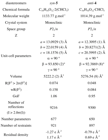
X-Ray structures of compounds 6a and 6s: Single X-ray quality crystals have been obtained for both diastereomers 6a and 6s in a mixture of chloroform and ethanol. Their crystallographic data of are summarized in Supporting Information (Table S5 in the Supporting Information). Both compounds crystallize in a P21/n space group. For
the two structures four molecules are present in the lattice. One CHCl3 molecule is present in the X-ray structure of 6a and another
CHCl3 molecule is present outside the cavity (Figure S6 in the
Supporting Information). The refined structure of 6a also reveals residual electronic density within the cavity that could correspond to a statistical occupation of half the cryptophane molecules by a CHCl3 molecule. The refined structure of 6s shows two CHCl3
molecules located outside the cavity and another CHCl3 molecule
present within the cavity of 6s (Figure S7 in the Supporting Information). The refined X-ray structure of compound 6a shows that some disorder is present. Indeed, as often observed in X-ray structures of the cryptophane derivatives, the three linkers can adopt different conformations. While the trans-trans-trans (ttt) and the
trans-trans-gauche conformations are present in the X-ray structure
of 6a, the X-ray structure of 6s does not show any disorder and the three linkers have an all-trans (ttt) conformation. The non-helical structure of 6s (C3 symmetry) and the ttt conformation give rise to a
large inner cavity, which has been estimated to be 147 Å3. By
contrast the mean value calculated for the inner cavity of 6a is 91 Å3.
For both compounds, the six methoxy groups show different orientations. Three methoxy groups are located in the plane of the bridges, whereas the three others are located out of the plane of the three benzene rings (cif files). This orientation is consistent with the X-ray structures of cryptophane-A (1) and compounds 9a, 9s, 12a,
12s previously reported by our group.[15,17] For instance, the X-ray
structure of the cryptophane-A derivative shows that the methoxy groups are in the plane of the benzene rings. In contrast, the introduction of additional methoxy substituents results in a change of orientation of these groups since they are located out of the plane of the benzene rings. A steric hindrance can explain this difference of orientation.
Scheme 2. synthesis of the cryptophane diastereomers anti-6a (4) and syn-6s (5).
Figure 1 X-ray structures of 6a-(4; left) (disordered parts have been omitted
for clarity) and 6s-(5; right) diastereomers. In both structures the CHCl3
molecule present inside the cavity as well as the hydrogen atoms have been removed for clarity.
129Xe NMR Spectroscopy: For this study,
1,1,2,2-tetrachloroethane-d2 has been chosen as a solvent because it is not supposed to enter
xenon. In these experiments, xenon gas is added after degassing the organic solutions that contain the different cryptophane derivatives. From the one-scan hyperpolarized 129Xe NMR spectra of
the compounds 6a, 6s, 9a, 9s, 12a and 12s recorded in the same conditions (Figure 2), it is worth noting that the xenon in-out exchange rate exhibits large variation depending on the nature of the cryptophane skeleton. A large peak at 225 ppm that corresponds to xenon free in 1,1,2,2-tetrachloroethane-d2 can be clearly identified in
all cases. A second up-field shifted signal of weaker intensity is sometimes located, thus revealing directly the caged xenon environment. For the anti cryptophane 6a at sub-millimolar concentration, in our experimental conditions, a fast exchange situation is observed (or no xenon encapsulation). In contrast, for the compounds 9a and 12a a slow exchange is encountered at sub-millimolar concentration and under the same experimental conditions, and the caged xenon signal is shifted towards higher field when the number of OCH3 groups increases. Somewhat counter intuitively a
broader line is observed for Xe@12a than for Xe@9a, indicating a faster xenon in-out exchange. On the other hand, compounds 6s, 9s and 12s do not show any signal corresponding to the complexes. However, it can be mentioned that the 129Xe NMR spectrum of
compound 6s shows a broadening of the free xenon signal suggesting that encapsulation of xenon occurs under not so fast exchange conditions. The xenon in-out exchange is likely to be slowed down by the six methoxy groups on one CTB unit. For compounds 9s and 12s, the free xenon exhibits sharp signals. Consequently, the ability of the xenon atom to enter the cavity of compounds 9s and 12s is questionable.
If one considers that the xenon in-out exchange is strongly dependent upon the anti or syn arrangement of the three linkers (slow exchange at the NMR scale for the anti-cryptophanes, fast exchange for the syn-cryptophanes), the two exceptions are cryptophanes 6a and 6s. Cryptophane 6a reveals a behavior that is different from what is observed with cryptophane-A (1) and cryptophane 3a (figure S10 in the Supporting Information). For instance, it has been reported that complexation of xenon with 3a – whatever the xenon/cryptophane concentration ratio - occurs under a slow exchange regime. For cryptophane 6s, xenon exhibits an intermediate exchange. This can be compared to the behavior of diastereomer 3s that shows a fast exchange regime at 293 K but for which a signal characteristic of the complex is observed at lower temperature.[18] A common cryptophane scaffold constituted by only
one highly methoxylated CTB seems to be responsible of this effect.
Figure 2. One-scan 129
Xe NMR spectra of the samples of 6 (a and s), 9 (a and
s), 12 (a and s). The subscripts a and s refer to anti and syn arrangement of
the linkers. Spectra have been recorded at 11.74 T and 293 K. Each sample contained 0.3 +- 0.03 mg of cryptophane (0.4 - 0.5 mM) in 600 µL of 1,1,2,2-tetrachloroethane-d2 and ca. 10 mg of xenon (representing a concentration of
ca. 100 mM).
1H NMR spectra of the cryptophane derivatives: the difference
observed with compounds 6a-s, 9a-s and 12a-s in the presence of xenon prompted us to examine more carefully their 1H NMR spectra.
We thus report in this Article the 1H NMR spectra of compounds
6a-s, 9a-s and 12a-s under degassed solution and in the presence of xenon. We also report the 129Xe-1H cross-relaxation (SPINOE)
experiments.[19] These experiments are very useful to establish
whether xenon enters or not the cavities of compounds 6a-s, 9a-s and 12a-s since the 129Xe-1H cross-relaxation only occurs at very
short distance. The lack of solvent peak (6.0 ppm) in the SPINOE spectra of compounds 6a-s, 9a-s and 12a-s is a good indicator that these experiments have been successfully performed.
As an example, Fig. 3 displays the 1H NMR spectra of cryptophanes
12a and 12s in degassed solution (Fig. 3a and 3b) and after
introduction of xenon into the solution (Fig 3c and 3d). Fig. 3e and 3f display the 129Xe NMR SPINOE spectra of compound 12a and 12s,
respectively. Degassing the solution before introducing xenon is a mandatory step to avoid fast 129Xe relaxation by paramagnetic
molecular oxygen and to avoid possible competing binding with other dissolved gases. Interestingly, we notice that introduction of xenon into the solutions deeply modifies the 1H NMR spectra:
important changes occur for both the anti and syn cryptophanes. For instance, for the degassed solution of 12a, the aromatic part of the proton spectrum shows two signals with very different intensities. For the sample in the presence of xenon this region now reveals a unique peak that corresponds to the minor NMR signal observed in the case of the degassed solution. The axial protons Ha of the CTB
units (4.2 ppm) are also strongly affected by the presence of xenon in solution. For instance, we can notice on Figure 3a that the Ha
signals originally observed for the degassed solution have been replaced by another up-field shifted doublet. It is noteworthy that signals characteristic of the three linkers (dashed line) are also strongly affected by the presence or not of xenon inside the solution. Indeed, the two signals initially observed at 4.14 ppm and 3.94 ppm are replaced by an intense multiplet located at 3.99 ppm. The 129Xe
NMR SPINOE sub-spectrum of 12a reveals a simplified spectrum where only the protons that are in close proximity to xenon appear. Dealing with the aromatic protons, for 12a, no change is observable compared to the 1H NMR spectrum of the solution with xenon. In
contrast, the aliphatic part is strongly simplified (in particular as expected the axial proton signals of the CTB are strongly attenuated).
Figure 3. Comparison of the 1
H NMR spectra for compounds 12a (left) and
12s (right). The samples were constituted by 0.3 mg of cryptophane 12a in
600 µL of 1,1,2,2-tetrachloroethane-d2 and 0.3 mg of cryptophane 12s in 600 µL of 1,1,2,2-tetrachloroethane-d2. From top to bottom, the green spectra were recorded with the degassed solution, the blue ones after introduction of xenon into solution, and the red ones correspond to the results of the SPINOE experiments obtained with the sequence of ref. [20].
The changes observed between the 1H spectra of the degassed
solution, of the solution in the presence of xenon and of the SPINOE (Fig. 3a, 3b and 3c) suggest that the introduction of xenon into the solution has a strong impact on the conformation of the three linkers. A comparison between these spectra indicates that compound 12a has to change the conformation of the three linkers to accommodate the xenon atom. This effect can clearly be evidenced in the aliphatic region of the spectrum.
For compound 12s, the spectral changes between the three spectra are even more important. The degassed solution of 12s exhibits a 1H
spectrum with numerous signals present in the aromatic and aliphatic regions. It reveals that compound 12s possesses at least three different conformations of the bridges in slow exchange regime. This is supported by the presence of three well-resolved signals in the aromatic part of the spectrum. The addition of xenon into the solution results in a simplification of the 1H NMR spectrum
and two NMR signals with close intensities are observed in the aromatic part. The aliphatic part of the 1H NMR spectrum is also
affected by the presence of xenon. However, this spectral region is not very informative due to the strong overlapping of the different signals present in that region.
From the single 129Xe NMR spectrum, there was no evidence of
inside the cavity of 12s could be ascertained from its 129Xe-1H
SPINOE spectrum, revealing some well-defined 1H signals. For
instance, the aromatic region reveals a single NMR peak, and a strong simplification in the aliphatic part of the spectrum is also clearly observed. Combined together these results show that compound 12s can easily adopt different conformations in solution depending on the experimental conditions. The presence of these conformations seems to be favored in the absence of guest inside the cavity of 12s. Upon addition of xenon, several conformations are still present in solution but only one conformation seems to be able to bind efficiently xenon.
The 1H NMR spectra of derivatives 9a-s and 6a-s (Figures. S8 and
S9 in the Supporting Information) that possess two CTB units with a different degree of substitution, also result in important spectral changes upon addition of xenon. These compounds possess a lower symmetry (C3) than cryptophanes 12a (D3) or 12s (C3h). Their
corresponding 1H NMR spectra are therefore more complicated to
interpret due to the presence of additional signals in the aromatic region of the 1H NMR spectrum. The 1H NMR spectrum of a
degassed solution of 9a shows several signals of with different intensities in the aromatic region (Figure S9a in the Supporting Information). Considering the molecular symmetry of 9a and the relative intensities of the signals, we expect three signals in the aromatic region. However five distinct peaks appear in this region, suggesting the presence of several conformations in a slow exchange regime. This is also supported by the presence of several doublets in the aliphatic region of the spectrum for protons Ha and
He. As observed for compounds 12a and 12s, for 9a a simplification
of the 1H NMR spectrum is observed after introduction of xenon into
the solution. For instance, the aromatic region of the Xe@9a complex is dominated by two intense signals (two protons of 9a resonate at the same frequency). Three other signals of much lower intensity are also present in this region and indicate the presence of another conformation in minor proportion. A simplification of the 1H
NMR spectrum of 9a in the aliphatic region is also observed (Figure S8b in the Supporting Information). The differences observed between these two spectra suggest that a strong rearrangement of the linkers takes place upon addition of xenon. The 129Xe-1H
SPINOE of 9a shows a simplified spectrum with respect to that of the Xe@9a complex. Once again, this simplification is clearly visible in the aromatic part of the spectrum since only two intense signals are now visible. Spectral changes are also observed in the aliphatic part.
As observed for its diastereomer, compound 9s also shows important spectral modifications upon addition of xenon into the solution. For instance, the 1H NMR spectrum of the degassed
solution of 9s (Figure S8d in the Supporting Information) shows the presence of at least two major forms in ca. 1:2 ratio and an additional minor form. This situation where two forms dominate, is different from that obtained for 9a, and can be interpreted considering the syn arrangement of the linker. Upon addition of xenon, this ratio is reversed, revealing a clear induction effect of xenon. Interestingly, the 129Xe-1H SPINOE spectrum of 9s shows a
simplified spectrum suggesting that a cryptophane with a unique conformation of the linkers can effectively accommodate xenon. Once again, as mentioned for compound 12s, the 129Xe-1H SPINOE
technique is a valuable tool to discriminate between the different conformations that bind or not xenon. In the case of compound 9s it can be clearly established that a single conformation of the bridges can accommodate xenon in solution, even though several conformations in a slow exchange regime can be detected on the 1H
NMR spectrum of 9s.
Among the different compounds studied in this Article, compound 6a and its diastereomer 6s have the lowest degree of substitution. The
1H NMR spectrum of a degassed solution of 6a (figure S9a in the
Supporting Information) is very simple; the presence of 4 aromatic signals is indicative of a single conformation. This is supported by the presence of only two sets of signals for the Ha and He protons. At
first glance, this result may appear surprising considering that compounds with a higher degree of substitution showed several conformations in slow exchange. However, it can be interpreted considering the higher flexibility of compound 6a in which one of the two CTB unit is devoid of substituents. Upon addition of xenon, only slight modifications of the spectrum occur more specifically in the aliphatic region of the spectrum. The 129Xe -1H SPINOE
sub-spectrum of 6a gives the same signals except the axial and
equatorial methylene protons. Consequently, compound 6a exhibits in solution a single conformation of the linkers whatever the xenon concentration (from 10 mM to 100 mM, data not shown). This conformation is able to accommodate xenon, even though no specific 129Xe NMR signal for the complex is detected at 293K. The
absence of spectral changes between these three spectra could indicate that the cavity size is too large to be modified upon xenon encapsulation. With compound 6s a different situation occurs: the aromatic region of the 1H spectrum for the degassed sample exhibits
two sets of signals (Figure. S9d in the Supporting Information). These two conformations are present in a similar proportion but the addition of xenon (Figure S9e in the Supporting Information) modifies their relative proportion, and the SPINOE sub-spectrum indicates a unique conformation able to accommodate xenon (Figure. S9f in the Supporting Information).
The helical arrangement of the linkers of the anti-cryptophanes is expected to give them more flexibility upon binding of guest molecules than syn-cryptophanes. It is surprising to observe that compound 6s can modify its conformation to interact with xenon, whereas the 1H NMR spectrum of its diastereomer 6a remains
unchanged. The 129Xe-1H SPINOE spectrum of 6s (figure S9f in the
Supporting Information) shows a simplification of the spectrum indicating that a single conformation can accommodate xenon. The NMR experiments performed with compounds 3a-s, 6a-s, 9a-s and 12a-s lead to conclusions that may seem counter-intuitive. Indeed, the study of these compounds reveals that a correlation between the degree of substitution of the CTB units and the in-out exchange dynamics of xenon cannot be clearly established as initially expected. Similarly, a correlation between the arrangement
syn or anti of the linkers and the exchange dynamics of xenon is not
direct. Consequently, the in-out exchange dynamics of xenon with these derivatives cannot be explained solely on considerations based on the cavity size of the host molecules and the steric hindrance induced by the presence of the additional methoxy groups. Other factors must be involved to interpret these results. The presence of numerous conformational forms observed for the majority of these derivatives and the ability of these compounds to change the conformation of their linkers give us a clue. The 1H NMR
spectra of these compounds reveal that the linkers can adopt several conformations (trans or gauche) in solution. Importantly, these conformations are in a slow exchange regime. This is attested by the presence of distinct signals in the aromatic part of these compounds. On the other hand, the 129Xe - 1H SPINOE spectra of
compounds 6a-6s, 9a-9s and 12a-12s show a clear simplification with respect to their 1H NMR spectra. Thus, these experiments
demonstrate without exception that all the derivatives studied in this article bind xenon, even though a 129Xe NMR signal characteristic of
the complexes cannot be always detected. The 129Xe - 1H SPINOE
spectra also indicate that a single conformation, among those detected by 1H NMR spectroscopy, can effectively accommodate
xenon. The presence of different conformations of the linkers, even after adding xenon into the solution, seems to be a consequence of the presence of the additional methoxy groups grafted on the CTB units. These substituents rigidify the cryptophane scaffold and constrain the linkers to adopt different conformations. This assumption is supported by the study of the 1H NMR spectra of 3a
and 3s (Figure S11 in the Supporting Information) that reveal a single conformation for the degassed and gassed samples. In the case of compounds 6s, 9a-9s and 12a-12s these different conformations are in a slow exchange regime. The anti derivatives 9a and 12a, thanks to the helical arrangement of their linkers are more flexible and they can more easily modify the conformation of their linkers to accommodate xenon. In contrast, the syn arrangement (compounds 6s, 9s and 12s) of the linkers seems detrimental and several conformations of the linkers are still present even after introducing xenon.
Interestingly, these results show that cryptophane derivatives with similar linkers can exhibit very different physical properties in the presence of xenon. Changing the nature of the substituents attached on the cryptophane skeleton appears to be a simple way to change drastically the in-out exchange dynamics of xenon. However, the substituents effect on the exchange dynamics of xenon is difficult to predict. It is worth-mentioning that these considerations do not take into account interactions that could eventually take place between solvent molecules and the host molecules. Interactions are probably more important in the case of compounds 6a and 6s considering the
larger apertures observed for these two compounds. Molecular dynamics calculations of the complexes using an explicit solvation model would be a valuable tool to interpret these data. Such theoretical approach appears is very challenging considering the large size of these molecules and the number of solvent molecules needed for the calculations.
Conclusions
We report a study of the Xe@6a-s, 9a-s and 12a-s complexes by 1H
NMR and 129Xe NMR spectroscopy. These compounds differ from
their congeners 1-3 by the number of methoxy substituents attached on the cryptophane-222 skeleton and the arrangement syn or anti of the linkers. We show that introduction of additional methoxy groups on the cryptophane backbone has a strong impact on the in-out exchange dynamics of xenon, whereas the lengths of the linkers are the same. Among this series, two cryptophanes have an unexpected behavior. Compound 6a is the only anti-cryptophane in which xenon is in fast exchange on the 129Xe NMR spectrum, 6s is the only
syn-cryptophane in which xenon displays an intermediate exchange regime, as evidenced by the broadening of the low-field signal. However for these two compounds we cannot exclude a contribution of the solvent molecules to explain these results.
The introduction of methoxy substituents positioned from each side of the linker rigidifies the cryptophane-222 scaffold. Thus, in solution these compounds show several conformations of their bridges in slow exchange dynamics. This effect seems more pronounced with the syn derivatives, which are more rigid. Addition of xenon into the solution modifies the relative distribution of these conformations. The
anti derivatives having a higher flexibility show a major conformation
for the linkers after introduction of xenon. In contrast, the 1H NMR
spectra of the syn derivatives show several conformations in slow exchange even after introduction of xenon. The 129Xe - 1H SPINOE
spectra provide additional information and are valuable to distinguish the different conformations that effectively accommodate xenon that the other ones that do not. In our case, the 129Xe - 1H SPINOE
spectra reveal that only a single conformational present in solution can bind xenon.
Combined together these results demonstrate that cryptophane derivatives possessing identical linkers can possess very different properties towards xenon encapsulation. This effect can possibly be exploited for 129Xe NMR-based biosensing application. It would be a
way to tune the in-out exchange dynamics that play a key role for sensitive NMR detection, helped in that by recent solutions from the literature based on quantitative analysis of saturation-transfer experiments.[21]
Experimental Section
Mass spectra (HRMS LSIMS) were performed by the Centre de Spectrométrie de Masse, University of Lyon, on a Thermo-Finnigan MAT 95XL spectrometer. 1H and 13C NMR spectra were recorded on
a 300 MHz NMR spectrometer at 300 and 72 MHz, respectively. Chemical shifts are in d values from Me4Si (1H, 13C). Column
chromatographic separations were carried out over Merck silica gel 60 (0.040-0.063 mm). Analytical thin layer chromatography (TLC) was performed on Merck silica gel TLC plates F-254. The solvents were distilled prior to use: DMF and CH2Cl2 from CaH2, THF from
Na/benzophenone and pyridine from KOH.
Synthesis of compound 12. Iodo derivative 11 (2.11 g, 6.1 mmol) was added in one portion to a stirred solution of 10,15-Dihydro-5H-tribenzo[a,d,g][9]annulene-2,7,12-triol 7 (0.58 g, 1.82 mmol), cesium carbonate (1.98 g, 6.1 mmol) in DMF (35 mL). The mixture was stirred overnight at 80°C under an argon atmosphere. The mixture was poured in water and the product was extracted four times with AcOEt. The combined organic layers were then washed twice with brine and dried over Na2SO4. Filtration and evaporation of the
solvent under reduced pressure to give yellow oil. The product was then purified on silica gel (eluent: AcOEt/Petroleum Ether: 50/50 then 75/25). The second spot was collected. The solvent was
evaporated under reduced pressure to give rise to an oily product. Compound 12 was obtained as a white glassy product (1.58 g; 72 %) was then obtained by removing trace of solvent with the vacuum line. 1H NMR (300 MHz, CDCl 3) d 7.21 (3 H, d, J = 8.5 Hz), 6.89 (3 H, d, J = 2.7 Hz), 6.61 (3 H, dd, J = 8.5 Hz, J = 2.7 Hz), 6.55 (s, 6 H), 4.72 (3 H, d, J = 13.5 Hz), 4.69 (3 H, d, J = 11.5 Hz), 4.68 (3 H, m), 4.41 (3 H, d, J = 11.5 Hz), 4.30 - 4.10 (12 H, m), 3.90 (3 H, m), 3.74 (18 H, m), 3.59 (3 H, d, J = 13.5 Hz), 3.52 (3 H, m), 1.90 – 1.50 (18 H, m). 13C NMR (75.475 MHz, CDCl 3, 25°C) d 157.5 (3C), 153.3 (3C), 141.0 (3C), 136.1 (3C), 134.0 (3C), 131.6 (3C), 131.0 (3C), 116.2 (3C), 112.7 (3C), 104.9 (3C), 97.8 (3C), 71.2 (3C), 69.1 (3C), 66.9 (3C), 62.3 (3C), 56.0 (3C), 36.6 (3C), 30.6 (3C), 25.4 (3C), 19.5 (3C). HRMS (ESI) calcd for C69H84O18Na [M+Na+], 1223.5550 found
1223.5503.
Synthesis of cryptophanes 4 (6a) and 5 (6s). In a 1000 mL round bottom flask, formic acid (300 mL) was added in one portion to a solution of cryptophane precursor 12 (0.55 g, 0.46 mmol) dissolved in chloroform (300 mL). The solution was stirred for 5 hours at 55°C. The solvents were removed under reduced pressure. Then CHCl3
(100 mL) was added and the solvent was removed under reduced pressure. This procedure was repeated three times in order to remove traces of formic acid via azeotropic distillation. The crude product was then purified on silica gel (eluent: CH2Cl2/Acetone –
90/10) to give rise to a white glassy product (0.58 g). This product contains the two cryptophanes anti-4 (6a) and syn-5 (6s) in the same amount. A second column chromatography (eluent: CH2Cl2/Acetone – 99/01) allows the separation give rise to the pure
syn-5 (6s) derivative (0.12 g; 29 %). This compound was then recrystallized in a mixture of CHCl3 and EtOH. Compound 6: 1H
NMR (300 MHz, CDCl3) d 7.11 (3H, s), 7.07 (3H, d, J = 8,7 Hz), 6.67 (3H, d, J = 2,4 Hz), 6.55 (3H, dd, J = 2,4 Hz, J = 8.7 Hz), 4.58 (3H, d, J = 13,5 Hz), 4.28 (3H, d, J = 13,5 Hz), 4.35 – 4.25 (3H, m), 4.25 – 4.10 (3H, m), 4.10 – 3.95 (3H, m), 3.91 (9 H, s), 3.87 (3H, d, J = 13,5 Hz), 3.73 (9H, s), 3.60 – 3.50 (3H, m), 3.46 (3H, d, J = 13,5 Hz). 13C NMR (75.475 MHz, CDCl3, 25°C) d 157.14 (3C), 152.03 (3C), 151.31 (3C), 140.7 (3C), 139.1 (3C), 136.5 (3C), 132.1 (3C), 130.6 (3C), 125.3 (3C), 118.4 (3C), 113.1 (3C), 110.5 (3C), 69.8 (3C), 65.8 (3C), 60.2 (3C), 55.7 (3C), 36.0 (3C), 30.0 (3C). HRMS (ESI) calcd for C54H55O12 [M+], 895,3688 found 895,3674.
This product was then followed by compound 4 (6a), still contaminated by the presence of a small amount of compound 5 (6s). Another column chromatography on silica gel (eluent: CH2Cl2/Acetone – 99/01) gives rise to the pure diastereomer 4 (0.12
g; 29 %). This product was then recrystallized in a mixture of CHCl3
and EtOH. 1H NMR (300 MHz, CDCl 3) d 7.08 (3H, d, J = 8.3 Hz), 7.05 (3H, s), 6.74 (3H, d, J = 2.4 Hz), 6.39 (3H, dd, J = 2,4 Hz, J = 8.3 Hz), 4.62 (3H, d, J = 13,5 Hz), 4.28 (3H, d, J = 13,5 Hz), 4,2 – 4,0 (12 H, m), 3.85 (9H, s), 3.83 (3H, d, J = 13.5 Hz), 3,72 (3H, s), 3.49 (3H, d, J = 13.5 Hz). 13C NMR (75.475 MHz, CDCl 3, 25°C) d 157.6 (3C), 151.1 (3C), 150.8 (3C), 141.0 (3C), 139.4 (3C), 135.6 (3C), 132.6 (3C), 130.7 (3C), 125.4 (3C), 119.1 (3C), 115.5 (3C), 110.8 (3C), 70.1 (3C), 68.2 (3C), 60.1 (3C), 55.6 (3C), 36.3 (3C), 29.9 (3C). HRMS (ESI) calcd for C54H55O12 [M+], 895.3688 found
895.3682.
X-ray crystallography: Suitable crystals for all four compounds were selected and mounted on a Gemini kappa-geometry diffractometer (Agilent Technologies UK Ltd) equipped with an Atlas CCD detector and using Cu radiation ( l= 1.5418 Å). Intensities were collected at 150 K for anti-5 and syn-6 by means of the CrysalisPro software Reflection indexing, unit-cell parameters refinement, Lorentz-polarization correction, peak integration and background determination were carried out with the CrysalisPro software.[22] An analytical absorption correction was applied using
the modeled faces of the crystal.[23] The resulting set of hkl was used
for structure solution and refinement. The structures were solved by direct methods with SIR97 and the least-square refinement on F2
was achieved with the CRYSTALS software.[24,25] All non-hydrogen
atoms were refined anisotropically. The hydrogen atoms were all located in a difference map, but those attached to carbon atoms were repositioned geometrically. The H atoms were initially refined with soft restraints on the bond lengths and angles to regularize their geometry (C---H in the range 0.93--0.98 Å) and Uiso(H) (in the range
refined with riding constraints. For compound anti-5, a chloroform solvent molecule could be located outside the cryptophane cage. Nevertheless this structure contains additional solvent molecules inside the cryptophane cages that could not be localized. The contribution of the disordered solvent molecules was removed using the SQUEEZE algorithm.[26]
CCDC 1525399, 1525400, contains the supplementary crystallographic data for anti-4 and syn-5, respectively. These data can be obtained free of charge from The Cambridge Crystallographic Data Centre via www.ccdc.cam.ac.uk/data_request/cif.
Laser-Polarized 129Xe NMR spectroscopy: Hyperpolarized xenon
was produced through the spin-exchange method in the batch mode using a home built apparatus based on two coupled 30 W laser diodes.[27] Frozen xenon was stored and transported in a glass
reservoir immersed in liquid nitrogen. For the transfer to the NMR tube this reservoir was heated and installed in a vacuum line in the fringe field of the NMR magnet. A hollow NMR spinner enabled us to condensate hyperpolarized xenon on top of the solution without freezing the solution.
Acknowledgements
Support from the French Ministry of Research (project ANR-12-BSV5-0003 MAX4US) is greatly acknowledged.
Keywords: Cryptophanes • hyperpolarized xenon • molecular recognition • SPINOE • NMR
[1] a) C. Landon, P. Berthault, F. Vovelle, H. Desvaux, Protein Science.
2001, 762-770. b) L. Dubois, S. Parrès, J. G. Huber, P. Berthault, H. Desvaux,
J. Phys. Chem. B 2004, 108, 767-773.
[2] T. Brotin, J. P. Dutasta, Chem. Rev. 2009, 88-130
[3] M. El Haouaj, M. Luhmer, Y. H. Ko, K. Kim, K. Bartik, J. Chem. Soc. Perkin. Trans. 2, 2001, 804-807.
[4] K. Bartik, M. Luhmer, S. J. Heyes, R. Ottinger, J. Reisse, Journal of Magnetic Resonance, serie B 1995, 109, 164-168.
[5] K. Bartik, M. Luhmer, J. P. Dutasta, A. Collet, J. Reisse, J. Am. Chem. Soc. 1998, 120, 784 – 791.
[6] a) L. L. Chapellet, J. R. Cochrane, E. Mari, C. Boutin, P. Berthault, T. Brotin, J. Org. Chem. 2015, 80, 6143-6151. b) E. Dubost, J. -P. Dognon, B. Rousseau, G. Milanole, C. Dugave, E. Boulard, E. Léonce, C. Boutin, P. Berthault, Angew. Chem. Int. Ed. 2014, 53, 9837-9840. c) E. Dubost, N. Kotera, S. Garcia-Argote, Y. Boulard, E. Léonce, C. Boutin, P. Berthault, C. Dugave, B. Rousseau, Org. Lett. 2013, 15, 2866-2868.
[7] a) M. M. Spence, S. Rubin, I. E. Dimitrov, R. J. Ruiz, D. E. Wemmer, A. Pines, S. Q. Yao, F. Tian, P. G. Schultz, Proc. Natl. Acad. Sci. U.S.A. 2001, 98, 10654-10657. b) L. Schroder, T. J. Lowery, C. Hilty, D. E. Wemmer, A. Pines, Science, 2006, 314, 446-449. c) T. J. Lowery, S. Garcia, L. Chavez. E. J. Ruiz, T. Wu, T. Brotin, J.-P. Dutasta, D. S. King, P. G. Schultz, A. Pines, A.; D. E. Wemmer, ChemBioChem 2006, 7, 65-73. d) S. Klippel, J. Dopfert, J. Jayapaul, M. Kunth, F. Rossella, M. Schnurr, C. Witte, C. Freund, L. Schröder, Angew. Chem. Int. Ed. 2014, 126, 503-506. e) C. Witte, V. Martos, H. M. Rose, S. Reinke, S. Klippel, L. Schröder, C. P. R. Hackenberger, Angew. Chem. Int. Ed. 2015, 54, 2806-2810. f) N. S. Khan, B. A. Riggle, G. Seward, Y. Bai, I. J. Dmochowski, Bioconjugate Chem. 2015, 26, 101-109.
[8] T. Brotin, A. Lesage, L. Emsley, A. Collet, A. J. Am. Chem. Soc.
2000, 122, 1171-1174.
[9] a) C. Boutin, A. Stopin, L. Fatimazorha, T. Brotin, J. P. Dutasta, N. Jamin, A. Sanson, Y. Boulard, F. Leteurtre, G. Huber, A. Bogaert-Buchmann, N. Tassali, H. Desvaux, M. Carriere, P. Berthault. Bioorganic and Medicinal chemistry 2011, 19, 4135-4143. b) N. Kotera, N. Tassali, E. Léonce, C. Boutin, P. Berthault, T. Brotin, J. P. Dutasta, L. Delacour, D. A. Buisson, F. Taran, S. Coudert, B. Rousseau. Angew. Chem. Int. Ed. 2012, 51, 4100-4103. [10] H. A. Fogarty, P. Berthault, T. Brotin, G. Huber, H. Desvaux, J. -P. Dutasta, J Am. Chem. Soc. 2007, 129, 10332–10333.
[11] R. M. Fairchild, A. I. Joseph, K. T. Holman, H. A. Fogarty, T. Brotin, J. -P. Dutasta, C. Boutin, G. Huber, -P. Berthault, J. Am. Chem. Soc. 2010, 132, 15505-15506.
[12] M. Luhmer, B. Goodson, Y. Q. Song, D.-D. laws, L. Kaiser, M. C. Cyrier, A. Pines, J. Am. Chem. Soc., 1999, 121, 3502-3512.
[13] a) N. Daugey, T. Brotin, N. Vanthuyne, D. Cavagnat, T. Buffeteau, J. Phys. Chem. B 2014, 118, 5211-5217. b) T. Brotin, N. Vanthuyne, D. Cavagnat, L. Ducasse, T. Buffeteau, J. Org. Chem. 2014, 79, 6028-6036.
[14] E. Dubost, J. -P. Dognon, B. Rousseau, G. Milanole, C. Dugave, Y. Boulard, E. Léonce, C. Boutin, P. Berthault, Angew. Chem. Int. Ed. 2014, 53, 9837-9840.
[15] T. Brotin; D. Cavagnat, E. Jeanneau, T. Buffeteau, J. Org. Chem. 2013, 78, 6143 - 6153.
[16] T. Brotin, V. Roy, J. -P. Dutasta, J. Org. Chem. 2005, 70, 6187 – 6195. [17] D. Cavagnat, T. Brotin, J.-L. Bruneel, J. P. Dutasta, A. Thozet, M. Perrin, and F. Guillaume, J. Phys. Chem. B, 2004, 108, 5572-5581.
[18] G. Huber, L. Beguin, H. Desvaux, T. Brotin, H. A. Fogarty, J. -P. Dutasta, P. Berthault, J. Phys. Chem. A, 2008, 112, 11363 - 11372. [19] M. Haake, A. Pines, J. A. Reimer, R. Seydoux, J. Am. Chem. Soc.
1997, 119, 11711-11712.
[20] H. Desvaux, T. Gautier, G. Le Goff, M. Petro, P. Berthault, Eur. Phys. J. D. 2000, 12, 289-296.
[21] a) M. Kunth, C. Witte, L. Schröder, J. Chem. Phys. 2014, 141, 194202. b) M. Kunth, C. Witte, A. Hennig, L. Schröder, Chem. Sci. 2015, 6, 6069– 6075. c) S. Korchak, W. Kilian, L. Mitschang, Chem. Commun. 2015, 51, 1721–1724. d) S. Korchak, W. Kilian, L. Schröder, L. Mitschang, J. Magn. Reson. 2016, 265, 139–145.
[22] CrysAlisPro, Agilent Technologies, Version 1.171.36.28 (release 20-01-2011 CrysAlis171 .NET) (compiled Feb 1 2013,16:14:44).
[23] R. C. Clark, J. S. Reid, Acta Cryst. A 1995, 51, 887-897.
[24] A. Altomare, M. C. Burla, M. Camalli, G. L. Cascarano, C. Giacovazzo, A. Guagliardi, A. Grazia, G. Moliterni, G. Polidori, R. J. Spagna, App. Cryst.
1999, 32, 115-119.
[25] P. W. Betteridge, J. R. Carruthers, R. I. Cooper, K. Prout, D. J. Watkin, J. Appl. Cryst. 2003, 36, 1487.
[26] P. V. D. Sluis, A. L. Spek, Acta Cryst. A 1990, 46, 194-201.
[27] C. Chauvin, L. Liagre, C. Boutin, E. Mari, E. Léonce, G. Carret, B. Coltrinari, P. Berthault, Rev. Sci. Instrum. 2016, 87, 016105.
Supporting Information
Role of the methoxy groups in cryptophanes for complexation of xenon.
Conformational Selection Evidenced by
129
Xe-
1
H
NMR SPINOE
Experiments.
Patrick Berthault,
*[b]Céline Boutin,
[b]Estelle Léonce,
[b]Erwann Jeanneau,
[c]Thierry Brotin
*[a][a]
Laboratoire de Chimie de l’ENS LYON, UMR 5182 - CNRS, École Normale Supérieure de Lyon, 46
Allée d’Italie, 69364 Lyon, France
[b]
NIMBE, CEA, CNRS, Université Paris-Saclay, CEA Saclay 91191 Gif-sur-Yvette, France
[c]
Centre de Diffractométrie Henri Longchambon, Université Lyon 1, 5 rue de La Doua, 69100
Villeurbanne, France.
S1:
1H NMR (300 MHz) spectrum of compound (4)-6a in CDCl
3
solution at 298 K.
S2:
13C NMR (75.5 MHz) spectrum of compound (4)-6a in CDCl
3solution at 298 K.
S3:
1H NMR (300 MHz) spectrum of compound (5)-6s in CDCl
3solution at 298 K.
S4:
13C NMR (75.5 MHz) spectrum of compound (5)-6s in CDCl
3solution at 298 K.
S5: crystallographic data for diastereomers (4)-6a and (5)-6s.
S6: (top) View of cryptophane (4)-6a (hydrogen atoms and disordered parts have been
omitted for clarity). (bottom) Projection af the packing of molecule 6 along the a unit-cell axis.
S7: (top) View of cryptophane (5)-6s (hydrogen atoms and disordered parts have been
omitted for clarity). (bottom) Projection af the packing of molecule (5)-6s along the a unit-cell
axis.
S8:
1H NMR spectra of compounds 9a (left) and 9s (right) for the degassed solutions (spectra
a and d); in the presence of xenon (spectra b and e).
129Xe-
1H SPINOE spectra of compounds
9a (spectrum c) and 9s (spectrum f).
S9:
1H NMR spectra of compounds 6a (left) and 6s (right) for the degassed solutions (spectra
a and d) ; in the presence of xenon (spectra b and e).
129Xe-
1H SPINOE spectra of compounds
6a (spectrum c) and 6s (spectrum f).
S10:
129Xe NMR spectra of compounds 3a (left) and 3s (right) at 293K
S11:
1H NMR spectra of compounds 3a (left) and 3s (right) for the degassed solutions
(spectra a and d) ; in the presence of xenon (spectra b and e).
129Xe-
1H SPINOE spectra of
compounds 3a (spectrum c) and 3s (spectrum f).
S1 :
1H NMR (300 MHz) spectrum of compound (4)-6s in CDCl
3solution at 298 K.
6.4 6.6 6.8 7.0 7.2 7.4 7.6 7.8 ppm 6.373 6.382 6.401 6.409 6.736 6.744 7.046 7.064 7.092 7.240 3.00 3.00 6.08 3.2 3.4 3.6 3.8 4.0 4.2 4.4 4.6 4.8 ppm 3.474 3.519 3.716 3.815 3.845 3.860 4.038 4.058 4.073 4.082 4.121 4.138 4.158 4.182 4.200 4.254 4.299 4.589 4.634 3.04 9.07 12.07 15.34 2.95 O O O O O O R R R R R R R = OMe
CHCl
3CHCl
3H
2O
9.5
9.0
8.5
8.0
7.5
7.0
6.5
6.0
5.5
5.0
4.5
4.0
3.5
3.0
2.5
2.0
1.5
1.0
ppm
1.543
3.474
3.519
3.716
3.815
3.845
3.860
4.038
4.058
4.073
4.082
4.121
4.138
4.158
4.182
4.200
4.254
4.299
4.589
4.634
6.373
6.382
6.401
6.409
6.736
6.744
7.046
7.064
7.092
7.240
3.04 9.07 12.07 15.34 2.95 3.00 3.00 6.08Compound
4 (6a)
S2 :
13C NMR (75.5 MHz) spectrum of compound (4)-6a in CDCl
3solution at 298 K.
20
30
40
50
60
70
80
90
100
110
120
130
140
150
160
170
180
190
ppm
29.92 36.33 55.57 60.12 68.18 70.09 76.57 77.00 77.42 110.82 115.51 119.05 125.35 130.73 132.61 135.60 139.43 141.01 150.81 151.08 157.61 O O O O O O R R R R R R R = OMeCDCl
3Compound
4 (6a)
S3 :
1H NMR (300 MHz) spectrum of compound (5)-6s in CDCl
3solution at 298 K.
9.5 9.0 8.5 8.0 7.5 7.0 6.5 6.0 5.5 5.0 4.5 4.0 3.5 3.0 2.5 2.0 1.5 1.0 ppm 1.520 3.443 3.488 3.532 3.547 3.564 3.581 3.597 3.734 3.845 3.891 3.906 4.020 4.037 4.052 4.070 4.146 4.163 4.180 4.197 4.229 4.255 4.274 4.288 4.305 4.321 4.338 4.555 4.600 6.534 6.542 6.562 6.571 6.664 6.672 7.060 7.089 7.108 7.240 6.18 9.09 11.63 3.03 9.01 3.03 2.94 2.96 6.00 6.6 6.7 6.8 6.9 7.0 7.1 ppm 6.534 6.542 6.562 6.571 6.664 6.672 7.060 7.089 7.108 3.6 3.8 4.0 4.2 4.4 4.6 ppm 3.443 3.488 3.547 3.564 3.734 3.845 3.891 3.906 4.020 4.037 4.163 4.180 4.197 4.255 4.274 4.288 4.305 4.321 4.555 4.600 CHCl 3 H2 O O O O R R R O O O R R R R = OMe Compound (5)-6s