Decoding Structure-function Relationships of Glycans
By
Nathan Wilson Stebbins
B.S., Biochemistry, Cellular and Molecular Biology
MAY
092017
University of Tennessee, 2011
LIBRARIES
ARCHIVES
Submitted to the Department of Biological Engineering In partial fulfillment of the requirements for the degree of
Doctor of Philosophy in Biological Engineering at the
MASSACHUSETTS INSTITUTE OF TECHNOLOGY
January 2017
7
@ 2017 Massachusetts Institute of Technology. All Rights Reserved.
Signature of Author
Signature redacted
Nathan~tebbins Department of Biological Engineering January 2017
Certified by
Signature redacted
Ram Sasisekharan, Ph.D. Alfred H. Caspary Professor of Biological Engineering
Thesis Supervisor
Accepted by
Signature redacted
N
_VMathe,Ph.D.
Professor, Department of Biological Engineering Chair, Graduate Program
This doctoral thesis has been examined by a committee of the Department of Biological Engineering
Katharina Ribbeck, Ph.D. Eugene Bell Career Development Professor of Tissue Engineering Department of Biological Engineering
Thesis Committee Chair
Signature redacted
Ram Sasisekharan, Ph.D. Alfred H. Caspary Professor of Biological EngineeringThesis Supervisor
David Berry, M.D. Ph.D General Partner Flagship Ventures
Decoding structure-function relationships of glycans
By
Nathan Stebbins
Submitted to the Department of Biological Engineering on January, 2017 in partial fulfillment of the
requirements for the degree of
Doctor of Philosophy in Biological Engineering
Abstract
Glycans are an important class of biological molecules that regulate a variety of physiological processes such as signal transduction, tissue development and microbial pathogenesis. However, due to the structural complexity of glycans and the unique intricacies of glycan-protein interactions, elucidating glycan structure-function
relationships is challenging. Thus, uncovering the biological function of glycans requires an integrated approach, incorporating structural analysis of glycans, and glycan-proteins interactions with functional analysis. In this thesis, I develop new tools and implement integrated approaches to study glycans and glycan-binding proteins (GBPs). I apply these approaches to study glycans and GBPs in two areas: i) the role of hemagglutinin-glycan receptor specificity in human adaptation and pathogenesis of influenza and ii) the function of glycan regulation of cell-microenvironment interaction in cancer
progression.
Section 1: Influenza poses a significant public health threat and there is a constant looming threat of a pandemic. Pandemic viruses emerge when avian viruses acquire mutations that enable human adaptation, leading to infection of an antigenically naive host. Influenza Hemagglutinin (HA), and HA-glycan receptor interactions, play a central role in host tropism, transmissibility, and immune recognition. In section one, I develop and apply an integrated approach comprised of structural modeling, inter-amino acid network analysis, biochemical assays, and bioinformatics tools to study the
hemagglutinin-glycan interaction and, in some cases, HA's antigenic properties. Using this approach, we i) identify the structural determinants required, and potential
mutational paths, for H5N1 to quantitatively switch it's binding specificity to human glycans receptors, ii) identify the mutations that enable the 2013 outbreak H7N9 HA to
improve binding to human glycan receptors in the upper respiratory tract, iii) uncover
H3N2 strains that are currently circulating in birds and swine that possess features of a
virus that could potentially re-emerge and cause a pandemic, and iv) characterize the glycan binding specificity of a novel 2011 Seal H3N8 HA. The approaches implemented here and the findings of these studies provide a framework for improved surveillance of influenza viruses circulating in non-human hosts that pose a pandemic threat.
Section 2: Glycans are abundant on the cell surface, and at the cell-ECM interface where they mediate interactions between cells and their microenvironment. Despite this, the function of glycans in cancer progression remains largely understudied. Here, I develop an integrated approach to characterize the cell surface glycome,
including N-linked, 0-linked glycans, and HSGAGs. This approach integrates glycogene expression data, analytical tools, and glycan binding protein reagents. I demonstrate that this platform enables rapid and efficient characterization of the N- and 0-linked glycome in a model cell system, representing metastatic versus non-metastatic cancer cells. Next, I apply this integrated approach to uncover new roles of glycans. I study the role that HSGAGs play in regulating cancer stem cell (CSC) activity in breast cancer. Here, we report that SULF1, an HSGAG modifying enzyme, is required for efficient tumor initiation, growth and metastasis of CSCs. Furthermore, we identify a putative mechanism by which SULF1 regulates interactions between CSCs and their
microenvironment. The approaches implemented here and the finding of these studies have important implications for the development of cancer therapeutics.
Overall, this thesis provides important tools, approaches and insights to enable and improve the study of glycans and glycan binding proteins. Together the work here provides a framework for decoding structure-function relationship of glycans.
Thesis Supervisor: Ram Sasisekharan, Ph.D.
Acknowledgements
First and foremost, I would like to thank my thesis advisor Ram Sasisekharan. The completion of this thesis work would not have been possible without his constant support, positivity and guidance. Ram, from our very first meeting MIT, you instilled such great confidence in me. You gave me a place to learn and the freedom to grow. Your outstanding mentorship and steadfast focus on impact shaped how I think about problems and create solutions. From studying bird flu's path to pandemicness, to investigating sugars on cancer cells, to making drugs from dust & bugs, to creating MITx courses and so much more, you provided so many unique and meaningful
opportunities that have helped me grow into the scientist I am today. Furthermore, your teaching out of the lab is something I greatly appreciate. Our countless candid
conversations and your 'think-big' approach on things like science, industry, education, and public health truly changed the way I see the world and gave me the confidence to shape it into a better place. Again, I thank you for the genuine bond we've fostered over the years; you have had an immeasurable impact on my life and future endeavors, and I look forward to the day that we get to work together again.
I would also like to thank the other members of my thesis committee, Katharina
Ribbeck and David Berry. Your feedback and guidance was essential to the completion of this thesis work. Your diverse perspectives and expertise were invaluable to this
process and I am grateful to have been able to share my work with you over the years. Additionally, I would like to acknowledge my funding sources, The NIEHS Toxicology Training Grant and The Lemelson Presidential Fellowship, for their generous financial
support during my PhD.
I would like to thank all of the members of the Sasisekharan lab that I
encountered during my tenure. In particular, I would like to thank several people that were key to the completion of this work. To Rahul Raman, Karthik Viswanathan, Akila Jayaraman and Kannan Tharakaraman, thank you all for your mentorship, training, and guidance. You all taught me everything I know about glycobiology and influenza as well as the ins and outs of working in the Sasisekharan lab. To Andrew Hatas, your scientific and technical insight was instrumental in the completion of my work and we are
fortunate to have such a stellar lab manager. To Devin Quinlan, you have been a
fantastic colleague and friend. Your energy, humor and support have made this journey unforgettable. To Ada Ziolkowski, you are a constant source of positivity and
productivity for the lab, but also for me personally. Thank you for all your help and support during my time in lab. To the rest of the Sasisekharan lab members, Vidya Subramanian, Troy Rurak and all former lab members and visiting scientists, thank you for making the early mornings and long nights in lab worthwhile. From our day-to-day discovery, to our mischievous lab antics, you all made our space more of a home than a lab for the last 5 years, so thank you for that.
To make an impact in science, collaboration is key and I've had the pleasure of working with a wide collection of outstanding individuals from here in Cambridge, to Thailand and Germany. Thank you to all my collaborators: Bob Weinberg, Christine Chaffer, Erika von Mutius, Eric Ma, Islam Hussein, and John Rundstadler. It was a pleasure to work with such enthusiastic and intelligent individuals. You all taught me a great deal and shared your expertise to give me a broader understanding of the
scientific world.
In addition to providing students with world-class academic training, MIT is deeply invested in the personal and professional development of its community. I am thankful to MIT for the support and freedom to create new opportunities for professional growth of its students (vis-c-vis MIT Biotech Group & 20.930J). To Dean Ian Waitz, Donna Savicki, Suzanne Glassburn: Thank you for all your support, resources, and your shared commitment to our vision. To Doug Lauffenburger, thank you for welcoming me to the BE community at MIT and providing me with a platform to give back to the
department that gave so much to me. To Raven Reddy: I could not have accomplished any of this without you. You are an inspiring friend and I hope we find a reason to work together again. Lastly, thank you to the MIT Biotech Group leadership (current and former) and member base. This group is a huge source of energy and creativity at the MIT and I'm excited to see its future growth.
To everyone from the BE department and the greater MIT community that I've come into contact with over the years, thank you for being part of my experience. I am
privileged to be a member of such a devoted and curious community that is dedicated to solving big problems and making a big impact. It was an honor to be a part of a
community whose affiliation is not defined by culture, geography, or background, but by a singular aim to make the world a better place. You are creative and bold, and you've taught me to be daring in my approach to change this world.
Another huge thank you goes out to my amazing friends. To the boys: James Weiss, Brian Bonk, Raja Srinivas, Vyas Ramanan, Andrew Warren, Rob and Erika Kimmerling, Alec Nielson and Baris Sevinc. I am so lucky to have such an inspiring groups of friends. Thank you for being there through it all and managing to make enough memories with me to last a lifetime. To my BE classmates: Thomas Segall-Shapiro, Daniel Rothenberg, Erica Ma, Allison Claas, Gabi Pregernig, Tony Kulesa, Jacob Barrajo, Fei Chen and many more not mentioned here, thank you for your
friendship over the years. I look forward to see where your future takes you. To the rest of my MIT crew: Andreas Miller, Dan Congreve, Markus Eizinger, Oliver Dodd, Christa Milley and to my UTK friends that have been there from the start, Brandon Birckhead, Ryan, Amy and Zelda Rickels, Mike Jungwirth, Marina Mikhailovna, and Tucker
Netherton, your friendship means the world to me. Thank you for everything; I couldn't imagine a better group of friends to call my own.
It would not have been possible for me to be where I am without the support and encouragement of my family, near and far. I would like to thank my Mom and Dad for their unconditional love and support. You were always there to help or lend an ear when
I needed it. Thank you to my sister Emily and all my Grandparents for their love and
encouragement.
Finally, to Sylvia, thank you so much for being there through this process. I could not have done this without your constant love and support. Words truly cannot express how lucky I feel to have you in my life and I am so excited to see where the future takes us. I love you.
Table of Contents
A b s tra c t ... 5
Acknowledgem ents ... 7
Chapter 1 : Introduction... 13
Motivation and Overview ... 13
Section 1: Integrated approaches to study hemagglutinin and hemagglutinin-glycan interactions: A strategy to improve pandemic risk assessments of influenza from non-human h o s ts ... 2 3 Section 2: Integrated approaches to study cell surface glycans: Uncovering the role of heparan sulfate glycosaminoglycans in modulating cancer stem cell activity through regulation of cell-m icroenvironment interactions ... 45
Thesis objectives ... 59
Chapter 2 : Implementation of a residue interaction network analysis tool to study glycan-protein interactions ... 61
Sum m ary and Significance... 61
In tro d u c tio n ... 6 2 Experimental Methods... 64
R e s u lts ... 6 9 C o n c lu s io n s ... 8 1 Chapter 3 : Structural determinants for naturally evolving H5N1 hemagglutinin to sw itch its receptor specificity ... 83
Sum m ary and Significance... 83
In tro d u c tio n ... 8 5 Experimental Methods... 88 R e s u lts... 9 4
Chapter 4 : Glycan Receptor binding of the Influenza A Virus H7N9 Hemagglutinin
... ,...111
Sum m ary and Significance... 111
In tro d u c tio n ... 1 1 2 Experimental Methods... 115
R e s u lts ... 1 1 8 C o n c lu s io n ... 1 2 6 Chapter 5 : Antigenically intact hemagglutinin in circulating avian and swine influenza viruses and potential for H3N2 pandemic ... 132
Sum m ary and Significance... 132
In tro d u c tio n ... 1 3 3 Experimental Methods... 134
R e s u lts ... 1 3 7 C o n c lu s io n ... 1 4 9 Chapter 6 : New England harbor seal H3N8 influenza virus retains avian-like receptor specificity ... 153
Sum m ary and Significance... 153
Chapter 7 : Development of Integrated Approaches to Study the Glycome...166
Sum m ary and Significance... 166
In tro d u c tio n ... 1 6 7 Experim ental Methods... ... 174
Results... ... 179
C o n c lu s io n ... 1 8 4 Chapter 8 : SULF1 modulates the activity of breast cancer stem cells ... 189
Sum m ary and Significance... 189
In tro d u c tio n ... 1 9 0 Experimental m ethods... 193 R e s u lts ... ... . --.... 1 9 6
C o n c lu s io n ... 2 1 1
Sum mary and Significance... 214 In tro d u c tio n ... 2 1 5
From dust to drug: A regulatory framework to characterize farm dust extract... 222 C o n c lu s io n ... 2 2 7
Chapter 10 : Thesis Sum m ary ... 229
Chapter 1 : Introduction
Motivation and Overview
Fundamentals of glycobiology
After sequencing the human genome, the most puzzling finding was the small number of protein encoding genes identified (-30,000). Given the phenotypic complexity
of Homo sapiens compared to D. melanogaster, C. elegans, or S. cereivisae, initial
estimates of the human genome size were 2-5 times greater than what was ultimately reported by Venter et al. [1]. This finding led authors to consider the importance of non-genetically encoded mechanisms in generating the functional diversity of proteins,
namely, post-translational modifications (PTMs) [1]. The modified central dogma now includes PTMs as key mediators of cellular phenotype and, in this post-genomics era, PTMs have become a central focus of study. Glycosylation, defined as the covalent attachment of a carbohydrate to a glycoconjugate carrier, is the most abundant and diverse PTM observed in nature [2]. The attachment of a glycan moiety encodes a vast array of information associated with cellular function. In mammals, glycans are found attached to cell surface proteins and lipids, although they may also exist as free reducing sugars (e.g. human milk glycans), unconjugated in the extracellular matrix
(ECM) (e.g. Hyaluronic acid), or attached to intracellular proteins (e.g. 0-GlcNAcylation)
[2]. Structurally, glycans are either branched or linear, and further classified by the nature of linkage to the glycoconjugate. Branched glycans can be Asn-linked (N-linked) or Ser/Thr-linked (0-linked) to a glycoprotein, or attached to a glycolipid [2,3]. In
contrast, linear glycans, that are predominantly glycosaminoglycan (GAGs), are Ser/Thr-linked to a proteoglycan core [2,4,5] (Figure 1.1).
a) N-Linked Glycans b) 0-Linked Glycans I I r-c) Glycosarminosglycans Complex Hybrid High Mannose Core 1 Core 2
wwIr~
wivwwminiv7w777777
I
U m Heparan sulfate Chondroitin sulfate Core 3 Core 4 w www ww www WW wwwMGlycan 0 Mannose 0 Galactose E N-acety galactosamine
U
N-acetyl glucosamine *XyloseNomenclature
*
N-acetyl neuraminic acid 4 Iduronic Acid 4 Glucuronic Acid & Fucose 0 Sulfate Figure 1.1 Structural diversity of glycans. Glycans are classified based on topology and linkage the glycoconjugate. They can be branched (N-linked and 0-linked glycans) or linear (Glycosaminoglycans). Glycans structures are depicted using CFG cartoon notation. (a) N-linked glycans are characterized by the carbohydrate attachment to a protein via the asparagine of the polypeptide sequon, N-X-S/T, where X is any amino acid other than proline. All N-glycans share a common chitobiose core (Man3GlcNAc2) and are categorized into three subtypes based on the natureof the extension of the core: high-mannose, complex, and hybrid. (b) Mucin-type 0-linked glycans are characterized by the covalent linkage of GaINAc to a serine or threonine, and are classified into 8 different core structures (core 1-4 depicted). Unlike N-linked glycans, O-glycans don't have a single peptide consensus sequence and several GaINAc polypeptide transferase (pp-GaiNAc-Ts) enzymes utilize unique motifs for O-GaNAcylation. (c) Glycosaminoglycan's (GAGs) are linear polysaccharides composed of repeating hexosamine-uronic acid disaccharide unit and are O-linked to a proteoglycan core. Several classes of GAGs exist and are classified based on their disaccharide composition; heparin/heparan sulfate (ldoA/GlcA-GlcN), and chondroitin sulfate (GicA-GaINAc) are depicted here. Adapted from [76]
Glycans interact with many components of the extracellular milieu including
ECM proteins, growth factors, receptors, enzymes, other cells, and pathogens [2,4].
Through these interactions glycans regulate diverse processes including cell growth
[6,7], development [8,9], morphogenesis [10], cell adhesion [11,12], cell-cell interaction IT lrlrTr 717
1 - 71
[13], immunity [2,14], and host-pathogen interactions [15,16]. The glycome is highly
dynamic and is remodeled biosynthetically or post synthetically during various biological processes or changes in cellular state [7,17]. As such, changes in composition or
abundance of glycans are observed during development, tumorigenesis and metastatic progression [18-21], auto-inflammatory disease [22], and in the immune evasion of pathogens [23].
In addition to the regulation of cell physiology in both normal and disease states, glycans exert their function at a molecular level. Glycosylation influences the structural properties of proteins, regulating features such as protein folding, solubility, stability,
immunogenicity, and clearance [24-26]. These features are of interest to pharmaceutical industries and have been studied extensively in the context of
developing effective biologic therapeutics. For example, monoclonal antibodies contain N-glycosylation sites in the Fc region. Interestingly, glycosylation in the Fc domain of
IgG1 is required for antibody dependent cell-mediated cytotoxicity (ADCC) or
complement dependent cytotoxicity (CDC) [26,27]. Furthermore, alterations in the structure or composition of Fc glycans can enhance or diminish ADCC/CDC (e.g. hypersialylation or increased core fucosylation results in decreased ADCC/CDC [25]).
Challenges with decoding structure-function relationships of complex glycans
Glycosylation plays an integral role in a diverse array of biological and disease processes, yet decoding structure-function relationships of complex carbohydrates has proved difficult (Table 1.1). Key aspects of glycan chemical properties and fundamental
modes of action suggest a need for a unique experimental toolkit and a systems level approach for determining biological function. First, glycans possess several unique
structural elements (i.e. branching, anomericity, varied monosaccharide linkages) that give rise to diverse chemical structures and high degree of isomerism [28-30]. These features pose significant barriers pertaining to the development of analytical techniques capable of elucidating fine chemical structure of glycans. Next, glycan biosynthesis is a non-template driven, non-proofreading process, and involves the concerted activity of
several hundred enzymes, including glycosyltransferases, glycosidases, and enzymes involved in sugar-nucleotide biosynthesis and transport [2,4,30].
Their biosynthetic complexity complicates the use of classical molecular biology or functional genomic strategies for interrogating glycan structure-function relationships.
Furthermore, the lack of a template or proof reading mechanism leads to the expression of a heterogeneous, often related, set of glycan structures, and results in
microheterogeneity and variation in site occupancy at the level of the glycoconjugate. Considering these attributes, cell surface glycans are presented as a heterogeneous ensemble of complex structures that modulate cellular function. The functional
information contained in this glycan ensemble is interpreted through interactions with glycan binding proteins (GBPs). While proteinprotein interactions are high affinity (109
-1012 M) 'digital' modulators of function, glycan-protein interactions are generally low
affinity (10-3 - 10-6 M), and affect function in an 'analog' fashion [30]. Proteins that
interact with the glycan ensemble may engage multiple discrete glycan structural epitopes. High affinity and specificity, which lead to signaling, are achieved through multivalent interactions (avidity) [31]. As such, glycan sequence alone is not the sole determinant of glycan-protein interactions. Features like glycan density or localization play important roles in determining activity. Thus, understanding the biochemical basis for glycan-protein interaction is challenging and conventional biochemical methods cannot accurately capture such interactions. Therefore, in order to elucidate the structure-function relationships of complex carbohydrates, we require a unique
approach which leverages the integration of a structural analysis of glycans, and glycan-protein interactions with functional analysis across genetic, cellular, and organismal levels (Figure 1.2).
Key challenge Features Impact on study of glycans Non-template-driven process, unlike Replication- or translation-like 'rules'
DNA/RNA and protein cannot be easily applied; no direct methods
to amplify glycans, unlike DNA (PCR) and protein (recombinant expression)
Limited availability of glycans from Without amplification tools, analytical and Glycan natural sources (e.g., cells, tissues) functional methods often require high
biosynthesis sensitivity
Tissue-, developmental-, and Glycan structure is sensitive to cellular metabolic-dependent expression of conditions, tissue type, and developmental glycan biosynthetic machinery stages
Lack of proofreading in glycan Increases structural diversity of glycans to biosynthetic process be analyzed
Presence of isomers and different Properties generally not present in DNA anomeric configurations and proteins; challenges structural
characterization by single method
Microheterogeneity - a range of Highly similar physicochemical properties glycan structures (length, of glycan microheterogeneities challenges composition, branching) found at their characterization
Glycan any given glycosylation site on a structural glycoprotein
complexity and Branching Unambiguous designation of branches and
heterogeneity their locations challenged by analytical
approaches
Presence of multiple modifications Chemical synthesis is difficult and limited to (sulfation, acetylation, methylation) small oligosaccharides due to the need of and high diversity of linkages complex protecting and deprotecting
strategies
Site of attachment to protein/lipid Requires glycan-protein and/or glycan-lipid characterization in addition to glycan
structure
Presentation of an ensemble of Studies must account for a population of different (often related) structures glycans with similar structures, rather than within a biological system or an 'average' single structure
Glycan interaction
presentation Glycan-protein interactions often Correct presentation of glycan and glycan-and glycan- achieve high affinity and specificity binding protein/domain(s) is critical for
protein by multivalency experimental design
interactions High torsional flexibility of glycans Sequence of glycan is often not sufficient mediates presentation of a range of to characterize glycan-protein interactions; conformations for a particular analysis of conformations and topologies
glycan should be considered
Table 1.1 Challenges with decoding structure-function relationship of glycans. Adapted from [32].
KC&C
AMuu Spec&~m
awn
LZoreZZs
rulsablat AnOdliMoikcular ModelnV
-1 *Pi-:s
AWP
s
I
cdli amaia an bedik ues honeing-sshsesos
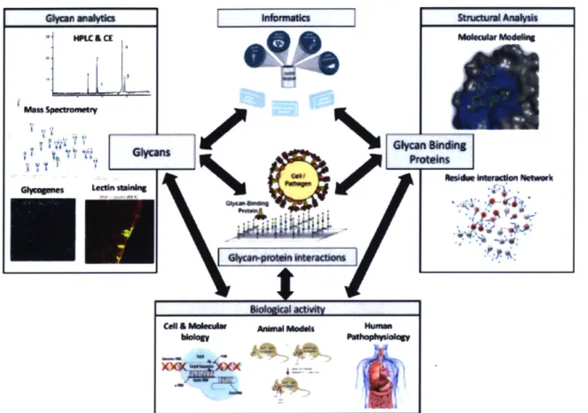
Figure 1.2 Schematic depicting an integrated approach to decode structure function relationships of glycans. Decoding the biological function of glycans requires an integration of
multiple tools (representative examples of tools are depicted inside boxes). From the glycan perspective (left), attributes such as: glycan fine-structure or "sequence", the ensemble of glycans expressed (glycome) can be measured using analytical tools. From the GBP perspective (right),
GBP-glycan interaction attributes such as specificity, and affinity as well as the structural basis for these
interactions can be measured using glycan arrays and structural modeling. Biological activity should be
assessed using experimental systems (i.e. cell or animal model systems) where glycan structure, GBPs, or glycan-protein can be perturbed (i.e. genetic, chemical, or enzymatic manipulation).
Ultimately, integration and correlation across each of these axes are needed to converge on
structure-function relationships.
Thesis outline
Motivated by these challenges discussed above, the goals of this thesis are to i) further develop and improve integrated approaches to study glycans and glycan-binding
proteins, and ii) leverage these approaches to uncover new biological roles of glycans and GBPs in disease. Given that glycans exert their function through interactions with glycan binding proteins, decoding glycan structure-function relationships requires technologies to study glycans, glycan binding proteins and their interaction. Thus, this thesis is divided into two sections: Integrated approaches to study glycan binding
proteins, and integrated approaches to study glycans. Furthermore, each section can be broadly divided into two parts: development of tools, and application of those tools.
In section one of this thesis work, I focus on integrated approaches to study glycan binding proteins (GBPs). Glycan binding proteins, through their direct interaction with glycans, decode the information encoded within the glycan milieu. Thus,
understanding the structural basis for affinity and specificity of GBP-glycan interactions is crucial to decode structure-function relationships. To elucidate the structural basis for GBP-glycan interactions from the perspective of the glycan binding protein, it is
important to characterize key structural determinants including (but not limited to): i) identification of the glycan binding site, ii) enumeration of key molecular contact made in the glycan-protein interface, and iii) identification of key functional residues facilitating the GBP-glycan interaction. To accomplish this, an approach that integrates structural analysis, with biochemical assays and functional readouts is required. Importantly, there is still a great need to develop new tools and improve approaches to characterize these structural determinants.
In part one, I develop tools and approaches to study the structural basis for GBP-glycan from the perspective of GBP-glycan binding proteins. Specifically, I implement a
computational tool enabling inter-residue network analysis (alongside structural analyses, biochemical and functions assays) and investigate the tool's utility in identifying residues in the glycan binding site of a model GBP, FGF-2, that play a functional role in FGF-2's interaction with heparin (Chapter 2).
In part two, I apply this integrated approach to study influenza A virus (IAV) hemagglutinin (HA), a glycan binding protein that regulates host tropism, airborne transmissibility, and host immune recognition. Influenza pandemic pose a significant global threat. Influenza pandemics can occur when IAVs from non-human hosts, which are antigenically novel to humans, undergo a host switch and gain the ability to infect humans. In these cases, the HA has acquired mutations enabling a switch in its
receptor specificity from avian glycan receptors to human glycan receptors. Here, using the integrated approach developed previously, I investigate the HA-glycan binding specificity of IAVs and uncover the structural determinants required for HA to switch its glycan receptor specificity (Chapter 3: H5N1, Chapter 4: H7N9). Furthermore, I
integrate the previous two analyses with an informatics approach to assess antigenic novelty of H3 HAs (Chapter 5: H3N2, and Chapter 6: H3N8). Ultimately, the work here provides insights and strategies for influenza surveillance and enables early
identification of IAVs with pandemic potential that are currently circulating in non-human
hosts.
In section two of this thesis work, I turn my attention to the study of glycans and their biological function. Glycans are abundant on the cell surface, and at the cell-ECM interface where they mediate interactions between cells and their microenvironment. Importantly, glycans are expressed as a heterogeneous ensemble of structures where they interact with glycan binding proteins to mediate biological processes in an "analog" fashion. To elucidate the functional role of glycans, a key first step is to measure their structural attributes, including: glycan fine-structure or "sequence", the ensemble of glycans expressed (glycome), the glycoprotein to which it's attached, and its expression in cells or tissue. Due to glycan complexity there is no single tool capable of capturing all these features and multiple analytical tools and methods are needed. Importantly, measurements of glycan structure must be integrated with functional measurement from cell or organism model systems. Importantly, assessing glycan biological activity
requires the ability to perturb the glycome and its interactions with GBPs (i.e. using
genetic or chemical approaches). Ultimately, using this integrated approach (i.e. a
relationships. Owing to a growing interest in understanding cell-microenvironment interactions, there is still a great need to implement functional glycomics approaches, and apply them to decode the biological roles of glycans in disease processes.
In part one, I develop an integrated approach to characterize the cell surface milieu of glycans (Chapter 7). This approach integrates glycogene expression data, analytical tools such as mass spectrometry, and lectin arrays to characterize the glycome. I demonstrate the utility of a functional glycomics approach by applying it to characterize glycomics changes that cause metastatic progression in cell lines derived from a mouse model of multistage lung cancer.
In part two, I apply this functional glycomics framework to study the function that HSGAGs play in regulating cell-microenvironment interactions in a model of breast cancer stem cells. Almost all cancer related deaths are due to metastasis and resistance to therapy. The latter has been attributed to the existence of cancer stem cells, a cell population characterized by their increased resistance to chemotherapeutics and ability to seed new tumors and metastasis. Thus, targeting cancer stem cells
therapeutically could lead to more durable clinical responses. Consequently, there is a great need to uncover the signaling pathways involved in cancer stem cell activity. The activity of cancer stems cells depends on their interactions with factors in their
microenvironment. Despite knowledge that HSGAGs play a critical role in
cell-microenvironment interactions, the function of HSGAGs in cancer stem cell activity is largely unknown. In Chapter 8, I study the role of HSGAGs in the activity of cancer stem cells. Specifically, I study how SULF1, an HSGAG modifying enzyme, regulates cell-microenvironment interactions to influence tumor initiation, metastasis, and
maintenance of breast cancer stem cells.
Next, I leverage my expertise in integrated analytics to explore an intellectually adjacent area, namely, developing an analytical framework to assess product
consistency/potency for therapeutic extracts. Exposure to certain environments with high microbial exposures, such as traditional farm environments, can protect against
allergic type disease. Recent evidence suggests that the protective effect can be
burgeoning interest in using dust extracts as therapeutics to prevent allergic-type disease. As dust extracts are complex mixtures comprised of various bioactive molecules, including complex polysaccharides, they pose a significant regulatory challenge. Here, I conceptualize an integrated analytical strategy to assess product consistency/potency for therapeutic farm dust extracts, thus addressing regulatory concerns surrounding the development of farm dust extract-derived therapeutics (Chapter 9).
The remainder of this chapter will focus on detailing the motivation and provide the relevant introductory material for the applications areas discussed in this thesis:
1. Integrated approaches to study hemagglutinin and hemagglutinin-glycan
interactions: A strategy to improve pandemic risk assessments of IAVs from non-human hosts.
2. Integrated approaches to study cell surface glycans: Uncovering the role of heparan sulfate glycosaminoglycans in modulating cancer stem cell activity through regulation of cell-microenvironment interactions
Section 1: Integrated approaches to study hemagglutinin and hemagglutinin-glycan interactions: A strategy to improve pandemic risk assessments of influenza from non-human hosts
Motivation and Objectives
Influenza A Virus (IAV) poses a significant public health threat. Each year
influenza epidemics result in approximately 3-5 million cases of illness and between 200,000 and 500,000 deaths worldwide. In high-risk groups, such as elderly, young, pregnant or immunocompromised individuals, influenza infection can result in
hospitalization or death. Perhaps more concerning than annual epidemics are influenza pandemics. There have been four well-documented influenza pandemics over the last century; the 1918 H1N1 Spanish flu, which killed -20-40 million people, the 1957 H2N2 Asian flu, which killed 2-3 million, the 1968 H3N2 Hong Kong flu, which killed ~2 million, and the 2009 swine flu pandemic, which killed -200,000 people [33,34]. Because
pandemics pose such as serious public health threat, there is significant motivation to develop tools and strategies to predict the emergence of future influenza pandemics. Successfully doing so could enable early interventions, such as the creation of vaccines or therapeutics, which could limit the devastating morbidity and mortality associated with pandemics. In this context, it is important to understand the molecular determinants of IAV pathogenesis.
Pandemics are the result of antigenic shift, where an antigenically novel IAV strain is introduced into the human population [35]. Because birds are the natural reservoir for influenza, pandemics are thought to emerge when avian influenza viruses (or avian-derived viral components via viral reassortment) gain the ability to infect
antigenically naive human populations. Fortunately, this event is rare as IAVs that infect birds cannot infect humans without acquiring mutations.
IAV host tropism and airborne transmissibility in humans is regulated by the viral coat protein hemagglutinin (HA), which regulates the first step of infection through its interaction with sialylated glycan receptors on the cell surface [36]. Importantly, the glycan binding specificity of HA is a critical determinant of host tropism. Humans
express a2->6-linked sialylated glycans and birds express a2->3-linked glycans. In order for avian-adapted IAVs to infect humans, mutations must occur in HA that facilitate a shift in glycan binding specificity from a2->3 to a2->6-linked glycans [37].
Using sequence information from circulating IAVs, a molecular surveillance approach enables our ability to predict the emergence of new viral pandemics by identifying IAV strains circulating in non-human hosts that pose a significant pandemic threat [38,39]. In the context of HA, an effective molecular surveillance approach
depends on uncovering the structural determinants that underlie HA-glycan specificity. Accomplishing this allows for the identification of mutations that could give rise to a switch in receptor specificity.
Much of the current research studying the structural determinants of HA-glycan interactions has significant limitations. Many studies in this field adhere to an over-simplified view of the HA-glycan interaction where specificity is governed only by the terminal sialic acid linkage. Furthermore, many current experimental tools to study HA-glycan interaction do not account for important features such as HA-glycan conformation, multivalent presentation of the HA, or HA-glycan affinity [15,37,40]. By ignoring these properties of HA-glycan interactions, it has been difficult to define the structural determinants of HA-glycan interactions and correlate these with outcomes like host tropism switch or airborne transmissibility.
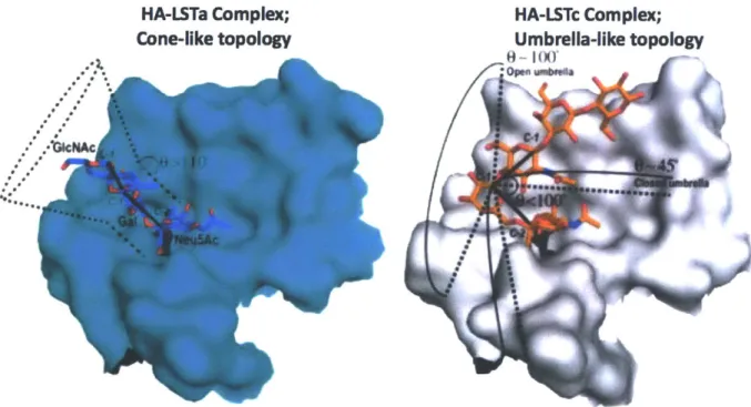
Previous work performed in our lab uncovered that HA-glycan specificity depends on the topological presentation of the glycan, where avian-adapted HAs recognize a2->3 linked sialylated glycans that adopt a cone-like topology and human-adapted HAs recognize (x2-6 linked glycan that adopt an umbrella-like topology [15]. Furthermore, our lab has previously developed biochemical assays capable of
assessing the quantitative affinity of multivalent HAs to bind to avian or human glycan receptors that adopt their respective topologies [41].
Leveraging these insights, the work in Chapters 3-6 of this thesis focuses on studying Hemagglutinin and the HA-glycan interaction to uncover insights and develop new strategies to aid in molecular surveillance of influenza. I apply a unique approach, which integrates structural modeling of the HA-glycan receptor complex, inter-residue
network analysis of HA, biochemical analysis of HA-glycan interactions, and informatics analysis of antigenic properties. Specifically, the following are the objectives of Section
1:
a. Identify the structural determinants for naturally evolving H5N1 HA to switch its receptor specificity from avian to human (Chapter 3)
b. Determine the physiologic glycan receptor binding properties of a 2013 outbreak H7N9 HA and identify the mutations required to enable binding to human glycan
receptors. (Chapter 4)
c. Develop bioinformatics tools and experimental methods capable of measuring antigenic properties of HA. Leverage these tools to identify avian and swine-adapted H3s that could re-emerge into the human population and potentially cause a pandemic (Chapter 5).
d. Characterize the glycan binding specificity of an HA isolated from the 2011 New
England harbor seal H3N8 (Chapter 6)
Influenza virus structure
Influenza is a single stranded
negative-sense RNA virus that belongs to the family
Orthomyxoviridae. Influenza virions are
spherical or elongated particles that are
approximately 80-120nm in diameter. Although
there are three influenza genera (Influenza A, B,
C), Influenza A viruses will be the focus of this
thesis as they are predominantly responsible for
seasonal outbreaks, and pandemics. The
influenza A genome is comprised of eight
segments encoding 10 proteins (Figure 1.3)
which are discussed below in detail. Unless
cited otherwise, information for this section was
obtained from [40,42]
:
Negaie
sv Ns PB1
Figure 1.3 Influenza A Virus structure. Influsenz rs sngle stranded negatv
gene segments. The function of each
viral component is enumerated in the
Viral glycoproteins
Two viral glycoproteins coat the surface of influenza virions: Hemagglutinin (HA) and Neuraminidase (NA). Importantly, both of these envelope glycoproteins are the major site of immune recognition. In fact, Influenza A Virus subtypes are classified serologically based on the presence of HA and NA subtypes (e.g. H1 N1, H3N2, or
H5N1). There are 16 different HAs and 9 different NAs, yielding 144 possible
combinations. Only a subset of HA/NA combination have been observed in nature. While all these subtypes circulate in waterfowl, only a few have been observed in the human population [36]. H1 N1, H2N2, and H3N2 have been observed to infect humans,
and initially gained foothold in human populations during past pandemic outbreaks:
1918 H1 N1, 1957 H2N2, 1968 H3N2 [43]. Importantly, H1 N1 and H3N2 continue to circulate in humans, causing seasonal outbreaks. Recently, several cases of highly pathogenic avian influenza viruses, like H5N1, H7N7 and H9N2 have been shown to infect humans [44-46]. These outbreaks are usually small, and result from direct contact with infected animals; however, their high case fatality rate (e.g. 60% for H5N1)
make these transmission events worrisome from a public health standpoint [46].
Functionally, HA and NA coat the viral envelope, giving rise to roughly 500 spike-like projections coming off of the capsid. The HA spike is composed of an HA trimer, while the NA spikes are tetramers. The HA is responsible for entry into the host cell, with its two key functions being viral attachment to host glycan receptors on the cell
surface, and membrane fusion in the endosome. The NA protein cleaves cell surface sialic acids. NA allows for the release of newly formed virions, which also prevents the re-infection of the virus producing cell [47].
Internal proteins
The influenza A virus contains seven internal proteins: RNA polymerase (PB1, PB2, PA), Nucleoprotein (NP), Matrix protein (Ml and M2), and Non-structural proteins
(NS1 and NS2/NEP). This thesis predominately focuses on the HA viral glycoprotein, so
Viral Ribonucleoprotein (RNP) complex
Influenza's eight gene segments of single stranded RNA are packaged as ribonucleoprotein complexes (RNPs). Each RNP is comprised of a viral polymerase complex (composed of PB1, PB2, and PA) bound to a short hairpin of vRNA, and multiple copies of viral nucleoprotein (NP) that also bind vRNA, acting as a structural protein [48].
Non-structural proteins
Non-structural protein, NS1, is known to play several important roles such as enhancement of viral mRNA translation, inhibition of host mRNA processing, and
inhibition of the host cell immune response. NS2, which is also known as nuclear export protein (NEP), facilitates nuclear export of viral RNP complexes.
M proteins:
The M1 matrix protein is involved in the export of viral RNP from the nucleus to the cytosol. M2 is an ion channel responsible for the acidification of the endosome, which is critical for fusion of the viral and host membranes, and release of vRNPs in the
cytosol.
Influenza viral life cycle
The influenza lifecycle, shown in Figure 1.4, begins with a virion associating with the host cell surface, where the viral hemagglutinin binds to sialylated glycan receptors
[36,40]. Next, the bound virus is endocytosed, predominantly through clatharin-coated pits, but other mechanisms have been described [49]. Next, the endosome is acidified
by the M2 ion channel matrix protein. This promotes a conformational change in the HA
protein which mediates fusion of the viral and endosomal membranes. Following
endosomal fusion, the uncoated viral ribonucleoprotein (RNPs) complex is released into the cytosol of the host cell. The ribonucleoprotein complex is transported into the
nucleus, where incoming negative sense viral RNA (vRNA) is transcribed into mRNA. During viral replication, a full-length complementary RNA is first made (i.e. a positive
sense copy of the vRNA) then used as a template to produce more vRNA. Next, the newly synthesized viral RNAs are exported to the cytoplasm by NEP, where the viral proteins are translated. The viral proteins are expressed, processed and targeted to budding sites on the host cell membrane. The viral surface glycoproteins, protein complexes, RNPs, vRNAs are assembled in viral particles and bud from the host cell membrane. The viral neuraminidase cleaves cell surface sialic acid receptors on host cells, which facilitate the release of new virions and simultaneously prevents re-infection of the same cell [36].
The work in this thesis focuses on the first step in the viral lifecycle; the binding of viral HA to host cell surface glycan receptors. Specifically, this work focuses on the viral hemagglutinin-glycan receptor interaction and its role in tropism and transmissibility in human hosts.
Nucleus
Host receptors
(a2.3- or a2.6-linked slafic acid)
Figure 1.4 Pathway to Viral Infection by Influenza A. Viral infection is initiated by binding of viral
glycoproteins to the sialylated receptors on the host cell membranes. Entry of the virus into the host is facilitated by endocytosis. Next the viral capsid fuses with the host endosome, releasing its genetic content into the host. Viral RNA enters the host nucleus where transcription and replication occur via RNA polymerase. Viral mRNA is produced and used to synthesize viral proteins in the cytoplasm which
are then assembled into viral ribonucleoproteins (vRNPs) in the nucleus. Virus particles are assembled at the cell membrane where budding occurs to release the particles into the extracellular space.
Adapted from [36].
Influenza virus ecology
Influenza A viral ecology is complex, yet critical for understanding how
pandemics emerge. Influenza is primarily a zoonotic pathogen and has been shown to infect a variety of animals
including birds, pigs, dogs, horses and humans. In its natural ecology, the Influenza
A virus exists as a commensal water birds
in the gut of migratory aquatic H 1-6
15 16 12
birds, including shorebirds and 14 -133
13
4-water fowl [36]. In these hosts,
the virus does not cause 11 6
disease and thus a significant 9 7 Domestic
amount of genetic diversity is N
introduced. Mutations can 2
7, arise that enable the influenza 6
viruses to infect domestic mammals or humans (Figure
1.5). In order to "jump" into
domestic mammals or
humans, aquatic birds must bids
first contact and infect Figure 1.5 Origin of Antigenic Shift and Pandemic Influenza.
The majority of influenza A viruses and existing HA subtypes (H1
-domestic birds, such as 16 and N 1-9) circulate in migratory water birds. Two ways a virus with a new HA subtype may be introduced into the human chickens or ducks. population are direct transmission of an avian virus to humans or
genetic re-assortment between an avian and human virus through
Subsequently, these avian infection of domestic pigs. Adapted from [297]
viruses can be transmitted
from domestic fowl to domestic pigs, which are susceptible to infection from both avian and human viruses. Swine are hypothesized to act as intermediate hosts that facilitate
viral re-assortment by acting as genetic "mixing vessels", leading to flu novel subtypes that can infect humans [50,51].
Occasionally, avian or swine influenza viruses gain the ability to infect human hosts (Figure 1.5). This can result in a pandemic in the instance that the new viral subtype is antigenically novel to humans (known as antigenic shift) [34]. In fact, it is thought that the previous pandemics all originated from avian or swine viruses, which were antigenically novel to humans, that gained the ability to infect humans [36].
Origin of influenza pandemics
There are three hypothesized mechanisms by which new viral subtypes emerge in human populations and give rise to a pandemic:
1. Direct transmission from avian to human hosts. In this instance, viruses
acquire mutations in key viral proteins such as hemagglutinin (enabling human infection) or viral polymerases (enabling replication in human tissue) which are necessary for human adaptation.
2. Genetic reassortment can occur when two Influenza A viruses infect the same
host cell. Upon infection, both vRNA sets are replicated and during viral
assembly the RNA segments from the two viral strains become mixed to produce a novel virus with a unique combination of genes [34]. Pigs are often considered "mixing vessels" for viral reassortment as they have the capability to be infected with both avian and human viruses [51]. The 2009 swine flu pandemic H1 N1, as thought to have emerged through genetic reassortment of bird, swine and human viruses [52].
3. Reintroduction of an "old" strain into the human population. Viral strains, for a variety of reasons, may disappear from the human population, and can
possibly re-emerge. If this occurs when the immunity in the infected population has waned, this virus will be perceived as antigenically novel. This was thought to be the case for the 1977 H1N1 "Russian flu", which was found to be
Although not discussed in detail here, antigenic novelty can yield pandemics and contribute to seasonal epidemics. Antigenic novelty arises through two major
mechanisms; Antigenic shift (discussed above), and antigenic drift [54]. Antigenic drift refers to the gradual accumulation of mutations on the antigenic domains of
Hemagglutinin or Neuraminidase. The replication of vRNA is an error prone process. In fact, the mutation rate for influenza is ~1/100,000 nucleotides, which means many of the nascent vRNA copies will likely contain one or more mutations [55]. Pressure from host immune systems can drive the gradual evolution of antigenic sites on viruses. In cases of significant antigenic divergence, it is possible that a population may not be protected
by their pre-existing immunity. This can yield more severe mortality and morbidity
events in a given flu season [56]. The rapid evolution of influenza viruses via shift or drift means constant monitoring of viral evolution is required (i.e. surveillance). Surveillance is important and serves to inform seasonal vaccine composition and helps monitor the geographic spread of influenza. It can also aid in predicting supply needs and can help prioritize allocation of vaccines.
Host glycan receptors
Glycans are known to play a critical role in host-pathogen interactions. One of the most well-characterized interactions is between Influenza A Virus (IAV) hemagglutinin and the sialylated glycans receptors on the surface of cells. As mentioned previously, the first critical step of viral entry is the binding of HA to glycan receptors. Importantly, the glycan-receptor specificity of IAV HA determines host adaptation and tissue tropism.
IAVs circulate as commensals in the gut of aquatic birds. In avian hosts, the gut and respiratory tract predominantly express N- and 0- linked glycans which contain terminal sialic acids that are a2->3-linked to galactose (referred to interchangeably as avian receptors) (Figure 1.6) [57]. In humans, however, the primary site of infection is the upper respiratory tract, which predominately express N- and O-linked sialylated glycans with an a2--6 terminal linkage (referred to interchangeably as human
specificity for a2-+3-linked sialylated glycans, while HAs from human-adapted viruses have specificity for a2->6-linked sialylated glycans.
a2,3
a2,6
a2,3/a2,6
-40
m ItdIch inA b
ina
SP cia Cityftorn avian to hurnan mrsor
Figure 1.6 Glycan Receptors for Influenza HA in Various Hosts. (Bottom) Chemical structure of the glycan receptors. Sialic acid is linked to galactose via a2-3 or a2-6 linkage. a2-3 and a2-6-linked glycan are the avian and human receptors for HA, respectively. Viral Host tropism is determined by the glycan receptor distribution in the host. (Top) Birds express a2-3, Human express a2-6, and pigs express a2-3/62-6. A switch in HAs receptor binding specificity from a2-3 to a2-6 is necessary for
human adaptation of the virus. Adapted from [40].
The distribution of glycan receptors in the upper respiratory tract of human tissue is also important, especially when considering cellular tropism. In order to investigate this question, lectins (a class of glycan binding proteins with well-characterized
specificity) have been used to investigate the physiologic distribution of glycans in human lung tissue. Using two lectins, SNA-l (specificity for sialic acid that is a2-+6-linked to Gal or GaINAc) and MAL-Il (specificity for a2-+3-a2-+6-linked glycans), the upper respiratory tract of human, namely the tracheal epithelium, was shown to express a2-+6-linked glycans [15]. Conversely, a2-÷3-linked sialylated glycans were mostly present in alveolus in the lower respiratory tract. Indeed this distribution of a2->3 vs a2->6 glycan receptors strongly correlated with cellular tropism of human-adapted vs
avian-adapted viruses [58]. HAs that are derived from human-adapted viruses bind to non-ciliated goblet cells and ciliated epithelial cells in the upper respiratory tract, whereas HAs from avian-adapted viruses do not bind the upper respiratory tract and
localize to deep lung and alveolar tissue. While the exact mechanism is unknown, the cellular tropism of pandemic viruses (i.e. binding to non-ciliated goblet cells and ciliated epithelial cells) seems to be important for their efficient airborne transmission [58].
In contrast to bird and humans, the epithelial cells on the upper respiratory tracts of pigs express both a2->3 and a2->6-linked sialylated glycan receptors. Consequently, pigs can be infected with both avian- and human-adapted IAV. For this reason, pigs are considered an evolutionarily intermediate host enabling viruses to jump from domestic birds to humans via acquisition of mutations or by acting as a "mixing vessel" for genetic re-assortment of IAV (Figure 1.6).
Influenza A virus hemagglutinin: Structure and interaction with glycans
HA is the most abundant protein of the influenza viral surface. It plays several critical
roles during the viral lifecycle and disease pathogenesis:
1. HA mediates viral entry and endosomal fusion.
2. HA determines host tropism through it preference for cx2-+3 or a2->6 linked sialylated glycan receptors.
3. HA-glycan specificity regulates respiratory droplet transmission.
4. HA is the major site of host immune recognition.
HA is produced as a precursor polypeptide (HAO), which is a type 1 transmembrane
glycoprotein ~ 550 amino acids. The HAO precursor becomes activated via proteolytic
cleavage into two peptides HA1 and HA2, which are disulfide linked. Ultimately, HA is displayed on the viral surface as a trimer of three monomers of HA1 -HA2. The first crystal structure of the HA ectodomain was reported in the early 1980s [59]. The protein
is generally divided into two domains: The membrane proximal stem domain, and the membrane distal globular head domain (Error! Reference source not found.a). The
globular head is of interest in this thesis as it contains the glycan receptor binding site (RBS) and the antigenic region [43].
Molecular insights into the HA-glycan interaction
The glycan receptor binding site (RBS) is a shallow pocket present on the tip of each monomer in the globular head region. The RBS is composed of a base, including several conserved amino acids (Tyr-98, Trp-1 53, His-1 83, and Tyr-1 95), and three structural elements around the edges of the pocket (the 130- and 220- loop, and the
190-a helix) (Error! Reference source not found.a) [36]. When the sialylated glycan
receptor is bound to HA, the terminal sialic acid residue makes critical hydrogen bonding contacts with the base residue, Y98, and the 130 loop. The monosaccharides distal from the terminal sialic acid interact with the 220-loop and the 190 helix.
Furthermore, W153, H183 and Y195 make additional van der Waals interactions with
the glycan receptor (Error! Reference source not found.a).
HAs specificity for a2->3 versus a2->6 sialylated glycans determines hosts tropism. Therefore, it is important to understand the structural determinants which govern the receptor specificity. Crystal structures have been obtained for avian-adapted and human-adapted viruses bound to LSTa (an cx2->3 linked pentasaccharide) or LSTc
(an a2->6 linked pentasaccharide)(Error! Reference source not found.). When
bound to HA the avian and human receptors adopt different conformations. Co-crystal structures of avian-adapted HAs in complex with LSTa, shows that the a2-+3 linked glycans exhibit a trans conformation, where the Gal and GIcNAc residues are in an extended conformation relative to the sialic acid residue. Furthermore, the glycosidic oxygen atom faces the 220-loop, which presents hydrogen bond between the GIn 226 and the 4-hydroxy group of Gal-2 and the oxygen in the glycosidic linkage (Figure 1.7b)
[36]. These interactions are present in all avian-adapted HAs. In contrast, the a2-+6
human glycan receptor (LSTc) bound to human-adapted HAs show a cis conformation and appear "folded". In this conformation the glycosidic oxygen points away from the
RBS and the hydrophobic C6 atom of galactose points towards the RBS base (Figure ReceptorbIndIng site 190-helix Y195 -H183 W153 -a 0 SEi 0E '0 peptide
Figure 1.7 Hemagglutinin and the Glycan Receptor binding site. a) Shown here is the crystal structure of HA (PDB ID= 4JUG). The two domains of the HA ectodomain are labelled: the globular head domain and the stem domain. The globular head domain contains the receptor-binding site which forms a shallow pocket comprising three structural elements: the 130-loop, 190-helix and 220-loop. Four highly conserved residues (Y98, W153, H183 and Y195) form the
base of the receptor-binding site (shown in orange). b,c) Human and avian receptor analogues show differing structural conformation upon binding to HA (PDB ID= 4JUH and 4JUJ). The three terminal monosaccharide of the glycan receptor are shown: terminal sialic acid SAl, galactose Gal2, and N-acetylglucosamine GlcNAc3. [36]
1.7c).
As mentioned earlier, pandemics often emerge from avian viruses that undergo a host switch. In this case, avian-adapted HAs must switch their glycan receptor binding preferences from a2->3 to a2-6-linked glycans. For this to occur, influenza viruses must acquire mutations, which cause changes in the HA glycan receptor binding sites that facilitate a switch in receptor binding specificity. Therefore, understanding the
b Avian receptor analogue 190-helix GIcNA3 SA IA40 220-loop y (130-loop Hydrophilic glycosidic oxygen atom
C Human receptor analogue 190-helix GlcNAc3 SAI 220-loop 130-loop Hydrophobir C .itoii Y98
A 3.Th GIc5 GaI4 GIcNAc3 GaI2 51.1 Lam
Sial GaI2 GkcNAc3 Gai4 GIc5
a2.3 03 03 14 GaM4 GIcNAc3 Glc5 sia1 LSTc
Slal Gai2 GIcNAc3 GaI4 GIc5
+a2.6 04 3 4
Figure 1.8 Human and Avian Receptor Analogs in Free Confirmation. a) Free confirmation of pentasaccharides LSTa, the avian receptor analog, and LSTc , the human receptor analog, show
LSTa in trans conformation and LSTc in cis conformation. b) Cartoon representations of LSTa and LSTc structures are show (CFG nomenclature). Adapted from [57]
structural determinants and specific mutations that govern a "switch" are critical to enumerate as they enable identification of viruses with 'pandemic potential'.
B
Challenges in elucidating HA-glycan interactions
Studying the "host switch" and human-to-human transmission is critical for our understanding of influenza pathogenesis and viral pandemics. Towards this, much work has been conducted using reverse genetics approaches to study these properties of Influenza A biology. Using reverse genetics, it is possible to recreate viruses and test their infection, replication, and transmission properties in ferrets [60,61]. Ferrets contain similar glycan receptors as humans and exhibit similar disease symptoms. In ferrets, various modes of transmission such as contact or respiratory droplet transmission can be studied by controlling ferret contact (i.e. co-housing or housed separately with a perforated wall, respectively) [40]. Using this model system, previous studies have been performed to investigate the virulence & transmissibility properties of single gene
reassortments of the 1918 pandemic H1N1 (A/South Carolina/1/18 or SC18). Initially, studies demonstrated that the HA protein played a dominant role in IAV virulence
(relative to other genes like NA or PB2) [62,63]. In order to further enumerate the role of
HA in virulence, viruses were created that contained identical genes but swapped in
various naturally occurring or mutant HAs. These studies investigated respiratory droplet transmission and viral replication in ferrets, and glycan receptor binding
HA derived from SC18, NY18 (a single point mutant of SC18), and AV18 (a double point
mutant) [37]. Using RBC agglutination, it was determined SC18 binds X2-+6 glycans, NY18 binds both cx2-+3/2-+6, and AV18 only binds a2->3 glycans. SC18 was able to transmit efficiently in respiratory droplets, while NY1 8 was inefficient, and AVI 8 did not transmit (Figure 1.9).
Presence or absence of hemagglutination Respiratory
a2-6 a2-3 Droplet
CRBCs CRBCs Untreated CRBCs Transmission SC18 + - + Efficient NY18 + + + Inefficient AV18 - + + None DK/Alb - + + None Tx/91 + + N/A* Efficient
Figure 1.9 Glycan receptor binding specificity and respiratory droplet transmission of H1 N1 Viral HA reassortants. Note that NY18 is a single amino acid mutant of SC18 (D225G) and AV18 is a double amino acid mutant of SC18 (E190D/D225G). Adapted from [37].
These results suggested that the loss of cx2-+3 binding is necessary for efficient transmission, while gaining specificity cx2-+6 was necessary but not sufficient to enable efficient transmission. Interestingly, confounding cases emerged where mixed
a2->3/a2->6 binders showed variable rates of transmission. For instance, NY1 8 was a mixed binder and showed inefficient transmission, whereas an HA from A/Texas/36/91 (or TX91) was a mixed binder but transmitted efficiently [37]. These confounding results led many to hypothesize that there was something more complex governing
transmissibility beyond this simple a2-3/ac2->6 paradigm.
Integrated approach to decode HA-glycan receptor specificity
As discussed above, aspects of glycan chemical properties and their
fundamental mode of necessitate a unique approach to determine biological function. Glycan-protein interactions are low affinity (uM-mM), and glycans possess a high
![Table 1.1 Challenges with decoding structure-function relationship of glycans. Adapted from [32].](https://thumb-eu.123doks.com/thumbv2/123doknet/14670516.556713/17.917.103.811.103.877/table-challenges-decoding-structure-function-relationship-glycans-adapted.webp)