D
D
e
e
g
g
r
r
e
e
e
e
C
C
o
o
u
u
r
r
s
s
e
e
L
L
i
i
f
f
e
e
T
T
e
e
c
c
h
h
n
n
o
o
l
l
o
o
g
g
i
i
e
e
s
s
M
M
a
a
j
j
o
o
r
r
:
:
B
B
i
i
o
o
t
t
e
e
c
c
h
h
n
n
o
o
l
l
o
o
g
g
y
y
D
D
i
i
p
p
l
l
o
o
m
m
a
a
2
2
0
0
1
1
4
4
--
c
c
o
o
n
n
f
f
i
i
d
d
e
e
n
n
t
t
i
i
a
a
l
l
-
-L
L
o
o
r
r
i
i
c
c
P
P
e
e
t
t
r
r
u
u
z
z
z
z
i
i
C
C
a
a
t
t
a
a
l
l
y
y
t
t
i
i
c
c
p
p
e
e
r
r
f
f
o
o
r
r
m
m
a
a
n
n
c
c
e
e
a
a
n
n
d
d
e
e
n
n
a
a
n
n
t
t
i
i
o
o
s
s
e
e
l
l
e
e
c
c
t
t
i
i
v
v
i
i
t
t
y
y
o
o
f
f
e
e
n
n
g
g
i
i
n
n
e
e
e
e
r
r
e
e
d
d
o
o
x
x
i
i
d
d
o
o
r
r
e
e
d
d
u
u
c
c
t
t
a
a
s
s
e
e
s
s
a
a
s
s
b
b
i
i
o
o
c
c
a
a
t
t
a
a
l
l
y
y
s
s
t
t
s
s
Professor Marc Pfeifer ExpertProf. Stefan Lutz
Submission date of the report
2 5 . 0 8 . 2 0 1 4
Catalytic
performance
and
enantioselectivity
of engineered
oxidoreductases
as biocatalysts
Petruzzi Loric
Objectives
The thesis will evaluate the catalytic activity and enantioselectivity of native Old Yellow Enzyme from S.pastoranius in comparison with three engineered enzyme variants. Substrates will be bioactive compounds with a particular emphasis on terpenoids and fragrances.
Methods | Experiences | Results
Three variants and wild type (WT, shown figure up left) Old Yellow Enzyme (OYE) from S.pastonarius (OYE1) were used. One mutant, W116I, contained a site-directed mutation that changed Trp-116 to Ile. The active site volume was increased and resulted in a reveresed enantioselectivity for (S)-carvone compared to WT OYE1. Then, cp303 was modified by circular permutation. The N- and C-termini were changed but the overall amino acid sequence was kept. The flexibility of the enzyme was increased as well as the size of active site entrance (see figure bottom right). Previous studies showed faster catalytic rates. The last mutant, cp303W116I, is an OYE that combined both peptide alterations (no published results). The enzymes were purified from transformed E.coli by Anion Exchange Chromatography and Size Exclusion Chromatography. Enzymatic assays were done in 200 µL final volume with 1 µM enzyme using 1 µM phosphite dehydrogenase and 10 mM sodium phosphite as a regenerating system and 100 µM NADP+ as cofactor. Time points were taken by extracting substrates with organic solvent before being analyzed by chiral GC with an optimized method. Results showed that for rigid linear substrates, mutants were not efficient, showing no enantioselectivity switch or increase in cataytic rates. It was hypothesized that these molecules needed more stabilization in the active site in order to be efficiencly catalyzed. However, mutants showed high rates with (S/R)-carvone.
Bachelor’s Thesis
| 2 0 1 4 |
Degree course Life Technologies Field of application Biotechnology Supervising professor Dr. Pfeifer Marc [email protected] PartnerProf. Lutz Stefan Emory University [email protected]
Crystal structure of wild type Old Yellow Enzyme from S.pastonarius. In the middle is the entrance of the active site with the FMN cofactor in yellow.
Crystal structure of cp303 mutant of the Old Yellow Enzyme from S.pastonarius. In the middle is showed the increased entry of the active site.
This document is the original report written by the student. It wasn’t corrected and may contain inaccuracies and errors.
“Learn from yesterday, live for today, hope for tomorrow.
The important thing is not to stop questioning.”
2/70
Table of contents
1.
! Abbreviations ... 4!
2.
! Introduction ... 5!
3.
! Project objectives ... 10!
4.
! Material and methods ... 11!
4.1
!
Material ... 11!
4.1.1!
Equipment ... 11!
4.1.2!
Cells ... 11!
4.1.3!
Reagents ... 12!
4.1.4!
Softwares ... 14!
4.2!
Methods ... 15!
4.2.1
!
Cell transformation by electroporation ... 15!
4.2.2
!
Overexpression of cp303W116I and W116I ... 16!
4.2.3
!
Purification of cp303W116I and W116I from E.coli BL21(DE3) by anion exchange chromatography ... 16!
4.2.4
!
Size exclusion chromatography ... 17!
4.3
!
Estimating purity with SDS-PAGE ... 19!
4.4
!
Determination of purified enzyme concentration ... 20!
4.5
!
Enzymatic Assays ... 21!
4.5.1
!
Calculations of relatives concentrations ... 22!
4.5.2
!
Calculation of enantiomeric excess ... 22!
4.5.3
!
Calculation of conversion rates ... 22!
4.6
!
Gas Chromatography analysis ... 23!
4.6.1
!
(S/R)-carvone ... 25!
4.6.2!
Trans -methyl-cinnamaldehyde ... 25!
4.6.3!
Myrtenol ... 25!
4.6.4!
Methyl 2-hydroxymethylacrylate ... 25!
4.6.5!
4-phenyl-3-butyn-2-one ... 25!
4.6.6!
(E/Z)-citral ... 25!
5.
! Results ... 26!
5.1!
Enzyme purification ... 26!
5.3
!
Catalytic rate and enantioselectivity of mutants ... 27!
5.3.1
!
(S)-carvone ... 27!
5.3.2!
(R)-carvone ... 28!
5.3.3!
Trans -methyl-cinnamaldehyde ... 29!
5.3.4!
Myrtenol ... 31!
5.3.5!
Methyl 2-hydroxymethylacrylate ... 33!
5.3.6!
4-phenyl-3-butyn-2-one ... 35!
5.3.7!
(E/Z)-Citral ... 37!
6.
! Discussion ... 42!
6.1!
(S)-carvone ... 42!
6.2!
(R)-carvone ... 43!
6.3!
Trans -methylcinnamaldehyde ... 44!
6.4!
Myrtenol ... 45!
6.5!
Methyl 2-hydroxymethylacrylate ... 46!
6.6!
4-phenyl-3-butyn-2-one ... 46!
6.7!
(E/Z)-citral ... 47!
7.
! Conclusion ... 48!
8.
! Acknowledgements ... 49!
9.
! Literature ... 50!
10.
! Appendix ... 53!
10.1
!
Recipes for reagent used during the experiment ... 53!
10.2
!
Anion Exchange Chromatography ... 55!
10.3
!
Size Exclusion Chromatography ... 56!
10.4
!
Relatives concentrations and quantitative additional data ... 57!
10.4.1
!
(S)-carvone ... 57!
10.4.2!
(R)-carvone ... 59!
10.4.3!
Trans -methyl-cinnamaldehyde ... 61!
10.4.4!
Methyl 2-hydroxymethylacrylate ... 63!
10.4.5!
4-phenyl-3-butyn-2-one ... 64!
10.4.6!
(E/Z)-citral ... 66!
10.5!
Equipment pictures ... 68!
10.5.1
!
Anion and Size exclusion Äkta module ... 68!
10.5.2
!
Gas Chromatography ... 69!
4/70
1. Abbreviations
Index of the abbreviations used in the text listed in alphabetical order. AEX ... Anion Exchange Chromatography CP ... Circular Permutation
cp303 ... OYE Circular Permutant n°303 from Daugherty et al.[1] study cp303W116I ... OYE Circular Permutant plus tryptophan mutant
CV ... Column Volume DMSO ... Dimethyl sulfoxide DNA ... Deoxyribonucleic acid
E.coli ... Escherichia coli
EWG ... Electron Withdrawing Group FAD ... Flavin Adenine Dinucleotide FID ... Flame Ionization Detector FMN ... Flavin Mononucleotide GC ... Gas Chromatography IPTG ... β-D-1-thiogalactopyranoside kDa ... Kilo Daltons
LB ... Lysogeny Broth MeOH ... Methanol
NADP ... Nicotinamide Adenine Dinucleotide NaPi ... Sodium phosphite
OYE ... Old Yellow Enzyme
OYE1 ... Old Yellow Enzyme from S.pastonarius PCR ... Polymerase Chain Reaction
PTDH ... Phosphite dehydrogenase RT ... Room Temperature
S.cerevisiae ... Saccharmoyces cerevisiae
SEC ... Size Exclusion Chromatography SDS ... Sodium Dodecyl Sulfate
SDS-PAGE ... Polyacrylamide Gel Electrophoresis in presence of SDS
S.pastonarius ... Saccharomyces pastonarius
W116I ... OYE tryptophan mutant from Padhi et al.[2] study WT ... Wild Type OYE1
2. Introduction
Nowadays, enzyme mediated biotransformations, which are chemical modifications involving a biological catalyst, are very useful and helpful processes. These have several advantages compared to chemical transformations, like remarkable chemo-, regio- and stereoselectivity and mild reaction conditions (temperature, pH). These properties meet the growing demand for sustainable and cost effective “green chemistry” processes.
Removing or adding an electron to a molecule belongs to the so-called redox reactions. It is one of the most investigated topics in biocatalysts research because it may lead to the creation of a chiral center. This is happening when the four substituents of a tetrahedral atom (usually carbon) are different. There are two possible dispositions of the substituents in space per each chiral center. One is the R and the other is the S-enantiomer, depending on the relative position of the substituents. These molecules have the same formula and atoms composition. The only detail that differs is the disposition of the substituents around a chiral center, which cause them to be the mirror image of each other, like the right and the left hand are. All biologically active molecules are chiral, and so are the majority of today's drugs [3].
Two enantiomers have similar chemical and physical properties. However, it is known that their effect as drug may be different depending on their different conformation. Thalidomide [4], Naproxen [5] or L-α-methyldopa [6] are good examples of why producing enantiopure drugs has become a real concern. Enantiomers, obtained by bioreduction, like the Roche-Ester [7, 8], may also be used as starting material for several industrially relevant molecules like vitamins, fragrance compounds, antibiotics or natural products.
Today’s helpful molecules are becoming more and more complex, because of the increasing number of chiral centers they feature. As seen before, classic chemistry cannot be as efficient as biocatalysts in creating enantiopure solutions. Biocatalysis is where the enzymes like the ones from the Old Yellow Enzyme (OYE) family found their usefulness. This kind of enzyme is able to reduce activated alkenes, which are organic molecules containing a double bonds and an electron withdrawing group (EWG) that weaken the double bond. The electron transfer is done with the help of a cofactor incorporated in the OYE, which can be: a flavin mononucleotide (FMN), a flavin adenine dinucleotide (FAD) or a nicotinamide adenine dinucleotide (NAD(P)). Enzymes are generally efficient for one or a group of substrates. Frequently, the enzyme can only either slightly catalyze, or not catalyze at all, the target molecule for which it is not adapted. Because of this, the enzyme must be optimized.
Today, thanks to protein engineering it is possible to modify the amino acids sequence of enzymes in order to affect (i.e. improve) their characteristics like their selectivity, their activity or even their physicochemical characteristics. The purpose of these modifications is to enhance those small catalytic units in order to optimize processes or investigate new synthetic pathways for industrially relevant molecules. Usually, changes of enzymatic properties have been recently achieved with different techniques, which can be divided into two approaches: rational design and directed evolution. Rational design is based on pre-acquired knowledge and focused on deleting, substituting or re-arranging one or several amino acids within the protein sequence.
6/70
Instead, the aim of directed evolution is to try to mimic the effect of evolution on several thousand years by using molecular biology and applying a selective pressure to find enzyme with improved properties. The effect of these changes within enzyme’s structure are difficult to predict [9] but, it is possible to study them through techniques such as crystallography and biophysical methods.
Recently, a new approach of protein engineering, the circular permutation (CP), has been introduced. It is based on the modification of the tertiary structure of an enzyme by re-arranging the overall amino acid composition. That means that the overall amino acids composition do not change but the N and C-termini are located elsewhere [1].
The aim of this work is to study the substrate and enantioselectivity of the native OYE from
Saccharomyces pastonarius and three variants obtained by protein engineering. Wild Type OYE1
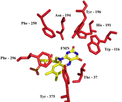
(WT) is 400 amino acids long and weights 40 kDa. The active site is showed in figure 1 where eight amino acid side chains are highlighted: Thr–37, Trp-116, His-191, Tyr-196, Asn-194, 250, Phe-296 and Tyr-375. Most of them are contributing to create a hydrophobic environment in the pocket, while others have a bigger importance. Indeed, Massey and Kohli have investigated the role of Tyr-196 [10], it was suggested that tyrosine Tyr-196 helps with the proton transfer during the oxidative half-reaction with the reduced flavin and an activated alkene. Massey also studied the role of asparagine 194 and histidine 191 [11]. Those residues were identified to make hydrogen bonds with phenolic compounds in order to stabilize them in the active site.
Figure 1: Active site of the Wild Type Old Yellow Enzyme 1 (OYE1) from S.pastonarius. Some amino acids
that take part to the formation of the active site are highlighted in red. They are close to where the substrate binds with the FMN, which is shown on the bottom of the active site pocket.
The first mutant, W116I, has been modified by site-directed mutagenesis. The tryptophan in position 116 was replaced by an isoleucine [2] (Trp is showed on the middle right of figure 1). This variant was chosen because it showed a change in enantioselectivity by producing enantiomeric products from the reduction of (S)-carvone instead of diastereoisomeric products [2]. Padhi et al.[2] made the assumption that replacing Trp by Ile permit a new orientation of the (S)-carvone into the active site and also increased its volume by allowing the access to a hydrophobic pocket.
The second variant is the cp303 and was modified by circular permutation. CP was done with a tandem of two attached enzyme genes (oye1 in the figure 2), which were linked with a tri-peptide. Then, with a polymerase chain reaction (PCR) technique, it was possible to create various mutants by selecting a beginning and an end at different positions on the tandem. The trick resides in designing primers that will always give a fragment of the same length (figure 2). Therefore, the overall amino acids composition is kept.
Figure 2: Creation of circular permutants of OYE1 [1]. The two genes are plugged together with a nine-nucleotide sequence encoding the -Gly-Thr-Ser- linker. PCR amplification with site-specific primers will give different fragments of same length each (1188 nucleotides) but with different N and C-termini. Picture from Daugherty et al [1].
One of the variants (cp303) thus obtained was showing significant improvement in activity for (S)-carvone and ketoisophorone, up to 10 fold higher than the wild type [1]. Statements were that removing the loop containing Phe-296 (figure 1) resulted in a significantly higher oxidative half reaction rate and in a better flexibility of the enzyme [1]. The opening to the active site was enlarged, as it is possible to see in figure 3.
8/70
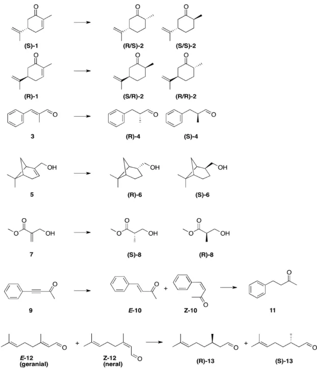
The last mutant, cp303W116I is a variant containing the tryptophan mutation as well as the same N and C-termini as cp303 (unpublished results). This last mutant was produced assuming it will have additives properties with the effect of the site directed mutagenesis as well as the circular permutation. In order to investigate the previous statements made on each OYE1 variant, different substrates with unique properties will be used. (S/R)-carvone (S/R-1) are molecules that have an industrial interest in flavors and fragrances industry [12]. Due to their high volatility, (S)-carvone reduced products, (2R,5S)- or (2S,5S)-carvone, respectively (R/S)-2 and (S/S)-2, are used as inhibitors of bacteria and filamentous fungi repellents [13]. They are also used as starting compounds in the synthesis of bioproducts like antimalarial drugs [14] or valuable chiral synthons (synthetics building blocks) [15, 16]. Carvone is also used as a reference and control because is one of the most common substrate used with OYEs [1, 2, 7]. Trans α-methyl-cinnamaldehyde (3) is a conjugated system, which (S/R)-α-methyl-dihydrocinnamaldehyde, (S/R)-4 are molecules with an industrial relevance for perfumery as starting material for fragrance compounds [17-19]. Myrtenol (5) is a fragrance component used in decorative cosmetics (fine fragrance, shampoos, soaps, etc.) as well as in non-cosmetic products like detergents or household cleaners [20]. Unlike the others molecules used in this work, myrtenol is not a flat component because it contains a bicyclic structure. Because the modifications made on the active site of OYE1 [1, 2], it is expected that one of the variants will be able to process that bulky substrate. Bioreduction of methyl-2-hydroxymethylacrylate (7) was investigated with OYE1 variants because that leads to (R)-Roche-Ester, (R)-8, which is a popular chiral building block for the synthesis of various compounds [7, 8]. It can be used to get vitamins (vitamin E [21]), fragrance molecules (muscone [22]), antibiotics (calcimycin [23]) and other natural products (spiculoic acid [24]) [8]. 4-phenyl-3-butyn-2-one (9) contains a triple bond; therefore it is an example for the use of OYE in a reaction that implies two consecutives bioreductions. This molecule was chosen because of the possibility to compare with previous results from BASF [25].
Finally, the (E/Z)-Citral, (E/Z)-12, is another reference substrate as it is a largely used molecule used for bioreductions. Furthermore, E-12 (geranial) and Z-12 (neral) forms are two different substrates for OYE. The native OYE1 catalyzes the conversion to the enantiopure products (E-12 to (R)-citronellal,
(R)-13 and Z-12 to (S)-citronellal, (S)-13 [26]). Both enantiomers, (S/R)-13 are used in the synthesis
of menthol [27].
Figure 4: Structure of the substrates used in this thesis. (S/R)-1 is (S/R)-carvone. (R/S)- and (S/S)-2 stands for (2R,5S)- and (2S,5S)-carvone. 3 is trans-α-methylcinnamaldehyde. (S/R)-4 is for (S/R)-α-methyldihydrocinnamaldehyde. 5 is for myrtenol. (S/R)-6 stands for (S/R)-dihydromertanol. 7 is for methyl 2-hydroxymethylacrylate. (S/R)-8 is for (S/R)-methyl-3-hydroxy-2-methylpropionate “Roche Ester”. 9 is for 4-phenyl-3-butyn-2-one. (E/Z)-10 is for (E/Z)-4-phenyl-3-buten-2-one. 11 is for 4-phenyl-3-butanone. (E/Z)-12 is
10/70
3. Project objectives
The goal of this study will be to determine the enantioselectivity and the catalytic activity for wild type OYE1 and three engineered variants among a set of various substrates.
Previous work by Padhi et al. [2] lead to development of the W116I mutant, after it was observed that WT was presenting diminished rates for 3-ethyl-2-cyclohexanone (figure 5). It was believed that these lower rates were a consequence of unfavorable steric interactions within the active site. In order to reduce these interactions, the active site volume was increased. Amino acids residues that were closest to the active site were investigated and attention was focused on residues that were within a distance of 5 Å of where the substrate binds. Relying on previous studies by Massey [10, 11, 28], the known amino acids that are essential for catalytic purposes were removed from the list. From the remaining amino acids, Trp-116 was chosen because it was the closest to where the β-substituents of 2-cyclohexenone were. Several mutations were made, but the isoleucine mutant was kept. The result on (S)-carvone showed a switch in enantioselectivity by producing (2S,5S)-carvone as major product instead of (2R,5S)-carvone as the wild type does. However, the mutant was not tested against substrates significantly different than (S)-1. This work will try to better understand the features of this new enzyme as well as possible enantioselectivity switch with other substrates.
Figure 5: 3-ethyl-2-cyclohexenone.
Circular permutation [29] was used by Daugherty et al. [1] to create a library of OYE variants. In order to find mutants with a higher activity among the library, OYE circular permutants were synthesized with an in vitro transcription/translation (IVTT) system. This ex vivo protein engineering strategy allowed the exploration of a library of hundreds of enzyme variants to highlight the ones with a higher activity. Among the selected enzymes, circular permutant #303 (cp303) was found to have an increased activity, up to > 10x for (S)-carvone compared to WT. Further studies showed that cp303 was allowing a faster regeneration of the cofactor. These results were in accordance with previous studies [30] that identified the loop region VI (the truncated region, amino acids from 290 to 310) as taking part in the reductive half reaction. Also, the active site entrance was significantly increased (see figure 3). Through this work, this variant will be tested with different substrates in order to formulate a better understanding of the nature of modifications presented in the cp303 scaffold.
The last variant is an OYE containing the cp303 and the Trp-116 changes. No studies were done on this enzyme before the beginning of this work. It has been suggested that this mutant would show additives properties regarding cp303 and W116I.
Other ongoing studies are investigating alternative amino acids positions for site-directed mutagenesis, such as an Ala instead of Pro-296 (located in the same region as cp303), which showed an increase in conversion rates with (S)-carvone by an order of magnitude about 5x higher than WT (Lutz unpublished data based on IVTT system).
4. Material and methods
4.1 Material
4.1.1
Equipment
Table 1: Listing of the specific equipment used for this work.
Model Provider Use
GenePulser Xcell BioRad Electroporation of competent cells (E.coli BL21 and DH5α)
Äkta purifier Amersham pharmacia biotech
Anion-Exchange Chromatography to purify proteins
HiTrap Q HP 5 ml (34 µm bead size,
bed dimension 7x25 mm) GE Healthcare Anion-Exchange Chromatography Agilent 6850 series Agilent
Technologies Gas Chromatography analysis Cyclosil-B
113-6632, 30 m, film 0.25 µM
Agilent
Technologies Gas Chromatography analysis Sonic Dismembrator Model 100 Fisher Scientific Cell lysis in purification step Millex-HV (0.45 µm)
article: SLHV033RS Millipore Clean clear lysate in purification step Amicon Ultra-15 Centrifugal Filter
Device
article: UFC901024
Millipore Concentration of protein before size exclusion separation
HiPrep 16/60 Sephacryl S-100 High
resolution 120 ml GE Healthcare Size Exclusion Chromatography Ressource Q 1 ml Pharmacia Biotech Anion-Exchange Chromatography Orbital Shaker S-500 VWR Enzymatic Assay
4.1.2
Cells
Table 2: Listing of cell lines used in this work.
Cell line Provider Use
BL21(DE3)pLysS Invitrogen Host cell for the transformation and expression of recombinant OYE
12/70
4.1.3
Reagents
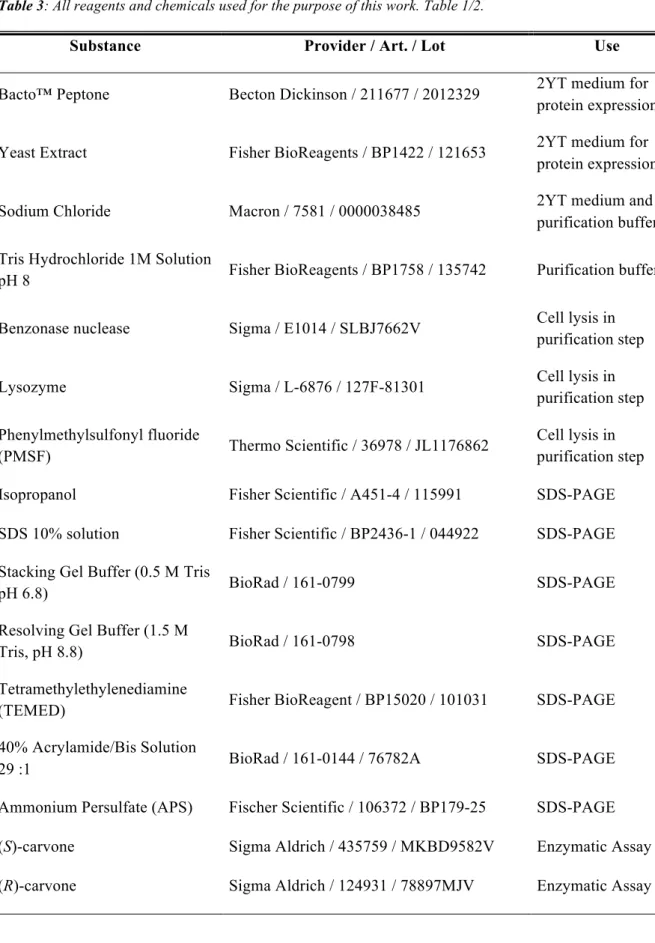
Table 3: All reagents and chemicals used for the purpose of this work. Table 1/2.
Substance Provider / Art. / Lot Use
Bacto™ Peptone Becton Dickinson / 211677 / 2012329 2YT medium for protein expression Yeast Extract Fisher BioReagents / BP1422 / 121653 2YT medium for
protein expression Sodium Chloride Macron / 7581 / 0000038485 2YT medium and
purification buffers Tris Hydrochloride 1M Solution
pH 8 Fisher BioReagents / BP1758 / 135742 Purification buffers Benzonase nuclease Sigma / E1014 / SLBJ7662V Cell lysis in
purification step Lysozyme Sigma / L-6876 / 127F-81301 Cell lysis in
purification step Phenylmethylsulfonyl fluoride
(PMSF) Thermo Scientific / 36978 / JL1176862
Cell lysis in purification step Isopropanol Fisher Scientific / A451-4 / 115991 SDS-PAGE SDS 10% solution Fisher Scientific / BP2436-1 / 044922 SDS-PAGE Stacking Gel Buffer (0.5 M Tris
pH 6.8) BioRad / 161-0799 SDS-PAGE Resolving Gel Buffer (1.5 M
Tris, pH 8.8) BioRad / 161-0798 SDS-PAGE Tetramethylethylenediamine
(TEMED) Fisher BioReagent / BP15020 / 101031 SDS-PAGE 40% Acrylamide/Bis Solution
29 :1 BioRad / 161-0144 / 76782A SDS-PAGE Ammonium Persulfate (APS) Fischer Scientific / 106372 / BP179-25 SDS-PAGE (S)-carvone Sigma Aldrich / 435759 / MKBD9582V Enzymatic Assay (R)-carvone Sigma Aldrich / 124931 / 78897MJV Enzymatic Assay
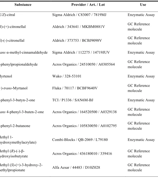
Table 4: All reagents and chemicals used for the purpose of this work. Table 2/2.
Substance Provider / Art. / Lot Use
(E/Z)-citral Sigma Aldrich / C83007 / 7819MJ Enzymatic Assay (R)-(+)-citronellal Aldrich / 343641 / MKBM0881V GC Reference
molecule (S)-(-)-citronellal Aldrich / 373753 / BCBJ9098V GC Reference
molecule
trans α-methyl-cinnamaldehyde Sigma Aldrich / 112275 / 14719JUV Enzymatic Assay 3-phenylpropionaldehyde Acros Organics / 24510050 / A0305564 GC Reference
molecule
Myrtenol Wako / 328-53101 Enzymatic Assay (+)-trans-Myrtanol Fluka / 70117 / BCBF9640V GC Reference
molecule
4-phenyl-3-butyn-2-one TCI / P1336 / SAN6M-BJ Enzymatic Assay
trans 4-phenyl-3-buten-2-one Acros Organics / 164520500 / A0329138 GC Reference molecule 4-phenyl-2-butanone Acros Organics / 105830050 / A0102795 GC Reference
molecule Methyl
1-(hydroxymethylacrylate) Combi-Blocks / QB-2069 / L79180 Enzymatic Assay Methyl
(R)-(-)-β-hydroxyisobutyrate Acros Organics / 436180010 / 339416
GC Reference molecule Methyl
(S)-(+)-3-hydroxy-2-methylpropionate Alfa Aesar / 44483 / D10Z028
GC Reference molecule
14/70
4.1.4
Softwares
Name / Version Developer Use
Microsoft Excel /
14.3.8 Microsoft
Gas chromatography data analysis, enantiomeric excess and quantitative data calculation
Prism / 6.0e GraphPad
Softwares, Inc. Conversion rate calculation and figures creation PyMOL / 1.6.0.0 Schrödinger LLC. Figures creation
GC ChemStation / B.03.02 [341]
Agilent
4.2 Methods
4.2.1
Cell transformation by electroporation
Genes of the targeted OYE1 enzymes have been cloned into a pET14b (4.7 kb) vector. This plasmid contains a T7 expression system as well as an Ampicillin resistance. It allows the induction of the recombinant protein expression by the presence of isopropyl β-D-1-thiogalactopyranoside (IPTG). This molecule binds the lac repressor (see figure 6) and leads to the transcription of the regulated gene. For a proper expression an E.coli strain containing the DE3 gene (such as the BL21(DE3)pLysS used in this work), which carries the information to express the T7 RNA polymerase as well as its regulation by the lac promoter. This engineered strain of E.coli allows an increased stability of the expressed proteins because is deficient in lon and aspartyl proteases. It also contain a pLysS plasmid [31], which is an additional control for the expression of the recombinant gene. It encodes for the T7 lysozyme, which is a natural inhibitor of T7 RNA polymerase [32].
Figure 6: Control elements of the pET System [32].
E.coli BL21(DE3)pLysS competent cells are transformed with pET14b-cpOYE303W116I and
pET14b-OYEW116I by electroporation. 150 ng of DNA is added to 100 µL of cells before the suspension is transferred in the electroporation cuvette. The transformation is performed with one pulse (capacitance 25 µF, resistance 200 Ω, 2 mm cuvette) and 1 mL of lysogeny broth (LB, Bacto™ Peptone 10 g/L, Yeats Extract 5 g/L, NaCl 10 g/L at pH 7) is immediately added to the cells and transferred to a sterile Eppendorf. Then, transformed cells are incubated 30 min at 37°C to allow the reconstitution of the membranes and the expression of the genes responsible for the antibiotic resistance. Finally, 200 µL are used to inoculate an agar LB plate (containing ampicillin and chloramphenicol), which is incubated overnight at 37°C.
16/70
4.2.2
Overexpression of cp303W116I and W116I
A single colony of transformed E.coli BL21 is inoculated in 2 mL of LB with antibiotics (chloramphenicol and ampicillin at 34 µg/ml and 100 µg/ml respectively) and incubated overnight at 37°C. Then, the whole volume is used as inoculum for 600 mL of 2YT (Bacto™ Peptone 16 g/L, Yeast Extract 10 g/L, NaCl 5 g/L at pH 7). This culture is incubated at 37°C with shaking at 200rpm until its optical density at 600nm (OD600) reaches 0.5 – 0.7. Overexpression of the recombinant protein
is induced by the addition of 1 M IPTG to a final concentration of 0.4 mM for 18 hours at 20°C. After that, the cells are harvested by centrifugation at 4°C for 20 min 4000 g. The cell pellet is washed in 20 mL of buffer A (NaCl 20 mM, Tris-HCl 40 mM at pH 8) in 50 mL Falcon tubes. These tubes are then centrifuged with the same conditions as before. Once the supernatant is removed, the cell pellet can be stored at -20°C.
4.2.3
Purification of cp303W116I and W116I from E.coli BL21(DE3) by
anion exchange chromatography
OYE enzymes were purified by anion exchange chromatography (AEX). A clear lysate solution was obtained by sonication, which promotes the disruption of cell membranes by the generation of sonic waves. This phenomenon involves the formation of a vapor phase in a liquid phase, in the same way as boiling does. The difference is that it is not the heat that induces vapor formation but high pressures. The bubbles, or cavities, formed will implode and generate intense shockwaves when subjected to higher pressures [33]. After centrifugation, the clear lysate is harvested and purified through the anion exchange column. The stationary phase is functionalized with quaternary ammonium radicals, -NH4+,
which are strong anion exchanger groups (see figure 7). The capture of OYE1 on the column is achieved with the help of negatively charged amino acids (Glu, Asp) regions on the enzyme, which will interact with the positively charged stationary phase. Once the column retains the compound, the system is washed in order to remove from the column non-specific bound molecules. Then, a gradient of a stronger anion competitor (NaCl or KCl) is passed through the system in order to displace the molecules attached to the stationary phase. At the column’s end, a spectrophotometric detector that can be set to a desired wavelength analyzes the buffers. The flavin cofactor bound in OYE1 enzymes has two distinct absorbance peaks at 360 and 460 nm; therefore the elution of the enzyme can be easily monitored. In order to follow the presence of the overall proteins, a third wavelength is set at 274 nm.
Figure 7: Diagram showing the anion exchange chromatography principle. In this experiment, the
immobilized cation surface is made of ammonium groups (-NH4+) and the negatively charged balls
Cell pellet containing an expressed OYE were suspended with a ratio of 1 gcells/6 mL in lysis buffer (1
mM PMSF, 1750 U benzonase nuclease and 10 mg lysozyme in buffer A) and stored on ice for 30 min. Cells were kept in ice and lysed by sonication (9x with 10 s pulse and 20 s pause) and cell extract was then centrifuged at 4°C for 30 min 16100 g. The clear lysate obtained was loaded at 5 mL/min into a pre-equilibrated HiTrap Q FF (5 mL) column with 3 column volumes (CV) of buffer A (Tris-HCl 40 mM at pH 8; depending on the protein concentration, two of those columns could be plugged in series on an Äkta purifier). The column was washed with 2 CV of buffer A until the A274nm was
stable. The elution of OYE was obtained with a gradient from 0 to 100% in 10 CV of buffer B (1 M NaCl, 40 mM Tris-HCl at pH 8), collecting the peak corresponding to the increase in the absorbance at 360 and 460 nm. The collected fractions were pooled together and concentrated by centrifugation at 4°C for 20 min, 4000 g with an Amicon Ultra-15 Centrifugal Filter Devices by Millipore (molecular weight cut-off: 10 kDa).
4.2.4
Size exclusion chromatography
In order to remove the last contaminants, a size exclusion chromatography (SEC) is performed as final step. This technique is usually used to remove small contaminants in an already purified solution. Through the column, particles will be separated depending on their molecular size. Therefore, it is important to notice that this technique is efficient only for molecules with a significant difference in their size. The column is packed with porous beads through which small molecules will travel (green path on figure 8). Obviously, if a molecule is too large to enter a pore, it will continue to flow through the system without being retained (red path). The result of this is that the small compounds have a higher retention time than the large ones.
Figure 8: Size exclusion chromatography principle. Small particles (green line) will enter porous beads and thus stay longer in the column, while larger molecules (red line) will pass through without being retained.
18/70
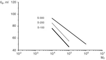
SEC column efficiency can be visualized with selectivity curves. These graphics are obtained by plotting the elution volume [mL] against the molecular weight [kDa]. The lines can inform the user on the molecular weight range where the column will be efficient. The selectivity curve corresponding to the column used in this work (HiPrep 16/60 Sephacryl S-100 High Resolution) is shown in the figure 9. Knowing that the OYE has an approximate molecular weight of 44 kDa, it should be eluted after 45-50 ml.
Figure 9: Selectivity curve for globular proteins on HiPrep 16/60 Sephacryl High Resolution columns.
For a standard run of SEC, the sample must be centrifuged initially at 4°C for 10 min at 16100 g to avoid loading precipitated protein in the system. Then, the sample is injected in a HiPrep 16/60 Sephacryl S-100 High-resolution (160 mL) column, pre-equilibrated in buffer C (300 mM NaCl, 40 mM Tris-HCl at pH 8). SEC is performed with a constant flow rate of 0.5 mL/min. The OYE detection is done the same way as for the AEX chromatography.
Finally, the protein is split into aliquots by using buffer C as diluent and with a final concentration of 10% glycerol. Aliquots are stored at -20°C.
▼
HiPrep 16/60 and 26/60
Sephacryl S-100 High Resolution
HiPrep 16/60 and 26/60
Sephacryl S-200 High Resolution
HiPrep 16/60 and 26/60
Sephacryl S-300 High Resolution
▼
instructions
iiiii 71-7117-00 Edition AG ● p1
▼
Introduction
HiPrep™ 16/60 and 26/60 Sephacryl™ S-100 High Resolution, Sephacryl S-200 High Resolution, and Sephacryl S-300 High Resolution are prepacked gel filtration columns designed for preparative purification of proteins and peptides. Sephacryl HR is a cross-linked copolymer of allyl dextran and
N,N-methylenebisacrylamide. This cross-linking gives the matrix good ridgity and chemical stability.
Steep selectivity curves give excellent resolution power for peptides and proteins in the molecular weight range, Mr 1 000–10 000 (Sephacryl S-100), Mr 5 000–250 000 (Sephacryl S-200), and Mr 10 000–1 500 000 (Sephacryl S-300). See Figure 1 and “Column data” for column characteristics.
Fig 1. Selectivity curves for globular proteins on HiPrep 16/60 Sephacryl HR
columns.
Column data
Matrix Cross-linked copolymer of allyl dextran and N,N-methylenebisacrylamide Mean particle size 47 µm (25–75 µm)
Separation range (Mr)
globular proteins 1 x 10³–1 x 10⁵ (Sephacryl S-100 HR)
5 x 10³–2.5 x 10⁵ (Sephacryl S-200 HR) 1 x 10⁴–1.5 x 10⁶ (Sephacryl S-300 HR) dextrans 1 x 10³–8 x 10⁴ (Sephacryl S-200 HR) 2 x 10³–4 x 10⁵ (Sephacryl S-300 HR) Column volume 120 ml (16/60) 320 ml (26/60)
Sample volume1 Up to 5 ml (HiPrep 16/60) Up to 13 ml (HiPrep 26/60)
Recommended flow rate 15 cm/h at room temperature (0.5 ml/min for 16/60 or 1.3 ml/min for 26/60) Maximum flow rate 30 cm/h at room temperature (1 ml/min
for 16/60 or 2.6 ml/min for 26/60) Maximum pressure over the 0.15 MPa, 1.5 bar, 21 psi
packed bed during operation2
HiPrep column hardware 0.5 MPa, 5 bar, 73 psi pressure limit2
Theoretical plates >5 000 m-1
pH stability
long term and working range 3–11 short term 2–13 Storage 20% ethanol
1 Optimal sample volume depends on the complexity of the sample, and flow rate. If the sample contains substances with small differences in size, either decrease sample volume, or decrease flow rate (in very difficult cases, it may be necessary to decrease both).
2 Many chromatography systems are equipped with pressure gauges to measure pressure at a particular point in the system, usually just after the pumps. The pressure measured here is the sum of pre-column pressure, pressure drop over the gel bed, and post-column pressure. This is always higher than the pressure drop over the bed alone. Keeping the pressure drop over the bed below 1.5 bar is recommended. Setting the upper limit of the pressure gauge to 1.5 bar will ensure that the pump shuts down before the gel is over-pressured. If necessary, post-column pressure of up to 3.5 bar can be added to the limit without exceeding the column hardware limit. To determine post-column pressure, proceed as follows:
To avoid breaking the column, post-column pressure must never exceed 3.5 bar.
1. Connect a piece of tubing in place of the column.
2. Run the pump at the maximum flow you intend to use for chromatography. Use a buffer with the same viscosity as you intend to use for chromatography. Note the backpressure as total pressure.
3. Disconnect the tubing and run at the same flow rate used in step 2. Note this backpressure as pre-column pressure.
4. Calculate post-column pressure as total pressure minus pre-column pressure. If post-column pressure is higher than 3.5 bar, take steps to reduce it (shorten tubing, clear clogged tubing, or change flow restrictors), and perform steps 1–4 again until the post-column pressure is below 3.5 bar. Note post-column pressure when it has reached a satisfactory level, add 1.5 bar to this value, and set this as the upper pressure limit on the chromatography system.
First-time use
The column is sealed with the top and bottom pieces which support the nylon nets welded to the column tube after the gel has been packed. Red plastic caps are snapped over both ends of the column. Do not attempt to remove these caps. The column cannot be opened, emptied or re-packed.
Connecting the column
1. Before connecting the column to a chromatography system, start the pump to remove all air from the system, particularly in tubing and valves.
2. Stop the pump.
3. Mount the column vertically, remove the domed nut, and connect the inlet tubing to the system “drop-to-drop”. 4. Remove the transport syringe and connect the column outlet tubing to, for example, a monitor cell. Save the transport syringe for use when storing the column. The column is now ready for use.
Equilibration of the column
Ensure an appropriate pressure limit has been set.
Equilibrate the column for first-time use, or after long-term storage as follows:
1. One-half column volume of distilled water at a flow rate of 15 cm/h (0.5 ml/min for 16/60 or 1.3 ml/min for 26/60). 2. Two column volumes of start buffer, e.g. 0.05 M sodium
phosphate, 0.15 M NaCl, pH 7.2 at 30 cm/h (1 ml/min for 16/60 or 2.6 ml/min for 26/60).
Try these conditions first
Flow rate: 15 cm/h (0.5 ml/min for 16/60, or 1.3 ml/min for 26/60). Sample volume: 1% of the column volume
(1.2 ml for 16/60 or 3.2 ml for 26/60) Buffer: 0.05 M sodium phosphate, 0.15 M NaCl,
pH 7.2 or select the buffer in which the sample should be solubilized for the next step. To avoid pH-dependent non-ionic
interactions with the matrix, include at least 0.15 M salt in the buffer (or use buffer with equivalent ionic strength).
Read the section “Optimization” for information on how to optimize a separation.
Buffers and solvent resistance
De-gas and filter all solutions through 0.22 µm filter to increase column lifetime. Buffers and solvents with increased viscosity will affect the backpressure and flow rate.
Daily use
All commonly used aqueous buffers, pH 3–11
Cleaning Acetonitrile, up to 30% Sodium hydroxide, up to 0.5 M Ethanol, up to 24% Acetic acid, up to 1 M Isopropanol, up to 30% Guanidine hydrochloride, up to 6 M Urea, up to 8 M Avoid Unfiltered solutions Sample recommendations
Recommended sample load 0.5%–4% of the column volume (0.6 –4.8 ml for 16/60, or 1.6–12.8 ml for 26/60)
Note: The sample volume is
critical for the separation. Preparation Dissolve the sample in start buffer,
filter through 0.22 µm filter, or centrifuge at 10 000 x g for 10 min.
Delivery and storage
The prepacked column is supplied in 20% ethanol. If the column is to be stored more than 2 days after use, wash the column with four column volumes of distilled water, and equilibrate with four column volumes of 20% ethanol.
To avoid air bubble formation in the column, use the transport syringe. Connect the transport syringe to the capillary tubing at the column outlet. Start the pump, and fill the syringe to approximately 50% of the total syringe volume.
DO NOT OPEN THE COLUMN!
Choice of buffer
Selection of buffering ion does not directly affect resolution. Select a buffer in which the purified product should be collected, and which is compatible with protein stability and activity. Buffer concentration must be sufficient to maintain buffering capacity and constant pH.
Ionic strength should be at least 0.15 M NaCl in the start buffer, to avoid non-specific ionic interactions with the matrix.
Optimization
Perform a first run as described in the section “Try these conditions first”. If the obtained results are unsatisfactory, consider the following:
Action Effect
Decrease flow rate Improved resolution Decrease sample volume Improved resolution
Figs. 2–5 show the influence of sample volume, flow rate and sample loading on the resolution
Buffer: 50 mM NaPO4, 0.15 M NaCl, 0.02% NaN3, pH 7.0
Sample: IgG, ovalbumin, cytochrome C, 1:2:1 Flow rate: 0.66 ml/min
Total protein load: 8 mg
Equilibration before a new run
Regenerate the column after each run with one column volume of start buffer at 25 cm/h (0.8 ml/min for 16/60 or 2.2 ml/min for 26/60).
Instructions
HiPrep 16/60 or 26/60 Sephacryl S-100 High Resolution HiPrep 16/60 or 26/60 Sephacryl S-200 High Resolution HiPrep 16/60 or 26/60 Sephacryl S-300 High Resolution Inlet tubing, Domed nut,
Unions M6 female/1/16” male
Transport syringe
Fig 2. Resolution (Rs) between IgG and ovalbumin at different sample volumes.
4.3 Estimating purity with SDS-PAGE
A polyacrylamide gel electrophoresis (PAGE) in denaturing condition with the presence of sodium dodecyl sulfate (SDS) can give an approximate idea on how pure the solution is. First, the SDS, which is an anionic detergent, is used in order to linearize the proteins. It applies negatives charges elsewhere on the protein resulting in the straightening of the biomolecule because each charge repulses each other of the same sign. Once the SDS linearizes the proteins, they can be separated by their size through an acrylamide gel. It works like a grid containing several holes, depending on the percentage of acrylamide. The more holes there is, the slower bigger proteins will travel and the fastest the small one will migrate through the grid. An electric current going from the negative cathode toward the positive anode produces the flow. Since the proteins are negatively charged, they will be attracted toward the positive pole. A blue coomassie dye is added to samples in order to be able to follow the elution front. When it reaches the bottom of the gel, the migration is stopped. To see the results, the gel must be dyed. For this experiment, the gel is stained with a blue coomassie dye. An overall view of the migration and coloration is showed by the figure 10.
Figure 10: Overall schematic view of a SDS-PAGE analysis [34].
SDS-PAGE gel is prepared in order to get a final 10% acrylamide. Samples (cells after sonication, clear lysate, fraction after AEX and fraction after SEC) are mixed with deionized water and 4x SDS buffer before being denatured at 95°C for 10 minutes. After loading samples and the markers (Broad Range Ladder from BioRad), the gel is run at 200 V for 40 min. For the coloration, the gel is put in a box with the staining solution (1 g coomassie blue in 50% methanol) and warmed up in a microwaves oven for 30 s at max potency. After that, the gel is destained with a solution of 10% MeOH, 7% Glacial Acetic Acid in water. A piece of absorbing paper can be added in the box to trap the coomassie. Then, the box is slightly rocked for 5 min before removing the solution. Additional destaining steps may be required. When the gel is destained, it is stored digitally using a geldoc imager (Biorad). An OYE’s band is visible corresponding to its molecular weight at 44 kDa.
20/70
4.4 Determination of purified enzyme concentration
Protein concentration can be calculated by measuring the absorbance in the UV-vis region and applying equation 1:
!! = !!∙ ! ∙ ! ↔ ! = !! !!∙ !
Equation 1: Re-arranged Beer-Lambert law in order to be able to deduce concentration of a solution from its absorbance. Where Ax [-] is the absorbance at a wavelength of X [nm]. εx is the molar extinction coefficient
[mM-1*cm-1] at a wavelength of X [nm]. [C] is the concentration of the solution [mM] and L is the length of the optical path [cm].
However, to use this formula, the molar extinction coefficient of each variant has to be experimentally determined. OYE1 has a FMN cofactor bound in its active site with a known extinction coefficient of the free form at 450 nm as 11.3 mM-1cm-1. By denaturing the enzyme, the flavin will be released and the absorbance of the free cofactor could be measured accurately. Assuming a ratio 1:1 (see equation 2), the concentration of the enzyme is equal to the concentration of FMN.
!"!!"!!"#$!%"&+ !"!!"## ↔ !"!!"#
Equation 2: Stoichiometric equation between the OYE1 with and without its flavin cofactor.
The molar extinction coefficient of the enzyme is calculated using the equation 1.
Initially, the enzyme solution is centrifuged at 4°C for 10 min 16100 g. Supernatant is used for the measurement of the soluble OYE’s spectrum from 300 to 800 nm. Secondly, the protein is denatured by adding 0.5% SDS final concentration and by boiling in the microwave 1 min at max potency. The solution is centrifuged at RT for 10 min 16100 g. The free flavin spectrum from is then measured. The max absorbances of the OYE1 (~450 nm) and of the free flavin (445 nm) are used for the calculation.
4.5 Enzymatic Assays
Because oxygen can be turned over by the enzyme consuming NADPH, the assays are done under anaerobic conditions in a hermetic chamber at room temperature with an orbital agitation. Within the reaction, the coenzyme NADPH is recycled by phosphite dehydrogenase, as shown in the following figure:
Figure 11: Representation of the enzymatic system applied during this work. The substrate is catalyzed by the OYE1 in the left half, while the cofactor is regenerated by the phosphite dehydrogenase in the right half.
For each measurement, 30 µL of reaction were taken from the reaction vials at pre-determined time points. 50 µL of ethyl acetate containing cyclohexanone (GC internal standard) are immediately added to quench the enzymatic reaction. The solution was well homogenized by shaking vigorously for 1 minute. To enhance phase separation, the tubes were centrifuged at RT for 2.5 min and 16100 g. Finally, the organic phase was analyzed by GC with the corresponding method. Table 5 shows the concentration of each constituent in the reaction vial per substrate.
Table 5: Composition of enzymatic reactions according to the substrate used. NaPi stands for sodium phosphate. NADP+ for Nicotinamide adenine dinucleotide phosphate and PTDH for phosphite dehydrogenase. S/R-1 are for
S/R-carvone. 3 is for trans α-methylcinnamaldehyde. 5 is for myrtenol. 7 stand for methyl 2-hydroxymethylacrylate. 9 is for 4-phenyl-3-butyn-2-one and (E/Z)-12 is for E/Z-citral.
(S)-1 (R)-1 3 5 7 9 (E/Z)-12 [Substrate] 200 µM 200 µM 2 mM 500 µM 600 µM 200 µM 600 µM [Enzyme] 1 µM 1 µM 1 µM 1 µM 1 µM 1 µM 1 µM [NaPi] 10 mM 10 mM 10 mM 10 mM 10 mM 10 mM 10 mM [FMN] 5 µM 5 µM 5 µM 5 µM 5 µM 5 µM 5 µM [NADP+] 100 µM 100 µM 100 µM 100 µM 100 µM 100 µM 100 µM [PTDH] 1 µM 1 µM 1 µM 1 µM 1 µM 1 µM 1 µM [Cyclohexanone] 500 µM 500 µM 1 mM 500 µM 500 µM 500 µM 500 µM
22/70
4.5.1
Calculations of relatives concentrations
Results shown as relative concentrations are calculated according equation 3. !!
!!+ !!+ ⋯ + !!
∙ 100 = !"#$%&'"!!"#!$#%&'%("#!!"!!!![%]
Equation 3: Calculation of relative concentration of a compound X in a reaction with N components.
4.5.2
Calculation of enantiomeric excess
Enantiomeric excess (ee) are calculated according equation 4. ! − !
! + !∙ 100 = !!!
Equation 4: Calculation of the enantiomeric excess for A, which is the predominant specie among products.
4.5.3
Calculation of conversion rates
Conversion rates are determined from quantitative data (figures in appendix 10.4.1 to 10.4.6) by using the slope of the linear regression of the consumption of substrate. Regressions are done with GraphPad Prism 6.
!"#$%! !!!"#!$%&$'!"#
!!"#$%!![!"#!!"#$%!]= !"#$%&'("#!!"#$!
!!! !"# ∙ !"#!!
Equation 5: Calculation of conversion rate from the slope of a linear regression of quantitative data of components against time.
4.6 Gas Chromatography analysis
Enantioselectivity and catalytic activity were determined following the consumption of starting material and the appearance of product by gas chromatography (GC) analysis. This instrument allows the separation of components through a capillary column. The mobile phase is a gas (i.e. hydrogen) and the compounds are vaporized in the injector so they also enter the system in gas phase. Then, the sample arrives in the column, which is installed in an oven. The instrument is programmable allowing the creation of defined methods that control the temperature of the system. The operator can choose between various modes: isothermal (same temperature over a selected duration of time), gradient (temperature increase with a chosen slope) or even a mix of both modes.
The column used in this work (Cyclosil B, Agilent, 30 m long, 0.25 µM film thickness) is coated with 30% hepatkis (2,3-di-O-methyl-6-O-t-butyl-dimethylsilyl)-ß-cyclodextrin (figure 12), which is a functionalized cyclodextrin.
Figure 12: Scheme of a Heptakis(2,3-di-O-methyl-6-O-t-butyl-dimethylsilyl)-ß-cyclodextrin, which is a functionalized cyclodextrin found on a Cyclosil-B (Agilent) stationary phase.
The principle is very similar to the SEC. Cyclodextrins can be imagined like barrels, which allows compounds (usually, the desired analyte) to get in it to form a complex, before being released (figure 13). This interaction depends on several factors. There are the hydrophobic interactions, the van der Waals attractions, any hydrogen bonding or electrostatic interactions that can retain a molecule guest inside a cyclodextrin. The molecules that enter the cyclodextrins will travel longer through the column than the ones that do not get inside the barrels. The difference of speed obtained between compounds will result in a separation of the components throughout the column.
24/70
At the end of the column, there is a flame ionization detector (FID). As its name suggest, the detector uses a flame to ionize analyte molecules coming out of the column. The ions formed then interact with the electrodes of the detector to generate electrical signals. This process is depicted in figure 14.
Figure 14: Flame ionization detector scheme. « A » stands for Analyte. Once it is burnt, it releases an electron (e-) and thus, become positively charged (A+), those ions are detected by the electrodes in the combustion
chamber. Then, the electrical detector gives a signal corresponding to the excitation of the electrodes.
In every sample, an internal standard was added. It is a molecule that does not interact with any other component in the injected solution and that is present at the same concentration. The utility of this standard is to reduce the impact of the error created by the injector, which does not always inject the exact same volume in the column. Since the concentration of internal standard is identical in every sample, injector inconsistencies can be overcome by normalizing analyte peak areas relative to the internal standard peak areas. Once this is done, areas from separate injections can be confidently compared between each other.
4.6.1
(S/R)-carvone
The method is the following: Start at 90°C and hold 5 min. Then increase temperature 1°C/min until 120°C and hold for 15 min, total run time of 35 min. Injector and detector are set at 250 and 250°C respectively. The H2 flow is set constant at 1.8 ml/min with a front pressure of 36.6 kPa. 1 µL is
injected with a split of 1:1. Retention times for (S/R)-carvone are 27.5 and 27.6 min respectively. (2S,5S)- (2R,5S)-carvone have retention times of 22.9 and 23.1 min, while retention time for (2R,5R)-carvone is 22.7 min.
4.6.2
Trans α-methyl-cinnamaldehyde
The method is the following: Isothermal, 150°C hold 10 min. Injector and detector are set at 250 and 200°C respectively. The H2 flow is set constant at 1.8 ml/min with a front pressure of 45.7 kPa. 1 µL is
injected with a split of 25.1:1. The retention time for trans α-methyl-cinnamaldehyde is 6.3 min. S-cinnamaldehyde has a retention time of 3.8 min.
4.6.3
Myrtenol
The method is the following: Start at 90°C and hold for 1 min. Then increase temperature 3°C/min until 120°C and hold 19 min, total run time 20 minutes. Injector and detector are set at 250 and 310°C respectively. The H2 flow is set constant at 4 ml/min with a front pressure of 71.2 kPa. 1 µL is injected
with a split of 1:1. Retention time for myrtenol is 11.7 min. (+)-trans-myrtanol has a retention time of 16.1 min.
4.6.4
Methyl 2-hydroxymethylacrylate
The method is the following: Starting at 60°C and hold 1 min. Then, 1°C/min until 75°C, hold 4 min, total run time 20 minutes. Injector and detector are set at 280 and 200°C respectively. The H2 flow is
set constant at 4 ml/min with a front pressure of 68.6 kPa. 1 µL is injected with a split of 1:1. Retention times for trans methyl 2-hydroxymethylacrylate is 16.8 min. Methyl (R)-(-)-β-hydroxyisobutyrate and methyl (S)-(+)-3-hydroxy-2-methylpropionate have retention times of 15.2 and 17.0 min.
4.6.5
4-phenyl-3-butyn-2-one
The method is the following: Start at 70°C and hold 1 min. Then, increase temperature 2°C/min until 110°C and hold 15 min, total run time 36 minutes. Injector and detector are set at 250 and 310°C respectively. The H2 flow is set constant at 4 mL/min with a front pressure of 71 kPa. 1 µL is injected
with a split of 1:1. Retention time for 4-phenyl-3-butyn-2-one is 23.4 min. For the 4-phenyl-3-butene-2-one and the 4-phenyl-2-butanone, retention times are 34.6 and 21.9 min.
4.6.6
(E/Z)-citral
The method is the following: Start at 80°C, increase temperature 2.5°C/min until 115°C and hold 2 min, total run time 16 minutes. Injector and detector are both set at 270°C. The H2 flow is set constant
at 4 ml/min with a front pressure of 68.5 kPa. 1 µL is injected with a split of 1:1. E/Z-citral peaks could not be identified because enantiopure solutions are not commercially available. However, the separation was possible and the E/Z-mixture gave two peaks, which retention times are 13.1 and 14.9 min. (S/R)-citronellal retention time are 9.057 min and 9.018 min respectively.
26/70
5. Results
5.1 Enzyme purification
Following expression the harvested cells (6.4 g) were lysed for a final 40 mL of clarified lysate. The sample was loaded on the Anion Exchange and purification proceeded as described into the materials and methods. Fractions were collected between 80 and 100 mL corresponding to the elution of the flavin peak at 460nm. An AEX chromatogram is showed in the figure 40in the appendix 10.2.
Harvested fractions were concentrated down and then injected on the SEC. OYE1 elute between 46 and 55 mL corresponding to a MW of about 88 kDa as OYE1 is a dimer in solution (SEC chromatogram showed in figure 41 appendix 10.3).
Collected protein fractions were loaded on a 10% acrylamide SDS-page gel and migrated for 40 min at 200 V. OYE’s band is visible at ~44 kDa, consistent with its reported molecular weight.
Figure 15: 10% acrylamide SDS-page gel migrated for 40 min at 200 V and stained with coomassie blue. 1) Broad Range Ladder from BioRad, 2) Wild Type OYE, 3) cp303OYE, 4) W116IOYE, 5) cp303OYEW116I.
5.2 Calculation of enzyme concentration
The molar extinction coefficients of W116I and cp303W116I variants were determined with spectrophotometric assays as described in the materials and methods.
Table 6: Values obtained and used during the experiment for the determination of the molar extinction coefficient of W116I and cp303W116I enzymes. Calculations are based on the equation 1.
!
W116I%
cp303W116I%
OYE1!A
max!!
0.116!
0.056!
Free!FMN!A
445!
0.0651!
0.0522!
ε
FMN![mM
71cm
71]!
12.5!
12.5!
Dil.!Factor!
2!
1!
FMN=OYE1![mM]!
0.0104!
0.0042!
ε
OYE![mM
71cm
71]!
11.1!
13.4!
5.3 Catalytic rate and enantioselectivity of mutants
Relative concentrations values used for figures 16, 17, 20, 26, 29 and 35 as well as quantitative data are shown in appendix 10.4.1 to 10.4.6.
5.3.1
(S)-carvone
Both the enzymatic assay and GC method have been previously optimized [1]. The initial concentration of 200 µM of substrate was chosen to ensure the most representative conversion in 3 hours. Retention times were obtained from reference molecules. Quantitative data were calculated from a standard curve. However the enantiomericaly pure products of the reaction are not commercially available, quantitative data of the products is calculated using (S)-carvone calibration curve (which could cause not accurate measures). Figure 16 shows evolution of relative concentrations over time.
Figure 16: Conversion of (S)-carvone into (2S,5S)- or (2R,5S)-carvone by WT and its variants. Experiments are done in buffer A (20 mM NaCl, 40 mM Tris-HCl, pH 8 at 20°C) with 200 µM of (S)-1 as starting material using phosphite dehydrogenase and NaPi as cofactor regenerating system.
28/70
Table 7 shows the calculated enantiomeric excess (ee) and conversion rates for (S)-1.
Table 7: Enantiomerics excess (ee) in percent and conversion rate expressed as !!!
!"#∙!"#!! for each variant with
(S)-carvone as substrate.
Enzyme ee Conversion Rate Wild Type 91 (R) 3.8 ± 0.3 cp303 79 (R) 7.5 ± 0.4 W116I 91 (S) 17.6 ± 2.6 cp303W116I 88 (R) 13.8 ± 1.1
5.3.2
(R)-carvone
Due to the similarities with the other enantiomer, (R)-1, experiments were done with 200 µM starting material, 1 µM enzyme and the same time points. As mentioned before, the enantiomericaly pure products (2R,5R)- and (2S,5R)-carvone are not commercially available. Therefore, quantitative data of the products is calculated from (R)-carvone calibration curve (which could cause not accurate measures). Figure 17 shows evolution of relative concentrations over time.
Figure 17: Conversion of (R)-carvone into (2R,5R)-carvone by WT and its variants. Experiments are done in buffer A (20 mM NaCl, 40 mM Tris-HCl, pH 8 at 20°C) with 200 µM of (R)-1 as starting material using phosphite dehydrogenase and NaPi as cofactor regenerating system.
![Figure 2: Creation of circular permutants of OYE1 [1]. The two genes are plugged together with a nine- nine-nucleotide sequence encoding the -Gly-Thr-Ser- linker](https://thumb-eu.123doks.com/thumbv2/123doknet/14994916.674254/11.892.214.689.288.528/figure-creation-circular-permutants-plugged-nucleotide-sequence-encoding.webp)