HAL Id: hal-03103744
https://hal.archives-ouvertes.fr/hal-03103744
Submitted on 8 Jan 2021
HAL is a multi-disciplinary open access
archive for the deposit and dissemination of
sci-entific research documents, whether they are
pub-lished or not. The documents may come from
teaching and research institutions in France or
abroad, or from public or private research centers.
L’archive ouverte pluridisciplinaire HAL, est
destinée au dépôt et à la diffusion de documents
scientifiques de niveau recherche, publiés ou non,
émanant des établissements d’enseignement et de
recherche français ou étrangers, des laboratoires
publics ou privés.
chemical vapor deposition
Ioannis Paradisanos, Shivangi Shree, Antony George, Nadine Leisgang, Cédric
Robert, Kenji Watanabe, Takashi Taniguchi, Richard Warburton, Andrey
Turchanin, Xavier Marie, et al.
To cite this version:
Ioannis Paradisanos, Shivangi Shree, Antony George, Nadine Leisgang, Cédric Robert, et al..
Control-ling interlayer excitons in MoS2 layers grown by chemical vapor deposition. Nature Communications,
Nature Publishing Group, 2020, 11 (1), �10.1038/s41467-020-16023-z�. �hal-03103744�
Controlling interlayer excitons in MoS
2layers grown by chemical vapor deposition
Ioannis Paradisanos1∗, Shivangi Shree1∗, Antony George2, Nadine Leisgang3,
Cedric Robert1, Kenji Watanabe4, Takashi Taniguchi4, Richard J. Warburton3, Andrey Turchanin2,5, Xavier Marie1, Iann C. Gerber1,∗ and Bernhard Urbaszek1†
1
Universit´e de Toulouse, INSA-CNRS-UPS, LPCNO, 135 Avenue Rangueil, 31077 Toulouse, France
2
Friedrich Schiller University Jena, Institute of Physical Chemistry, 07743 Jena, Germany
3Department of Physics, University of Basel, Basel, Switzerland
4
National Institute for Materials Science, Tsukuba, Ibaraki 305-0044, Japan and
5
Abbe Centre of Photonics, 07745 Jena, Germany
Combining MoS2 monolayers to form multilayers allows to access new functionalities. In this
work, we examine the correlation between the stacking order and the interlayer coupling of valence
states in MoS2 homobilayer samples grown by chemical vapor deposition (CVD) and artificially
stacked bilayers from CVD monolayers. We show that hole delocalization over the bilayer is allowed in 2H stacking and results in strong interlayer exciton absorption and also in a larger A-B exciton separation as compared to 3R bilayers, where both holes and electrons are confined to the individual layers. Comparing 2H and 3R reflectivity spectra allows to extract an interlayer coupling energy of
about t⊥= 49 meV. Obtaining very similar results for as-grown and artificially stacked bilayers is
promising for assembling large area van der Waals structures with CVD material, using interlayer exciton absorption and A-B exciton separation as indicators for interlayer coupling. Beyond DFT calculations including excitonic effects confirm signatures of efficient interlayer coupling for 2H stacking in agreement with our experiments.
Introduction.— Transition metal dichalcogenides (TMDs) with the form MX2 (M = Mo, W, Ti, etc and
X = S, Se, Te) have tunable electronic properties from metallic to semiconducting depending on the crystal sym-metry, composition and number of layers [1–9]. The band structure of TMD semiconductors is drastically modi-fied by changing the sample thickness by just one atomic monolayer [10–12]. For instance, the combination of two different monolayer materials such as MoSe2-WSe2 into
a heterobilayer results in type II band alignment and opens new research perspectives on periodic moir´e po-tentials for carriers in the different layers and the result-ing interlayer excitons [13–17]. Twisted homobilayers of graphene, WSe2 and MoSe2 allow accessing new
super-conducting phases and correlated insulating states [18–
20].
To access the new functionalities provided by assem-bling monolayers to form multilayers it is necessary to identify physical parameters that strongly depend on in-terlayer coupling and to experimentally control them. Our approach is to compare CVD grown MoS2
bilay-ers with artificially stacked bilaybilay-ers made from CVD monolayers. We study for both as-grown CVD and in-dividually assembled cases the 2H (180◦ twist angle, see schematic in Fig.1c) and 3R (0◦ twist angle) stacking which gives precise control over the interlayer coupling and hence interlayer exciton formation. Studying these two precise alignments is also relevant for samples ini-tially assembled with other twist angles as reconstruc-tion results also in these experiments in the formareconstruc-tion of µm wide 2H and 3R areas [21, 22], which are energeti-cally most stable. We show that the valence states for 2H bilayers are strongly impacted by interlayer coupling
as the hole is delocalized over the 2 layers [23–25]. This results in important changes in the optical spectra gov-erned by K-K transitions as we observe strong absorption from interlayer excitons and a clear change in separation between A- to B-exciton transition in differential white light reflection at T= 4 K. These observations are made possible due to the drastically improved optical quality of CVD samples removed from the growth substrate and encapsulated in hBN [26]. We show that both indicators for interlayer coupling are absent in the measured 3R bi-layer spectra as hole hopping between the bi-layers is sym-metry forbidden [27]. Comparing for 3R (no interlayer coupling) and for 2H the A-B exciton absorption spectra allows us to extract an experimental value of the perpen-dicular hopping (coupling) term of t⊥≈ 49 meV,
impor-tant for moir´e superlattices [28] and so far only roughly estimated from theory [27]. To artificially stack large area CVD layers and control interlayer coupling through stacking (i.e. 0◦ or 180◦ twist angle) is technologically relevant for 2D materials optoelectronics [17], as CVD substrates are covered by a large number of monolay-ers and are very practical to stack (twist) due to their symmetric triangular shape and well characterized edge termination.
In addition to our optical spectroscopy experiments we show in density functional theory (DFT-GW ) calcu-lations that at the independent particle and the quasipar-ticle (exciton) level description of the system the valence band splittings for 2H as compared to 3R are different due to interlayer coupling. In our calculated absorption spectra with excitonic effects solving the Bethe-Salpeter-Equation (BSE) for 2H stacking we show strong inter-layer exciton absorption, absent for 3R stacking.
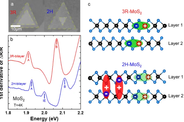
FIG. 1: Spectroscopy of CVD grown bilayers encapsulated in hBN. (a) Optical microscope images of as-grown 3R
(left) and 2H CVD MoS2 bilayers (right) on SiO2/Si before pick-up. (b) First derivative of white light reflection spectrum for
as-grown 2H-bilayer (blue) and as-grown 3R-bilayer (red), recorded at T= 4 K, both bilayers are encapsulated in high quality
hBN for optical spectroscopy [29]. (c) Schematic of 3R stacked bilayer with intralayer excitons (top) compared to 2H stacked
bilayer where in addition interlayer excitons are observed as in panel (b).
Interlayer excitons in as-grown CVD MoS2
homobilayers.— The thermodynamically most stable configurations of TMD homobilayers are the 2H and the 3R stacking [24, 30]. In practice, most naturally occur-ring molybdenite shows 2H not 3R stacking. In this work we focus on high quality CVD grown flakes for several reasons : during CVD growth of MoS2 both 2H and 3R
stackings for bilayers can occur [31] and we are therefore able to compare the optical response for samples grown under identical conditions. Secondly, as all CVD flakes on our substrate show the same edge termination, we can artificially stack two layers in 2H and 3R configuration to compare with the as grown samples - see discussion be-low. Third, many monolayers cover the SiO2 substrate
and can all be picked-up in a single step, which makes fabrication of bilayer structures very efficient.
Optical microscope images of as-grown CVD bilay-ers on SiO2/Si with 3R and 2H stacking are presented
in Fig.1a. The stacking can be determined already by the relative rotation of the triangular monolayers and was confirmed in second harmonic generation (SHG) ex-periments. The SHG signal for 2H stacking was not detectable (inversion symmetry restored) but we could perform detailed angle dependent SHG for the 3R
stack-ing (broken inversion symmetry), see supplement. The high quality MoS2 bilayers and monolayers were grown
by a modified CVD process in which a Knudsen-type effusion cell is used for the delivery of sulfur precursor [33]. Using a water-assisted pick-up technique [32], the as-grown CVD bilayers have been deterministically trans-ferred and encapsulated in hBN to achieve high optical quality [26], which has recently been shown to be crucial for optical spectroscopy on CVD samples lowering the typical emission linewidth from about 50 meV to below 5 meV at T= 4 K. The thickness of the top and bottom hBN has been carefully selected to optimize the oscillator strength of the interlayer excitons (IEs) [24]. After en-capsulation, the samples were cooled down to T= 4 K in a closed-cycle cryostat and a series of differential reflectiv-ity measurements with a home-built confocal microscope have been performed at different locations of the sam-ples, see methods. We define differential reflectivity as (Rsam− Rsub)/Rsub, where Rsamis the intensity
reflec-tion coefficient of the sample with the MoS2 layers and
Rsubis the same structure without the MoS2. Note that
the overall shape and amplitude of the differential reflec-tivity signal also depends on cavity effects (thlayer in-terference) given by top and bottom hBN and SiO2layer
3
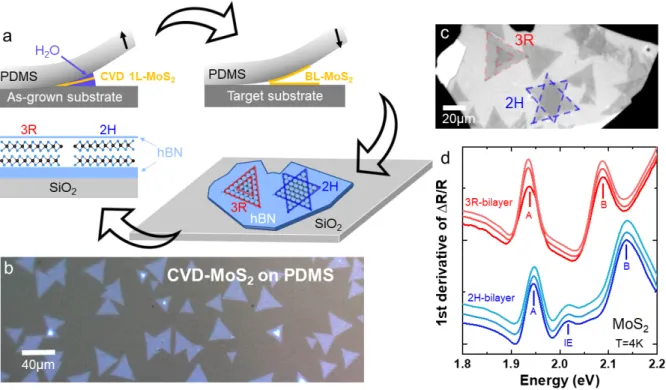
FIG. 2: Artificial stacking of CVD monolayers into bilayers. (a) Schematic of sample pick-up, bilayer assembly and
encapsulation for optics. (b) Optical micrograph of CVD-grown MoS2 monolayers and a few homobilayers, transferred to the
PDMS stamp following water-assisted pick-up [32] from the growth substrate. (c) Artificially-assembled 3R and 2H MoS2
homobilayers, fabricated by an all-dry deterministic transfer process. (d) First derivative of reflectivity spectra collected from
three different areas of the artificially stacked 3R (red) and 2H (blue) MoS2 homobilayers, shown in (c). Spectra have been
shifted for clarity.
thickness (see [34] for details). In Fig.1b, the first deriva-tive of the differential reflectivity spectra for as-grown CVD 2H and 3R MoS2bilayers can be compared. There
are two striking differences between the 2H and 3R bi-layer spectra: (i) While A and B intrabi-layer excitons are identified for both configurations, a pronounced feature at ≈ 2 eV appears exclusively in the 2H bilayer. This feature is assigned to an interlayer state, its energy being in good agreement with the very recently identified IEs in high quality and hBN encapsulated exfoliated MoS2
bi-layers with 2H stacking [23,24, 35], in contrast to CVD grown samples studied here. This observation of IEs was made possible by our specific CVD sample preparation for the optical spectroscopy experiment [26]. IE absorp-tion was not detectable in very detailed earlier works due to considerably larger optical linewidth or detection of emission and not absorption [36–39]. In contrast to 2H stacking, in the 3R configuration no additional states are detected between the A - and B-excitons, thus indicating that delocalization of holes is not allowed in this partic-ular stacking order [24], see below for a more detailed discussion. (ii) The separation between the A - and B - exciton transitions is considerably larger in the 2H bi-layers (about 185 meV) as compared to the 3R bilayer (about 150 meV, mainly given by the spin-orbit splitting
in the valence band). This is a second indication for ef-ficient interlayer coupling of A-B valence states for 2H stacking, as the separation of the valence states mainly governs the A-B exciton separation [23,27,40].
Control of the interlayer coupling in artificially-assembled MoS2 homobilayers.— In Fig.1 we show
that as-grown CVD MoS2 bilayers experience interlayer
coupling resulting in interlayer exciton formation, here observed for a non-contaminated interface between the top and bottom layer. Contamination from secondary transfer processes could potentially suppress the coupling between the layers and hence IE formation. By choos-ing 2H or 3R orientation manually when stackchoos-ing CVD monolayers to form bilayers one can allow hole tunnel-ing between the layers or not. This requires to pick-up the CVD monolayers from their growth substrate while maintaining their structural integrity, optical quality and a sufficiently clean interface after transfer. Furthermore, fine control of the twist angle between the top and bot-tom layer is needed, since the IE formation is allowed only in a precise stacking order, see sample prepara-tion schematic in Fig.2a. Here we use water-assisted deterministic transfer that allows the ability to control-lably assemble CVD bilayers with a desired twist angle [32]. First, CVD monolayers have been carefully
picked-up from the growth substrate and transferred to PDMS (Fig.2b) [32, 41]. The structural integrity of the CVD monolayers is preserved in this case and the following step is to slowly assemble 2H and 3R bilayers and encap-sulate them in hBN as shown in Fig.2c. Small deviations from 0◦ or 180◦ twist angle are expected but natural reconstruction of the bilayer will again favor the lowest energy arrangement, 3R and 2H, respectively [21,22].
Differential reflectivity spectra have been collected from ten different areas of the assembled 2H and 3R bilayers of Fig.2c. In Fig.2d, three typical examples of the assembled 2H and 3R spectra are presented. The spectra show a striking resemblance with the as-grown bilayer spectra discussed before in Fig.1b. So also for the assembled 2H bilayers we identify clear interlayer exci-ton absorption and an increased separation between the A- and B-excitons. It is important to note that the IE transition was clearly observed over the whole surface area of the manually constructed 2H bilayer. We take it as a strong indication of efficient interlayer coupling and possibly efficient reconstruction/self-rotation to the 2H configuration. We therefore further confirm the forma-tion of IEs exclusively in the 2H stacking. By manually choosing the stacking configuration i.e. twist angle, it is possible to tune the valence band splitting and the for-mation of interlayer excitons in large area, high quality CVD samples.
Beyond-DFT band structure calculations and exciton absorption.— In addition to optical spec-troscopy we perform beyond DFT calculations to study the striking differences between 2H and 3R MoS2
bilay-ers, see methods for the computational details. Please note that our GW +BSE calculations are performed for MoS2 bilayers in vacuum for simplicity and not in hBN.
The general target of our calculations is to understand the microscopic origin of the optical transitions and to re-produce the energetic order qualitatively. In GW calcula-tions we compare band structures corrected by screening effects, the exciton description being added later. In the vicinity of the K-point of the Brillouin zone, see Fig.3a and schematic in Fig.3c, differences between 2H and 3R stacking in VB, VB−1 states are clear. We find a VB
splitting in the 2H bilayer that is 19 meV larger than in the 3R bilayer. By solving the BSE we obtain the absorp-tion for the 2H and 3R bilayers shown in Fig. 3b. The main characteristics are (i) the presence of a strong in-terlayer exciton peak in 2H and (ii) a larger A-B exciton separation for 2H than for 3R configuration, exactly as found in the experiments in Figs.1 and 2. Interestingly, in 3R stacking, there is a minor departure from degener-acy that splits VB and VB−1 states from distinct layers
when they remain degenerate in 2H configuration. As a consequence, two distinct A-type excitons separated by only 14 meV constitute the A-peak of the 3R-bilayer ab-sorption spectrum in our calculation, explaining its larger
width compared to the 2H case, see Fig.3c. Discussion.—
To summarize the main experimental findings, first we observe strong interlayer exciton absorption between the main A - and B - exciton transitions for CVD grown (Fig.1b) and artificially stacked (Fig.2d) 2H bilayers, whereas this interlayer transition is absent for 3R stack-ing as hole tunnellstack-ing is symmetry forbidden and only intralayer exciton transitions are observed. A second striking observation is that stacking of the layers also af-fects the energy difference between A - and B - exciton transitions. This is demonstrated in Fig.3d, where the A - B exciton energy difference is compared between as-grown and assembled 2H and 3R bilayers. It is apparent that 2H bilayers exhibit a significantly larger energy dif-ference between the A - and B - exciton states, compared to 3R bilayers.
Our next target is to experimentally extract the in-terlayer hopping term, t⊥ based on a k.p model of
bi-layers in the vicinity of K points [23, 27, 28] and com-pared it to post-DFT estimates. As indicated in Fig.3c, for 3R stacking the measured A- to B- exciton split-ting S3R is roughly given by the spin orbit splitting as
S3R = ∆SO. For 2H-stacking the A-B splitting in the
valence band depends in addition on the coupling energy t⊥ as S2H=p∆2SO+ 4t2⊥ and hence t⊥= r S2 2H− ∆2SO 4 , (1)
where S2H is the measured A - B exciton splitting of
the as-grown 2H MoS2 bilayer and as a value for ∆SO
we take the measured A-B separation in the 3R sam-ple. For S2H = 183 meV and ∆SO = 155 meV, we
obtain t⊥ ≈ 49 meV. This value can be compared to
the ones extracted from our standard DFT as previously done [27], or from more advanced GW and GW +BSE calculations that correct for screening effects and include excitonic effects. TableI summarizes calculated valence band splittings and A-B energy differences for monolayer, 2H and 3R stacking as well as the corresponding coupling strength, directly extracted from the VB splitting as in [28] or Eq. 1. The agreement between theoretical, in-cluding previous rough estimates [27], and experimental results is good as we reproduce the larger A-B splitting for the 2H bilayers as compared to 3R. Although we mea-sure exciton transitions and not directly the valence band splitting in the 2H bilayer, the agreement between theory and experiment strongly supports our interpretation for the reason behind the different A-B exciton splitting. It should be noted that for 3R bilayers, t⊥ = 0 since
in-terlayer hopping is not allowed in this case. This short numerical analysis highlights that the efficiency of this interlayer coupling will depend on the ratio of t⊥
ver-sus the spin-orbit valence band splitting [42], which is much smaller in MoS2 (∆SO ≈ 150 meV) as compared
5 3R bilayer 2H bilayer valence band A B energy k-vector K,K’ K,K’ D iff ere nce A -B e xci to n en erg ie s ( me V ) Experiment assembled 3R as-grown 2H exfoliated 2H 2H# 3R# as-grown 3R assembled 2H -0.2 -0.1 K 0.1 0.2 3.0 2.5 2.5 1.5 2.0 1.0 0.5 0.0 0.5 -0.2 -0.1 K 0.1 0.2 3.0 2.5 2.5 1.5 2.0 1.0 0.5 0.0 -0.5 -0.5 0.0 0.5 (Å-1) (Å-1) 2H bilayer 3R bilayer VB(L2) VB(L1) VB-1(L2) VB-1(L1) CB-1(L2) CB(L2) 40 20 20 0 40 20 20 0 1.8 1.9 2.0 2.1 2.2 1.8 1.9 2.0 2.1 2.2
B
A
A
B
IE
Ab so rb an ce (% ) Energy (eV) 3R bilayer 2H bilayer Ba nd En erg y (e V) VB# VB-1# CB# Theorya
b
c
d
FIG. 3: Interlayer coupling in theory and experiment. (a) Valence and conduction bands around K-point calculated
with DFT-GW for 2H and 3R stacking, with the energy value of the highest VB set to 0. (b) Calculated absorption using DFT-GW-BSE for both stackings. (c) Schematic of the A- and B valence bands for 3R bilayers (left) and 2H bilayers (right)
as a function of the Spin-Orbit splitting ∆SOand the interlayer coupling parameter t⊥. (d) Energy difference between B and
A exciton for the as-grown (orange), as well as artificially-assembled (black) 2H and 3R MoS2 homobilayers. The error bars
represent the standard deviation extracted over 10 different spectra in each case.
tunable interlayer coupling for MoS2 [43] and so called
spin-layer-locking for WSe2 bilayers [44].
The A-B exciton splitting in MoS2 bilayers has been
previously studied by several groups [36,45–48]. In these reports, the spin-orbit coupling and interlayer coupling have been discussed, but neither interlayer exciton forma-tion nor an experimental analysis of the coupling term t⊥.
In our study the direct comparison in the same set-up on the same substrate of 2H and 3R bilayers with good opti-cal quality due to encapsulation allows to determine the difference in A - B exciton energies precisely, which we ascribe to interlayer coupling, as supported by our quasi-particle GW and exciton absorption calculations. From an experimental point of view, our results suggest two practical test criteria for interlayer coupling following ar-tificial stacking : the strong interlayer exciton absorption and the clear difference in A-B exciton transition ener-gies. The physics discussed here for 2H and 3R bilayers is also relevant for samples with a twist angle slightly different from 0◦ or 180◦ as reconstruction/self-rotation
results in artificial stacks typically in large areas of 2H and 3R stacking, which will show the optical properties of the samples investigated here.
Methods.
CVD samples growth.- MoS2 crystals were grown
on thermally oxidized silicon substrates (Siltronix, ox-ide thickness 300 nm, roughness < 0.2 nm RMS) by a modified CVD growth method as described in Ref. [33] in detail.
Sample pick-up and encapsulation.- A clean PDMS stamp was first placed on a glass slide and the SiO2/Si
substrate containing the as-grown CVD MoS2
monolay-ers and homobilaymonolay-ers was brought in contact with the PDMS stamp [32]. The substrate was pressed against the PDMS stamp and distilled water droplets were injected at the perimeter of the substrate. Water droplets pene-trated into the SiO2/MoS2/PDMS interface and after 1
minute the SiO2/Si substrate was carefully lifted,
result-ing into the transfer of a large area of CVD-grown MoS2
ML 3R-bilayer 2H-bilayer t⊥
VB splitting 178 (189) 175 (189) 194 (203) 57 (42)
S 185 186 205 43
TABLE I: Valence band splittings, A-B transition energy differences (S) extracted from GW and GW +BSE calculations and the corresponding interlayer coupling parameters. Values extracted for standard DFT calculations are in parentheses. All values are given in meV.
Then the PDMS stamp (with the CVD-grown MoS2 on
top) was dried off using a nitrogen gun. Finally, hBN flakes were exfoliated from high quality bulk crystal [29] onto the target substrate and subsequent deterministic-dry transfer of the CVD-grown MoS2triangles from the
PDMS stamp on top of the hBN was applied.
Optical spectroscopy Set-up.- Low temperature re-flectance measurements were performed in a home-built micro-spectroscopy set-up assembled around a closed-cycle, low vibration attoDry cryostat with a temperature controller (T = 4 K to 300 K). The white light source for reflectivity is a halogen lamp with a stabilized power sup-ply focussed initially on a pin-hole that is imaged on the sample. The emitted and/or reflected light was dispersed in a spectrometer and detected by a Si-CCD camera. The excitation/detection spot diameter is ≈ 1µm, i.e. smaller than the typical size of the homobilayers.
Methods for DFT and GW
calculations.-The atomic structures, the quasi-particle band structures and optical spectra have been obtained from DFT cal-culations using the VASP package [49, 50]. The plane-augmented wave scheme [51,52] has been used to to treat core electrons. We have set the lattice parameter value of 3.22 ˚A for all the runs. A grid of 15×15×1 k-points has been used, in conjunction with a vacuum height of 21.9 ˚A, for all the calculation cells. The geometry’s optimization process has been performed at the PBE-D3 level [30] in order to include van der Waals interaction between lay-ers. All the atoms were allowed to relax with a force convergence criterion below 0.005 eV/˚A. Heyd-Scuseria-Ernzerhof (HSE) hybrid functional [53–55] has been used as approximation of the exchange-correlation electronic term, including SOC, to determine eigenvalues and wave functions as input for the full-frequency-dependent GW calculations [56] performed at the G0W0 level. An
en-ergy cutoff of 400 eV and a gaussian smearing of 0.05 eV width have been chosen for partial occupancies, when a tight electronic minimization tolerance of 10−8 eV was set to determine with a good precision the correspond-ing derivative of the orbitals with respect to k needed in quasi-particle band structure calculations. The total number of states included in the GW procedure is set to 1280, in conjunction with an energy cutoff of 100 eV for the response function, after a careful check of the direct band gap convergence (smaller than 0.1 eV as a function of k-points sampling). Band structures have been obtained after a Wannier interpolation procedure
Lab
E(ω)
armchair
ar
m
ch
ai
r
ω
ω
2ω
2ω
ω
S Mo
θ
0θ
FIG. 4: Schematic representation of the crystallographic
ori-entation of MoS2monolayer and the polarization of the SHG.
performed by the WANNIER90 program [57]. All opti-cal excitonic transitions have been opti-calculated by solving the Bethe-Salpeter Equation [58, 59], using the twelve highest valence bands and the sixteen lowest conduction bands to obtain eigenvalues and oscillator strengths on all systems. From these calculations, we report the ab-sorbance values by using the imaginary part of the com-plex dielectric function.
SUPPLEMENTARY INFORMATION Second Harmonic Generation
Second harmonic generation (SHG) spectroscopy, which is a second-order nonlinear process, has been per-formed to determine the stacking order of CVD grown MoS2 bilayers. Broken inversion symmetry and strong
light matter interaction in bilayer 3R MoS2enables us to
observe higher harmonics generation. In a SHG experi-ment, two photons of the same frequency are converted into a single photon with twice that frequency:
7 where, Pi is the induced polarization and Ei,k the
ap-plied electric field vector components. TMD monolayer crystal lattices in the 2H phase belong to the trigonal prismatic (D3h) point group. The second-order
nonlin-ear susceptibility tensor χ(2)ijk has four non-zero elements with one free parameter χ(2)0 in this symmetry [60, 61]. The TMD layers are excited with linearly polarized light under normal incidence. We detect the SHG response with a linear polarizer, oriented along the fundamental polarization angle. This results in a six-fold SHG inten-sity pattern ISHGk ∝ cos23(θ −θ
0) (SHG polarization
par-allel to the excitation polarization), where the angle θ0is
the rotation of the armchair direction of the crystal rel-ative to the laboratory axis. Polarization-resolved SHG experiments were performed using a home-built, confocal microscope set-up at room temperature. A Ti:Sapphire laser source with 76 MHz repetition rate at a wavelength of 804 nm was used. The laser (average power 8 mW) with a spot size (∼ 1.5 µm ) was focused on the sam-ple by a microscope objective lens (N A = 0.65) at nor-mal incidence and with a fixed linear polarization. The SHG signal was collected by the same objective and di-rected through a dichroic beamsplitter to a spectrometer with a 300 grooves/mm grating and a nitrogen cooled silicon charge-coupled device (CCD). The SHG inten-sity strongly depends on the polarization angle (θ - θ0)
between the laser polarization E(ω) and the armchair direction of the crystal shown in Fig. 4. To perform polarization-resolved SHG, the laser polarization was ro-tated using a half-wave plate, and the SHG intensity ISHGk was collected. The polar plot of 3R MoS2 bilayer in
Fig. 5 directly reveals the stacking order and the crys-tallographic orientation of the bilayer. It is important to note that for the 2H MoS2 bilayers no SHG signal at
all could be detected as inversion symmetry is restored. SHG spectroscopy therefore allows a clear distinction be-tween 3R and 2H stacked bilayers studied in this work.
Optical quality of CVD MoS2 monolayers
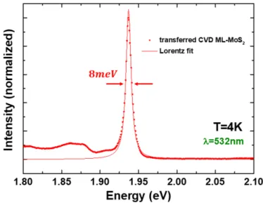
Photoluminescence (PL) spectroscopy has been em-ployed to evaluate the optical quality of the individual MoS2 MLs after the water-assisted transfer. The PL
ex-periments were carried out in a confocal microscope built in a vibration free, closed cycle cryostat from Attocube at T = 4K. The excitation/detection spot diameter of the 532 nm laser is below 1µm. The optical signal is dispersed in a spectrometer and detected with a Si-CCD camera. In Fig.6we present a typical PL spectrum col-lected from an hBN-encapsulated ML MoS2 next to the
manually assembled bilayers. The A-exciton emission has a linewidth of 8 meV at the full width at half maximum (FWHM), very close to the recently reported high optical quality encapsulated CVD ML MoS2 [26].
FIG. 5: Polar plot of the polarized SHG signal of CVD grown
3R MoS2 bilayer.
FIG. 6: PL spectrum of ML MoS2 transferred and
encapsu-lated in hBN following the water-assisted transfer method. A laser (532 nm) was used as an excitation source and the spectrum was collected at T = 4 K.
Clear observation of interlayer excitons for CVD grown samples is due to the improved optical quality as compared to earlier studies and due to performing the measurements at cryogenic temperatures. Temperature (i.e. thermal effects), contamination of sample-substrate interface, interference effects and intrinsic sample quality are factors that may have a detrimental contribution on the detection of interlayer excitons in differential reflectivity experiments. The spectral linewidth and the exciton oscillator strength are highly sensitive to the aforementioned factors. Encapsulation of as-grown and manually-assembled CVD bilayers combined with a careful selection of the top and bottom hBN thickness
FIG. 7: G0W0 band structures of 2H and 3R bilayers.
significantly reduces the inhomogeneous broadening and maximizes the oscillator strength of interlayer excitons. This results in high optical quality samples for a precise determination of the interlayer interaction in different stacking orders.
DFT-GW -BSE calculations
In the main text we only discuss the conduction and valence states around the K-point of the Brillouin zone, here we show in Fig. 7 the band structure calculations over the full Brillouin zone using Γ-M-K-Γ path of 2H and 3R bilayers at the G0W0level of theory. The degeneracy
leave that appears when going from the 2H to the 3R case is present almost over the entire path.
Acknowledgements
(*) I.P. and S.S. contributed equally to this work. Toulouse acknowledges funding from ANR 2D-vdW-Spin, ANR VallEx, ANR MagicValley, ITN 4PHOTON Marie Sklodowska Curie Grant Agreement No. 721394 and the Institut Universitaire de France. Growth of hexagonal boron nitride crystals was supported by the Elemental Strategy Initiative conducted by MEXT, Japan, and CREST (JPMJCR15F3), JST. I.C.G. thanks the CALMIP initiative for the generous allocation of computational times, through the project p0812, as well as the GENCI-CINES and GENCI-IDRIS for the grant A006096649. For FSU Jena this project received funding from the joint European Unions Horizon 2020 and DFG research and innovation programme FLAG-ERA under a grant TU149/9-1, DFG Collaborative Research Center SFB 1375 NOA Project B2 and the Th¨uringer MWWDG via FGR 0088 2D-Sens. N.L. and R.J.W. acknowledge funding from the PhD School Quantum Computing and Quantum Technology, SNF (Project No.200020156637)
and NCCR QSIT.
∗
Electronic address: igerber@insa-toulouse.fr
†
Electronic address: urbaszek@insa-toulouse.fr
[1] Novoselov, K. S., Mishchenko, A., Carvalho, A. &
Cas-tro Neto, A. H. 2d materials and van der waals
het-erostructures. Science 353 (2016).
[2] Mak, K. F. & Shan, J. Photonics and optoelectronics of 2d semiconductor transition metal dichalcogenides. Na-ture Photonics 10, 216–226 (2016).
[3] Schaibley, J. R. et al. Valley depolarization dynamics and valley hall effect of excitons in monolayer and bilayer
mos2. Nature Reviews Materials 1, 16055 (2016).
[4] Unuchek, D. et al. Room-temperature electrical control of exciton flux in a van der waals heterostructure. Nature 560, 340 (2018).
[5] Schneider, C., Glazov, M. M., Korn, T., Hfling, S. &
Urbaszek, B. Two-dimensional semiconductors in the
regime of strong light-matter coupling. Nature Comms. 3, 2695 (2018).
[6] Koperski, M. et al. Optical properties of atomically thin transition metal dichalcogenides: observations and puz-zles. Nanophotonics 6, 1289–1308 (2017).
[7] Dufferwiel, S. et al. Valley-addressable polaritons in
atomically thin semiconductors. Nature Photonics 11, 497 (2017).
[8] Scuri, G. et al. Large excitonic reflectivity of monolayer
mose2 encapsulated in hexagonal boron nitride. Phys.
Rev. Lett. 120, 037402 (2018).
[9] Hong, X. et al. Ultrafast charge transfer in atomically thin mos 2/ws 2 heterostructures. Nature nanotechnology 9, 682 (2014).
[10] Splendiani, A. et al. Emerging photoluminescence in
monolayer mos2. Nano Letters 10, 1271 (2010).
[11] Mak, K. F., Lee, C., Hone, J., Shan, J. & Heinz, T. F.
Atomically thin mos2: A new direct-gap semiconductor.
Phys. Rev. Lett. 105, 136805 (2010).
[12] Tonndorf, P. et al. Photoluminescence emission and ra-man response of monolayer mos 2, mose 2, and wse 2. Optics express 21, 4908–4916 (2013).
[13] Zhang, N. et al. Moire intralayer excitons in a
mose2/mos2 heterostructure. Nano letters 18, 7651–7657 (2018).
9 waals heterostructures. Nature 567, 71 (2019).
[15] Jin, C. et al. Observation of moir´e excitons in wse 2/ws
2 heterostructure superlattices. Nature 567, 76 (2019).
[16] Seyler, K. L. et al. Signatures of moir´e-trapped valley
excitons in mose 2/wse 2 heterobilayers. Nature 567, 66 (2019).
[17] Alexeev, E. M. et al. Resonantly hybridized excitons
in moir´e superlattices in van der waals heterostructures.
Nature 567, 81 (2019).
[18] Cao, Y. et al. Unconventional superconductivity in
magic-angle graphene superlattices. Nature 556, 43
(2018).
[19] Wang, L. et al. Magic continuum in twisted bilayer wse2 (2019). 1910.12147.
[20] Shimazaki, Y. et al. Moir superlattice in a
mose2/hbn/mose2 heterostructure: from coherent
cou-pling of inter- and intra-layer excitons to correlated mott-like states of electrons (2019). 1910.13322.
[21] Weston, A. et al. Atomic reconstruction in twisted
bilayers of transition metal dichalcogenides (2019). 1911.12664.
[22] Sung, J. et al. Broken mirror symmetry in excitonic
re-sponse of reconstructed domains in twisted mose2/mose2
bilayers (2020). 2001.01157.
[23] Slobodeniuk, A. et al. Fine structure of k-excitons in multilayers of transition metal dichalcogenides. 2D Ma-terials 6, 025026 (2019).
[24] Gerber, I. C. et al. Interlayer excitons in bilayer mos2
with strong oscillator strength up to room temperature. Physical Review B 99, 035443 (2019).
[25] Deilmann, T. & Thygesen, K. S. Interlayer excitons with large optical amplitudes in layered van der waals materi-als. Nano letters 18, 2984–2989 (2018).
[26] Shree, S. et al. High optical quality of mos2 monolayers grown by chemical vapor deposition. 2D Materials 7, 015011 (2019).
[27] Gong, Z. et al. Magnetoelectric effects and
valley-controlled spin quantum gates in transition metal dichalcogenide bilayers. Nature communications 4, 2053 (2013).
[28] Tong, Q. et al. Topological mosaics in moir´e superlattices
of van der Waals heterobilayers. Nature Physics 13, 356– 362 (2016).
[29] Taniguchi, T. & Watanabe, K. Synthesis of high-purity boron nitride single crystals under high pressure by using ba-bn solvent. Journal of Crystal Growth 303, 525 – 529 (2007).
[30] Grimme, S., Antony, J., Ehrlich, S. & Krieg, H. A con-sistent and accurate ab initioparametrization of density functional dispersion correction (DFT-D) for the 94 ele-ments H-Pu. J. Chem. Phys. 132, 154104–19 (2010). [31] Xia, M. et al. Spectroscopic signatures of aa’ and ab
stacking of chemical vapor deposited bilayer mos2. ACS nano 9, 12246–12254 (2015).
[32] Jia, H. et al. Large-scale arrays of single-and few-layer mos 2 nanomechanical resonators. Nanoscale 8, 10677– 10685 (2016).
[33] George, A. et al. Controlled growth of transition metal dichalcogenide monolayers using knudsen-type effusion cells for the precursors. Journal of Physics: Materials 2, 016001 (2019).
[34] Robert, C. et al. Optical spectroscopy of excited exciton
states in mos2 monolayers in van der waals
heterostruc-tures. Phys. Rev. Materials 2, 011001 (2018).
[35] Niehues, I., Blob, A., Stiehm, T., de Vasconcellos, S. M. & Bratschitsch, R. Interlayer excitons in bilayer mos 2 under uniaxial tensile strain. Nanoscale 11, 12788–12792 (2019).
[36] Shinde, S. M. et al. Stacking-controllable interlayer cou-pling and symmetric configuration of multilayered mos 2. NPG Asia Materials 10, e468 (2018).
[37] Huang, S. et al. Probing the interlayer coupling
of twisted bilayer mos2 using photoluminescence spec-troscopy. Nano letters 14, 5500–5508 (2014).
[38] Yeh, P.-C. et al. Direct measurement of the tunable elec-tronic structure of bilayer mos2 by interlayer twist. Nano letters 16, 953–959 (2016).
[39] van Der Zande, A. M. et al. Tailoring the electronic
structure in bilayer molybdenum disulfide via interlayer twist. Nano letters 14, 3869–3875 (2014).
[40] Kormanyos, A. et al. k.p theory for two-dimensional tran-sition metal dichalcogenide semiconductors. 2D Materi-als 2, 022001 (2015).
[41] Castellanos-Gomez, A. et al. Deterministic transfer of two-dimensional materials by all-dry viscoelastic stamp-ing. 2D Materials 1, 011002 (2014).
[42] Horng, J. et al. Observation of interlayer excitons in
mose2 single crystals. Phys. Rev. B 97, 241404 (2018).
[43] Wu, S. et al. Electrical tuning of valley magnetic mo-ment through symmetry control in bilayer mos 2. Nature Physics 9, 149 (2013).
[44] Jones, A. M. et al. Spin–layer locking effects in optical orientation of exciton spin in bilayer wse2. Nat. Phys 10, 130–134 (2014).
[45] Latzke, D. W. et al. Electronic structure, spin-orbit cou-pling, and interlayer interaction in bulk mos2 and ws2. Physical Review B 91, 235202 (2015).
[46] Du, L. et al. Temperature-driven evolution of critical
points, interlayer coupling, and layer polarization in
bi-layer Mos2. Physical Review B 97, 165410 (2018).
[47] Zhang, Y. et al. On valence-band splitting in layered mos2. ACS nano 9, 8514–8519 (2015).
[48] Jin, W. et al. Direct measurement of the
thickness-dependent electronic band structure of mos 2 using angle-resolved photoemission spectroscopy. Physical review let-ters 111, 106801 (2013).
[49] Kresse, G. & Hafner, J. Ab initio molecular dynamics for liquid metals. Phys. Rev. B 47, 558–561 (1993).
[50] Kresse, G. & Furthm¨uller, J. Efficient iterative schemes
for ab initio total-energy calculations using a plane-wave basis set. Phys. Rev. B 54, 11169–11186 (1996).
[51] Bl¨ochl, P. E. Projector augmented-wave method. Phys.
Rev. B 50, 17953 (1994).
[52] Kresse, G. & Joubert, D. From ultrasoft pseudopotentials to the projector augmented-wave method. Phys. Rev. B 59, 1758–1775 (1999).
[53] Heyd, J. & Scuseria, G. E. Assessment and validation of a screened Coulomb hybrid density functional. J. Chem. Phys. 120, 7274 (2004).
[54] Heyd, J., Peralta, J. E., Scuseria, G. E. & Martin, R. L. Energy band gaps and lattice parameters evaluated with the Heyd- Scuseria-Ernzerhof screened hybrid functional. J. Chem. Phys. 123, 174101 (2005).
[55] Paier, J. et al. Screened hybrid density functionals ap-plied to solids. J. Chem. Phys. 124, 154709 (2006). [56] Shishkin, M. & Kresse, G. Implementation and
perfor-mance of the frequency-dependent gw method within the paw framework. Phys. Rev. B 74, 035101 (2006).
[57] Mostofi, A. A. et al. wannier90: A tool for
obtain-ing maximally-localised Wannier functions. Computer
Physics Communications 178, 685–699 (2008).
[58] Hanke, W. & Sham, L. J. Many-Particle Effecs in the optical Excitations of a semiconductor. Phys. Rev. Lett. 43, 387 (1979).
[59] Rohlfing, M. & Louie, S. G. Electron-hole Excitations in Semiconductors and Insulators. Phys. Rev. Lett. 81, 2312–2315 (1998).
[60] Mennel, L., Paur, M. & Mueller, T. Second harmonic generation in strained transition metal dichalcogenide monolayers: Mos2, mose2, ws2, and wse2. APL Pho-tonics 4, 034404 (2019).
[61] Leisgang, N. et al. Optical second harmonic generation in encapsulated single-layer inse. AIP Advances 8, 105120 (2018).


