HAL Id: tel-03118553
https://tel.archives-ouvertes.fr/tel-03118553
Submitted on 22 Jan 2021HAL is a multi-disciplinary open access
archive for the deposit and dissemination of sci-entific research documents, whether they are pub-lished or not. The documents may come from teaching and research institutions in France or
L’archive ouverte pluridisciplinaire HAL, est destinée au dépôt et à la diffusion de documents scientifiques de niveau recherche, publiés ou non, émanant des établissements d’enseignement et de recherche français ou étrangers, des laboratoires
UDP-glucose pyrophosphorylase (UGP) : import dans
les glycosomes et implication dans la biosynthèse
glycosomale et cytosolique des sucres nucléotidiques
chez Trypanosoma brucei
Oriana Villafraz
To cite this version:
Oriana Villafraz. UDP-glucose pyrophosphorylase (UGP) : import dans les glycosomes et implication dans la biosynthèse glycosomale et cytosolique des sucres nucléotidiques chez Trypanosoma brucei. Microbiologie et Parasitologie. Université de Bordeaux, 2020. Français. �NNT : 2020BORD0215�. �tel-03118553�
Thèse présentée pour obtenir le grade de
DOCTEUR
DE L’UNIVERSITÉ DE BORDEAUX
École Doctorale Sciences de la Vie et de la Santé
Microbiologie - Immunologie
Par Oriana Villafraz
UDP-glucose pyrophosphorylase (UGP) : import
dans les glycosomes et implication dans la
biosynthèse glycosomale et cytosolique des sucres
nucléotidiques chez Trypanosoma brucei
Sous la direction de : Frédéric Bringaud
Membres du jury:
Président: Dr. Sébastien Mongrand Rapporteur: Pr. Wolfgang Schliebs Rapporteur: Pr. Mick Urbaniak
Résumé
Trypanosoma brucei, un protiste responsable de la Trypanosomose Humaine
Africaine, également connue sous le nom de la maladie du sommeil, est transmis par la mouche tsé-tsé (Glossina sp.). La découverte d'organites de type peroxysome spécialisés dans la glycolyse, appelés glycosomes, a soulevé un certain nombre de questions sur le rôle de cet organite dans la biologie des trypanosomes. Plusieurs voies métaboliques présentes dans le cytosol d'autres eucaryotes, comme la glycolyse et la biosynthèse des sucres nucléotidiques, sont compartimentées dans les glycosomes. Les raisons et les avantages de la présence des enzymes glycolytiques dans l'organite ont été largement discutés, mais la fonctionnalité et le rôle des voies de biosynthèse des sucres nucléotidiques glycosomales ne sont pas connus. Notre étude s'est focalisée sur l'UDP-glucose pyrophosphorylase (UGP), une enzyme impliquée dans la synthèse de l'UDP-glucose (UDP-Glc). Sur la base de la double localisation glycosomale et cytosolique de l'UGP mise en évidence ici à l'aide de plusieurs techniques de localisation subcellulaire, nous avons abordé deux questions en utilisant comme modèle les formes procycliques de T. brucei présentes dans l'insecte vecteur. La première est liée au mécanisme d'import de l'UGP dans les glycosomes, car cette protéine ne possède aucun signal d'adressage aux peroxysomes de type PTS1 ou PTS2. Nous avons montré que l'UGP est importée dans les glycosomes par "piggybacking" en s'associant à la phosphoénolpyruvate décarboxylase (PEPCK) possédant un signal d’adressage PTS1. Les interactions entre l'UGP et la PEPCK ont été montrées in situ et l'identification les régions impliquées dans ces interactions ont été identifiées. Nos résultats suggèrent que le complexe UGP-PEPCK est formé de manière transitoire lors de son import dans les glycosomes nouvellement produits et compétents pour l'import des protéines. La seconde question concerne le rôle de l'UGP dans les glycosomes. Nous avons montré que l'UGP est essentielle à la croissance des trypanosomes et que les voies métaboliques glycosomales et cytosoliques dont l'UGP fait partie sont fonctionnelles. En effet, des mutants viables contenant l'UGP exclusivement dans les glycosomes ou dans le cytosol sont viables et produisent des quantités similaires d'UDP-Glc. La raison d'être de la production glycosomale d'UDP-Glc par l'UGP reste inconnue, mais elle n'est probablement pas liée aux réactions de glycosylation, étant donné qu'aucune glycosyltransférase n'a été détectée dans l'organite.
Un autre aspect de ce travail concerne le rôle des intermédiaires du cycle de l'acide tricarboxylique (TCA) dans le métabolisme mitochondrial des formes procycliques. Dans le tractus digestif de son insecte vecteur, les trypanosomes dépendent de la proline pour alimenter leur métabolisme énergétique. Cependant, la disponibilité d'éventuelles autres sources de carbone pouvant être utilisées par le parasite est actuellement inconnue. Nous avons montré que les intermédiaires du cycle TCA, i.e. succinate, malate et α-cétoglutarate, stimulent la croissance des formes procycliques incubées dans un milieu contenant 2 mM de proline, concentration se situant dans la gamme des quantités mesurées dans l'intestin de la mouche. De plus, le développement de nouvelles approches ont permis d'étudier une branche peu explorée du cycle TCA convertissant le malate en α-cétoglutarate, précédemment décrite comme peu ou pas utilisée par le parasite, quelles que soient les quantités de glucose disponibles. L'activité de cette branche suggère qu'un cycle TCA complet peut être mis en œuvre dans les formes procycliques et probablement dans les autres formes parasitaires de l'insecte. Nos données élargissent le potentiel métabolique des trypanosomes et ouvrent la voie vers une meilleure compréhension du métabolisme de ce parasite dans divers organes de la mouche tsé-tsé, où il évolue.
Mot clés : Trypanosoma brucei; formes procycliques; glycosome; "piggybacking";
Abstract
Trypanosoma brucei, a protist responsible for human African trypanosomiasis, also
known as sleeping sickness, is transmitted by the tsetse fly (Glossina sp.). The discovery of peroxisome-related organelles specialised in glycolysis called glycosomes, has raised a number of questions about the role of this organelle in the biology of trypanosomes. Several metabolic pathways present in the cytosol of eukaryotes, like glycolysis and sugar nucleotides biosynthesis, are compartmentalised within glycosomes. While the reasons and advantages of having glycolytic enzymes compartmentalised in the organelle have been extensively discussed, little is proposed for sugar nucleotides biosynthetic pathways. This study is focused on the glucose pyrophosphorylase (UGP), an enzyme involved in the synthesis of UDP-glucose (UDP-Glc). Based on the UGP's dual glycosomal and cytosolic localisation evidenced here by using several subcellular localisation techniques, we addressed two questions using as a model the procyclic form of T. brucei present in the insect vector. The first question is related to the mechanism of UGP import into glycosomes, since this protein lacks any known peroxisomal targeting signal (PTS1 and PTS2). We demonstrated that UGP is imported into the organelle by piggybacking on the glycosomal PTS1-containing phosphoenolpyruvate carboxykinase (PEPCK). Interactions between UGP and PEPCK were shown in situ and the interacting regions were identified. Our data suggest that the complex UGP-PEPCK is formed transiently to facilitate the import of UGP and that it is detected in newly formed import-competent glycosomes. The second question concerns the role of UGP in glycosomes. We demonstrated that UGP is essential for the growth of trypanosomes and that mutants containing UGP exclusively in the glycosomes or in the cytosol still produce UDP-Glc at similar levels and are viable, which implies that the glycosomal and cytosolic metabolic pathways involving UGP are functional. The glycosomal function of UDP-Glc is currently unknown and probably not related to glycosylation reactions since no glycosyltransferases have been detected in the organelle.
Another aspect of this work concerns the role of tricarboxylic acid (TCA) cycle intermediates in the mitochondrial metabolism of the procyclic trypanosomes. In the midgut of its insect vector, trypanosomes rely on proline to feed their energy metabolism. However, the availability of other potential carbon sources that can be used by the parasite is currently unknown.
We showed that TCA cycle intermediates, i.e. succinate, malate and -ketoglutarate, stimulate growth of procyclic trypanosomes incubated in medium containing 2 mM proline, which is in the range of the amounts measured in the midgut of the fly. In addition, we have implemented new approaches to study cell growth and metabolic pathways in order to investigate mitochondrial metabolism. These new tools have allowed us to study a poorly explored branch of the TCA cycle converting malate to -ketoglutarate, which was previously described as non-functional or little used in the parasite, regardless of the glucose levels available. The discovery of this branch reveals that a full TCA cycle can operate in procyclic trypanosomes and probably in the other trypanosome forms present in the fly. Our data broaden the metabolic potential of trypanosomes and pave the way for a better understanding of the parasite's metabolism in various organ systems of the tsetse fly, where it evolves.
Keywords: Trypanosoma brucei; procyclic forms; glycosome; piggybacking;
UDP-glucose; mitochondria; TCA cycle; glucose and proline metabolism
Laboratoire de Microbiologie Fondamentale et Pathogenicité - UMR5234
Acknowledgements
I would like to express my sincere gratitude to Dr. Frédéric Bringaud for letting me be part of this incredible Laboratory, for his kind supervision, consistent support and guidance during my PhD. It has been a great honour to work with you and to learn from you.
I am also grateful to the LabEx ParaFrap PhD Program for its financial support and for organising remarkable workshops that let me grow both professionally and personally. A special thanks to the members of my thesis committee Dr. Sébastien Mongrand, Pr. Wolfgang Schliebs, Pr. Mick Urbaniak and Pr. Paul Michels for reviewing my thesis and participating in my defence.
I acknowledge all members of the iMET Team for their constant support and encouragement. I feel I have all the more reason to extend my thanks to Erika, Sarah and Simone, for their invaluable advice, for being always there and making me feel home. A very special thanks to Nico, Corinne and Magali for their contribution to this thesis project, for being always kind and willing to help me. To Stefan for training me at the very beginning of this journey. Thanks also to Manu, Loîc, Chloé, Magamba, Yoann and Pauline for their support and helpful discussions during the Lab meetings. To Rodolpho for giving me a helping hand during his stay in Bordeaux. Thank you to all members of the MFP, UMR 5234 for their assistance and support, specially to Dr. Mélanie Bonhivers, Nicolas Landrein and Doranda who have usefully contributed to my work.
I extend my gratitude to all the people who collaborated on this project. To Marc Biran, for his invaluable contribution with analyses of NMR spectra, to Jean-William Dupuy, Hanna Kulyk, Dr. Edern Cahoreau, Dr. Jean-Charles Portais, Dr. Michael Boshart, Dr. Ariel Silber, Dr. Alena Zíková, Dr. Michael Barrett, Dr. Daniel Inaoka and the members of the GlycoNov network for their constructive discussions.
Finally, many thanks to my family and all my friends for their continuous encouragement and love.
CONTENTS Résumé ... ii Abstract ... iv Acknowledgements ... vi 1. INTRODUCTION ... 2 1.1 Trypanosomes ... 2
1.2. Human African Trypanosomiasis (HAT) ... 6
1.2.1. Diagnosis, Prevention and Treatment ... 6
1.2.2. Challenges for HAT elimination ... 8
a) Drug resistance ... 8
b) Human and animal reservoirs ... 10
1.3. Animal African Trypanosomiasis (AAT)... 12
1.4. Trypanosoma brucei: a remarkable biological model ... 14
1.4.1. Life cycle and transmission ... 14
1.4.2. Cell architecture ... 18
1.4.4. Gene expression and regulation ... 18
1.4.5. Cell surface ... 22
a) VSG ... 22
b) Procyclins ... 24
c) Other surface glycoproteins ... 26
d) GPI anchors ... 26
1.5. Intermediate and energy metabolism ... 28
1.5.1. Bloodstream vs procyclic, an overview ... 30
1.5.2. Energy metabolism of PCF ... 31
a) Glucose metabolism ... 31
b) Glycerol metabolism ... 39
c) Proline metabolism and TCA cycle ... 39
d) Metabolism of other amino acids ... 44
e) Respiratory chain and mitochondrial ATP production... 44
1.5.3. Energy metabolism of BSF ... 46
a) Glucose metabolism ... 50
b) Mitochondrial metabolism and ATP production ... 51
1.6. Glycosomes ... 57
1.6.1. Morphology and composition ... 57
1.6.1. Origin and evolution ... 59
2. RESULTS PART I: Glycosomal import of UDP-glucose pyrophosphorylase and its functionality in glycosomal and cytosolic UDP-glucose producing
pathways ... 83
2.1. Abstract... 86
2.2. Introduction ... 87
2.3. Results ... 91
a) UDP-glucose pyrophosphorylase (UGP) has a dual glycosomal and cytosolic localisation ... 91
b) PEPCK-dependent import of UGP into glycosomes ... 93
c) Production and analysis of UGP-MYC and TY-PEPCK tagged cell lines ... 95
d) UGP interacts with PEPCK in some glycosomes ... 99
e) Determination of critical parts for PEPCK-UGP interaction ... 101
f) The UGP protein is essential for T. brucei ... 107
g) Targeting a recombinant UGP exclusively to the glycosomes ... 109
h) Expression of the glycosomal rUGP-GPDH rescues the lethality of the RNAiUGP mutant ... 111
i) The Δugp/EXPrUGP-GPDH cell line is viable ... 113
j) The glycosomal and cytosol UGP-containing pathways are functional... 115
2.4. Supplementary results ... 121
2.5. Discussion ... 123
3. RESULTS PART II. Analysis of TCA intermediates and metabolites excreted from glucose metabolism as alternative carbon sources ... 130
3.1. Abstract... 132
3.2. Introduction ... 133
3.3. Results ... 139
a) Procyclic trypanosomes can re-metabolise end products excreted from glucose degradation ... 139
b) Succinate, pyruvate and alanine are metabolised in the presence of glucose or proline ... 141
c) TCA cycle intermediates stimulate growth of the PCF in in vivo-like conditions ... 145
d) The TCA cycle is used to metabolise malate in procyclic trypanosomes . 149 e) Metabolism of -ketoglutarate in the presence of proline ... 151
f) -Ketoglutarate rescued the growth defect of the RNAiPRODH.i and RNAiAAT.i mutants ... 155
g) -Ketoglutarate is toxic if not metabolised at a high rate ... 157
3.4. Supplementary results ... 163
3.5. Discussion ... 166
4. GENERAL DISCUSSION AND PERSPECTIVES ... 171
4.1. Overview Part I: UDP-glucose pyrophosphorylase (UGP) ... 171
4.4. The UGP-PEPCK interaction, a new way to study import of glycosomal
proteins ... 175
4.5. Is piggybacking a rule or an exception? ... 176
4.6. Overview Part II: Alternative carbon sources for PCF ... 177
4.7. PCF trypanosomes metabolise carbon sources other than glucose and proline ... 177
4.8. Unravelling new mitochondrial capabilities ... 178
4.9. Role of metabolites in trypanosomes differentiation ... 178
5. MATERIALS AND METHODS ... 180
5.1. Materials ... 180
5.1.1. Trypanosomes ... 180
5.1.2. Plasmids and bacteria ... 180
5.2. Methods ... 182
5.2.1. Molecular biology ... 182
a) Cloning and sequencing ... 182
b) DNA purification ... 184
- Plasmid DNA ... 184
- Genomic DNA ... 184
5.2.2. Cell biology ... 184
a) Growth of T. brucei: curves and alamar blue assays ... 184
b) Transfection ... 186
c) Inhibition of gene expression by RNA interference (RNAi) ... 188
d) Production of null mutants ... 190
e) Expression of tagged proteins ... 190
- Endogenous tagging ... 190
- Over-expression ... 192
- Glycosomal recombinant UGP proteins ... 194
5.2.3. Biochemistry ... 194
a) Differential centrifugation ... 194
b) Digitonin permeabilization ... 196
c) Cell fractionation by hypotonic lysis ... 196
e) Blue-native PAGE (BN-PAGE) ... 196
f) SDS-PAGE and Western Blots ... 198
g) Enzymatic activity assays ... 198
5.2.4. Imaging ... 200
a) Immunofluorescence Microscopy ... 200
LIST OF FIGURES
Figure 1.1. Classification of Trypanosoma species. ... 1
Figure 1.2. Morphology and phylogeny of kinetoplastids, diplonemids, and euglenids. ... 3
Figure 1.3. Trypanosomes of mammals. ... 5
Figure 1.4. Progress in the elimination of HAT. ... 7
Figure 1.5. Morphological characteristics of T. brucei, T. congolense and T. vivax bloodstream form. ... 11
Figure 1.6. Life cycle of T. brucei. ... 13
Figure 1.7. Life cycle stages of T. b. brucei within the tsetse fly. ... 15
Figure 1.8. Morphology of Trypanosoma brucei. ... 17
Figure 1.9. Gene expression mechanisms in kinetoplastids. ... 19
Figure 1.10. The dynamic Variant Surface Glycoprotein (VSG) coat of T. brucei bloodstream forms... 21
Figure 1.11. Surface glycocalyx of T. brucei procyclic cells. ... 23
Figure 1.12. Developmental expression of major surface glycoproteins. ... 25
Figure 1.13. The GPI biosynthetic pathway of T. brucei. ... 27
Figure 1.14. ATP production in PCF and BSF trypanosomes. ... 29
Figure 1.15. Intermediate metabolism of PCF trypanosomes in glucose rich conditions. ... 33
Figure 1.16. [U-13C] enrichment of key glycolytic intermediates from proline, glycerol or glucose. ... 35
Figure 1.17. Proline metabolism of PCF trypanosomes in glucose-depleted conditions. ... 38
Figure 1.18. Respiratory chain of T. brucei. ... 43
Figure 1.19. FoF1-ATP synthase/ATPase complex in T. brucei mitochondria... 45
Figure 1.20. Glucose and glycerol aerobic metabolism of BSF trypanosomes. ... 48
Figure 1.21. Mitochondrial morphology and proteome of PCF and BSF ... 49
Figure 1.22. Glycosomal metabolic pathways – Part 1... 53
Figure 1.23. Structure and distribution of glycosomes... 56
Figure 1.24. A proposed metabolic model and peroxisome remodeling during evolution in Euglenozoa. ... 58
Figure 1.25. Glycosome biogenesis in Trypanosoma brucei. ... 62
Figure 1.26. Mechanisms for peroxisome formation in mammalian cells. ... 64
Figure 1.27. Proliferation of glycosomes. ... 66
Figure 1.28. Piggyback import model of peroxisomal matrix proteins. ... 68
Figure 1.29. Biosynthesis of UDP-Glc, UDP-Gal, UDP-GlcNAc, Man and GDP-Fuc in T. brucei. ... 74
Figure 1.30. 3D structure of TbUGP in complex with UDP-Glc. ... 78
Figure 1.31. Sequence alignments of UGP orthologues. ... 80
Figure 2.34. Production of UGP-MYC and TY-PEPCK tagged cell lines. ... 96
Figure 2.35. UGP interacts transiently with PEPCK. ... 98
Figure 2.36. Analysis of UGP oligomerization in native gel. ... 100
Figure 2.37. The N-terminal 123 residues of UGP are required for import into the glycosomes. ... 102
Figure 2.38. PCR analysis to confirm endo-tagging at the UGP locus. ... 103
Figure 2.39. A 34-residues peptide of PEPCK is required for glycosomal import of UGP. ... 104
Figure 2.40. Production and functional analyses of RNAiUGP cell lines. ... 106
Figure 2.41. Functional analysis of RNAiUGP cell lines in the presence or the absence of glucose. ... 108
Figure 2.42. Subcellular localisation of UGP in the presence or the absence of glucose. ... 108
Figure 2.43. Expression of a glycosomal recombinant UGP. ... 110
Figure 2.44. Production and functional analyses of Δugp cell lines. ... 112
Figure 2.45. Analysis of cell lines expressing UGP in different compartments. ... 114
Figure 2.46. PCF produces UDP-Glc in glycosomes and cytosol. ... 116
Figure 2.47. IC-HRMS analyses of intracellular metabolites ... 119
Figure 2.48. Test of anti-UGP antiserum. ... 120
Figure 2.49. Sequence alignment of L. major, T. brucei and T. cruzi PEPCK protein sequences. ... 120
Figure 2.50. Sequence alignments of UGP orthologues ... 122
Figure 3.51. Proline metabolism of the PCF trypanosomes in the presence of other carbon sources. ... 137
Figure 3.52. Kineticanalyses of end products excretion from [U-13C]-glucose and proline metabolism. ... 138
Figure 3.53. Proton (1H) NMR analyses of end products excreted from the metabolism of 13C-enriched succinate, alanine, pyruvate and acetate. ... 140
Figure 3.54. 1H-NMR analyses of end products excreted from the metabolism of 13 C-enriched succinate. ... 142
Figure 3.55. Alamar Blue assays with a series of metabolites. ... 144
Figure 3.56. Succinate, malate and -ketoglutarate stimulate growth of the PCF.. 146
Figure 3.57. 1H-NMR analyses of end products excreted from the metabolism of malate. ... 148
Figure 3.58. 1H-NMR analyses of end products excreted from the metabolism of -ketoglutarate. ... 150
Figure 3.65. Malate is toxic for the ∆aco cell line. ... 164
Figure 5.66. Sequence alignment of the recoded UGP sequence (rUGP) and native UGP. ... 181
Figure 5.67. UGP knock-down by RNAi in T. brucei. ... 187
Figure 5.68. Production of glycosomal recombinant rUGP proteins. ... 191
Figure 5.69. Principal techniques used for subcellular localisation of proteins. ... 193
Figure 5.70. UGP activity assay. ... 197
LIST OF TABLES
Table 1.1. Summary of treatment choices for patients with T. gambiense HAT. ... 9
Table 1.2. Effect of proline metabolism and TCA cycle enzymes disruption on PCF growth. ... 40
Table 1.3. Function of peroxins identified in mammals, yeast and T. brucei. ... 60
Table 1.4. Peroxisomal matrix proteins imported as oligomers. ... 70
Table 1.5. Characterised glycosyltransferases from T. brucei ... 72
Table 1.6. Enzymes of sugar nucleotide biosynthetic pathways. ... 76
Table 2.7. Protein expression levels by label-free mass spectrometry proteomic analysis of glycosomal enriched fractions. ... 92
Table 3.8. Excreted end products from the metabolism of carbon sources in the PCF trypanosomes. ... 161
Table 5.9. PCR primers sequences and features concerning PEPCK mutant cell lines. ... 183
Table 5.10. PCR primers sequences and features concerning UGP mutant cell lines. ... 185
Table 5.11. Mutant cell lines disrupted in expression of enzymes of proline degradation, TCA cycle and acetate production pathways used in this work. ... 189
Table 5.12. Primary and secondary antibodies used for western blot and immunofluorescence analysis. ... 195
Figure 1.1. Classification of Trypanosoma species. Euglenozoa Symbiontida Euglenida Diplonemea Kinetoplastea Prokinetoplastina Metakinetoplastina Eubodonida Parabodonida Neobodonida Trypanosomatida
Phylum Class Subclass Order Family Subfamily Genus Subgenus Species
Trypanosomatidae Leishmaniinae Leishmania Phytomonadinae Strigomonadinae Blechomonadinae Paratrypanosomatinae Trypanosomatinae Trypanosoma Herpetosoma Megatrypanum Schizotrypanum Tejeraia Dunotella Nannomonas Pycnomonas Trypanozoon T. equiperdum T. evansi T. brucei T. vivax T. congolense T. cruzi
1. INTRODUCTION
In this chapter, I describe the biological model used in this thesis: the parasite
Trypanosoma brucei. Since this parasite causes the sleeping sickness in Africa, a brief
description of the diagnosis, prevention and treatment, with special focus on the challenges for the human African trypanosomiasis (HAT) elimination will be done. I explain in detail cellular and metabolic features of particular interest with focus on glycolytic and mitochondrial metabolism. A special section is dedicated to the very important organelle called glycosome, which is key for the development of this thesis. At the end, I present a comparative analysis of UDP-glucose pyrophosphorylases, the enzyme this work is focused on.
1.1 Trypanosomes
Trypanosomatids are uniflagellate parasites that belong to the class Kinetoplastea, defined by the presence of mitochondrial DNA organised in a unique and extensively studied structure called kinetoplast (k) which is composed of DNA circles (Shapiro and Englund, 1995; Cayla et al., 2019). Among kinetoplastids, there are free-living and symbiotic organisms in addition to parasitic protists including the well-studied pathogens from Trypanosoma and Leishmania genera. The most recent classification of the class Kinetoplastea (Figure 1.1) divides it into two groups. The first one is the subclass Prokinetoplastina, which includes an ectoparasite infecting the skin and gills of fishes called Ichthyobodo (Robertson, 1985) and the non-photosynthetic symbiont
Perkinsela that resides inside amoebozoans of the genus Paramoeba (Tanifuji et al.,
2017). The second subclass of kinetoplastids, Metakinetoplastina, includes three orders of predominantly free-living bodonids (Eubodonida, Parabodonida, and Neobodonida) and the parasitic Trypanosomatida (Moreira, 2004; Simpson et al., 2006; Deschamps et al., 2011; Butenko et al., 2020).
Remarkably, these organisms are considered to have large amounts of organellar DNA (kDNA) with a high structure variability (Figure 1.2). While trypanosomatids kDNA mini and maxicircles are densely packaged, in bodonids and outer groups the
Figure 1.2. Morphology and phylogeny of kinetoplastids, diplonemids, and euglenids.
In the common ancestor shared by diplonemids and kinetoplastids, the organellar DNA content might have increased dramatically soon after the divergence from euglena. Then, Trypanosoma may have either undergone massive reduction of its kDNA or has not expanded it to the levels seen in the sister lineages. Cell morphology is shown on the left and a schematic phylogenetic tree is shown on the right. Brown: parasitic or endosymbiotic species, blue: free-living species (From Lukeš et al., 2018).
It has been recently proposed that the large amounts of kDNA evolved through neutral processes or constructive neutral evolution (CNE), suggesting that it does not represent any adaptive benefit for these organisms (Lukeš et al., 2018). This hypothesis is supported by recent studies performed with the outer group of free-living diplonemids. The authors showed that this abundant and diverse group of organisms has very complex mitochondrial genome architecture, structure and post-transcriptional processes despite the absence of a parasitic lifestyle, which is commonly considered full of peculiarities (Kaur et al., 2020).
Many factors must have participated in the development of parasitism within trypanosomatids. A phylogenomic study recently explored the role of gene gain and gene loss in the development of trypanosomatid genomes. A reduction in complexity of numerous catabolic pathways, macromolecular degradation and ion transport was evidenced, in addition to the gain of gene families involved in host invasion. This work clearly points out the importance of interaction between the protist and the host immune system as a selective pressure that led to the specialization of the parasites cell surface (Jackson et al., 2016).
The trypanosomatid order contains a single family: Trypanosomatidae, which comprises parasites of invertebrates, vertebrates and plants (Maslov et al., 2019). Several species from the genus Trypanosoma are of particular medical and veterinary interest (Figure 1.3). This genus is divided in two main groups: stercoraria and salivaria, based on the mode of transmission by their insect vector (Hoare, 1966; Gibson, 2016). The Stercoraria group, represented by the etiological agent of Chagas disease T. cruzi, is characterised by transmission through the excretion of faeces (Kaufer et al., 2017). On the other hand, Salivaria group parasites are transmitted by the bite of an infected insect and is represented by T. vivax, T. congolense and T.
brucei. The latter is by far the most well studied Trypanosoma and the model used in
this work. The three T. brucei subspecies are transmitted through the bite of an infected tsetse fly. T. b. gambiense and T. b. rhodesiense infect humans and are the causative agents of Human African Trypanosomiases (HAT), while T. b. brucei infects
Figure 1.3. Trypanosomes of mammals.
Hosts, transmission and relation to mammal diseases (adapted from (Gibson, 2016)). Two main groups, stercoraria and salivaria, have been stablished based on the mode of transmission by their insect vector(Hoare, 1966).
1.2. Human African Trypanosomiasis (HAT)
HAT, also known as sleeping sickness, is a neglected tropical disease that occurs in sub-Saharan Africa and can be fatal if left untreated. There are two forms of the disease, depending on the T. brucei subspecies causing the infection. The first one,
T. brucei gambiense, is the most important one as it is responsible for 98% of cases
in western and central Africa. Gambiense HAT progresses slowly causing a chronic infection that can be asymptomatic for years. Secondly, T. brucei rhodesiense is responsible for the remaining 2% of cases in eastern and southern Africa. Rhodesiense HAT progresses faster causing an acute infection that can affect the central nervous system within months.
The disease has two stages with a different set of symptoms. In the first or early stage, trypanosomes remain in the blood and lymph system and the patients present mild symptoms like fever, headaches, joint pains and itching. In the second or late stage, parasites are detected in the cerebrospinal fluid and patients can develop disturbed sleep pattern, confusion, sensory disturbances, extreme lethargy, poor condition and, in some severe cases, progression to coma. (Steverding, 2008; Büscher et al., 2017; World Health Organization, 2019). Over the past years, the number of HAT cases has been reduced significantly. The last WHO report in 2019 states that there were less than 1000 cases reported in 2018, targeting elimination of HAT as a public health problem possible by this year (Figure 1.4).
1.2.1. Diagnosis, Prevention and Treatment
There are several approaches to diagnose HAT and the choice depends on the country and available facilities. A serological test is performed first to detect trypanosome-specific antibodies followed by confirmation of parasites present in blood. Finally, the stage of the disease is determined by a lumbar puncture.
Recently introduced rapid tests (Büscher et al., 2013, 2014) and classical card agglutination test for trypanosomiasis (CATT) developed in the late 1970s are the serological tests performed in the field. A new rapid test has been developed with
Figure 1.4. Progress in the elimination of HAT.
The total number of reported cases of HAT (gambiense and rhodesiense) per year is shown. Less than 1000 cases were reported in 2018, most of which caused by infection with T. b. gambiense. HAT was targeted for elimination as a public health problem by this year. A further target is to fully eliminate the occurrence of gambiense HAT (i.e. zero cases) within the next ten years. The green line and the green bar show the milestones and target set in the WHO Roadmap for HAT elimination (From Franco et al., 2020).
Combining two or more screening tests seems to be required in order to achieve elimination of HAT since the specificity and sensitivity of individual tests might not be high enough to improve the detection of cases now that the disease prevalence is decreasing (Jamonneau et al., 2015; Lumbala et al., 2018).
In addition to case detection and treatment, the inclusion of vector control is also crucial to achieve HAT elimination. An affordable cost-effective tiny target technology was recently developed consisting of visual devices (blue colour) made with textiles impregnated with the insecticide deltamethrin (Tirados et al., 2015). These ‘Tiny Targets’ have been successfully used in different regions and represent an effective control strategy of tsetse populations (Courtin et al., 2015; Tirados et al., 2015; Mahamat et al., 2017).
Currently there are six drugs available for the treatment of HAT: pentamidine, suramin, melarsoprol, eflornithine, nifurtimox and fexinidazole. Pentamidine and suramin were the first-line drugs against stage 1 gambiense HAT and rhodesiense HAT, respectively. For the second stage of the disease, a nifurtimox–eflornithine combination therapy (NECT) is used for gambiense HAT, while melarsoprol is restricted to rhodesiense HAT. Recently, the guidelines on therapeutic choices were updated after the approval in 2018 of the new drug fexinidazole (Torreele et al., 2010; Mesu et al., 2018) (Table 1.1). The big advantages of Fexinidazole is that it is orally administered, in contrast to previous treatments, therefore it does not require intravenous or intramuscular infusions which reduces treatment costs and, importantly, is effective against both early and late stages of HAT (World Health Organization, 2019).
1.2.2. Challenges for HAT elimination
Despite the significant reduction of HAT cases, the disease still represents a public health problem in some African countries, especially in the Democratic Republic of the Congo. The factors considered as potential limitations towards elimination of HAT include:
Age, body weight Clinical examination Cerebrospinal fluid findings Treatment
1st choice 2nd choice* Relapse**
< 6 years or <20 kg
≤ 5 WBC/µl, no
trypanosomes Pentamidine - NECT > 5 WBC/µl or
trypanosomes NECT Eflornithine NECT-long
≥ 6 years and ≥ 20 kg No suspicion of severe HAT Lumbar puncture not needed Fexinidazole Pentamidine (First stage) NECT NECT (Second stage) Suspicion of severe HAT < 100 WBC/µl Fexinidazole Pentamidine (First stage) NECT NECT (Second stage) ≥ 100 WBC/µl or failed lumbar puncture NECT Fexinidazole NECT-long or melarsoprol
Table 1.1. Summary of treatment choices for patients with T. gambiense HAT.
*2nd choice treatment corresponds to the alternative treatment recommended in cases where the 1st choice treatment is not available or is not appropriate. **Relapse treatment is given in cases of treatment failure.
WBC: White Blood Cells
NECT: nifurtimox–eflornithine combination therapy
NECT-long: nifurtimox (15mg/kg per day) in 3 doses for 10 days, eflornithine (400mg/kg per day) in 2 infusions for 14 days.
fexinidazole has already been shown in vivo and in vitro (Sokolova et al., 2010). Fexinidazole is cross-resistant with nifurtimox which raises questions about a possible selection of resistance to this new drug. However, the progressive reduction of cases could reduce the probability of selecting resistant parasites (Dickie et al., 2020). The search for new treatments is still going on. For instance, clinical trials with an additional oral treatment consisting of only one dose of the drug acoziborole seem promising (Jacobs et al., 2011; Dickie et al., 2020). Furthermore, adenosine analogues showing trypanocidal activity were recently identified. One of these compounds showed to be highly active in both stages of HAT and to not have cross resistance with other drugs, representing a suitable candidate for treatment (Hulpia et al., 2019).
b) Human and animal reservoirs
The importance of asymptomatic infections in serologically positive patients where no trypanosomes are detected in the blood has been recently addressed (Capewell et al., 2019). The authors propose that trypanosomes found in the skin (Caljon et al., 2016; Capewell et al., 2019) and adipose tissue (Trindade et al., 2016) may constitute a crucial reservoir that contributes to transmission. Indeed, a very recent study proved that HAT confirmed and unconfirmed seropositive subjects carry extravascular trypanosomes in their skin, supporting the role of human skin as a reservoir for trypanosomes (Camara et al., 2020). The aparasitaemic cases do not receive any treatment, which might hamper the possibility of eliminating HAT, therefore, a revision of the WHO recommendations was strongly suggested in the light of this new data. Similar to asymptomatic human infections, the role of animal reservoirs requires further attention. Clearly there is a need to develop more sensitive and specific detection techniques to cope with discordant results produced as a consequence of the low parasitaemia generally observed in T. b. gambiense infections. T. b.
gambiense can infect a variety of domestic animals and wildlife, including cattle and
pigs recently identified as potential reservoirs (N’Djetchi et al., 2017). Importantly, a number of animals have been successfully infected with patient-derived T. b.
Figure 1.5. Morphological characteristics of T. brucei, T. congolense and T.
vivax bloodstream form.
T. brucei group trypanosomes (T. b. brucei, T. b. evansi, T. b. equiperdum)
characteristics are shown (From Giordani et al., 2016).
Lenght 20-30 µm 9-22 µm 18-26 µm
Posterior end Pointed Rounded Rounded
Nucleus (N) Central Central Central
Undulating membrane (UM) Conspicuous Modest Modest
Flagellum (F) Free at anterior end No free Free at anterior end
Kinetoplast (K) Small Subterminal, medium
size
Terminal, large
T. brucei T. congolense T. vivax
K N F UM UM K N F K N F UM
1.3. Animal African Trypanosomiasis (AAT)
AAT also known as nagana is caused by T. congolense, T. vivax and, to a lesser extent, T. brucei spp. The morphological differences between these parasites are shown in Figure 1.5. In addition to tsetse fly, other insects can mechanically transmit
T. vivax parasites, like tabanid and stable (Stomoxys sp) flies, resulting in a wider
geographical distribution (Wells, 1972; Jones and Dávila, 2001). The disease is widespread in sub-Saharan Africa and affects valuable domestic livestock including bovines, ovines, caprines, equids, camelids and suids, causing a huge economic and social impact in terms of economic loss. In the absence of treatment, infection with one or more Trypanosoma species can lead to acute or chronic disease resulting in weaken animals (Steverding, 2008; Giordani et al., 2016). The compounds diminazene aceturate (DA) and isometamidium chloride (IC) are the most used veterinary trypanocides against AAT. Since the 1960s, resistance to these chemicals has been reported in 17 African countries (Delespaux et al., 2008). Despite the increase of resistance, the treatments are still effective in controlling the parasite and the corresponding disease at an acceptable level (Chitanga et al., 2011).
Interestingly, T. b. brucei subspecies as well as T. congolense and T. vivax are only infectious to animals. The lack of infectivity in humans is due to the trypanolytic activity of a human-specific serum apolipoprotein called Apolipoprotein L-I (APOL-I) which is bound to high-density lipoprotein particles known as trypanolytic factors (TLFs). These particles enter the cell through endocytosis at the flagellar pocket progressing towards the lysosomes. The acidic lysosomal pH induces conformational changes of APOL-I which binds to the lysosomal membrane creating pores that lead to lysis and death of the parasites (Hager et al., 1994; Pays et al., 2006).
In contrast, T. b. rhodesiense and T. b. gambiense have different ways to escape this trypanolytic activity. T. b. rhodesiense expresses a serum-resistance associated protein (SRA) which interacts with APOL-I preventing its insertion in the lysosomal membrane (Van Xong et al., 1998; Stephens et al., 2012). T. b. gambiense expresses a specific glycoprotein (TgsGP) whose incorporation into the lysosomal membranes
Figure 1.6. Life cycle of T. brucei.
The five major stages are shown. Circular arrows represent the proliferative capacity of procyclics and epimastigotes in the tsetse fly, as well as slender bloodstream forms in the mammal (From Wheeler et al., 2019).
1.4. Trypanosoma brucei: a remarkable biological model
Several aspects of T. brucei brucei biology make it an ideal model for studying trypanosome biology. The genome sequence was published in 2005 (Berriman et al., 2005) and is freely available on TriTrypDB (http://tritrypdb.org), a database providing access to genome-scale datasets for kinetoplastid parasites (Aslett et al., 2010).
T. b. brucei is not pathogenic for humans and both insect and mammalian stages are
relatively easy to culture in vitro (Brun and Schönenberger, 1979; Hirumi and Hirumi, 1989). It is also possible to generate different life cycle stages present in the mammals or in the fly using in vitro systems (Kolev et al., 2012; Qiu et al., 2018).
A variety of advanced genetic tools have been developed to manipulate T. brucei, which can be efficiently transfected by electroporation-based methods. These tools include inducible systems for the regulated ectopic expression of genes, for RNAi mediated knock-down and for the expression of proteins such as Cas9 to use CRISPR-Cas9 technology (Wirtz and Clayton, 1995; Biebinger et al., 1997; Ngô et al., 1998; Bringaud et al., 2000; Beneke et al., 2017; Rico et al., 2018). Up to five drug-resistance markers are available, therefore it is possible to generate mutant cell lines targeting or expressing multiple genes (ten Asbroek et al., 1990; Poon et al., 2012). TrypTag (http:// tryptag.org) is another resource available which contains subcellular localisations of over 2000 endogenously-tagged proteins and aims to localise every protein encoded in the genome (Dean et al., 2017). All these characteristics make T.
brucei an ideal model in fundamental cell biology and applied research looking for drug
targets and new potential treatments for HAT.
1.4.1. Life cycle and transmission
T. brucei is transmitted to mammals through the bite of a tsetse fly and has a complex
developmental cycle life (Figure 1.6). The mammalian host gets infected when a tsetse fly bite delivers metacyclic trypomastigotes to the bloodstream. Then, growth-arrested metacyclics differentiate into slender forms that proliferate in the blood as well as other fluids and, additionally, accumulate in the interstitial spaces of several tissues like
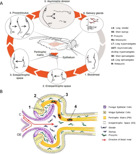
Figure 1.7. Life cycle stages of T. b. brucei within the tsetse fly.
In panel A, red arrows join life cycle stages within the fly while grey arrows represent transit between insect and mammalian host (Walshe et al., 2009). Panel B shows trypanosomes entering the ectoperitrophic space (ES) via the proventriculus of the tsetse fly. Trypanosomes ingested during the blood meal transform into procyclic forms within the proventricular lumen (1) and then migrate to the ES through a more fluid PM in the proventriculus (2). Alternatively, parasites may become trapped between PM layers forming cyst-like bodies (3). These bodies are carried through the midgut as the PM continues to be secreted (4). Blood flow from oesophagus (OE) or crop (C) are represented (From Rose et al., 2020).
Foregut Epithelial Cells
Midgut Epithelial Cells
Peritrophic Matrix (PM)
Ectoperitrophic Space (ES) Slender
Stumpy Procyclic
Direction of blood meal
A
B
LS: Long slender SS: Short stumpy P: Procyclic Ms: Mesocyclic LT: Long trypomastigotes ADT: Asymmetrically dividing trypomastigotesSE: Short epimastigotes LE: Long epimastigotes M: Metacyclic
skin, visceral adipose tissue and eventually the brain (Langousis and Hill, 2014; Caljon et al., 2016; Capewell et al., 2016; Trindade et al., 2016; Wheeler et al., 2019; Szöőr et al., 2020).
Slender bloodstream forms differentiate into the cell-cycle arrested short stumpy form that is pre-adapted for survival in the tsetse fly (Dewar et al., 2018). A quorum sensing-like mechanism involving oligopeptide signals received via a transmembrane protein called TbGPR89 triggers differentiation after accumulation of the stumpy-induction factor (SIF) (Rojas et al., 2019).
Bloodstream trypanosomes including slender and stumpy cells are taken up with a blood meal when a tsetse fly bites the infected host. Once in the fly midgut, short stumpy forms differentiate into the proliferative procyclic form that establish a midgut infection. To establish a new infection in mammals, trypanosomes in the fly must reach the salivary glands and be injected within a next blood meal, a journey that includes going through physical barriers (Figure 1.7A). The standard mechanism proposes that trypanosomes penetrate the peritrophic matrix (PM) in the anterior midgut to reach the ectoperitrophic space (ES). This PM is a proteoglycan matrix continuously produced by a specialised region of the anterior midgut that separates the midgut epithelium from the food bolus, protecting the cells of the intestine from mechanical damage, pathogens and toxins (Lehane, 1997).
The trypanosomatid journey has been recently explored using innovative microscopy techniques (Rose et al., 2020). According to this recent model, T. brucei procyclics reach the ES after penetrating an immature, freshly secreted PM within the proventiculus (Figure 1.7B). In the proventriculus, parasites go through asymmetric division to generate one long epimastigote and one short epimastigote. Then, epimastigotes go forward to the mouthparts, salivary ducts and ultimately into the salivary glands, where short epimastigotes attach to the gland epithelium, replicate and differentiate into metacyclic trypomastigotes. Finally, metacyclic cells detach from the epithelium and are released to the lumen. These cells are preadapted for transmission and survival in the mammalian host (Tetley and Vickerman, 1985;
Figure 1.8. Morphology of Trypanosoma brucei.
Panel A shows the morphology of a procyclic cell based on electron microscopy and tomography. The localisation of organelle components corresponds to reports using fluorescence microscopy of both endogenous fluorescent protein tagging and immunofluorescence (From Wheeler et al., 2019). Morphological changes in trypanosomes during the parasite cycle are shown in panel B. Scale bar: 5 µm, old (arrow) and new (arrowhead) basal body positions are indicated, DAPI is in red. SL: slender trypomastigote; ST: stumpy trypomastigote; PC: procyclic trypomastigote; MS: mesocyclic trypomastigote; E: proventricular epimastigote; DE: asymmetrically dividing epimastigote; LE: long epimastigote; SE: short epimastigote; AE: attached epimastigote; MT: metacyclic trypomastigote (From Rotureau et al., 2011).
1.4.2. Cell architecture
The characteristic shape of T. brucei (Figure 1.8A) is defined by its ordered cytoskeleton composed of a packed sub‐pellicular microtubule array linked to the plasma membrane (Hemphill et al., 1991). This array of microtubules confers a very pronounced polarity to the cell thanks to its anterior minus ends and posterior plus ends (Robinson et al., 1995).
The single-copy organelles have precise positions within the cell and include the flagellar pocket, flagellum, kinetoplast, mitochondrion and nucleus. The flagellar pocket is an invagination of the plasma membrane where endo and exocytosis take place and where the flagellum emerges onto the cell surface (Overath and Engstler, 2004; Lacomble et al., 2009). The flagellum is attached to the cell body via the flagellum attachment zone (FAZ) composed of a filament and a set of four specialised microtubules originating close to the basal bodies. The distal extremity of the flagellum is free and determines the direction of movement (Robinson et al., 1995).
The mitochondrial genome within the kinetoplast is connected with the basal body where the flagellum originates. The replication of kDNA is coordinated with the cell cycle through the correct segregation of the new and the old basal body (Robinson and Gull, 1991; Ogbadoyi et al., 2003). Like other trypanosomatids, T. brucei has a single mitochondrion extended through the whole cell that can adapt during the developmental cycle to take advantage of the different environmental conditions (Priest and Hajduk, 1994).
T. brucei has particular peroxisome-related organelles called glycosomes, where the
6-7 first steps of glycolysis are compartmentalised (Opperdoes and Borst, 1977a).This organelle is described in detail in section 1.6. Morphological changes during the parasite cycle are shown in Figure 1.8B.
1.4.4. Gene expression and regulation
The genome of T. brucei has 11 megabase chromosomes (~35 Mb total) containing, with few exceptions, all protein coding genes. In addition, 5 intermediate (200-300 kb)
Figure 1.9. Gene expression mechanisms in kinetoplastids.
1: Modified histones in an RNA polymerase II initiation region located in a divergent strand-switch intergenic region. 2: RNA polymerase II elongation. 3: RNA polymerase II termination located in a convergent strand-switch intergenic region. 4: Endonuclease cleavage of precursor. 5: Trans-splicing and polyadenylation. 6: Incompletely processed mRNAs can be degraded by the exosome. 7: Export of a completed mRNA, with bound poly(A) binding protein (PABP), exon junction complex (EJC) and nuclear cap-binding complex (CBC). 8: Emergence of a mature mRNA including proteins on the coding region (a) and a specific, stabilizing protein (b) bound to the 3'-untranslated region (3'-UTR). 9: Binding by a silencing or aggregating RNA-binding protein (c) and condensation into granules. 10: Binding of EIF4E, EIF4G and EIF4A, and translation. 11: Protein (b) is replaced by a destabilizing RNA-binding protein (d) and deadenylation starts. 12: Decapping by ALPH1. 13: Degradation by XRNA and the exosome. 14: Rapid decay pathway-immediate decapping promoted by protein (e) (From Clayton, 2019). 1 2 2 4 5 6 7 8 9 10 11 12 13 14 3
Among the unusual aspects that characterise gene expression in trypanosomatids is the fact that the genes are arranged in large multi-gene (polycistronic) transcription units, therefore genes lack individual promoters (Figure 1.9).
The sites of RNA polymerase II initiation located in a divergent strand-switch region (region located between divergent polycistronic transcription units) are epigenetically marked, being enriched in the histone modifications H4K10ac and H3K4me3, the histone variants H2AZ, H2BV and the bromodomain factor BDF3 (Siegel et al., 2009; Wright et al., 2010). Transcription termination is regulated by the histone H3 variant (H3.V) and, in bloodstream forms, the base J (ß-D-glucosyl-hydroxymethyluracil). This modified thymidine is the result of a hydroxylation by J-binding proteins JBP1 and JBP2 followed by a glycosylation by the J-associated glucosyl transferase (JGT) (Siegel et al., 2009; Schulz et al., 2016).
RNA polymerase II transcribes most protein-coding genes in polycistronic units that become mature mRNA after two coupled processes: 5' trans-splicing and 3' polyadenylation (Clayton, 2019). Trans-splicing involves addition of a capped 39-nt sequence called ‘spliced leader’ (SL) at the 5’ extremity of all mRNAs. This is performed by the spliceosome, which contains U1, U2, U4, U5, and U6 snRNAs in addition to several proteins (Günzl, 2010). Polyadenylation and trans-splicing are guided by the position of pyrimidine-rich tracts located between these two sites (Clayton and Michaeli, 2011). The polyadenylation complex including a major functional poly(A) polymerase was recently characterised (Koch et al., 2016). RNA polymerase I is responsible for the transcription of ribosomal rDNA, while a modified version transcribes the genes coding for the major surface proteins procyclins and VSG (Günzl et al., 2003).
The extranuclear mitochondrial genome (kDNA) is composed of thousands of minicircles (around 10,000 of 1-kb) and maxicircles (50 of 22-kb) condensed in one structure. Maxicircles encode rRNA and respiratory chain subunits, most of which require post-transcriptional RNA editing to be functional. This RNA editing phenomenon was first discovered in trypanosomes and consists of addition and
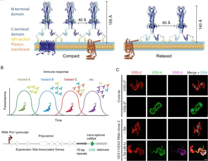
Figure 1.10. The dynamic Variant Surface Glycoprotein (VSG) coat of T. brucei bloodstream forms.
VSGs are homodimers that are attached to the plasma membrane via GPI anchors. The VSGs tightly packed (left) and relaxed conformations (right) are shown in panel A (From Bartossek et al., 2017). The antigenic variation (panel B) depends upon monoallelic and switchable VSG expression (From Horn, 2014). The allelic exclusion that governs the expression of VSG genes is coordinated by the VEX-complex (panel C). Depletion of this complex (VEX1/VEX2 RNAi mutant) yields multi-VSG expression (From Faria et al., 2019).
1.4.5. Cell surface
The surface of T. brucei is covered by a dense coat of glycoconjugates forming a protective glycocalyx against the host defence systems that changes depending on the life cycle stage. The bloodstream form (BSF) is coated with a dense layer of variant surface glycoprotein (VSG) (Figure 1.10) while the procyclic form (PCF) is coated with procyclins (Figure 1.11). These families of proteins are anchored to the membrane via GPI anchors (Figure 1.13).
a) VSG
VSG is a highly abundant dimeric protein that represents 95% of all BSF surface protein molecules. Its N-terminal domain is connected to the C-terminal domain through a linker region that confers flexibility between domains. VSG can adopt a compact or relaxed conformation (Figure 1.10A) depending on the protein density along the surface and as a response mechanism to changes. Interestingly, VSG forms a mobile and flexible shield that acts as a protective barrier for other less variable or invariant proteins in the plasma membrane to avoid recognition by the immune system (Grunfelder et al., 2002; Horn, 2014; Bartossek et al., 2017).
Given that VSG is immunogenic, the parasites developed an immune evasion strategy involving a switch to express an antigenically distinct VSG, known as antigenic variation (Figure 1.10B). There are three mechanisms of antigenic variation: 1) a coupled transcriptional activation/inactivation of VSG expression site (ES), 2) replacement of the active VSG by homologous recombination and 3) homologous recombination between active and inactive ES (chromosome exchange) (Horn, 2014). Around 300 functionally complete VSG genes are available for expression, in addition to other ~2200 incomplete or partial genes (Cross et al., 2014), but only one VSG gene is transcribed at a time from one of the ~20 telomeric ES until switching. RNA polymerase I is recruited to VSG-ES promoters constituting the expression site body (ESB) (Navarro and Gull, 2001). VSG genes are adjacent to telomeres and most of them are preceded by 70-bp repeats that facilitate recombination and duplication of
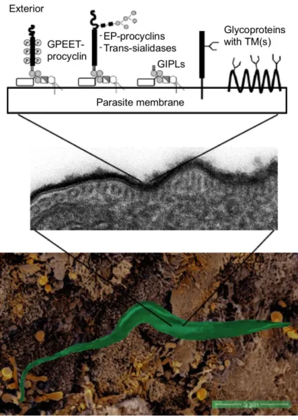
Figure 1.11. Surface glycocalyx of T. brucei procyclic cells.
A parasite interacting with the cell microvilli in the tsetse fly proventriculus is shown in the bottom panel. Transmission EM showing the surface glycocalyx (central panel). The main surface glycosylphosphatidylinositol (GPI)-anchored (EP- and GPEET-procyclins and trans-sialidases) and transmembrane (including polytopic) glycoproteins and glycolipids are shown in the top panel. Open rectangles linked to GPI molecules represent side chains. GIPLs: glycoinositolphospholipids, or free GPIs (From Rodrigues et al., 2015).
The allelic exclusion mechanism was recently shown to depend on the protein complex VEX1-VEX2 that associates with VSG, promotes transcription and transmits a silencing signal to negatively control transcription of other VSGs (Figure 1.10C). Additionally, it was shown that maintenance of the VEX complex and recruitment of the conserved chromatin assembly factor (CAF-1) during the S-phase allow inheritance of VSG exclusion (Faria et al., 2019).
VSGs can be N-glycosylated with different N-linked oligosaccharides depending on the VSG variant. Type I VSGs contain one N-glycosylation site where oligomannose structures (Man9–5GlcNAc2) are added. Type II VSGs generally have two N-glycosylation sites, one at the C-terminus occupied by a mixture of oligomannose structures similar to those from type II and larger polylactosamine-containing, while the internal site contains small structures like Man4–3GlcNAc2, GlcNAcMan3GlcNAc2 and in some cases, biantennary complex glycans. Type III VSGs have three sites of glycosylation occupied with a combination of oligomannose and complex biantennary glycans (Zamze et al., 1990, 1991; Mehlert, 1998). Additionally, VSGs can be O-glycosylated with a chain of zero to three O-linked hexoses on the top surface of the protein, which can play an immunomodulatory role (Pinger et al., 2018).
b) Procyclins
During differentiation of stumpy to procyclic forms, the VSG molecules are cleaved by a protease and the coat is replaced with procyclic acidic repetitive proteins (PARPs), named procyclins, within hours (Matthews and Gull, 1994). There are two major forms of procyclin, EP and GPEET, which differ in the type of amino acid repeats in their C-terminal domains. EP procyclins have Glu-Pro repeats (Mowatt and Clayton, 1987; Roditi et al., 1987) and GPEET procyclins have Gly-Pro-Glu-Glu-Thr repeats (Mowatt et al., 1989).
Figure 1.12. Developmental expression of major surface glycoproteins.
The top panel shows representative immunostaining images of the parasite stages. Nuclei and kinetoplasts (magenta) and cell surface (white) detected with antibodies corresponding to each surface molecule are highlighted. The bottom bars define the duration and intensity of the protein expression in the corresponding stage. Bloodstream forms (BSF), procyclic forms (PF), mesocyclic forms (MSC), short and long epimastigotes (EMF PV), attached epimastigotes colonising the salivary glands (EMF SG), pre-metacyclic forms (P-MCF) and metacyclic forms (MCF) (From preprint Casas-Sanchez et al., 2018).
There are eight copies of the PARP gene sequence per diploid genome encoding three PARP types: two EPs with or without an N-glycosylation site, respectively, and one GPEET with N-glycosylation sites (Mowatt and Clayton, 1987). Both types of procyclins adopt a highly extended rod-like structure (Figure 1.11) and their GPI anchors have an unusual lipid structure and large carbohydrate side chains made of branched polylactosamine repeats. In parasites grown in culture, a trans-sialidase substitutes the β-galactose termini of these side chains with sialic acid present in the fetal calf serum glycoconjugates (Mehlert, 1998). Interestingly, it was shown that the procyclins coating of parasites is not required to establish mature infections and complete the developmental cycle in the fly. However, the mutant analysed in this work lacking all the PARP genes showed a 10-fold reduced capacity of transmission and lower prevalence of salivary gland infections than with the wild type, suggesting that alterations of the membrane probably reduce migration and/or adherence of epimastigotes to the salivary glands (Vassella et al., 2009).
c) Other surface glycoproteins
In addition to VSGs and procyclins, T. brucei surface has other glycoproteins in minor amounts like the transmembrane invariant surface glycoproteins (ISGs) (Ziegelbauer and Overath, 1992) or transporters, such as the transferrin receptor (TfR) (Mehlert et al., 2012). Epimastigote forms express a different family of GPI-anchored proteins called brucei alanine-rich proteins (BARPs) (Nolan et al., 2000). During metacyclogenesis, this BARP coat is progressively replaced by a novel family of hypothetical proteins called MISP, which are invariant surface proteins exposed on the surface of metacyclics, along with VSG (Figure 1.12) (Casas-Sánchez et al., 2018). All these proteins are mostly N-glycosylated.
d) GPI anchors
The essentiality of GPI biosynthesis was addressed in bloodstream and procyclic forms by disrupting the TbGPI10 gene, which codes for a mannose transferase that
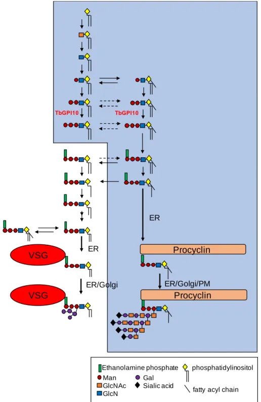
Figure 1.13. The GPI biosynthetic pathway of T. brucei.
The blue shaded area represents the pathway in procyclic cells and the nonshaded area represents the additional fatty acid remodelling steps (replacement by myristate) and attachment to VSG in bloodstream forms (From Ferguson, 2000).
Man Gal
GlcNAc Sialic acid
GlcN VSG VSG Procyclin Procyclin ER ER ER/Golgi ER/Golgi/PM
Ethanolamine phosphate phosphatidylinositol
fatty acyl chain
In BSF, the expression of an extra copy of the TbGPI10 gene was required for double knock-out cells to be viable, indicating that biosynthesis of GPI anchors is essential for BSF viability. In contrast, mutant PCF cells were able to grow and colonise the tsetse fly midgut, with lower efficiency than wild type cells (Nagamune et al., 2000). This is consistent with the results obtained for procyclin null mutants (Vassella et al., 2009).
T. brucei use different GPI structures to anchor VSGs and procyclins in the cell
membrane, and the structures of both anchors have been characterised. The complex structure includes a backbone of ethanolamine-phosphate-6Manα1-2Manα1-6Manα1-4GlcNac linked to an inositol phospholipid. In the VSG-GPI anchor, the diacylglycerol moiety suffers fatty-acid remodeling, by replacing the fatty acids by myristate, before transfer to VSG protein, with 1,2-dimyristylglycerol as final lipid (Ferguson et al., 1988). The preassembled GPI membrane anchor is then linked to a pre-VSG C-terminal domain by single-step reaction performed by a multisubunit transamidase complex. Finally, GPI can be glycosylated with branched side chains consisting of a variable number of α-galactose residues attached to the O-3 position of the mannose residue adjacent to GlcN (Figure 1.13) (Mayor et al., 1992).
In the case of procyclic cells, the structures of procyclins GPEET and EP GPI anchors are very similar, with lipid moiety of 1-stearoyl-2-lyso-glycerol and large complex carbohydrate side chains containing galactose, N-acetylglucosamine and sialic acid. Moreover, the procyclin GPI anchor contains a fatty acid attached to the inositol ring (Field et al., 1991; Ferguson et al., 1993; Bütikofer et al., 1997).
1.5. Intermediate and energy metabolism
T. brucei metabolism has been extensively studied due to its peculiar traits and the
continuous research of drug targets to develop treatments against HAT. Within the metabolomics approaches currently existing, the two techniques principally used in the majority of references described in the next section are nuclear magnetic resonance (NMR) and mass spectrometry (GC-MS and LC-MS). Indeed, 13C isotopic
Figure 1.14. ATP production in PCF and BSF trypanosomes.
Catabolism of proline and/or glucose is shown. Excreted end products are underlined. Arrows thicknesses represent metabolic flux. Only key enzymes involved in ATP production are indicated here. Mitochondrial production of ATP by succinyl-CoA synthetase is not shown in the right panel (BSF) (From Smith et al., 2017).
Enzymes: 1a, glycosomal phosphoglycerate kinase; 1b, cytosolic phosphoglycerate kinase; 2, pyruvate kinase; 3,
phosphoenolpyruvate carboxykinase; 4, glycosomal malate dehydrogenase; 5, cytosolic fumarase (for simplification this reaction is placed in the glycosome); 6, glycosomal NADH-dependent fumarate reductase; 7, pyruvate phosphate dikinase; 8, acetate:succinate CoA-transferase; 9, acetyl-CoA thioesterase; 10, succinyl-CoA synthetase; 11, trypanosome alternative oxidase; 12, respiratory chain; 13, F0/F1-ATP synthase; 14, mitochondrial ADP/ATP exchanger. Abbreviations: AcCoA, acetyl-CoA A; DHAP,
Procyclic form
(Glucose-depleted medium)
Procyclic form
T. brucei adapts its metabolism to the available extracellular nutrients encountered
during the life cycle within a mammalian host and tsetse fly insect vector. The long-slender bloodstream form mainly catabolises glucose while procyclic forms rely on amino acids metabolism, mostly proline. In the following section, I describe the main metabolic differences between these two life cycle stages.
1.5.1. Bloodstream vs procyclic, an overview
The energy and carbon metabolism of PCF and BSF has been compared in a number of reviews (Bringaud et al., 2006; Michels et al., 2006; Tielens and van Hellemond, 2009; Bringaud, 2012; Creek et al., 2012; Smith et al., 2017). The next few lines highlight the main features of energy metabolism in these two parasitic forms of T.
brucei. The single mitochondrion, approximately 40 - 60 glycosomes and cytosolic
compartment are crucial to understand the differences between bloodstream (BSF) and procyclic (PCF) trypanosomes in terms of energy metabolism. As shown in Figure 1.14, PCF trypanosomes have an elaborate mitochondrion and are able to metabolise amino acids like proline, abundantly present in the fly. In contrast, the BSF mitochondrion is less developed and the parasites have developed a glycolysis-based metabolism from glucose abundantly present in the blood of the mammalian host. The production of ATP in BSF depends on glycolysis, whereof the first seven enzymes are compartmentalised in the glycosomes. The glycosomal membrane is impermeable to large (300 – 400 Da) metabolites, therefore consumption and production of ATP is balanced with no net ATP production inside the organelles. It is in the cytosol where the net ATP production from glycolysis takes place resulting in production and excretion of pyruvate. Besides, relatively low amounts of ATP are generated in the mitochondrion since oxidative phosphorylation does not occur. ATP production in the PCF varies depending on the carbon sources available, as it can be cultured in the presence or the absence of glucose. When glucose is absent, proline is metabolised in the mitochondrion and alanine is excreted as the main end product, in addition to glutamate, succinate and acetate. The reduced cofactors produced during proline


![Figure 1.16. [U- 13 C] enrichment of key glycolytic intermediates from proline, glycerol or glucose](https://thumb-eu.123doks.com/thumbv2/123doknet/14523128.722700/49.892.128.766.229.642/figure-enrichment-key-glycolytic-intermediates-proline-glycerol-glucose.webp)


