A Biochemical Study of Class
II
MHC and
Cytochrome P-450scc Proteins
By
Aaron K. Sato
B.Sc., Chemistry with Honors, 1993 University of Puget Sound, Tacoma, WA
Submitted to the Department of Chemistry in Partial Fulfillment of the Requirements for the Degree of
DOCTOR OF PHILOSOPHY in Biological Chemistry
at the
Massachusetts Institute of Technology June 1999
@ 1999 Massachusetts Institute of Technology All rights reserved
Signature of Author Certified by L7 Department of Chemistry May, 1999 Lawrence J. Stem Assistant Professor of Chemistry Thesis Supervisor
Accepted by -Dietmar
Seyferth Chairman, Departmental Committee on Graduate Students
This doctoral thesis has been examined by a Committee of the Department of Chemistry as follows:
/
Professor Jun Liu /
Chairman
Professor Lawrence .Stem
Research 7tpervisor
Professor Alexander Klibanov___
I
doL-I I
A Biochemical Study of Class II MHC and
Cytochrome P-450scc Proteins
By
Aaron K. Sato
Submitted to the Department of Chemistry in Partial Fulfillment of the Requirements for the Degree of Doctor of Philosophy in Biological Chemistry, June 1999
ABSTRACT
Class H major histocompatibility complex (MHC) proteins play a key role in the specific immune response. This thesis explores how these proteins function on the molecular level.
HLA-DQ8 ( 1*0302) is strongly correlated with Insulin-dependent diabetes mellitus
(IDDM) onset. Most IDDM resistant alleles contain Asp at position P57, while most IDDM susceptibility alleles contain a residue other than Asp. Others have proposed that this correlation is due to an instability of these MHC molecules. Using circular dichroism (CD) and thermal denaturation studies, we show that DQ8 is not an unstable molecule and that it adopts a slightly different structure from DQ9 (P 1*0303), which has an Asp residue at P57.
A monoclonal antibody (KL-304) specific for the empty conformation of class II MHC
molecules revealed the presence of abundant empty molecules on spleen- and bone marrow-derived dendritic cells among various types of antigen presenting cells. The empty class II MHC molecules are developmentally regulated, and can capture peptide antigens directly from the extracellular medium and present them to T-lymphocytes. These results suggest that dendritic cells may utilize an unusual extracellular presentation pathway in which antigen processing and peptide loading occur entirely outside the cell.
As an extension of this cellular study, we also investigated the mechanism of the conformational change between empty and peptide-loaded HLA-DRI. We found that the KL-304 and related KL-295 antibody epitopes are only detected on the empty form of DR1 and are lost when the large, hydrophobic P1 pocket is filled, either with a G86Y mutation of the protein or with a residue from a bound peptide as small as 4 amino acids in length. This immunoassay data is supported by near-UV circular dichroism data that shows that the empty protein has a
different secondary structure from the mutant and peptide bound proteins.
Cytochrome P-4 50scc is a mammalian monooxygenase that catalyzes the conversion of cholesterol to pregnenolone in the steroid hormone biosynthetic pathway. Cholesterol is first hydroxylated at the C-22 pro-R position in a stereospecific manner to form
(22R)-22-hydroxycholesterol. This intermediate is then hydroxylated again at the C-20 pro-S of cholesterol to form (20R,22R)-20,22-dihydroxycholesterol. These hydroxyl groups are
introduced from two different molecules of oxygen. Finally, the glycol intermediate is cleaved between the hydroxyl carbons to form isocaproaldehyde and pregnenolone. The C-20 oxygen is not disturbed during this step and becomes the ketone oxygen of pregnenolone. In this study, we find that the C-22 oxygen also is not disturbed during the final oxidation step.
Thesis Supervisor: Dr. Lawrence J. Stern Title: Assistant Professor of Chemistry
DEDICATION and THANKS
I dedicate this thesis to the following people who have helped me professionally and
personally over the last six years.
Laura Santambrogio, M.D., Ph.D. I am deeply indebted to you for being my collaborator, friend, and teacher all rolled into one. Through working with you, I learned so much about the value of teamwork. I also learned the importance of placing my biochemical work into a more cellular context.
Jennifer Zarutskie. As my other collaborator, I am also indebted to you for working with me and allowing me to contribute to your projects. Also, thanks for listening to me over the last two years and giving me your thoughts on anything from lab stuff to world at large.
Jennifer Schmitke, Ph.D. Thanks for being such a great friend and confidant. I don't know how I would have survived the last couple years without talking about my fears and problems during our daily coffee breaks. Danny DeOliveira, Ph.D. Thanks for being a great satellite lab mate and teaching me so much about peptide synthesis.
Mia Rushe. Thanks for being such a great teacher and technical resource in the lab. Charles Wescott, Ph.D. Thanks for all your timely and helpful professional advice. Curt Lockshin, Ph.D. Thanks for being a good friend over the years.
Mike Amendola, M.S. and Diego A. Hoic, Ph.D. Thanks for being my friends.
Normand Cloutier, Ph.D. Thanks for helping me navigate through the world of P-450 enzymology.
William Orme-Johnson, Ph.D. Although I wasn't in your lab for an extended period of time, I thank you for giving me the opportunity and means to conduct research when no one else would.
Lawrence J. Stern, Ph.D. I thank you for being such an attentive advisor for my first two years and then allowing me to grow on my own for the last two. Through working with you, I learned not only a great deal of chemistry & immunology, but I also learned how to be a productive and independent scientist.
The rest of the Stern Lab (Jennifer Ogrodnick, Tom Cameron, Dikran Aivazian, Helen Chan, Ravi Joshi, Greg Carven). Best of luck in the future.
Kenneth Rousslang, Ph.D. Thanks for opening my eyes to the fascinating world of chemistry.
To my parents, Paul and Susie Sato. I could not have gotten here without your never-ending support and encouragement throughout my life. I love you both.
To my fiancde, Kyong A. Yi. Kyong Ah, you make my life so complete. Thank you for being so understanding and caring over the last two years. I love you dearly.
TABLE OF CONTENTS
Page Number
I. Introduction to Class II Proteins encoded by
the Major Histocompatibility Complex (MHC) 7
II. In Vitro Refolding of HLA-DQ Class II MHC Protein for Use in
Biophysical and Crystallographic Studies
A. Introduction 16
B. Materials and Methods 16
C. Results and Discussion 19
El. HLA-DQ8 (cx1*0201, P1*0302) and DQ9 (ccl*0201, P1*0303)
MHC Class II Proteins: The Role of P57 Polymorphism in Complex Structure and Stability
A. Introduction 23
B. Materials and Methods 24
C. Results 29
D. Discussion 37
E. Future Work 39
IV. KL-295 and KL-304: Monoclonal Antibodies that Specifically Bind Empty Class H MHC Proteins; A. Antigen Presentation by Empty Cell Surface Class 1I MHC Molecules on Dendritic Cells
A. Introduction 40
B. Materials and Methods 42
C. Results 50
D. Discussion 62
E. Future Work 63
V. KL-295 and KL-304: Monoclonal Antibodies that Specifically Bind Empty Class II MHC Proteins; B. A Conformational Change
in HLA-DR1 is Dependent on Both Main Chain Interactions and P1 Pocket Occupancy
A. Introduction 64
B. Materials and Methods 67
C. Results 73
D. Discussion 82
VI. The Cytochrome P-450scc Glycol Cleavage Reaction: Analysis of
180 and 2H Incorporation into Isocaproaldehyde and Evaluation of
Potential Mechanisms
A. Introduction 86
B. Materials and Methods 90
C. Results 94
VII. References
VIII. Appendix 122
A. Methods Developed or Extensively Used in the Lab 123
B. Archival DNA, Protein, and Peptide Samples 130
IX. Biographical / CV Note 132
I. Introduction to Class II Proteins Encoded by the
Major Histocompatibility Complex (MHC)
MHC molecules are membrane-bound glycoproteins that play an important role in specific immune responses to foreign antigens (Germain, 1994). Class I MHC proteins are responsible for presenting peptides derived from intracellular antigens and are expressed in the
all cell types. On the other hand, class II MHC proteins are responsible for presenting peptides derived from extracellular antigens (including membrane bound antigens) which are brought into specialized antigen-presenting cells (APC), including B-cells, dendritic cells, macrophages, monocytes, via receptor-mediated and/or fluid-phase endocytosis. These protein-peptide complexes bind to T cells with receptors specific for a particular complex of MHC protein and peptide. Once bound to the MHC-peptide complex, the T cell becomes activated and stimulates a specific immune response to the invading antigen (Korman, 1985).
Peptide Binding
MHC class II alleles are non-covalently associated ac heterodimers (Benacerraf, 1981; Kaufman, 1984; Korman, 1985). Each monomer has an extracellular domain of approximately 200 amino acids and a transmembrane region and cytoplasmic domain of approximately 40 amino acids. Together they form an extracellular region with two immunoglobulin fold domains and a unique peptide-binding site, which is composed of an antiparallel P-sheet and two a helices (Fig. lA, B). Peptides bind between the o helices in a type II polyproline helix
conformation. The MHC protein contacts the peptide through both sequence independent and dependent interactions. The sequence independent interactions consist of hydrogen bonds, electrostatic and Van der Waals interactions with the bound peptide main chain (Fig. IC). The sequence dependent interactions, however, are mediated between the peptide side chains and
several hydrophobic and/or charged pockets (P1, P4, P6, P7, P9; the numbering is derived from
N to C terminal numbering of the bound peptide, where P1 is the first N-terminal residue bound
population and many of the resulting polymorphic residues in the expressed protein are
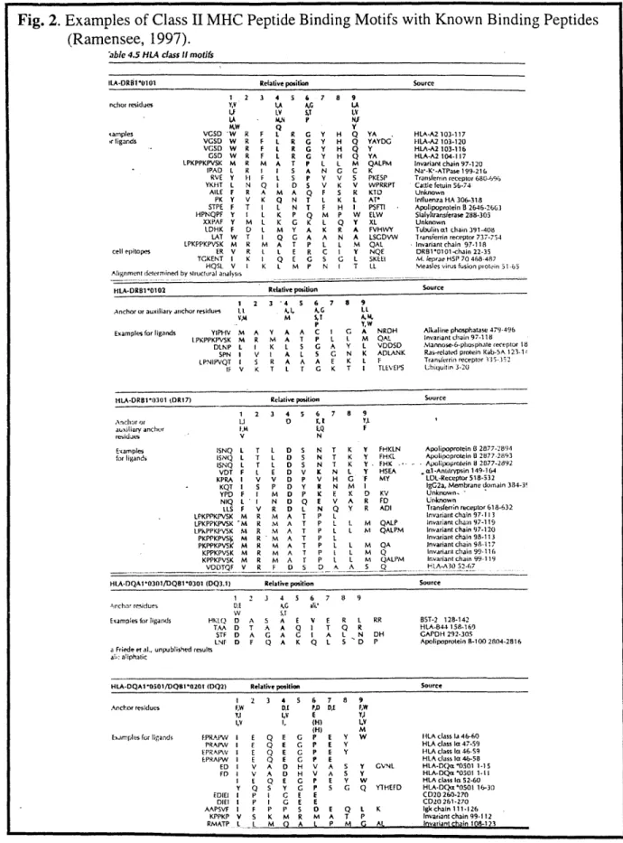
concentrated in these binding pockets (see next section). These changes in pocket residues lead to modified peptide binding preferences for different MHC proteins, or alleles [Fig. 2; For review, see (Rammensee, 1995, 1997; Stem. 1994b; Sinigaglia, 1994).] In this study, several human (HLA-DR1, DQ8, DQ9) and murine (I-As) are expressed in recombinant form and
analyzed to see how they may function in the immune system.
Polymorphism
The human MHC class II gene complex consists of many loci, which are designated DM,
DO, DP, DQ, and DR. Within each locus site, there are a and
P
genes. In the case of DR, thereare several P gene subloci sites, which can express functional subunits. These genes are highly polymorphic in the general population, with the polymorphic residues concentrated in the peptide-binding site (Fig. 3; Bodmer, 1991). These genes are labeled using a specific nomenclature, (species label)-(locus site)*(serology number)(allele number). For example,
HLA-DQB1*0301 is a human class II gene derived from the DQB1 locus with a serology/allele
number of 0301. Before sequencing was as streamlined as it is today, MHC proteins were identified by their reactivity towards antibodies specific for groups of proteins, or serotypes (Marsh, 1989). Although the nomenclature numbers refer to cDNA sequences, these proteins are still grouped together on classification charts and in the above nomenclature by serology number. As expected by their common reactivity towards the same antibodies, the members of each serological group contain many of the same polymorphic residues and as a result, may show a great deal of tertiary structural homology. These allelic variations in a polymorphic pair of a and
P
subunits lead to different binding motifs for each particular MHC allotype, and may contribute to why two individuals can have completely different immune responses to certain antigens.(A)
(B)
(2
P2
(P
Otl1> ApS Arg aB
u53 An* \ o HH Fig L. Cls II MHC. R (t , 1 R-4 H C Asp P57 14 3. Nsa A W H s 028 Trp 061
(D
Arg PFig. 1. Class 11IMHC
Structure. (A) Side-view ribbon diagram of
HLA-vjg K .34DRI '5
(Stern, 1994a); (B)
Top-view ribbon diagram
of HLA-DR1 (Stem, 1994); (C) Sequence independent main chain interactions between HLA-DRI and A2(103-117)
' 04 peptide (Murthy, 1997);
7 (D) Sequence-dependent
pocket interactions between HLA-DR1 and
HA(306-318) peptide
Fig. 2. Examples of Class II MHC Peptide Binding Motifs with Known Binding Peptides (Ramensee, 1997).
'able 4.5 HLA class I motifs
ILA-ORSPil*0 Relative position Source
1.2 3 4 5 6 7 8 9 nchor residues YV ,A A, LA i.F ,y 5, 1,Y ,A - , P NJ KW Q Y samples VGSD 'W R F L R G Y H Q YA HLA-A2 103-117
'rligands VGSD W R F L R G Y H Q YAYDG HLA-A2 103-120 VGSD W R F L R C Y H Q Y HLA-A2 103-116 GSD W R F L R G Y H ( YA HLA-A2104-i7
LPKPPKPVSK M R M A T P L L M QALPM Invariant chain 97-120
[PAD L R I I S A N G C K Na-K--ATPase 199-216
RVE Y H- F L S P Y V S PKESP TransfLerrin receptor 680-694 YKiT L N Q I D S V K V WPRRPT Cattle fetuin 56-74
AILE F R A M A Q F S R KID Unknown PK Y V K Q N T L K L AT* Influenza HA 306-318
STPE F T I L N T F H I PSFTI Apolipoprotein B 2646-2663 HPNQPF Y I L K P Q M P W ELW Sialyltransferase 288-305
XXPAF Y M L K G K L Q Y XL Unknown
LDHK F D L M Y A K R A FVHWY Tubulin o. chain 391-408 LAT W T I Q G A A N A LSGDVW Transferrin receptor 737-754
LPKPPKPVSK M R M A T P L L M QAL Invariant chain 97-118 cell epitopes ER V R I L E R C I Y NQE DR1*0101-chain 22-35
TGKENT I K I Q E G S G L SKEEI M. leprte HSP 70 468-487
HQSL V I K L M P N I T LL Measles virus fusion protin 51.6 Alignment determined by siuctural analysis
tiLA-DRBI*0102 Relative position Source
1 2 3 4 S 6 7 8 9
Anchoror auxiliary anchor residues I .1, AG I.L V,4I M 5,Y A.A
P Y,W
Examples for ligands YIPHV M A Y A A C I G A NROH Alkaline phosphatase 479-496
L.PKPPKPVSK M R M A T P L L M QAL Invariant chain 97-118 13LNP L I K L S G A Y L VDDSD Mannose-6-phosphate receptor 18
SPN I V I A L S G N K ADLANK Ras-related protein Kab-5A 123.1
LPNIPVQT I S R A A A E K L F Transierrin receptor 55. IF V K T L r C K T I TLEVEPS Ubiquitin 3-20
tiLA-DRB1*0301 tDR17) Relative position Source 1 2 3 4 5 6 7 8 9
Anch or ,1 K.. I YI
auxiliary anchor I'M EQ F
resiues V N
Examples !SNQ L T L D S N T K y FHKLN Apolipoprotein I 2877-2894
for ligands ISNQ L T L D 5 N T K Y FHKL Apolipoprotein 8 2877-2893
ISNQ L T L o S N T K Y F FHK -. Apolipoprotein 8 2877-2692 VDT F L E D V K N L Y HSEA .l1-Antitrypsin149-1&4
KPRA I V V D P V H G F MY LDL-Receptor 518-532
- KQT I S P D Y R N Mi I IgG2a, Membrane domain 334-3!
YPD F I M D P K E K D KV Unknown,
NIQ L I N D Q E V A R F'D Unknown
LLS F V R D L N Q Y R ADI Transferrin receptor 618-632
. LPKPPKPVSK M R M A T P L Invariantchain97-113
LPKPPKPVSK 'M R N4 A T P , 1 M QALP invariant chain 97-119 I.PkPPiKPV5K hi R M A T P L L M QALPM Invariant chain 97-120 PKPPKPVS i M R h A T P L Invariant chain 99-113 PKPPKPVSK M R M A T P L L M QA invariant chain 9-117 KPPKPVSK M R M A T P L L M Q Invariant chain 99-116 KPPKPVSK M R M A T P . L M QALIM Invariant chain 99-119
VOITQF V R F 1 5 0 A A 5 Q HI.A-A30 52-67
HLA-DQAI10301/DQ81*0301 (DQ3.1) Relative position Source
S12 3 4 5 6 7 8 9
Ancho residues 0.1 AC ai,
W SJT
Exampiesforligands HKLQ 3 A 5; A E V E R . RR 85T-2 128-142
TAA D T A A Q I T Q R HLA-844 158-169 STF D A G A G I A L N OH GAPDH 292-305
LNF D F Q A K Q 1 5 D P Apolipoprotein B-100 2804-2816 a Friede et al., unpublished results
at: aliphatic
HLA-DQA1*0501/DQ8I1-201 (DQ2) Relative position Source
I 2 3 4 5 6 7 8 9
Anchor residues F,W
Y,2
1,V
Examples for ligands EPRAIMW I
PRAPW I EPiRAPV I EPRAPW I EDi I FD I Y EDIEI I DIE1 I AAPSVF I KPPKP V RMATP L E E E V V E Q P P F DC PD DAE 4V E I, (H) (H) Q E G P E Y Q E G P 1E Y Q E G P E Y E G D H D H E G Y G GK E G E P S M R 0 A P E V A V A P E P S E E M L Q A A P K Mh S Y G FW Y) 1,V M W Y GVNL y W Q YTHEFD E Q L K A T P P M r. At HLA class la 46-60 HLA class Ia 47-59 HLA class la 46-59 HLA class la 46-58 HLA-oQn *O5sO 1-IS
HLA-DQas'0501 1-11 HLA class lo 52-60 HIA-DQi's0501 16-30 C020 260-270 CD20 261-270 igk chain 111-126 Invariant chain 99-112 Invariant ihrin I1R8- 23
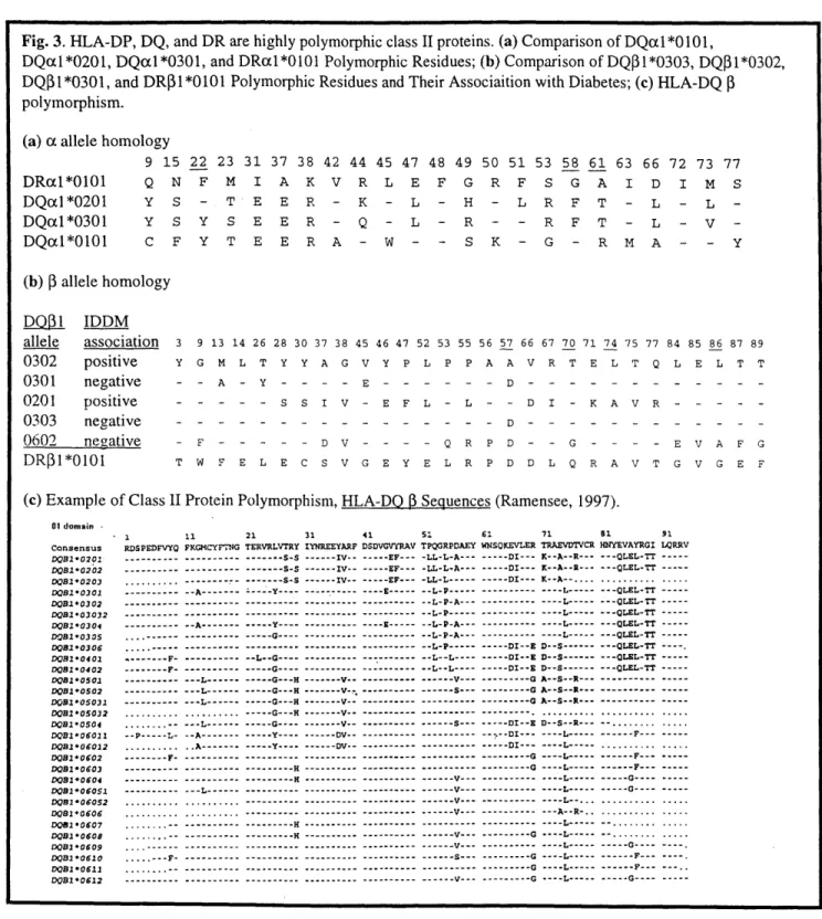
Fig. 3. HLA-DP, DQ, and DR are highly polymorphic class II proteins. (a) Comparison of DQLL *0101,
DQal*0201, DQacl*0301, and DRX1*0101 Polymorphic Residues; (b) Comparison of DQP1*0303, DQ$1*0302, DQI 1*0301, and DR3 1*0101 Polymorphic Residues and Their Associaition with Diabetes; (c) HLA-DQ
polymorphism. (a) (x allele homology
9 15 22 23 31 37 38 42 Q N F M I A K V Y S - T E E R -44 45 47 48 49 50 51 53 58 61 63 66 72 73 77 R L E F G R F S G A I D I M S K - L - H - L R F T Y S Y S E E R - Q - L - R - R F T L - L -L - V -C F Y T E E R A - W - - S K - G - R M A - -(b) P allele homology D0pl IDDM allele association 3 0302 positive Y 0301 negative -0201 positive -0303 negative -0602 negative -DR 1*0101 T 9 13 14 26 28 30 37 38 45 46 47 52 53 55 G M L T Y Y A G V Y P L P P 56 57 66 67 70 71 74 75 77 84 85 86 87 89 A A V R T E L T Q L E L T T - A - Y - - - - E - - - - - - D - - - - - - - - - - - -S - S S I V - E F L - L - - D I - K A V R - - - - -- - - - - - - - - - - - - - - D - - - - ---- - - - -F - - - - - D V - - - - Q R P D - - G - - - - E V A F G W F E L E C S V G E Y E L R P D D L Q R A V T G V G E F
(c) Example of Class 1I Protein Polymorphism, HLA-DQ D Sequences (Ramensee, 1997).
11 21 31 41 51 61 71 81 91
FKGMCYFDNG TERVRLVTRY IYNREEYARF DSDVGVYRAV TPQGRPDAEY WNSQKEVLER TRAEVDTVCR HUYEVAYRGI LORRV .--- ---- S-S --- --- s-S --- ---- S-s -- A--- --- Y --- A--- --- Y--- --- G---- G---- --- -- L--G ---- --- --- --- L--- L--- --L---... --L---... ...- -.- L---1 -- P---L- -- A---... .. A--- F- A--- --- --- --- --- L---... L---... ... ... , . . .-- ---. ---. . , -- ---.. --- ---.. .- -- ---. ---.---. . -- --- IV -- -- - - -IV-- IV IV --- --- - --- --- G---- --- G---H --- V--- G---H V--- V--. --- G---H --- V--- G--- H --- V--- G ---- V-- ---V---- Y---- --- DV--- Y---- --- DV-- --- H --- H --- ----...--- ----...--- --- --- ------ -- --H --- EF--- --- EF--- -LL-L-A--- EF--- -LL-L--- E--- -- L-P--- -- L-P-A--- --- P---E -- ---- -- L-P-A--- -- L-P-A--- -- L-P--- -- L--L--- -- L--- --- --- --- --- --- --- --- --- --- --- --- --- --- --- --- .--- V--- --- V--.. --- --- V--- V--- --- --- --- --- V--- --- S--- S--- --- --- V--- DI--- K--A--R--- DI--- K--A--R.--- DI--- K--A--....L --- --- L--- ---- L--- ---- L--- ---- L--- ---- L--- DI--E D--SL--- --- DI--E D--S--- D--S--- DI--E D--S --- G A--S--R--- G A--S--R---0 A--S--R---.-...--.. ... --- DI--E D--S--R --- DI--- ---- -- -- -- G ---G0 --- G --- G-- G-- G--- - G - --- --- --- S--L--- L--.. --- -- --.-- --.--- - -- - --- L--- -L--... --- --- --- --- --- --- --- --- --- --- - - - --- --- G----... G----...G----... . .. ... - - - -- - - - -- - - --- ----... ... -- -G--- -G---F --- - F ---DRxl*0101 DQcd *0201 DQal*0301 DQcil*0101 Y - 1 RDSPEDFVYQ ---.--- ---... ---. --- .--- F--- - -F- ---01 domain -Consensus DQB1'0201-DOB-0202 D0B1*0203 DOB*0301 D08*0302 DOB103032 DB*0304 D081O030S DOB10306 DOB10402 DOB1*0402 DB1*0501 DB1*0502 00B*05031 DOS1-05032 DB*0504 D0B*06011 0081*06012 D0B0*0602 D0B1*0603 D01*0604 D081*06051 D01*06052 D081*0606 D01*0607 D01*0605 DO1 0609 DOB1*0610 D0B1*0611 D01 *0612 --- QLEL-TT --- QLEL-TT ---... ... --- QLEL-TT --- OLEL-TT --- QLEL-TT --- QLEL-TT ---QLEL-TT --- -QLEL-TT---- QLEL-TT --- OLEL-TT --- --- ---... ... ... ... --.- DI---- ---- L--- ---
F----F-Structure
To date three-dimensional structures have been determined for several human and murine MHC class II proteins. The human protein structures include DRI bound to endogenous peptides (Brown, 1993), DRI bound to either HA(306-318) (Stern, 1994a) or A2(103-117) (Murthy,
1997), DR2 bound to MBP(85-99) (Smith, 1998), DR3 bound to CLIP(82-104) (Ghosh, 1995),
and DR4 bound to collagen 11(261-273) (Dessen, 1997). The murine protein structures include
I-Ek bound to either murine hemoglobin(68-76) or Hsp70(238-246) (Fremont, 1996), I-Ad bound
to either OVA(323-339) or HA(126-138) (Scott, 1998), I-Ak bound to HEL(50-62) (Fremont,
1998). Although each allele has a different peptide-binding motif, the large number of class II
structures has made it easier to understand the general mode of peptide binding and predict peptide-binding motifs for class II proteins of unknown structure. As will be shown in Chapter III, however, future structural efforts should be aimed at determining structures of alleles associated with autoimmune diseases, such as diabetes and celiac disease (Fig. 4A). Several of these alleles associated with diabetes susceptibility contain an Asp to Ala mutation at P57 (Todd,
1987), which destroys a salt-bridge between the a and
P
subunits (Fig. 4B; Brown, 1993). Sinceit is difficult to model the effect of such a mutation and how it may affect subsequent peptide binding, the structures of such alleles should be determined to fully understand the structure-function relationship in the development of these diseases.
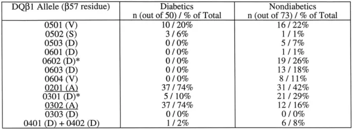
Fig. 4. Association of HLA-DQ Alleles with Diabetes Susceptibility. (A) Individuals with DQ
Alleles containing non-AspP57 residues greater risk of contracting diabetes (Todd, 1987); (B) AspP57 makes an important salt bridge with Arga76. Loss of this interaction may affect the complexes stability and/or structure (Reizis, 1998).
(A) HLA-DQ
P
alleles and IDDM Susceptibility. GenotypeIDDM patients and 73 randomly selected healthy controls. analysis done on 50 unrelated
DQ 1 Allele (P57 residue) Diabetics Nondiabetics
n (out of 50) / % of Total n (out of 73) / % of Total
0501 (V) 10/20% 16/22% 0502(S) 3/6% 1/1% 0503 (D) 0/0% 5/7% 0601 (D) 0/0% 1 / 1% 0602 (D)* 0/0% 19/26% 0603 (D) 0/0% 13/18% 0604 (V) 0/0% 8/11% 0201 (A) 37/74% 31/42% 0301 (D)* 5/10% 21/29% 0302 (A) 37/74% 12/16% 0303 (D) 0/0% 0/0% 0401 (D) + 0402 (D) 1/2% 6/8%
(B) Model of DQ8 Peptide Binding Site and Location of Asp57-Arga76 salt bridge
HLA-DQ9 (al*0301, 1*0303) Salt Bridge Intact HLA-D08 (a1*0301, 1*0302) No Salt Bridge
*J0
#$A7
Processing
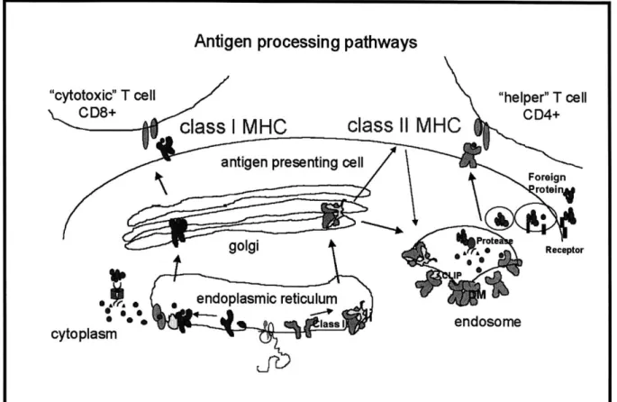
Much effort has been directed at understanding the final conformation of the peptide-class II complex, and many studies have tried to understand how bound peptides are derived from foreign proteins and how they are loaded onto empty class II proteins (Germain, 1993a,
1994; Cresswell, 1994). Foreign antigens in the extracellular matrix are brought into endosomal compartments via receptor-mediated and/or fluid phase endocytosis (Fig. 5). Upon uptake, these antigens are proteolyzed into small peptides and loaded onto newly-synthesized or recycled class II molecules. The newly-synthesized class II molecules are transported from the endoplasmic reticulum complexed to invariant chain (Ii), a trimeric protein that binds the empty molecule through the peptide-binding site and other areas and thus prevents unwanted peptide binding during transport (Cresswell, 1994; Cresswell, 1996). Upon reaching the endosomes, the class II-Ii complex is dissociated by II-Ii proteolysis to yield a DR-CLIP complex (CLIP is a fragment of invariant chain (89-101) that binds within the peptide-binding site). Subsequently, the CLIP peptide is released by interacting with another protein, HLA-DM, a non-polymorphic class II homologue which promotes CLIP peptide release, protects the empty protein from aggregation,
and promotes the binding of the antigenic peptides derived from proteolysis (Avva, 1994; Roche,
1995; Sanderson, 1995; Weber, 1996; Kropshofer, 1997; Denzin, 1995). Another
non-polymorphic class II homologue, HLA-DO, is a negative regulator of HLA-DM and inhibits DM action, possibly by inhibiting its interaction with other class II molecules (Karlsson, 1991;
Kropshofer, 1998; Denzin, 1998). Although this pathway is considered to be the primary class II loading pathway in B-cells, it possible that alternative pathways exist in dendritic cells (Rovere,
1998) and macrophages.
Although not discussed in this thesis, class I molecules obtain their bound peptides through a different mechanism (For review, see Germain, 1993a; Rammensee, 1997).
Cytoplasmic proteins (derived from endogenous proteins and/or foreign antigens transcribed within the cell) are proteolyzed by a 20S proteosome complex and the resulting peptide
binding of these peptides to newly-synthesized class I molecules, the peptide-class I complexes are transported from the endoplasmic reticulum, through the Golgi apparatus, and eventually to the cell surface for possible CD8+ T-cell activation.
Antigen processing pathways
"cytotoxic" T cell "helper" T cell
CD8+
class I MHC
class
Il
MHC
CD4+antigen presenting cell
Foreign roteinj ert golgi *Receptor *9 IP , ,0' endoplasmic reticulum * lass Iendosome cytoplasm
Fig. 5. Class I and Class II MHC Antigen Processing and Presentation Pathways.
This majority of this thesis addresses various aspects of class II MHC structure and antigen presentation. Within each chapter, a specific and concise introduction to the subject matter is provided. The references cited within the body of the text are at the end of the thesis (Chapter VII). The final P-450scc chapter contains unpublished data collected in the lab of William Orme-Johnson, Ph.D. during my first year at M.I.T.
II. In Vitro Refolding of HLA-DO Class II MHC Protein for
Use in Biophysical and Crystallographic Studies'
INTRODUCTION
During my first year in the Stem lab, we attempted to express several HLA-DQ proteins
(DQ1, DQ2, and DQ8) using a procedure previously developed to refold HLA-DR1 from E.
coli-produced subunits. In this procedure, a and
P
subunits are expressed as shorter extracellular versions of their wildtype form, isolated as insoluble inclusion bodies, and purified to homogeneity using anion exchange chromatography. By removing the connecting peptide, transmembrane, and intracellular regions of each chain, Frayser et al. (1999) refolded soluble peptide-DR1 complexes in high yield and purity. Since the protein was refolded outside of a cellin the presence of an added antigenic peptide, there was no chance of other contaminating peptides within the peptide binding groove, as is often seen in other expression systems that rely on the cellular machinery to fold the molecule. Once the protein was expressed and refolded, we were planning to conduct biophysical studies, as in the next chapter, and crystallization trials for structural determination. In the end, we were able to express but not refold several DQ proteins.
MATERIALS AND METHODS
Subunit Cloning and Expression of monomers
The modified versions of the DQcc and DQ genes were amplified (using NEB VentTM Polymerase) from either Sf9 insect cells, which had been infected with baculovirus carrying each the extracellular gene, or cDNA provided by collaborators and ATCC (see Table I for listing of cloned genes). Each PCR-modified gene terminates at the last homologous residue observed in the HLA-DR1 structure (cc184 and
P190)
and therefore does not contain any of thetransmembrane and cytoplasmic domain genes. The forward and reverse primers incorporated
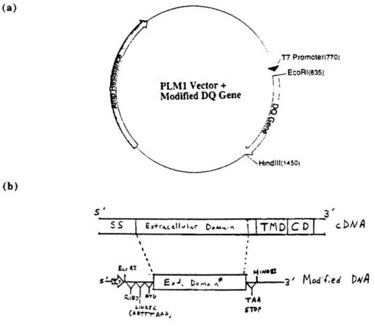
EcoRI and HindIII sites into the beginning and end of each modified gene, respectively. After the EcoRI site, a ribosomal binding site (AGGAGG) and spacer sequence (AATTTAAA) were added and the cDNA signal sequence was replaced with a Met start codon. Before the HindIII site, a stop codon (TAA) was inserted after the last codon (Fig. 1). PCR products were isolated, purified, and doubly digested with EcoRI and HindI. These gel purified fragments were then
ligated into a previously digested PLM1 vector behind a strong T7 promoter site. The ligations were transformed into DH5ct competent cells and grown on LB/Ampicillin (100 gg/ml) plates overnight. All colonies were picked and grown up overnight in LB/Ampicillin (100 tg/ml) media. The plasmid from each culture was isolated using the boiling prep method and EcoRI/HindIII, HincII (only cuts DQA gene inserts), and SmaI (only cuts DQB gene inserts) restriction enzyme digests were conducted. Two positive clones from each allele were transformed into BL21 (DE3) pLysS competent cells and grown on LB / Ampicillin /
Chloramphenicol (each antibiotic 100 pig/ml) plates overnight. Single colonies were picked into 5 ml of LB/0.2% glucose/Ampicillin/Chloramphenicol (same antibiotic concentrations) media and grown overnight to saturation. Fifty RI of saturated culture was added to 5 ml of the same media, grown to approximately 0.5 O.D., and then induced with addition of IPTG (0.5 mM).
After 1 h of induction, uninduced and induced cell samples were pelleted, lysed in SDS loading buffer at 100 degrees for 3', and then loaded onto an SDS-PAGE gel. All clones expressed well to give a protein product of approximately the correct molecular weight for each subunit (a =
20.8 KD, =22.5 KD).
One BL21 (DE3)pLysS clone from each allele was grown up and expressed on a 3 liter scale. The insoluble inclusion bodies were isolated from isotonically lysed cells, soluble
bacterial proteins and membrane fragments were removed with a deoxycholate wash and a series of Triton X-100/Tris (50 mM, pH 8.0) washes. SDS-PAGE analysis of the washes showed only
E. coli endogenous proteins. The inclusion bodies were dissolved in 8M urea, 1 mM DTT, 25
mM MES, and 10 mM EDTA, analyzed on SDS-PAGE, and found to be free of many E. coli endogenous proteins. Some inclusion bodies, however, were partially resistant to urea
solubilization and had to be dissolved in larger volumes of urea. Recombinant proteins were further purified by anion exchange HQ chromatography (Perseptives Biosystems).
Fig. 1. Construction of extracellular DQ a and
P
expression plasmids. (a) PLM1 expressionvector with T7 promoter and Amp resistance marker; (b) Truncated gene map; SS, signal sequence; TMD, transmembrane domain; CD, cytoplasmic domain; RIBO, ribosome binding site. (a) /T7 Promoter(770) EcoRI(835) PLM1 Vector + Modified DQ Gene HindIII(1450) (b) S S r~r~d~g... ~eM%~I Tt-DI c Dv sH ' 3 ' Peptide Synthesis
Peptides were either synthesized using standard t-Boc chemistry by the MIT biopolymer facility or using Fmoc chemistry by an outside provider (Anaspec). Couplings were checked at each cycle by ninhydrin assay and secondary amino acid couplings were done in the case of incomplete couplings. Transferin receptor (TfR) 334-343 (VQTISRAAAE), MB65KD 243-255
(KPLLIIAENVEGE), and FceR 104-122 (SQDLELSWNLNGLQADLSS) peptides were
biotinylated on the N-terminus using Pierce EZ-link Sulf-NHS-LC-LC-Biotin (equivalent to a two 6-aminocaproic acid linkers and a biotin molecule) (Chicz, 1994; Johansen, 1994). All
peptides were purified by reverse phase chromatography using C4 or C18 columns and were shown by MALDI-MS and amino acid analysis to contain a single peptide species.
Biotinylated Peptide Refolding Assay and Refolding Screen
Using the conditions established by Frayser et al. (1999), E. coli expressed HLA-DQ subunits (5 pg/ml each; 10 mg/ml total protein) and biotinylated peptide (100 gM) were diluted into an optimized buffer system consisting of 50 mM Tris (pH 8.5), 3 mM reduced glutathione,
0.3 mM oxidized glutathione, and 50% glycerol and then allowed to incubate for at least two
days. These optimized conditions were originally developed for the refolding of HLA-DRl, another human class II protein. In an attempt to increase the overall DQ refolding yields, the following parameters were varied in microscale refoldings: protein (0.2-20 jig/ml), peptide
(1-100 jiM), pH (5-9), glutathione [20-1 (reduced: oxidized ratio); keeping total concentration at 3.3
mM], and glycerol (0-50% w/v) concentrations. Since I did not have an antibody capture assay that detected a correctly folded molecule, a specific peptide binding assay was used. In this assay, a polystyrene plate was coated with streptavidin (10 ng/gl) and then blocked with phosphate-buffered saline (PBS), 3% BSA, 0.02% sodium azide. Fifty microliters of each refolding was diluted into 50 gl of dilution solution (PBS, 0.05% Triton-X-100, 0.1% BSA) in the streptavidin plate and allowed to incubate for 1 hour at room temperature. After washing with PBS, 0.05% Triton-X-100 (PBST), the amount of bound DQ was determined by incubating the plate sequentially with 1:10,000 rabbit-anti-DQ polyclonal serum, 1:10,000 goat anti-rabbit polyclonal serum labeled with horseradish peroxidase, and 2,2'-Azido-di-[3-ethylbenzthiazoline sulfonate (ABTS) substrate (Boehringer Mannheim). Between each step, the plate was washed thoroughly with PBST.
RESULTS AND DISCUSSION
We did not have any difficulties expressing the denatured inclusion bodies of the alleles listed in Table I and purifying them to homogeneity on anion exchange chromatography (Fig. 2).
When it came time to refold the molecules, however, we ran into several roadblocks (see Table II for ac combinations and peptides). Unlike the DRi system, we had no assays that detected the presence of correctly folded molecule (Fig. 3A). As a substitute, we developed an assay that detected whether biotinylated antigenic peptides were bound by DQ protein molecules (see Materials and Methods) and used it to screen conditions similar to those used by Frayser et
al.(1999) [20 mM Tris, 25% glycerol, 3 mM reduced glutathione, 0.3 mM oxidized glutathione,
pH 8.5]. As a positive control, we tested HLA-DR1 bound to biotinylated HA 306-318 (a
specific DRI binding peptide) and found that the assay detected the bound complex (Fig. 3B) [Note: The detection DQ polyclonal antibody serum also recognizes DR class II molecules]. Once folding conditions were found that led to high levels of specific peptide binding, we planned to set up larger refoldings with non-biotinylated peptide under the same refolding conditions. For all the u, -peptide combinations tested (Table II), however, we found no conditions that produced high levels of refolding compared to the DRI procedure.
For each expressed allele, we have bacterial glycerol stocks of DH5c cells for DNA isolation and BL21 (DE3) pLysS cells for subunit production in the -80' C freezer (Rm. 56-553). Within the same box, any purified plasmid DNA stocks are stored in the -80' C freezer. Finally, all purified DQ subunits, if any, are stored in the -20' C freezer (Rm. 56-567). All of these items are catalogued in the appendix of this thesis. Although we would not recommend refolding any of these DQ molecules, these DNA and protein stocks may prove useful for other class II studies, e.g. expression in the baculovirus system, antibody epitope studies, PCR/sequencing standards.
Fig. 2. Example of HLA-DQ a and $ protein expression and purification.
HLA-DQa1*0201 and 1*0302 BL21(DE3)pLys
Induction and Inclusion Body Purifications
a b c d e f
a, a1*0201 not induced; b, a1*0201, induced; c, ion-exchange purified
inclusion bodies (pH 7.0); d, P1*0302, not induced; e, 1*0302,
induced; f, ion-exchange purfied inclusion bodies (pH 9.0)
Table I: Compre ensive listing of available DQ constructs
Allele Haplotype cDNA E. Coli Construct Baculovirus?3
(short versions) (long versibns)
A1*0101 DQl(5) X X X1 A1 *0201 X X A1*0301 X2 A1*0501 X B1*0201 DQ2 X B1*0302 DQ3(8) X X B 1*0303 DQ3(9) X X B1*0402 DQ4.2 X B1*0501 DQ1(5) X X X Notes: 1 2 3
Both DQ1 genes cloned into one baculovirus transfer plasmid; both under polyhedrin promoter. A1 *0301 was cloned into a periplasmic expression vector (PET27b) under T7 promoter
These baculovirus constructs are not discussed in this chapter, but are included to provide a complete record of all available DQ constructs.
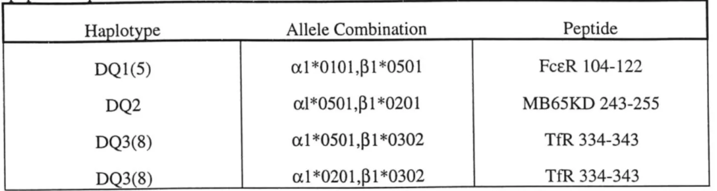
Table II: Subunit combinations tested in refolding screen. See Materials and Methods for peptide sequences.
Haplotype Allele Combination Peptide
DQ1(5) cl*0101,p1*0501 FcER 104-122
DQ2 al*0501,p1*0201 MB65KD 243-255
DQ3(8) a1*0501,P1*0302 TfR 334-343
DQ3(8) a1*0201,P1*0302 TfR 334-343
Fig. 3. DQ ELISA Construction - Detecting the formation of folded peptide complex. (A) Problems and solutions to ELISA development; (B) BioHA/HLA-DR1 standard curve using
Avidin ELISA as shown in (A).
(A)
DQ ELISA Problem and Solutions??
~Me 54cl
144j .' ?~.
Pnhipeq with mnnnelnnjit Ab ranture F1 14;A en fat-1-I SI'. AVIDiJA 6: I (B) E" C 0 a
I. Monoclonalantbody HB10 (anti-DQ) recognizes free a and b, as well as complex.
2. HSI" sandwhich ELISA is not as sensitive as comparable DR ELISA.
The init of detection greater than 3 ng.
3. Otheranti.DQ uwdonoc al sntibodi.n lab (HBIIO. SPVL3) eitherdo not recognize Kz DQ8 or are not sensitive enough.
1.2 0.8 0.6 0.4 0.2 0.1
BloHAIHLA-DR1 BINDING CURVE
Streptavidin ELISA SW O -S -0 SN HLA-DR (ng) 10 100 1 A
III. HLA-D08 (cl*0201, 1*0302) and DQ9 (cl*0201, 1*0303)
MHC Class II Proteins:
The Role of j57 Polymorphism in Complex Structure and Stability
2INTRODUCTION
Through extensive population studies, the incidence of insulin-dependent diabetes mellitus (IDDM) has been shown to be strongly correlated with the presence of particular MHC class II HLA-DQ genes (Morel, 1988; Todd, 1987). HLA-DQ8 (al*0301, 1*0302) and
HLA-DQ6 (al *0 102, 1*0602), for example, show predisposition and protection against IDDM
development, respectively. A comparison of many such pairs revealed that protection alleles have Asp at P57, while susceptibility alleles have Ala, Val, or Ser at P57. Among class II MHC proteins in general, Asp is present at position 157 in -60% of human alleles [HLA-DR (77%),
-DQ (53%), -DP (49%)]. In all class II structures solved to date (Brown, 1993; Dessen, 1997;
Fremont, 1996; Fremont, 1998; Ghosh, 1995; Murthy, 1997; Scott, 1998; Smith, 1998; Stem, 1994), this Asp makes a salt bridge with a conserved Arg at position 76 in the a chain. Since there are no structures of non-AspP57 alleles, the role that this particular polymorphism plays structurally and in IDDM etiology is still unclear.
Previously, others have observed that DQ8 shows a unique binding preference for peptides with acidic residues near the C-terminus (Kwok, 1995; Kwok, 1996a, Kwok, 1996b; Nepom, 1996), presumably by interacting'with Arga76 residue, which would be left unpaired in the absence of an acidic residue at P57, or with other residues within P9 pocket. Modeling of non-AspP57 alleles, however, shows that the Arga76 may be too far from the P9 side chain to influence peptide preferences at this position (Reizis, 1998). Previously, others have compared the relative stability of various complexes by comparing their ability to resist ap chain
dissociation in a non-boiled sample on SDS-PAGE (Gorga, 1987). This property is often referred to as the "SDS-stability" of a particular complex. Complexes of DQ8 (c 1*0301,
1*0302) with endogenous peptides are more prone to SDS-induced chain dissociation than for
DQ9 (a1*0301, 1*0303), which is identical to DQ8 except having Asp at 157 (Reizis, 1997).
Like DQ9, DQ6 also has an intact salt bridge between Asp 57 and ArgCC76 (see MHC Introduction). The majority of DQ6 is SDS-stable, while DQ8 exists in both SDS-sensitive and resistant populations (Reizis, 1997). Unexpectedly, DQ8 and DQ6 persist on cells for similar
lifetimes as measured by pulse chase experiments with surface iodinated molecules (t1/2 approximately 24 hours). These results suggest that SOS-stability may not be an accurate measure of overall stability of one complex versus another and that more detailed
thermodynamic analysis must be done to address this question.
In this study, we analyze the thermodynamic stability of empty and peptide loaded DQ8 (x1*0201, 1*0302) and DQ9 (xl*0201, 1*0303) complexes using circular dichroism and
thermal denaturation studies. We find that DQ8 and DQ9 are both thermodynamically stable molecules, but have somewhat different secondary structures. Since this change is most likely concentrated within the peptide binding site, these results may help to explain why these proteins bind a set of three peptides with different affinities.
MATERIALS AND METHODS
Preparation of HLA-DQ Molecules
Soluble HLA-DQ8 (cd*0201, 11*0302) and DQ9 (cl*0201, 11*0303) were produced in insect cells using separate baculovirus recombinants for a (EDIVAD ... IPAPMS) and
1
(RDSPED ... WRAQSE) subunits as described in Stem et al. (1992) and Raddrizzani et al.
(1997). The recombinant baculoviruses were a gift from Dr. Francesco Sinigaglia (Roche
Milano, Italy). For protein production, High Five (BTI-TN-5B1-4) insect cells were grown in 6 liter suspension cultures in Gibco BRL Sf900-II serum-free medium (see Appendix) and were
determined using a SPVL3 capture (anti-DQ complex specific antibody; Spits, 1984) and IVD12 detection (anti-DQ8/9 monoclonal antibody; also known as HB144; Giles, 1983) sandwich
ELISA by Raddrizzani et al (1997). This assay can specifically detect folded ac complex in the
presence of denatured subunits and other medium proteins (see Appendix). After 5 days, the cells were removed by centrifugation (4500 x g, 45 min.), and protease inhibitors (1 mM PMSF,
5 mM EDTA, 1 mM iodoacetamide) and sodium azide (0.02%) were added to the supernatant.
The supernatant was concentrated to 1/10 the original volume and then precipitated debris was removed by centrifugation. Finally, the DQ protein was purified from the clarified supernatant
by immunoaffinity chromatography using mAb IVD12 (anti-DQ3 P) coupled to CNBR sepharose
as described (Raddrizzani, 1997). Protein was eluted with 50 mM CAPS, pH 11.5 and
neutralized with one-third volume of 300 mM sodium phosphate (NaPi), pH 6.0 to give final pH
- 6.5. The same protease inhibitor cocktail was added to the eluted protein. The theoretical
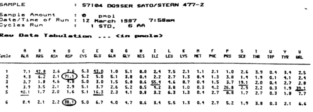
extinction coefficient of 66720 M1*cm- was used for all protein concentration and subsequent 9 calculations. This value was verified experimentally by N-terminal sequencing (Fig. 1) and amino acid analysis of empty DQ9 (data not shown). The overall purified yield was
approximately 0.5 mg per 6 liter culture for both DQ8 and DQ9.
To prepare peptide complexes, affinity-purified empty DQ protein (- 1 mg/ml) was immediately mixed with 100 gM peptide and allowed to incubate at 370 C for 3 days. Complexes were isolated using a Pharmacia Superdex 200 HR gel filtration column with a
Pharmacia HiTrap Q precolumn (PBS, pH 6.9, 0.4 ml/min). DQ peptide complexes do not bind
the HiTrap Q column in this buffer but some non-DQ protein contaminants were removed. DQ complexes separated clearly from aggregates and free peptide on the Superdex column. Any remaining empty DQ protein binds partially to the HQ column and elutes later than peptide complex. For low-affinity peptide complexes, 250 gM excess peptide was immediately added to the size exclusion fractions to maintain the equilibrium towards complex formation.
All empty and peptide loaded proteins were diluted into Laemili loading buffer
boiling. The percentage of SDS-resistant complex was estimated by scanning the not-boiled
(NB) lane and dividing the ac complex band intensity by the total band intensity in the same
lane. The NIH Image program was used to quantitate the intensity in the scanned gel bands. Using this procedure, we assume that free subunits stain with approximately the same intensity as complex bands.
Fig. 1. N-terminal sequencing of empty DQ9. The underlined residues correspond to signal
from either the a (EDIVAD ... ) and
P
(RDSPED ... ) chains of DQ9. For some sequencepositions, both a and
P
contain the same residue (circled residues); so the signal at that position is higher than the previous residue.SA-PL-E S71e4 DOSSER SATOZSTERN 477-2
Sample Amount : c mol
Date/Tme of Run: 12 Mmr-ch 1997 7:-B-som
Cycles Run I STD. 6 AA
M-L
D TA~,& n*_Lw --- <:Lu, pwmX 3
A R N a C E 0 6 H L L K ti P s 1 u y V Cvcle ALR ARG flm RSP CYS GLU 6.LH SLY KIS ILL LEU LYS MET PHE PRD SEY TIR TRP TYW U3
1 7.1 42.8 Z.6 S3 jLp 1.0 5.1 8.0 2.4 7.S 2.1 1.1 2.1 1.0 2.6 3.9 0. 3.1 Z.S 2 4.3 6.2 2.1 71.1 S.2 S.0 5.1 3.8 0.4 2.2 2.7 1.3 0.4 1.3 3.0 1.1 1.9 0.1 1.1 I., 3 3.7 1.8 4.6 5.0 3.0 1.6 6.8 0.6 55.4 4.6 0.7 0.1 1.5 3.7 19.1 2.0 0.4 2.? 2.8 4 3.9 3.S 2.1 2.9 5.1 3.7 2.6 5.2 0.5 4.2 3.8 1.0 .3 4.2 26.8 2.9 2.2 0.3 1.9 Ju1 S 40.1 1.7 2.0 1.6 5.1 16.3 2.3 4.1 0.8 3.2 6.3 1.3 0.4 2.7 8E.8B 1.7 2.7 0.3 1.8 7.7 6 e. 2.1 2.2 S.0 6.7 4.0 4.7 0.6 3.4 6.s 1.3 0.4 2.7 s.Z 1.9 3.8 0.3 2.1 6.6 Peptide Synthesis
Peptides were either synthesized using standard t-Boc chemistry by the MIT biopolymer facility or using Fmoc chemistry by the Roche peptide facility. Couplings were checked at each cycle by ninhydrin assay and secondary amino acid couplings were done in the case of
incomplete couplings. TfR (334-343) (VQTISRAAAE) and GAD (253-265)
(IARFKMFPEVKEK) peptides were biotinylated on the N-terminus using Pierce EZ-link Sulf-NHS-LC-LC-Biotin (LC, 6-amino-hexanoic acid; NHS, N-hydroxysuccinimide ester), and
GFKA7 (GFKAAAAAAA) was biotinylated by coupling two 6-aminocaproic acid spacers and
one free biotin molecule on the N-terminus, sequentially, using standard amino acid diisopropyl carbodiimide coupling conditions. All peptides were purified by reverse phase chromatography
using C4 or C18 columns and were shown by HPLC to contain a single peptide species that had the expected molecular weight by MALDI-MS.
Peptide Binding Assays
To determine the Kd for each peptide (TfR, GAD, and GFKA7) in a direct binding assay,
DQ was diluted to 50 nM in PBS, 0.2% azide, 1% octylglucoside, protease inhibitor cocktail
(same as above) and then 50 gl aliquots were prepared in silanized centrifuge tubes. Various concentrations of each biotinylated peptide were prepared (2 x 10⁻⁴ to 2 x 10⁻⁹ M) in the same binding buffer. Fifty gl of each peptide titration sample was added to triplicate DQ aliquots to give a final protein concentration of 25 nM. These samples were incubated at 370 C for 4 days,
followed by assay for bound peptides using an antibody capture and streptavidin-Europium detection sandwich ELISA. The ELISA plate was prepared as follows: 100 gl of 10 ng/gl
SPVL3 (anti-DQ complex specific; (Spits, 1984)) in PBS, 0.02% azide were added to each well
of a 96 well polystyrene plate, followed by incubation at 370 C, 2 hours. The plate was then
blocked with PBS containing 3% BSA and 0.02% azide at room temperature for 2 hours. Prior to loading the samples, the blocked ELISA plate was washed with Tris-buffered saline, 0.1% Tween-20 (TBST). Twenty-five gl of each sample was diluted into 75 gl dilution solution (PBS containing 0.05% Triton-X100 and 0.1% BSA) and incubated with the plate at 370 C for 30
minutes. After washing, the DQ bound peptide was detected by adding 100 tl of a 1:1000
TM
dilution of streptavidin-Eu in Wallac DELFIA Assay Buffer to each well. After the plate was incubated at 370 C for 15 minutes, the plate was washed again and developed with 200 jl/well of
TM
Wallac DELFIA Enhancement Solution at room temperature for 5 minutes. Bound Europium
TM
was detected using a Wallac VICTOR 1420 Multilabel Counter and the default Europium detection protocol (1 second/well counting time; normal aperture).
Circular Dichroism Analysis
For circular dichroism (CD) analysis, empty and peptide loaded complexes (0.5 mg/ml) were dialyzed in a 10K molecular weight cut-off Slide-A-Lyzer cassette (Pierce) into 10 mM sodium phosphate, pH 7. GFKA7 (100 gM), GAD (100 gM) or TfR (250 piM) free peptide was
added to the dialyzing buffer of each respective peptide complex to prevent peptide dissociation. Dichroism measurements were made using a 1 mm path length cuvette in an AVIV 60DS CD spectrophotometer. Wavelength scans were obtained using 1.5 nm bandwidth, constant 100 C temperature, and 1 nm sampling with 5 seconds dwell time per point. All experimental scans were adjusted for background signal by subtracting out the signal from a dialyzing buffer scan.
CD spectra were reported on a per residue basis; the empty, GFKA7, TfR, and GAD proteins
have 382, 392, 392, and 395 residues, respectively.
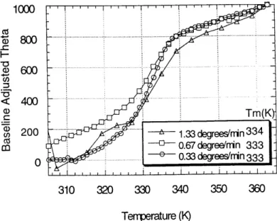
Thermal denaturation data was obtained using 2 nm bandwidth, 224 nm detection wavelength, 100 C to 90' C range with 1 C intervals, a 1 minute equilibration time and a 1 minute dwell time at each temperature. The relationship of the unfolding transition to an
irreversible denaturation that occurs in the same temperature range was investigated by recording the dependence of the midpoint temperature on the rate of the scan for overall scan rates 0.33' C/min to 1.33' C/min (Sanchez-Ruiz, 1992). No dependence was observed over the rates tested, indicating that the two-state approximation can be used at the experimental scan speeds (Fig. 2).
For determination of thermodynamic ACp and AH, thermal denaturation data were fit to a seven-parameter function that describes a two-state transition:
0 = (Ou +muT) + (Of - Ou)+ T(mf - mu)
I = exp[ AH+ ACp Tm T
+exp- -- l+ln-H
RT RTm )iJ
where Ou and mu describe the slope and y-intercept of the unfolded state baseline, Of and mf describe the slope and y-intercept of the folded state baseline, Tm is the midpoint of the
Fig. 2. The midpoint temperature is independent of the scan rate. The complex tested was DQ8
bound to the GAD peptide. The theta (0) units are deg*cm2/(dmol residues). The uncertainty in each Tm calculation is approximately +I 1 degree K.
1000 800 6 0 .-- --ci), < 400 T)rTm(Km cn 200 ... 1.33 degrees'n 33 4 Cz CO co67 degree/nin 333 0 ... ... 0.33degrees/nin 333 310 320 330 340 350 360 Tem-perature (K)
transition (where AG = 0), ACp is the heat capacity change upon unfolding, and AH is the
enthalpy of unfolding at the Tm. For each melt, the Tm was determined separately taking the first derivative of the melt data and selecting the temperature at which the slope is at its highest value. These thermodynamic values derived in this analysis are likely to be dependent upon the concentration at which the equilibrium is measured, and therefore only hold for the concentration ranges tested (0.1-1 mg/ml).
RESULTS
Synthetic Peptide Binding
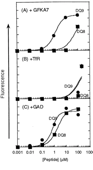
Previously, Raddrizzani et al (1997) showed that GFKA7 binds to many DR and DQ proteins and can be used as a promiscuous binder in parallel peptide competition assays. Since the peptide bound well to DQ7 (x1*0501, 0 1*0301), we tested its binding to DQ8 and DQ9 in a direct binding assay using biotinylated peptide. As shown in Fig. 3, bioGFKA7 binds to DQ9
with an apparent Kd of approximately 0.79 gM; whereas the same peptide binds poorly to DQ8 with a Kd of approximately 62 gM. Since their maximal binding signals are approximately the
same (~2x 10⁵) and the same amount of protein was used for each assay, the binding capacity of both proteins are approximately equivalent.
(A) + GFKA7 DQ9 Q8
-
-(B) +Tf R-DQ9 DQ8 C)+GAD S DQ DQ8 S(
- -0.001 0.01 0.1 1 10 100 eptide] ( M)Fig. 3. DQ8 and DQ9 Direct Binding Assay with Three Peptides [(A) GFKA7, (B) TfR,
and (C) GAD]. Kd values for each binding curve are listed in Table I.
1000
To further test this differential peptide binding between DQ8 and DQ9, we tested binding of TfR (334-343), a slightly truncated version of the peptide [TfR (332-347)] previously isolated from affinity purified wildtype DQ8 (cl*0301, 1*0302) (Chicz, 1994). Both molecules bound the peptide weakly with Kd's greater than 100 pM, but DQ9 did show a slightly higher binding signal than DQ8 at 100 gM peptide (Fig. 3).
A
C: U 0
Previously, Kwok et al. identified a GAD peptide (253-265) that showed differential binding between DQ7 (cl*0301,1*0301), DQ8 (ccl*0301,p1*0302), and DQ9
(cxl*0301,1*0303) (Kwok, 1996a). In a whole cell peptide binding assay using 10 gM
biotinylated peptide, DQ8 showed the highest relative binding, DQ9 showed approximately 25% of the maximal binding, and DQ7 showed no binding. Using our empty molecules, this peptide was tested in a direct binding assay as described above. Unlike Kwok's results, our DQ9 (a1*0201, 1*0303) bound the peptide with a three-fold higher affinity relative to DQ8
(cl*0201,1*0302) (Fig. 3). This difference in peptide binding specificity, however, could be the presence of the cl*0201 chain, instead of cl*0301. On the other hand, the previous results were obtained using peptide-bound DQ on whole cells at only one concentration of peptide. It is likely that our assay gives a closer approximation of the actual binding affinities of this peptide for DQ8 and DQ9.
SDS-Stability Analysis
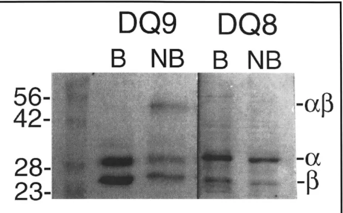
Since the biochemistry of empty DQ molecules has not been described, we first analyzed
DQ8 and DQ9 without peptides to determine the intrinsic SDS-stability of the proteins
themselves (SDS-stability previously defined on page 24). Surprisingly, DQ9 was partially SDS-stable (as much as 40%); whereas DQ8 was completely SDS-sensitive (Fig. 4). To our knowledge, this is the first instance of an apparently empty MHC class II molecule with SDS-stable character. This property, however, could be due to the presence of bound peptides obtained during insect cell expression. To address these concerns, DQ9 was subjected to N-terminal sequencing (Fig. 1) and amino acid analysis; both conformed to expected values with no extraneous signals from a bound peptide. To check if SDS-stability were dependent on the cell
type used for expression, Sf9 insect cells, instead of High Five, were used for expression and again the empty protein was partially SDS-stable (data not shown). We, however, cannot exclude the possibility that a heterogeneous peptide population binds DQ9, where each peptide represents a small percentage of the total bound peptide. For all the peptides tested in the direct
binding assays, none of them affected the above electrophoretic properties of either DQ8 or DQ9 (data not shown). Since all of the peptides bind with moderate (submicromolar) to weak
affinities, it is possible that the peptides dissociated from the complexes before a SDS-stable complex could be separated from free subunits in the gel.
DQ9
B
NB
DQ8
B
NB
Fig. 4. Reduced SDS-PAGE of Empty DQ8 and DQ9. Purified proteins were loaded
onto 12.5% gel before (NB) and after boiling for 2 minutes (B) in Laemili buffer. ac, a, and