HAL Id: hal-01063652
https://hal.archives-ouvertes.fr/hal-01063652
Submitted on 15 Sep 2014HAL is a multi-disciplinary open access archive for the deposit and dissemination of sci-entific research documents, whether they are pub-lished or not. The documents may come from teaching and research institutions in France or abroad, or from public or private research centers.
L’archive ouverte pluridisciplinaire HAL, est destinée au dépôt et à la diffusion de documents scientifiques de niveau recherche, publiés ou non, émanant des établissements d’enseignement et de recherche français ou étrangers, des laboratoires publics ou privés.
Diversity and catalytic potential of PAH-specific
ring-hydroxylating dioxygenases from a
hydrocarbon-contaminated soil.
Florence Martin, Laure Malagnoux, Fabien Violet, Jean Jakoncic, Yves
Jouanneau
To cite this version:
Florence Martin, Laure Malagnoux, Fabien Violet, Jean Jakoncic, Yves Jouanneau. Diversity and cat-alytic potential of PAH-specific ring-hydroxylating dioxygenases from a hydrocarbon-contaminated soil.. Applied Microbiology and Biotechnology, Springer Verlag, 2013, 97 (11), pp.5125-35. �10.1007/s00253-012-4335-2�. �hal-01063652�
Diversity and catalytic potential of PAH-specific ring-hydroxylating dioxygenases from a hydrocarbon-contaminated soil Florence Martin1,2 , Laure Malagnoux1,2 , Fabien Violet1,2 , Jean Jakoncic3 and Yves Jouanneau1,2
1CEA, DSV, Laboratoire de Chimie et Biologie des Métaux, F-38054 Grenoble Cedex 9,
France.
2CNRS UMR 5249, Grenoble, France.
3Brookhaven National Laboratory, National Synchrotron Light Source, Upton, NY, USA
Running title : Catalytic potential of PAH-specific RHDs from soil
*Corresponding author: Yves Jouanneau
LCBM/iRTSV, UMR 5249 CNRS/UJF/CEA F-38054 Grenoble Cedex 9, France.
Tel. : 33 (0)4.38.78.43.10 ; Fax : 33 (0)4.38.78.54 87 Email : yves.jouanneau@cea.fr
Abstract
1
Ring-hydroxylating dioxygenases (RHDs) catalyze the initial oxidation step of a range of 2
aromatic hydrocarbons including polycyclic aromatic hydrocarbons (PAHs). As such, they 3
play a key role in the bacterial degradation of these pollutants in soil. Several PCR-based 4
methods have been implemented to assess the diversity of RHDs in soil, allowing limited 5
sequence-based predictions on RHD function. In the present study, we developed a method 6
for the isolation of PAH-specific RHD gene sequences of Gram-negative bacteria, and for 7
analysis of their catalytic function. The genomic DNA of soil PAH degraders was labeled in 8
situ by stable isotope probing, then used to PCR amplify sequences specifying the catalytic 9
domain of RHDs. Sequences obtained fell into five clusters phylogenetically linked to RHDs 10
from either Sphingomonadales or Burkholderiales. However, two clusters comprised 11
sequences distantly related to known RHDs. Some of these sequences were cloned in-frame 12
in place of the corresponding region of the phnAIa gene from Sphingomonas CHY-1 to 13
generate hybrid genes, which were expressed in E. coli as chimerical enzyme complexes. 14
Some of the RHD chimeras were found to be competent in the oxidation of 2- and 3-ring 15
PAHs, but other appeared unstable. Our data are interpreted in structural terms based on 3D-16
modeling of the catalytic subunit of hybrid RHDs. The strategy described herein might be 17
useful for exploring the catalytic potential of the soil metagenome and recruit RHDs with new 18
activities from uncultured soil bacteria. 19
Keywords : Dioxygenases ; metagenomic DNA ; catalytic domain ; hybrid enzymes ; 20
bioremediation 21
Introduction 1
2
Ring-hydroxylating dioxygenases (RHDs) are bacterial metalloenzymes that catalyze the first 3
step in the biodegradation of a variety of aromatic hydrocarbons. Some of them are able to 4
oxidize highly resistant compounds such as polycyclic aromatic hydrocarbons (PAHs) 5
(Jouanneau et al. 2011; Jouanneau et al. 2006). RHDs are two or three-component systems, 6
consisting of an oxygenase which bears the enzyme active site, and specific electron carriers 7
which deliver reductant to the oxygenase for oxygen activation during catalysis (Parales and 8
Resnick 2006). The oxygenase component is most often an heterohexamer α3β3. Structural
9
studies on representative oxygenases showed that the α subunit is composed of two domains, 10
a Rieske domain that binds a [2Fe-2S] cluster, and the catalytic domain containing the 11
substrate-binding site and a mononuclear Fe(II) center where oxygenation of the substrate 12
takes place (Ferraro et al. 2005). Bacterial RHDs form a large enzyme family that can be 13
divided into four to five subgroups based on phylogenetic comparisons of their α subunit 14
amino acid sequences (Kweon et al. 2008; Nam et al. 2001). In this respect, PAH-specific 15
RHDs fall into two clusters of enzymes, those found in Gram-positive bacteria and those 16
found in Gram-negative bacteria (Jouanneau et al. 2011). 17
While the classification of RHDs essentially relies on enzymes found in cultured bacteria, 18
there is compelling evidence that bacterial species recalcitrant to cultivation outnumber 19
cultivable ones by several orders of magnitude in soils and sediments (Cole et al. 2010). 20
Hence, the biodiversity of bacterial enzymes in soil, including RHDs, is certainly much 21
greater than that considered in current classifications, as exemplified by a recent study of 22
dioxygenase genes by a metagenomics approach (Iwai et al. 2010). 23
The diversity of RHDs associated to PAH-polluted sites has been investigated by PCR-based 24
analysis of sequences encoding a portion of the α subunit gene. In marine sediments, RHD 25
genes appeared to be mainly represented by sequences related to the Cycloclasticus phnAI 1
gene (Lozada et al. 2008), whereas in marine microbial mats subjected to oil contamination, 2
gene sequences similar to nahAc found in Pseudomonads and to nagAc and pahAc found in 3
Betaproteobacteria were mostly detected (Bordenave et al. 2008). A variety of gene sequences 4
related to RHDs from Gram-negative bacteria have been found in soils and sediments 5
polluted by PAHs (Gomes et al. 2007; Ni Chadhain et al. 2006; Park and Crowley 2006; Zhou 6
et al. 2006). As a means to estimate the relative abundance of these genes in soil, quantitative 7
methods based on qPCR have been proposed (Cebron et al. 2008; Ding et al. 2010). 8
Differences in RHD biodiversity observed on PAH-polluted sites might reflect natural 9
variability, but they could also arise from the variety of primer sets used to PCR amplify 10
different portions of the α subunit genes (Iwai et al. 2011). 11
In a recent study, we explored the bacterial diversity of a PAH-contaminated site by stable 12
isotope probing, and found out that Betaproteobacteria were the main microorganisms 13
responsible for the degradation of the tricyclic hydrocarbon phenanthrene in soil (Martin et al. 14
2012). In order to examine the diversity of PAH-specific RHDs on the same experimental 15
site, 13
C-labeled DNA extracted from soil was used as a template to amplify a large segment 16
of the α subunit gene, extending over the catalytic domain. Phylogenetic analysis revealed 17
that the sequences obtained were related to RHDs found in Sphnigomonadales and 18
Burkholderiales. As a means to further investigate the enzymatic function associated to these 19
microbial sequences, some of them were inserted in place of a gene portion specifying the 20
catalytic domain of the RHD from Sphingomonas CHY-1 (Jakoncic et al. 2007b; Jouanneau 21
et al. 2006). Hybrid genes were expressed together with other components of strain CHY-1 22
enzyme in E. coli, and the activity of the resulting RHDs was assayed in the oxidation of 2- 23
and 3-ring PAHs. This study represents one of the first attempts to link partial sequences 24
retrieved from soil DNA to enzymatic function using a strategy based on exchange of 1
catalytic domains. 2
MATERIALS AND METHODS
1 2
Bacterial strains, plasmids and culture conditions
3
Bacterial strains and plasmids used in this study are listed in Tables 1 and 2. E. coli 4
strains were grown in rich medium (Luria-Bertani) at 37°C with appropriate antibiotics as 5
previously described (Demaneche et al. 2004). 6
7
Primer design and PCR amplification of sequences internal to RHD α subunit genes
8
Primers used in this study were designed after analysis of sequence alignments of 9
RHD alpha subunits from representative and well established PAH degraders taken among 10
Gram-negative bacteria. Sequences considered in the alignment are encoded by the following 11
genes : phnA1a from Sphingomonas sp. CHY-1 (Demaneche et al. 2004), bphA1f from 12
Sphingobium yanoikuyae B1 (Chadhain et al. 2007), bphA1f from Novosphingobium 13
aromaticivorans (Romine et al. 1999), phnA1f from Sphingomonas sp. LH128 (Schuler et al. 14
2009), phnA1 from Cycloclasticus sp. A5 (Kasai et al. 2003), the phnAc genes from 15
Burkolderia sp. RP007 (Laurie and Lloyd-Jones 1999), Acidovorax sp. NA3 (Singleton et al. 16
2009), Alcaligenes faecalis AFK2 (accession # AB024945), Delftia sp. Cs1-4 (formerly 17
Burkholderia Cs1-4; accession # AY367786), Burkholderia sp. DBT1 (Di Gregorio et al. 18
2004), and ndoB from Pseuomonas putida NCIB9816 (Simon et al. 1993). Two short, 19
partially conserved sequences were selected for primer design, corresponding to 20
FVCNYHGW and IGETSYR in PhnA1a from Sphingomonas sp. CHY-1. These sequences 21
delimited a portion of the protein (residues 98-412) encompassing the almost entire catalytic 22
domain (Jakoncic et al. 2007b). 23
One set of degenerate primers was intended to target a wide range of RHD genes from 24
Proteobacteria. The nucleotide sequences of the forward RHDf2) and reverse (Phn-25
RHDR3) primers were TTCRYBTGCAAYTATCAYGGYTGG and 1
CCYCKRTAVCWKGTYTCKCCRA, respectively. Another pair of primers, with a lower 2
level of degeneracy, was used to specifically amplify sequences relevant to RHDs from 3
Betaproteobacteria. The sequences of the forward and reverse primers were: 4
TTCASYTGCACYTATCACGGCTGG (RHD-Beta-Grp1f) and
5
CCKCKRTARGASGTYTCRCCAA (RHD-Beta-Grp1r). The template was a preparation of 6
13
C-labeled DNA obtained through stable isotope probing. The DNA sample was extracted 7
from a soil microcosm incubated for 5 days with 310 ppm 13
C-phenanthrene, then purified by 8
CsCl isopycnic ultracentrifugation as previously described (Martin et al. 2012). PCR was 9
conducted with a Tpersonal thermocycler (Biometra) in a final volume of 50 µl consisting of 10
100 ng template DNA, 1× PCR buffer, 1.5 mM MgSO4, 200 μM of each dNTP, 0.3 μM of
11
each primer, 1 U of Kod Hot start DNA polymerase (Novagen). Amplification conditions 12
were as follows: 95°C for 3 min, then 30 cycles of denaturation for 30 s at 95°C, annealing 13
for 20 s and extension at 70°C for 16 s. Annealing was performed at 52°C for 10 cycles then 14
at 50°C for 10 cycles and finally at 48°C for 10 cycles. When the RHD-Beta-Grp1f/r primer 15
pair was used, PCR conditions were similar except that annealing was carried out at 63°C for 16
20 s and extension at 70°C for 15 s. 17
18
Construction of clone libraries and sequence analysis of amplicons
19
PCR products obtained with the Phn-RHDf2/R3 primer set were cloned into pSTBlue-1 20
using the perfectly blunt cloning kit (Novagen, Merck). Clones were screened for the 21
occurrence of an insert of the correct size by PCR using the same primer set. A selection of 22
positive clones was grown in a 96-well microtiter plate, resulting in a first library named 23
DcatdioxA. A second library called DcatdioxB was obtained by cloning PCR products 24
obtained with the RHD-Beta-Grp1f/r primers into pJET1.2 (Fermentas). The cloned inserts of 25
both libraries were subjected to nucleotide sequencing on both DNA strands (as performed by 1
either Beckman Coulter genomics or Eurofins). The resulting sequences were translated, then 2
aligned and compared using Clustal X. Phylogenetic analysis was performed by the neighbor-3
joining clustering method with bootstrap analysis, using the One-click software available on 4
the www.phylogeny.fr website (Dereeper et al. 2008). The tree was formatted with Treedyn. 5
Nucleotide sequences described in this study were deposited in the EMBL-EBI database 6
under accession numbers HE795402-HE795451 (DcatdioxA library) and HE774402-7
HE774466 (DcatdioxB library). 8
9
Construction of hybrid RHD genes
10
Hybrid genes were constructed by replacing a portion of the CHY-1 phnA1a gene as 11
follows. First, the phnA1aA2a pair of genes encoding PhnI was subcloned as a XbaI-HindIII 12
fragment from pSD8 into pUC18 to give pFMA1. In this construction, the resulting PhnI α 13
subunit carried a His-tag at the N-terminus. Plasmid pFMA1 was then used to replace a 14
fragment internal to phnA1a by an appropriate sequence derived from the DcatdioxA clone 15
library as outlined in supplementary figure S1. To this end, PCR fragments were amplified by 16
using six cloned sequences, U3-18, U3-60, U3-72, U3-89, U3-112 and U3-116 as templates 17
and primers introducing restriction sites for RsrII and AgeI at the 5’- and 3’-end, respectively. 18
The fragments were digested by these two enzymes, and then cloned into corresponding sites 19
of pFMA1 to generate hybrid genes showing a replacement of a 695 bp segment between 20
nucleotide 545 and 1240 of the phnA1a coding sequence (Table 2). Two additional 21
constructions were made using U3-16 and U3-124 as templates and primers introducing RsrII 22
and MluI sites at the 5’- and 3’-end, respectively. The fragments were cloned into pFMA1, 23
resulting in hybrid genes with a 580-bp sequence substitution between nucleotide 545 and 24
1125 (Table 2). All constructions were checked by nucleotide sequencing on both strands. 25
1
Overexpression and in vivo assays of RHD chimeras
2
Plasmid pFMA1 and vectors derived thereof carrying hybrid genes (Table 2) were 3
introduced into strain JM109 together with plasmid pIBA34 by co-transformation. Plasmid 4
pIBA34 carries the phnA3 and phnA4 genes encoding the ferredoxin and reductase 5
components of CHY-1 RHD, respectively. The PhnA4 reductase was His-tagged at the N-6
terminus. Transformants were grown at 37°C until bacterial density reached 1.0 (OD600), then
7
induced with 1 mM isopropyl β-D-1-thiogalactopyranoside (IPTG) and further incubated at 8
25°C for about 20 h. Cultures were centrifuged, washed and resuspended to a bacterial 9
density of 2.0 (OD600) in M9 medium containing 20 mM glucose. Bacterial suspensions were
10
distributed in 2-ml Eppendorf tubes (1ml/tube), in which 0.5 µmole of PAH had been 11
introduced as a solution in acetone. After solvent evaporation, bacteria were incubated on a 12
rotary shaker at 25°C for 6 h. Tubes were centrifuged 2 min at 12,000 g, then 0.9 ml of 13
supernatant medium was mixed with 9 µl of 2,3-dihydroxybiphenyl (0.1 or 1 mM; Sigma-14
Aldrich) as an internal standard. The aqueous fraction was extracted with an equal volume of 15
ethyl acetate. Dried extracts were taken up in 0.2 ml acetonitrile and analyzed by GC/MS as 16
n-butylboronate (NBB) derivatives as previously described (Schuler et al. 2008). Anthracene 17
dihydrodiol was instead reacted with a silylation reagent (Silyl-991; Macherey-Nagel). 18
Dihydrodiol derivatives were quantified on the basis of peak area using calibration curves 19
generated with known amounts of naphthalene 1,2-dihydrodiol for NBB products, or 20
anthracene 1,2-dihydrodiol for trimethylsilyl derivatives (Jouanneau et al. 2006). Salicylate 21
hydroxylase activity was assayed as described previously (Demaneche et al. 2004). Briefly, 22
induced cells were resuspended in 10 ml M9 medium as above, then incubated with 1 mM 23
salicylate for 6 h. After pelleting bacteria by centrifugation and ethyl acetate extraction, 24
catechol was detected by GC/MS as a NBB derivative. 25
1
Purification of his-tagged oxygenase components
2
The his-tagged PhnI oxygenase produced in JM109(pFMA1), as well as enzyme 3
chimera produced from similar expression systems, were tentatively purified through a 4
procedure inspired from that described previously (Jouanneau et al. 2006), with 5
simplifications. Cultures of recombinant bacteria (0.8 L) were grown in 2-L flasks to an OD600
6
of 1, then supplemented with 50 µM Fe(NH4)2(SO4)2 and induced with 1 mM IPTG at 20°C
7
for 20 h. Bacteria were harvested by centrifugation and kept frozen at -20°C. Frozen pellets 8
were resuspended in twice the volume of PGE buffer consisting of 25 mM potassium 9
phosphate pH 7.5, 0.5 M NaCl, 5% glycerol, 5% ethanol and 2 mM β-mercaptoethanol. The 10
following steps were performed under anoxic conditions with argon-saturated buffers. After 11
treatment with 0.5 mg/ml lysozyme for 20 min, suspensions were subjected to ultrasonication for 5
12
min on ice with a Vibra Cell apparatus operated in pulse mode (Fisher Bioblock Scientific, 13
Illkirch, France). The lysates were centrifuged at 12,000 g for 20 min, and the resulting cell 14
extracts were applied onto small columns (1.5 ml) of immobilized-Co2+
affinity 15
chromatography resin (TALON, BD Biosciences Ozyme, France). The columns were washed 16
successively with 15 ml of PGE buffer and 10 ml of PGE containing 10 mM imidazole. The 17
proteins were eluted in approx. 1 ml of PGE lacking NaCl but containing 0.15 M imidazole 18
and kept frozen in liquid nitrogen. 19
20
Biochemical analyses
21
Expression of recombinant RHDs in E. coli was analyzed by sodium dodecyl sulfate-22
polyacrylamide gel electrophoresis (SDS-PAGE) on 10% slab gels using a SE 260 Mighty 23
Small II system (Hoefer®, Inc). Gels were processed and stained as previously described 24
(Jouanneau et al. 2006). For Western blot analysis, proteins were transferred to Hybond C 25
nitrocellulose membranes (Amersham Biosciences) in a Biometra apparatus as described 1
previously (Jouanneau et al. 1995). Membranes were processed for immunodetection of His-2
tagged proteins using mice anti-Histag IgG coupled to horse radish peroxidase at a 1/3000 3
dilution ratio (Sigma-Aldrich). Blots were revealed using a luminescent reagent (ECL; 4
Amersham Biosciences) and a Fusion Fx7 imaging system (Vilbert Lourmat). 5
Protein concentrations were determined using the Bradford reagent with bovine serum 6 albumin as a standard. 7 8 Molecular modeling 9
The crystal structure of the oxygenase component (PhnI) of Sphingomonas CHY-1 10
RHD (PDB code: 2CKF) was used as a template to generate 3D-models for the hybrid 11
enzymes. To this end, all necessary residue replacements were simultaneously performed in 12
the relevant region of the PhnI catalytic domain (between amino acids 185 and 415 of the 13
α subunit), using COOT (Emsley et al. 2010). The backbone structure of the catalytic domain 14
was therefore maintained. The position of replaced amino acids was inspected and sometimes 15
adjusted to minimize interactions, using coordinates and electron density maps of the original 16
PhnI structure as guides. Water molecules were not modeled. 17
RESULTS
1
Isolation of RHD sequences from soil treated with 13C-phenanthrene
2
In a previous study, we implemented a stable isotope probing approach to identify 3
PAH degrading bacteria in a hydrocarbon-contaminated soil from a road runoff collecting 4
facility (Martin et al. 2012). Analysis of a 13
C-labeled DNA fraction purified from soil 5
incubated for 5 days with 310 ppm 13
C-phenanthrene, showed that phenanthrene degraders 6
were dominated by Betaproteobacteria. Here, we made use of the same DNA fraction to 7
investigate the biodiversity of RHD sequences possibly involved in PAH oxidation. To this 8
end, we employed a PCR-based strategy to amplify a ca 940-bp internal fragment of the alpha 9
subunit gene. The sequences, delimited by nucleotides 291 and 1234 in the phnA1a gene from 10
strain CHY-1, extended between two partially conserved regions corresponding to the [2Fe-11
2S] cluster binding site on the 5’-end, and a region 130-bp from the 3’end (Fig. S1). It 12
covered part of the Rieske-cluster binding domain and the major part of the catalytic domain, 13
including the ligands of the Fe(II) center at the active site and residues forming the substrate-14
binding pocket (Jakoncic et al. 2007a; Jakoncic et al. 2007b). 15
Primers used for PCR amplification were designed after alignment of RHD alpha 16
subunits from representative PAH degraders. The Phn-RHDf2/R3 primer set, which showed a 17
high degree of degeneracy, was intended to target all RHD α-subunit genes found in a 18
selection of Proteobacteria (see Methods). PCR conditions affording specific amplification of 19
the expected phnA1a fragment from strain CHY-1 genomic DNA were applied to recover 20
RHD gene sequences from 13
C-labeled metagenomic soil DNA. From a total of 96 sequences 21
analyzed, fifty were predicted to encode RHD catalytic domains of the expected size. Among 22
the other clones, two contained RHD sequences bearing a stop codon, thirty-six contained 23
sequences unrelated to RHDs, and eight yielded incomplete sequences. 24
Phylogenetic analysis of relevant translated products revealed that the RHD sequences 1
were highly diverse and comparison with counterparts found in the databases showed that 2
they fell into four clusters (Fig. 1). Cluster 1 featured 11 sequences sharing a high level of 3
similarity (86-99% over 314 amino acids) with the α subunit of previously studied RHDs, 4
including NahA1f from Sphingomonas LH128 (Schuler et al. 2009) and PhnA1a from 5
Sphingomonas CHY-1 (Demaneche et al. 2004). Cluster 2 comprised 17 sequences, which 6
appeared to be more distantly related to known RHDs, as similarity levels did not exceed 67% 7
with the best matching α subunit (PhnA1a from strain CHY-1). Some of the sequences such 8
as U3-89 best matched with PhnA1 from Cycloclasticus A5 (62%, (Kasai et al. 2003)). 9
Cluster 3 contained 15 sequences showing less that 40% identity with relevant PAH-specific 10
RHDs. On the other hand, this group was found to share significant sequence similarity with 11
BphA1b from Novosphingobium aromaticivorans (87%; (Romine et al. 1999)) and AhdA1b 12
from Sphingobium sp. P2 (84%; (Pinyakong et al. 2003b)). The latter proteins resembled the 13
α subunit of oxygenases mainly found in Spingomonadaceae, which do not catalyze 14
dioxygenase reactions but instead function as salicylate hydroxylases (Jouanneau et al. 2007; 15
Pinyakong et al. 2003b). Finally, 7 sequences were related to RHDs from Burkholderiales, 16
four of which were closely similar and grouped with sequences in cluster 4 (see below). 17
These sequences showed limited similarities with α subunits of known RHDs. In particular, 18
none of the cloned sequences was related to the RHD from Acidovorax NA3 (Singleton et al. 19
2009), whereas bacteria belonging to this genus were found to be dominant among 20
phenanthrene degraders identified by SIP in the soil of interest (Martin et al. 2012). 21
Given that PCR amplification from environmental DNA is often subjected to biases 22
resulting in underrepresentation of some sequences in gene libraries, we used a second set of 23
less degenerate primers designed to target the RHD α subunit genes from Betaproteobacteria. 24
PCR amplicons obtained from the same sample of soil 13
C-DNA as template served to 25
generate a second library, from which 65 DNA sequences were analyzed. The deduced 1
protein sequences were all related to RHDs from Burkholderiales (Fig. 1), and could be 2
divided into two clusters. The majority of sequences belonged to cluster 4, and shared less 3
than 65% identity with the closest match in the UniProt database, the RHD α subunit from 4
Burkhloderia sp. DBT1 (Di Gregorio et al. 2004). Interestingly, this strain initially isolated on 5
dibenzothiophene as sole carbon source was recently shown to degrade phenanthrene and 6
other PAHs (Andreolli et al. 2011), suggesting that its dioxygenase could oxidize these 7
hydrocarbons. The last cluster (cluster 5) consisted of 8 sequences highly similar to the α 8
subunit of Acidovorax NA3 RHD (>94% identity over 314 amino acids)(Fig. 1). 9
In order to assess the catalytic activity associated to some of the RHD sequences 10
described above, construction of enzyme chimeras was undertaken using the well-described 11
dioxygenase from Sphingomonas CHY-1 as a model. 12
13
Construction of hybrid phnA1a genes and expression of RHD chimera in E. coli
14
In previous studies, we used a 12-kb plasmid (pSD9) for optimal expression of the 15
PhnI oxygenase from strain CHY-1 in either E. coli (Demaneche et al. 2004) or 16
Pseudomonas putida (Jouanneau et al. 2006). In order to facilitate the genetic constructions 17
described in this study (Table 2), a smaller expression plasmid was generated by cloning a 18
2.3-kb DNA fragment including the phnA1a and phnA2a structural genes in pUC18 under the 19
lac promoter (Fig. S1). The resulting plasmid (pFMA1) allowed consistent overproduction of 20
PhnI in E. coli JM109, and yielded RHD activity levels about 60% as high as the 21
BL21(DE3)(pSD9) expression system, when combined with plasmid pIBA34 expressing 22
accessory electron carriers PhnA4 and PhnA3 (data not shown). 23
Eight hybrid genes were constructed by exchange of a 695-bp fragment of phnA1a by 24
an equivalent fragment from sequences U3-18, U3-60, U3-72, U3-89, U3-112 and U3-116, or 25
by exchange of a 580-bp fragment for the U3-16 and U3-124 sequences (Table 2; see 1
Materials and Methods for details). Resulting plasmids were transferred into strain JM109 2
carrying pIBA34, and expression of RHD chimeras was examined. Analysis of whole cell 3
protein extracts from recombinant E. coli strains by Western blot using His-tag-specific 4
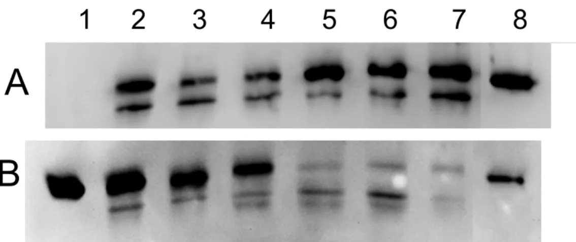
antibodies indicated that hybrid alpha subunits (Mr ≈ 52,000) were produced in all cases (Fig.
5
2A). The second his-tagged protein detected with a Mr of 45,000 was the co-expressed
6
reductase PhnA4. 7
Three RHD chimera derived from the U3-18, U3-72 and U3-116 sequences were 8
found to be catalytically active and transformed naphthalene to the corresponding 1,2-9
dihydrodiol at rates lower than the reference PhnI enzyme expressed under similar conditions 10
(Tables 2 and 3). All other constructions yielded no detectable dioxygenase activity. The 11
RHD derived from U3-18 and U3-116 also transformed phenanthrene, biphenyl and 12
anthracene at relative rates comparable to those found for PhnI (Table 3). The hybrid enzyme 13
made from the U3-72 sequence showed low but significant activity with phenanthrene and 14
biphenyl. Since the deduced amino acid sequences of U3-16 and U3-124 showed high 15
similarity with the catalytic domain of salicylate hydroxylases, we tested whether the relevant 16
hybrid enzymes would be competent in the hydroxylation of salicylate. No activity was 17
detected suggesting that hybrid enzymes were not stably produced in E. coli, as supported by 18
SDS-PAGE analysis of soluble extracts (data not shown). 19
Given that RHDs are multi-component enzymes, several reasons might explain the 20
observed poor activity of most of the studied chimeras, including wrong folding of the hybrid 21
alpha subunit, defective assembly of the α3β3 hexamer or incorrect interaction of the hybrid
22
oxygenase component with the PhnA3 ferredoxin. We first examined whether the enzyme 23
constructions were produced in soluble form in the cytosolic fraction of E. coli. Immunoblot 24
analysis of such fractions indicated that only hybrid RHDs endowed with high activity (U3-18 25
and U3-116) were detected at levels comparable to PhnI (Fig. 2B). On the other hand, hybrid 1
RHDs displaying little or no activity were barely detected in E. coli cytosol (Fig. 2B, lines 5-2
7). Since the His-tagged α-subunit of these hybrids was found in whole cell extracts (Fig. 3
2A), it is likely that they accumulated as inclusion bodies, suggesting that the lack of activity 4
of those hybrid RHDs was primarily due to instability or misfolding. 5
We then tried to isolate hybrid RHDs by affinity chromatography (IMAC), taking 6
advantage of the His-tag fused at the N-terminus of the α subunit. Only hybrids with high 7
enough dioxygenase activity could be isolated in significant amount. The absorbance 8
spectrum of the U3-116 hybrid isolated in this way was very similar to that of PhnI 9
(Jouanneau et al. 2006), indicating that Fe-S centers were correctly inserted in the protein 10
(data not shown). On the other hand, hybrid RHDs with no activity could not be isolated from 11
E. coli soluble extracts suggesting that these constructions were essentially produced as 12
insoluble proteins. 13
14
Structural insights based on 3D modeling of hybrid RHDs
15
To further analyze the structural consequences of the exchange of catalytic domain on 16
hybrid RHDs, we took advantage of the known crystal structure of PhnI from strain CHY-1 to 17
perform molecular modeling of the hybrids (Jakoncic et al. 2007b). The 4 hybrids derived 18
from U3-18, U3-60, U3-72 and U3-112 have closely similar alpha subunit sequences since 19
they differ from each other by less than 6 residues (Fig. S2). Compared to PhnI, these hybrids 20
showed 42 or 43 amino acid replacements (Table 2), some of which are likely to perturb 21
intramolecular interactions within alpha subunits because they are located at the interface 22
between the catalytic and Rieske domains. These changes include Met244, which replaces a 23
threonine in PhnI and Lys332, which replaces a serine. In addition, the sequence Val387-24
Gly388-Tyr389, which is located at the interface between the catalytic and Rieske domains of 25
adjacent alpha subunits in PhnI, is replaced by the highly charged Lys-Asp-Arg peptide in the 1
hybrids. Such substitutions might have marked destabilizing effects on the structure of the 2
oxygenase component. However, the U3-18 hybrid RHD displayed reasonable stability, 3
which seems to be correlated with a unique amino acid replacement, a cysteine in position 4
409 (Fig S2). In the crystal structure of PhnI, a serine residue is present at this position, which 5
is part of a α helix in the core of the catalytic domain and interacts with main chain O of 6
Val202 and Phe242. The former residue is part of the catalytic pocket, the latter is found in a 7
nine-stranded antiparallel β-sheet in the center of the domain. Additionally, Ser409 8
participates in a hydrogen bond network involving three water molecules and extending to 9
Asn295, one of the amino acid forming the substrate-binding pocket next to the active site Fe 10
atom (Fig. 3). This network is highly conserved across RHDs of known structure. Hence, 11
Ser409 appears to play a critical structural role, and it is clear that a bulkier amino acid in this 12
position would perturb interactions stabilizing the protein. In this respect, the lack of activity 13
of the U3-60 and U3-89 hybrids might be related to the presence in position 409 of an 14
arginine and a tryptophan, respectively. Indeed, by modeling of the serine-to-arginine 15
substitution, we observed large perturbations at positions 203, 241 and 251 and a shift of the 16
beta-sheet impairing interactions between the alpha and beta subunits. On the other hand, the 17
replacement of Ser409 by a cysteine, as observed in the U3-18 and U3-116 hybrids, would 18
not affect the inter- or intradomain interactions, inasmuch the cysteine is expected to adopt a 19
conformation similar to the serine, and H-bonds involving the Ser O atom in PhnI would be 20
replaced by bonds involving the Cys S atom (Fig. 3). Nevertheless, it is unclear what could 21
explain the superior stability of the U3-18 hybrid compared to closely similar hybrids, 22
especially those having a serine in position 409. 23
24
DISCUSSION
In this study, we explored the biodiversity of PAH-specific RHDs in a polluted soil by 1
combining DNA-SIP with selective PCR amplification of targeted gene sequences. Our 2
approach involved two successive screening steps, a SIP experiment allowing for the recovery 3
of genomic DNA from soil PAH degraders only, and amplification of a portion of RHD alpha 4
subunit genes encoding the catalytic domain. The set of sequences recovered in this way 5
appeared to depend on the degree of degeneracy of the primers used. With the highly 6
degenerate primer set, a significant proportion of the sequences (33%) were unrelated to 7
RHDs, but on the other hand, a greater diversity was observed among relevant sequences than 8
when a less degenerate primer set was used (Fig. 1). Most retrieved RHD sequences were 9
relatively distant from known enzymes available in the UniProt database, and were not 10
detected in previous studies targeting PAH-specific RHDs in soil (Cebron et al. 2008; Ding et 11
al. 2010) or marine environment (Lozada et al. 2008). The predominance of unknown RHD 12
sequences is consistent with the finding that the majority of bacteria identified as 13
phenanthrene degraders in the same soil sample were either from uncultured genera or 14
unknown for their ability to degrade PAHs (Martin et al. 2012). 15
We then searched for correlations between the expected bacterial hosts of RHDs found 16
in this study, and the taxa previously identified as the main phenanthrene degraders based on 17
16S rRNA sequence analysis. Among Alphaproteobacteria, Sphingomonads were the most 18
abundant degraders, consistent with the occurrence of 11 RHD sequences affiliated to this 19
taxon (cluster 1), representing 22% of the sequences in the DcatdioxA library. Cluster 2 20
sequences appeared to be best related to RHDs from Alphaproteobacteria, although the 21
similarity level was relatively low, so their affiliation to another taxonomic group such as the 22
Cycloclasticus genus cannot be ruled out. Cluster 3 sequences are similar to 3-component 23
salicylate hydroxylases, which play a role in the lower part of the PAH degradation pathway 24
(Jouanneau et al. 2007). Since the latter sequences were not included as targets for PCR 25
primer design, their retrieval was unexpected. Analysis of the cluster 3 sequences indicates 1
that the reverse primer does not match the 3’end. Nevertheless, the high degeneracy of the 2
primer allowed annealing within the 3’-end of the gene sequences, thus generating products 3
about 100-bp shorter than sequences in other clusters. Interestingly, the genomes of 4
Sphingomonads were found to contain multiple copies of highly similar genes related to 5
cluster 3 sequences (Pinyakong et al. 2003a; Schuler et al. 2009), suggesting that they might 6
be present in higher copy numbers than other catabolic genes in metagenomic DNA from the 7
studied soil. This might account for the relatively high proportion of cluster 3 sequences 8
(30%) in the Dcatdiox A library. 9
Using a second primer pair targeting Betaproteobacteria RHDs, sequences closely 10
related to Acidovorax RHD were obtained, consistent with the prevalence of this genus 11
among the soil phenanthrene degraders (Martin et al. 2012; Singleton et al. 2005). However, 12
the majority of the sequences in the DcatdioxB library (87%) showed no clear affiliation. 13
Since uncultured members of the Rhodocyclaceae also appeared as prominent degraders in 14
our previous SIP experiments, we suggest that the sequences of interest (cluster 4) might be 15
specific of this bacterial taxon. Rhodocyclaceae have been detected as potent PAH degraders 16
in other SIP studies (Singleton et al. 2007; Singleton et al. 2006), but no representative strain 17
has been isolated so far. Therefore, no RHD sequence specific for this taxon is yet available. 18
In a second part of this study, we demonstrated the feasibility of constructing RHD 19
chimeras to assess the catalytic activity and substrate specificity associated to partial gene 20
sequences retrieved from soil. Three RHD hybrids were proved competent in the 21
dioxygenation of 2- and 3-ring PAHs, whereas most other hybrids appeared unstable. The 22
reason for this instability is intriguing. Four of them have very similar sequences but variable 23
stabilities. The U3-18, U3-60, U3-72 and U3-112 sequences are almost identical to the 24
catalytic domain of strain LH128 RHD (Schuler et al. 2009). Mismatches with the LH128 25
sequence occur at positions where amino acids were neither strictly conserved nor involved in 1
catalytic activity, namely, Val182, Lys195, Gln273, Asn370, Ser409 (Fig. S2). Therefore, the 2
question arises as to why RHD chimeras resulting from a sequence combination between the 3
α subunits of two naturally occurring enzymes were so unstable. In this respect, the case of 4
the U3-18 hybrid is interesting in that it showed reasonable stability and activity, and 5
displayed one unique replacement compared to the other three hybrids in position 409. A 6
cysteine is found at this position whereas an arginine occurs in U3-60 and a serine in the other 7
hybrids as well as in most RHD sequences in the databases (Fig. S2). This substitution 8
appeared to be the sole sequence difference between the U3-60 and U3-18 hybrids, and 9
therefore this unique change has drastic effects on the enzyme stability. Structural 10
considerations could predict the strong destabilizing effect of a bulky residue such as arginine 11
in position 409 on the 3D fold of the RHD catalytic component and thus explain the 12
instability of the U3-60 hybrid. On the other hand, the instability of other hybrids with a 13
serine in position 409 could not be interpreted on structural grounds. 14
During the course of this study, Standfuß-Gabisch reported on the characterization of 15
hybrid biphenyl dioxygenases generated through a similar strategy (Standfuss-Gabisch et al. 16
2012). Sequences amplified from soil DNA encoded a portion of the catalytic domain similar 17
to that considered in the present work, but diversity was lower as most sequences fell into 2 18
main clusters and shared 97-100% identities within clusters and 85-88% identities between 19
clusters. Hybrid RHDs were constructed using the well-characterized biphenyl dioxygenase 20
from B. xenovorans LB400. Out of 21 hybrid RHDs tested, three were found to be unstable 21
and three were inactive. No obvious correlation was observed between lack of activity and 22
predicted structural changes resulting from amino acid substitutions. The high proportion of 23
active hybrids might be explained by the fact that, in their case, a majority of the hybrids 24
exhibited a catalytic domain closely similar (85-96% identity) to that of the LB400 enzyme. 25
In comparison, our constructions carried an inserted domain sharing less than 83% identity 1
with the corresponding region of the PhnI sequence. Hence, the stability and activity of 2
hybrid RHDs might depend on the relatedness between the sequences to be inserted and that 3
of the recipient enzyme system. Also, for the recovery of active hybrids, one should consider 4
the intrinsic stability of the enzyme system employed to generate hybrids or its propensity to 5
tolerate replacement of a large portion of its catalytic domain. In this respect, RHDs other 6
than PhnI from strain CHY-1 might prove more suitable for the construction of hybrid 7
enzymes with appropriate stability. Finding such enzymes is likely to be a prerequisite to the 8
functional exploration of RHD diversity in soil, based on the exchange of catalytic domains. 9
10
Acknowledgments
11
F. Martin received a grant from the Rhône-Alpes region. This work was supported by grants 12
from the Centre National de la Recherche Scientifique and the University of Grenoble I. 13
References
1
Andreolli M, Lampis S, Zenaro E, Salkinoja-Salonen M, Vallini G (2011) Burkholderia 2
fungorum DBT1: a promising bacterial strain for bioremediation of PAHs-3
contaminated soils. Fems Microbiol Let 319:11-18 4
Bordenave S, Goni-urriza M, Vilette C, Blanchard S, Caumette P, Duran R (2008) Diversity 5
of ring-hydroxylating dioxygenases in pristine and oil contaminated microbial mats at 6
genomic and transcriptomic levels. Environ Microbiol 10:3201-3211 7
Cebron A, Norini MP, Beguiristain T, Leyval C (2008) Real-Time PCR quantification of 8
PAH-ring hydroxylating dioxygenase (PAH-RHD alpha) genes from Gram positive 9
and Gram negative bacteria in soil and sediment samples. J Microbiol Meth 73:148-10
159 11
Chadhain SMN, Moritz EM, Kim E, Zylstra GJ (2007) Identification, cloning, and 12
characterization of a multicomponent biphenyl dioxygenase from Sphingobium 13
yanoikuyae B1. J Ind Microbiol Biotechnol 34:605-613 14
Cole JR, Konstantinidis K, Farris RJ, Tiedje JM (2010) Microbial diversity and phylogeny: 15
extending from rRNAs to genomes. In: Liu W-T, Jansson JK (eds) Environmental 16
molecular microbiology. Caister Academic Press, Norfolk, UK, pp 1-19 17
Demaneche S, Meyer C, Micoud J, Louwagie M, Willison JC, Jouanneau Y (2004) 18
Identification and functional analysis of two aromatic ring-hydroxylating 19
dioxygenases from a Sphingomonas strain degrading various polycyclic aromatic 20
hydrocarbons. Appl Environ Microbiol 70:6714-6725 21
Dereeper A, Guignon V, Blanc G, Audic S, Buffet S, Chevenet F, Dufayard JF, Guindon S, 22
Lefort V, Lescot M, Claverie JM, Gascuel O (2008) Phylogeny.fr: robust phylogenetic 23
analysis for the non-specialist. Nucleic acids res 36(Web Server issue):W465-469 24
Di Gregorio S, Zocca C, Sidler S, Toffanin A, Lizzari D, Vallini G (2004) Identification of 1
two new sets of genes for dibenzothiophene transformation in Burkholderia sp DBT1. 2
Biodegradation 15:111-123 3
Ding GC, Heuer H, Zuhlke S, Spiteller M, Pronk GJ, Heister K, Kogel-Knabner I, Smalla K 4
(2010) Soil type-dependent responses to phenanthrene as revealed by determining the 5
diversity and abundance of polycyclic aromatic hydrocarbon ring-hydroxylating 6
dioxygenase genes by using a novel PCR detection system. Appl Environ Microbiol 7
76:4765-4771 8
Emsley P, Lohkamp B, Scott W, Cowtan K (2010) Features and development of Coot. Acta 9
Cryst D66:486-501 10
Ferraro DJ, Gakhar L, Ramaswamy S (2005) Rieske business: Structure-function of Rieske 11
non-heme oxygenases. Biochem Biophys Res Commun 338:175-190 12
Gomes NCM, Borges LR, Paranhos R, Pinto FN, Krogerrecklenfort E, Mendonca-Hagler 13
LCS, Smalla K (2007) Diversity of ndo genes in mangrove sediments exposed to 14
different sources of polycyclic aromatic hydrocarbon pollution. Appl Environ 15
Microbiol 73:7392-7399 16
Iwai S, Chai BL, Sul WJ, Cole JR, Hashsham SA, Tiedje JM (2010) Gene-targeted-17
metagenomics reveals extensive diversity of aromatic dioxygenase genes in the 18
environment. Isme J 4:279-285 19
Iwai S, Johnson TA, Chai B, Hashsham SA, Tiedje JM (2011) Comparison of the specificities 20
and efficacies of primers for aromatic dioxygenase gene analysis of environmental 21
samples. Appl Environ Microbiol 77:3551-3557 22
Jakoncic J, Jouanneau Y, Meyer C, Stojanoff V (2007a) The catalytic pocket of the ring-23
hydroxylating dioxygenase from Sphingomonas CHY-1. Biochem Biophys Res 24
Commun 352:861-866 25
Jakoncic J, Jouanneau Y, Meyer C, Stojanoff V (2007b) The crystal structure of the ring-1
hydroxylating dioxygenase from Sphingomonas CHY-1. Febs J 274:2470-2481 2
Jouanneau Y, Martin F, Krivobok S, Willison JC (2011) Ring-hydroxylating dioxygenases 3
involved in PAH biodegradation : structure, function and biodiversity. In: Koukkou AI 4
(ed) Microbial bioremediation of non metals: current research. Caister Academic 5
Press, Norflok, UK, pp 149-175 6
Jouanneau Y, Meyer C, Jakoncic J, Stojanoff V, Gaillard J (2006) Characterization of a 7
naphthalene dioxygenase endowed with an exceptionally broad substrate specificity 8
toward polycyclic aromatic hydrocarbons. Biochemistry 45:12380-12391 9
Jouanneau Y, Meyer C, Naud I, Klipp W (1995) Characterization of an fdxN mutant of 10
Rhodobacter capsulatus indicates that ferredoxin I serves as electron donor to 11
nitrogenase. Biochim Biophys Acta 1232:33-42 12
Jouanneau Y, Micoud J, Meyer C (2007) Purification and characterization of a three-13
component salicylate 1-hydroxylase from Sphingomonas sp. strain CHY-1. Appl 14
Environ Microbiol 73:7515-7521 15
Kasai Y, Shindo K, Harayama S, Misawa N (2003) Molecular characterization and substrate 16
preference of a polycyclic aromatic hydrocarbon dioxygenase from Cycloclasticus sp 17
strain A5. Appl Environ Microbiol 69:6688-6697 18
Kraulis PJ (1991) Molscript: a program to produce both detailed and schematic plots of 19
protein structures. J Appl Cryst 24:946-950 20
Kweon O, Kim S-J, Baek S, Chae J-C, Adjei M, Baek D-H, Kim Y-C, Cerniglia C (2008) A 21
new classification system for bacterial Rieske non-heme iron aromatic ring-22
hydroxylating oxygenases. BMC Biochem 9:11 23
Laurie AD, Lloyd-Jones G (1999) The phn genes of Burkholderia sp. strain RP007 constitute 1
a divergent gene cluster for polycyclic aromatic hydrocarbon catabolism. J Bacteriol 2
181:531-540 3
Lozada M, Mercadal JPR, Guerrero LD, Di Marzio WD, Ferrero MA, Dionisi HM (2008) 4
Novel aromatic ring-hydroxylating dioxygenase genes from coastal marine sediments 5
of Patagonia. BMC Microbiol 8 6
Martin F, Torelli S, Le Paslier D, Barbance A, Martin-Laurent F, Bru D, Geremia R, Blake G, 7
Jouanneau Y (2012) Betaproteobacteria dominance and diversity shifts in the bacterial 8
community of a PAH-contaminated soil exposed to phenanthrene. Environ Pollut 9
162:345-353 10
Merritt EA, Murphy ME (1994) Raster3D version 2.0: a programforphotorealistic molecular 11
graphics. Acta Cryst D50:869-873 12
Moreno-Ruiz E, Hernaez MJ, Martinez-Perez O, Santero E (2003) Identification and 13
functional characterization of Sphingomonas macrogolitabida strain TFA genes 14
involved in the first two steps of the tetralin catabolic pathway. J Bacteriol 185:2026-15
2030 16
Nam JW, Nojiri H, Yoshida T, Habe H, Yamane H, Omori T (2001) New classification 17
system for oxygenase components involved in ring- hydroxylating oxygenations. 18
Biosci Biotechnol Biochem 65:254-263. 19
Ni Chadhain SM, Norman RS, Pesce KV, Kukor JJ, Zystra GJ (2006) Microbial dioxygenase 20
gene population shifts during polycyclic aromatic hydrocarbon biodegradation. Appl 21
Environ Microbiol 72:4078-4087 22
Parales RE, Resnick SM (2006) Aromatic ring hydroxylating dioxygenases. In: Ramos JL, 23
Levesque RC (eds) Pseudomonas. vol 4. Springer, pp 287-340 24
Park JW, Crowley DE (2006) Dynamic changes in nahAc gene copy numbers during 1
degradation of naphthalene in PAH-contaminated soils. Appl Environ Microbiol 2
72:1322-1329 3
Pinyakong O, Habe H, Omori T (2003a) The unique aromatic catabolic genes in 4
sphingomonads degrading polycyclic aromatic hydrocarbons (PAHs). J Gen Appl 5
Microbiol 49:1-19 6
Pinyakong O, Habe H, Yoshida T, Nojiri H, Omori T (2003b) Identification of three novel 7
salicylate 1-hydroxylases involved in the phenanthrene degradation of Sphingobium 8
sp. strain P2. Biochem Biophys Res Commun 301:350 - 357 9
Romine MF, Stillwell LC, Wong KK, Thurston SJ, Sisk EC, Sensen C, Gaasterland T, 10
Fredrickson JK, Saffer JD (1999) Complete sequence of a 184-kilobase catabolic 11
plasmid from Sphingomonas aromaticivorans F199. J Bacteriol 181:1585 - 1602 12
Schuler L, Chadhain SMN, Jouanneau Y, Meyer C, Zylstra GJ, Hols P, Agathos SN (2008) 13
Characterization of a novel angular dioxygenase from fluorene-degrading 14
Sphingomonas sp strain LB126. Appl Environ Microbiol 74:1050-1057 15
Schuler L, Jouanneau Y, Chadhain SM, Meyer C, Pouli M, Zylstra GJ, Hols P, Agathos SN 16
(2009) Characterization of a ring-hydroxylating dioxygenase from phenanthrene-17
degrading Sphingomonas sp. strain LH128 able to oxidize benz[a]anthracene. Appl 18
Microbiol Biotechnol 83:465-475 19
Simon MJ, Osslund TD, Saunders R, Ensley BD, Suggs S, Harcourt A, Suen WC, Cruden 20
DL, Gibson DT, Zylstra GJ (1993) Sequences of genes encoding naphthalene 21
dioxygenase in Pseudomonas putida strains G7 and NCIB 9816-4. Gene 127:31-37 22
Singleton DR, Hunt M, Powell SN, Frontera-Suau R, Aitken MD (2007) Stable-isotope 23
probing with multiple growth substrates to determine substrate specificity of 24
uncultivated bacteria. J Microbiol Meth 69:180-187 25
Singleton DR, Powell SN, Sangaiah R, Gold A, Ball LM, Aitken MD (2005) Stable-isotope 1
probing of bacteria capable of degrading salicylate, naphthalene, or phenanthrene in a 2
Bioreactor treating contaminated soil. Appl Environ Microbiol 71:1202-1209 3
Singleton DR, Ramirez LG, Aitken MD (2009) Characterization of a polycyclic aromatic 4
hydrocarbon degradation gene cluster in a phenanthrene-degrading Acidovorax strain. 5
Appl Environ Microbiol 75:2613-2620 6
Singleton DR, Sangaiah R, Gold A, Ball LM, Aitken MD (2006) Identification and 7
quantification of uncultivated Proteobacteria associated with pyrene degradation in a 8
bioreactor treating PAH-contaminated soil. Environ Microbiol 8:1736-1745 9
Standfuss-Gabisch C, Al-Halbouni D, Hofer B (2012) Characterization of biphenyl 10
dioxygenase sequences and activities encoded by the metagenomes of highly 11
polychlorobiphenyl-contaminated soils. Appl Environ Microbiol 78:2706-2715 12
Yanisch-Perron C, Vieira J, Messing J (1985) Improved M13 phage cloning vectors and host 13
strains: nucleotide sequences of the M13mp18 and pUC19 vectors. Gene 33:103-119 14
Zhou HW, Guo CL, Wong YS, Tam NFY (2006) Genetic diversity of dioxygenase genes in 15
polycyclic aromatic hydrocarbon-degrading bacteria isolated from mangrove 16
sediments. FEMS Microbiol Lett 262:148-157 17
18 19
Tables
1
Table 1: Bacterial strains and some of the plasmids used in this study 2
Strain or Plasmid Description/ genotype Reference or source
Escherichia coli Strains
NEB 5-α
fhuA2 Δ(argF-lacZ)U169 phoA glnV44 Φ80Δ (lacZ)M15
gyrA96 recA1 relA1 endA1 thi-1 hsdR17 New England Biolabs
JM109 recA1 supE44 endA1 hsdR17 gyrA96 relA1 thi-1∆(lac-proAB)
F(TraD36 proAB+ lacIq lacZ∆M15)
(Yanisch-Perron et al. 1985)
Plasmids a
pSD8 pET15b carrying phnA1aA2a from strain CHY-1 (Demaneche et al. 2004) pIZ1036 KmR, broad range expression plasmid derived from pBR1MCS-2 (Moreno-Ruiz et al. 2003) pIBA34 pIZ1036 carrying phnA3 and phnA4 from strain CHY-1 Jouanneau, unpublished
pUC18 AmpR, cloning plasmid (Yanisch-Perron et al. 1985)
pFMA1 pUC18 carrying phnA1aA2a This study
a
Other plasmids constructed in this study are listed in Table 2 3
Table 2 : Characteristics of chimerical RHDs and plasmids used for heterologous expression 1
2
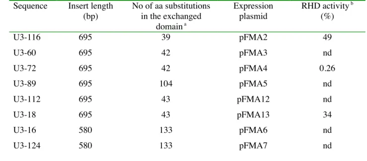
Sequence Insert length (bp) No of aa substitutions in the exchanged domain a Expression plasmid RHD activity b (%) U3-116 695 39 pFMA2 49 U3-60 695 42 pFMA3 nd U3-72 695 42 pFMA4 0.26 U3-89 695 104 pFMA5 nd U3-112 695 43 pFMA12 nd U3-18 695 43 pFMA13 34 U3-16 580 133 pFMA6 nd U3-124 580 133 pFMA7 nd a
Numbers refer to amino acid changes compared to the corresponding region of PhnA1a 3
b
Activities were determined with naphthalene as substrate and calculated as percentages of 4
the activity measured with the reference strain carrying pFMA1. nd means not detected. 5
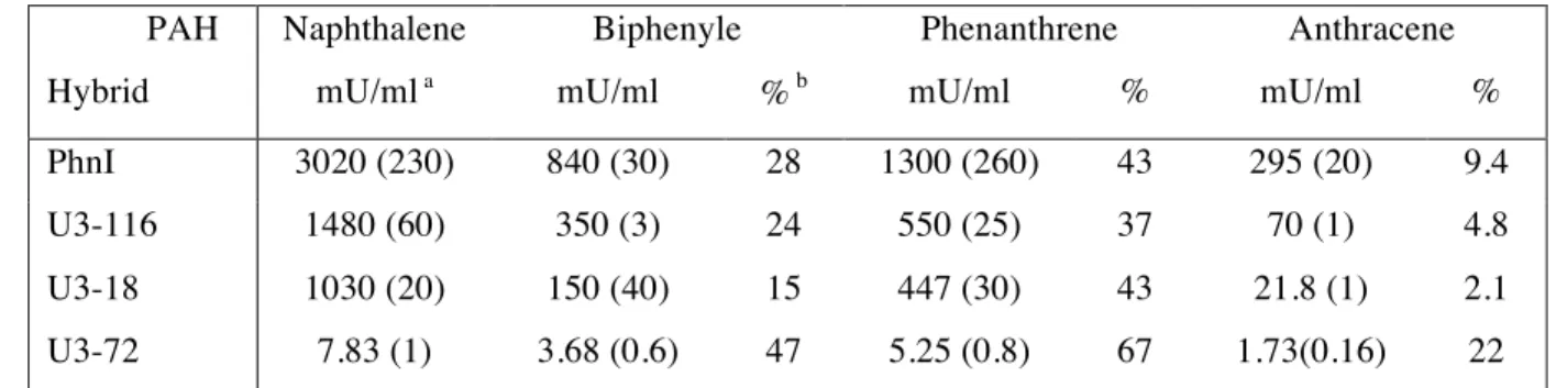
Table 3 : Relative activity of hybrid dioxygenases with 2- and 3-ring PAHs 1
2
PAH Naphthalene Biphenyle Phenanthrene Anthracene
Hybrid mU/ml a mU/ml
% b mU/ml % mU/ml %
PhnI 3020 (230) 840 (30) 28 1300 (260) 43 295 (20) 9.4
U3-116 1480 (60) 350 (3) 24 550 (25) 37 70 (1) 4.8
U3-18 1030 (20) 150 (40) 15 447 (30) 43 21.8 (1) 2.1 U3-72 7.83 (1) 3.68 (0.6) 47 5.25 (0.8) 67 1.73(0.16) 22
a One milliunit of activity (mU) is the amount of enzyme that produced one pmol of
3
dihydrodiol per hour at 25°C under the defined assay conditions. Numbers in parenthesis are 4
standard deviations. 5
b
Percentages were calculated relative to the activity measured with naphthalene as substrate. 6
Figure legends
1 2
Figure 1 : Phylogenetic tree of predicted partial sequences of RHD alpha subunits from soil 3
subjected to DNA-SIP. Sequences were from two clone libraries obtained by PCR 4
amplification of target RHD genes using labeled DNA extracted from soil treated with 13
C-5
phenanthrene for 5 days. Alignment was performed on a common portion of the translated 6
sequences (corresponding to residues 98-411 of PhnA1a) encompassing the catalytic domain 7
of RHD α-subunits. Only 47 of the 50 RHD sequences of the DcatdioxA library were 8
considered in the alignment. Five clusters of related sequences were defined (see text). 9
Triangles represent groups of sequences sharing >97% identity, with sequence number per 10
group indicated on the left. Arrows designate sequences used for the construction of hybrid 11
RHDs. Two labels present at the end of a branch (i.e. U3_72 U3_6) means that two identical 12
sequences were found at this position. Relevant accession numbers are indicated. 13
14
Figure 2 : Immunoblot analysis of protein cell extracts from strain JM109 expressing hybrid 15
RHDs. 16
Panel A: whole cell extracts prepared by lysing cells in SDS mix 10 min at 99°C. Protein 17
loading in each lane was adjusted based on equal bacterial density (OD600) as measured prior
18
to cell lysis. Lane 1 : non-induced cells carrying pFMA13 (U3-18); lane 8: purified ht-PhnI, 19
0.2 µg. Panel B: Cytosolic fractions prepared by cell ultrasonication and centrifugation. 20
Lanes were loaded with 6 µg (lanes 2-4) or 12 µg (lanes 5-7) of proteins. Lanes 1 and 8 were 21
loaded with 0.3 and 0.1 µg of purified ht-PhnI, respectively. Other lanes were loaded with 22
extracts from cells expressing PhnI (lane 2) or the following hybrid RHDs; lane 3, U3-116 ; 23
lane 4, U3-18 ; lane 5, U3-60 ; lane 6, U3-72 ; lane 7, U3-112. Western blots were revealed 24
with anti-Histag antibodies. The second band detected with Mr ≈ 45,000 is the reductase
ht-1
PhnA4 co-expressed in JM109. 2
3
Figure 3 : Close up view of the hydrogen bond network involving Ser409 and its connections 4
with residues at the active site of Phn1. 5
Ser409 is involved in a hydrogen bond network (red dashed lines) involving two water 6
molecules, and extending to Asn295, a residue lining the catalytic pocket. It is connected 7
through its hydroxyl group to the carbonyl of Val202 and Phe242. The network also includes 8
the Gly203-Asp204 dipeptide, where Asp204 is thought to be responsible for electron transfer 9
between the Rieske center of the adjacent alpha subunit and the catalytic site. The covalent 10
bonds between active site Fe atom and its three ligands are shown as green dashed lines. An 11
indole molecule is represented inside the substrate-binding site. The figure was prepared with 12
Molscript (Kraulis 1991) and Raster3D (Merritt and Murphy 1994). 13