HAL Id: hal-02896357
https://hal.univ-grenoble-alpes.fr/hal-02896357
Submitted on 21 Sep 2020HAL is a multi-disciplinary open access archive for the deposit and dissemination of sci-entific research documents, whether they are pub-lished or not. The documents may come from teaching and research institutions in France or abroad, or from public or private research centers.
L’archive ouverte pluridisciplinaire HAL, est destinée au dépôt et à la diffusion de documents scientifiques de niveau recherche, publiés ou non, émanant des établissements d’enseignement et de recherche français ou étrangers, des laboratoires publics ou privés.
The controversy on the ancestral arsenite oxidizing
enzyme; deducing evolutionary histories with phylogeny
and thermodynamics
Julie Szyttenholm, Florence Chaspoul, Marielle Bauzan, Anne-Lise
Ducluzeau, Mahmoud Hajj Chehade, Fabien Pierrel, Yann Denis, Wolfgang
Nitschke, Barbara Schoepp-Cothenet
To cite this version:
Julie Szyttenholm, Florence Chaspoul, Marielle Bauzan, Anne-Lise Ducluzeau, Mahmoud Hajj Chehade, et al.. The controversy on the ancestral arsenite oxidizing enzyme; deducing evolutionary histories with phylogeny and thermodynamics. Biochimica biophysica acta (BBA) - Bioenergetics, Elsevier, 2020, 1861 (10), pp.148252. �10.1016/j.bbabio.2020.148252�. �hal-02896357�
1 The controversy on the ancestral arsenite oxidising enzyme; deducing evolutionary histories
with phylogeny and thermodynamics
Julie Szyttenholm§, Florence Chaspoul£, Marielle Bauzan‡, Anne-Lise Ducluzeau#, Mahmoud
Hajj Chehade†, Fabien Pierrel†, Yann Denis¶, Wolfgang Nitschke§, and Barbara
Schoepp-Cothenet§*
Running Title: evolutionary history of the Arr and Arx enzymes
§ Aix-Marseille Univ., CNRS, BIP UMR 7281, FR 3479, IMM, 13402 Marseille Cedex 20, France
£ Aix Marseille Univ., CNRS, IRD, IMBE UMR 7263, Faculté de Pharmacie, 13005 Marseille, France
‡ Aix-Marseille Univ., CNRS, Plateforme fermentation, FR3479, IMM, 13402 Marseille Cedex 20, France
#Institute of Arctic Biology, University of Alaska Fairbanks, Fairbanks, AK 99775-7220, USA †Univ. Grenoble Alpes, CNRS, Grenoble INP, TIMC-IMAG, 38000 Grenoble, France
¶ Aix-Marseille Univ., CNRS, Plateforme transcriptomique, FR3479, IMM, 13402 Marseille Cedex 20, France
* Corresponding author: Barbara Schoepp-Cothenet
Aix-Marseille Univ., CNRS, BIP UMR 7281, FR 3479, IMM, 13402 Marseille Cedex 20, France Phone: (33) 4 91164672, fax: (33) 4 91164578, e-mail: [email protected]
2 Abstract
The three presently known enzymes responsible for arsenic-using bioenergetic processes are arsenite oxidase (Aio), arsenate reductase (Arr) and alternative arsenite oxidase (Arx), all of which are molybdoenzymes from the vast group referred to as the Mo/W-bisPGD enzyme superfamily. Since arsenite is present in substantial amounts in hydrothermal environments, frequently considered as vestiges of primordial biochemistry, arsenite-based bioenergetics has long been predicted to be ancient. Conflicting scenarios, however, have been put forward proposing either Arr/Arx or Aio as operating in the ancestral metabolism. Phylogenetic data argue in favor of Aio whereas biochemical and physiological data led several authors to propose Arx/Arr as the most ancient anaerobic arsenite metabolizing enzymes. Here we combine phylogenetic approaches with physiological and biochemical experiments to demonstrate that the Arx/Arr enzymes could not have been functional in the Archaean geological eon. We propose that Arr reacts with menaquinones to reduce arsenate whereas Arx reacts with ubiquinone to oxidize arsenite, in line with thermodynamic considerations. The distribution of the quinone biosynthesis pathways, however, clearly indicates that the ubiquinone pathway is recent. An updated phylogeny of Arx furthermore reinforces the hypothesis of a recent emergence of this enzyme. We therefore conclude that anaerobic arsenite redox conversion in the Archaean must have been performed in a metabolism involving Aio.
Key words
arsenic; arsenite oxidation evolutionary history; phylogeny; quinones; prokaryotes; thermodynamics
Highlights
●Phylogeny remain inconclusive as to a putative pre-LUCA character of Arr/Arx enzymes ●Quinone biosynthesis pathways correlate Arr and Arx to specific quinones, MK/MKH2 and
UQ/UQH2, respectively
●In vivo and in vitro directionality of Arr and Arx fully correlates with thermodynamics
●Evolutionary history of quinones strongly suggests that Arx entered later the evolutionary scene ●Aio appears as the single candidate for arsenite oxidation in the Archaean time
3 1. Introduction
Despite its low crustal abundance, arsenic (As) is widely distributed in nature. Arsenic is commonly found in an insoluble form but also occurs as soluble inorganic oxyanions, arsenite (H3AsO3) and arsenate (HAsO42-/H2AsO4-). The poisonous properties of As have been known since
the antiquity even if the element has also early on been exploited for medical purposes. To counter the deleterious effects of these oxyanions, organisms evolved detoxifying, energy-consuming systems [1]. However, certain prokaryotes are also able to use AsIII (arsenite) and AsV (arsenate) as electron donors and acceptors, respectively, for their energy conserving respiratory systems (for reviews, see [2-4]).
A number of parameters, both phylogenetic and functional, set Aio apart from the highly related dyad of the Arr/Arx enzymes. For example, Arr and Arx both contain very similar electron transfer subunits (ArrB/ArxB) corresponding to cubane [4Fe-4S]-proteins, while this role is played by a Rieske-[2Fe-2S] protein (AioB) in Aio (see[3] for review). Arr and Arx appear to be able to function in both the forward and the backward directions when assayed in vitro with non-physiological electron donors and acceptors whereas Aio seems to be strictly unidirectional [5, 6]. Reflecting these structural and functional differences, Aio is phylogenetically only distantly related to Arr and Arx [7].
Since AsIII has been found in hydrothermal habitats, considered as comparable to the archaean environment, AsIII-based bioenergetics has early on been proposed to be ancestral [1]. This has
been confirmed by geochemical studies of archaean rocks [8]. Phylogenetic studies on Aio yielded a tree which is characterized by a profound Bacteria/Archaea cleavage and which compares reasonably well with current species trees [3, 9-11]. Such tree topologies have led us to propose that AsIII oxidation in the anaerobic Archaean, and even in the Last Universal Common Ancestor (LUCA), was mediated by Aio. By contrast, we interpreted phylogenetic trees containing Arr/Arx [3, 11, 12], as suggesting that these enzymes are comparatively recent and have emerged from within the domain of Bacteria.
However, physiological and biochemical characterizations of Arr, revealing a reversible function of the enzyme, led Oremland and co-workers to propose the Arx/Arr enzyme as the most ancient As-metabolizing enzyme under anaerobic conditions [5, 7, 13-16]. Recently, a phylogenetic tree including two archaeal Arx sequences well-separated from the bacterial one has
4
been published [17] thus strengthening the hypothesis of a pre-LUCA origin of the enzyme. Therefore, two conflicting scenarios on the evolutionary history of bioenergetic usage of arsenicals by life presently oppose each other. Deducing the evolutionary pathway of enzymes solely based on their molecular phylogeny is hampered by the inherent probabilistic nature of this approach and often, as in the case of the Aio, Arr and Arx systems, by low statistical significance due to small sample sizes (of species containing the respective genes).
To overcome these limitations, we have in this work integrated the information provided by functional parameters and thermodynamic constraints into an updated phylogenetic analysis of Arr and Arx sequences. The result of this multidisciplinary approach strongly favors the scenario positing that the Arr/Arx enzymes appeared late in evolution, after the Grand Oxygenation Event (GEO).
2. Material and Methods
2.1. Bioinformatics. Open reading frames (ORFs; Sup Table A.1 for accession numbers) coding for subunits homologous to Arr/Arx were retrieved from the National Center for Biotechnology Information (http://www.ncbi.nlm.nih.gov) using the ArrA/B sequences from Shewanella ANA-3 as query templates in BLAST searches. Polysulfide reductase (Psr) sequences were retrieved by starting from PsrA/B/C sequences from Wolinella (W.) succinogenes [18]. Tetrathionate reductase (Ttr) sequences were retrieved by starting from TtrB/C/A sequences from Salmonella typhimurium [19]. Aio sequences, used as outgroup, have been sampled before [11]. We searched for ORFs coding for MK or UQ biosynthesis enzymes in all available sequenced genomes from Arr/Arx harbouring species. There are two MK biosynthesis pathways: Men and Mqn [20]. We analyzed therefore the genomes, using the MenA/B/C/D/E/F sequences from E. coli str. K-12 substr. W3110 and also the Mqn sequences from Streptomyces (S.) coelicolor as queries. Ubi is the only known pathway responsible for UQ biosynthesis [21]. We analyzed therefore the genomes, using the UbiA/B/C/D/E/F/G/H/X sequences from E. coli str. K-12 substr. W3110. Since two analogs of UbiC (Rv2949c and XanB2) are known and one analog of UbiF is known (Coq7) [22-24], we also analyzed the genomes using Rv2949c from Mycobacterium tuberculosis and XanB2 from
Xanthomonas campestris and Coq7 from Pseudomonas aeruginosa. Most of the quinone
biosynthetic genes belong to superfamilies of enzymes that perform reactions involved in various biochemical pathways besides quinone synthesis. Hence, it is not rare during homology searches
5
to retrieve homologs that actually aren’t involved in quinone biosynthesis but may be mistaken in the absence of careful scrutiny. Identifications of the quinone homologs were validated after reconstruction of superfamily global trees containing representatives of the different protein members and the hits retrieved. This procedure was applied in the case of the Polyprenyl-diphosphate transferase superfamily for MenA and UbiA, the SAM radical methyltransferase family for UbiE and UbiG, the flavin-monooxygenases family for UbiF,-H,-I,-M and -L, the radical SAM proteins MqnC, -E with the FO-synthase subunits (CofG and –H), and finally for the paralogs MqnA and MqnD. For Men proteins, we defined motifs based on alignments of taxonomically distant representatives of each protein and then looked for these motifs in the primary sequence of the retrieved hits for validation.
Multiple sequence alignments of recognized subfamilies of Mo-bisPGD subunits were automatically produced using ClustalX [25] and T-Coffee [26]. The automatically generated alignments were subsequently refined using Seaview [27] with respect to functionally conserved residues, to structural alignments and to secondary structures. Structures were obtained from the pdb database (http://www.rcsb.org/pdb/welcome.do). Structural alignments were obtained using
the root-mean-square fit option of the Swiss-Pdb Viewer (version 4.0.1.;
http://www.expasy.ch/spdbv).
Secondary structure prediction was performed by means of the pSAAM package (http://www.life.uiuc.edu/crofts/ahab/psaam.html) on crystal structure of 2VPZ for Psr. Tertiary
structure modeling was performed using the option of the ExPaSy site
(https://www.expasy.org/proteomics). Phylogenetic trees were reconstructed from these alignments using either the Neighbor-Joining (NJ)- algorithm or the Maximum Likelihood method implemented in MEGA7.
2.2. Growth of H. halophila. All growth experiments and corresponding quantifications were done
in triplicates. H. halophila SL1 (DSM244) was grown photosynthetically and anaerobically at 42 °C in media derived from the ATCC 1448 medium (typically at pH 8.8) in 100 mL bottles. For growth at pH 7, sodium carbonate buffer in the medium was replaced by HEPES buffer and NaHCO3 was added at 0.2%. Growth conditions combine Na2S, thiosulfate and arsenicals. When
added, AsIII and AsV were at 2 mM and 5 mM, respectively. Specific experiments at saturating cell density were performed. For this, bacterial growth was achieved at pH 8.8 under AsIII with the aim to favor enzyme expression. Cells were collected in the stationary phase, washed and resuspended
6
in new medium containing AsV or AsIII, at pH 7. To test the need of proton motive force (pmf) for
AsV reduction under these conditions, we added gramicidin (10 M) to cell suspensions.
2.3. Growth of A. ehrlichii. All growth experiments and corresponding quantifications were done
in triplicates. A. ehrlichii MLHE-1 (DSM 17681) was grown anaerobically at 30°C in DSMZ 1457 medium at pH 9.3 [13]. For growth at pH 7.3, sodium carbonate buffer was replaced by HEPES buffer and NaHCO3 was added at 0.2%. When added, AsIII and AsV were at 10 mM and 5 mM,
respectively. Specific experiments were performed at saturating cell density. For these essays, bacterial growth was achieved at pH 9.3 in the presence of AsIII with the aim to induce enzyme expression. Cells were collected in the stationary phase, washed and resuspended in new medium containing AsV or AsIII, at pH 7.3.
2.4. Growth of Desulfitobacterium hafniense. All growth experiments and corresponding
quantifications were done in triplicates. D. hafniense (DSM 10664) was grown anaerobically at 37 °C in DSMZ 720 medium at pH 7.3 [28], where pyruvate was replaced by lactate. For growth at pH 9.3, TAPS buffer and NaOH were added. When added, AsIII and AsV were at 2-5 mM and 10 mM, respectively. Specific experiments were performed at saturating density. For these essays, bacterial growth was first achieved at pH 7.3 in the presence of AsV with the aim to induce enzyme expression. In the stationary phase, TAPS buffer, NaOH was added to reach pH 9.3 and nitrate was added to serve as electron acceptor while AsIII (produced during the growth phase) served as
electron donor.
2.5. Growth of Sulfuritalea hydrogenivorans strain sk43H. All growth experiments and
corresponding quantifications were done in triplicates. S. hydrogenivorans (DSM 22779) was grown anaerobically at 25 °C in DSMZ 1304 medium at pH 7 under a 80% N2 and 20% CO2
atmosphere [29], with AsV (5 mM) as electron acceptor and lactate (10 mM) as electron donor, or
AsIII (1-2 mM) as electron donor and nitrate (20 mM) as electron acceptor. To test the need of a
pmf for As conversion, we added gramicidin (10 M) to cell suspensions.
2.6. Arsenic quantification using HPLC-ICP-MS. Detailed AsIII and AsV contents during bacterial growth have been quantified by coupling online liquid chromatography (HPLC) and Inductively Coupled Plasma/Mass Spectrometry (ICP/MS) as already described [30]. HPLC chromatography allowed to separate AsIII from AsV which were consequently quantified separately by ICP/MS. For specifications, the analytical setup was an HPLC (Agilent 1100) delivery pump with anionic-exchange column (Synchropak Q300), in-line with an ICP-MS detection system (ThermoElectron
7
ICAPQ). The chromatographic conditions of elution were as follows: mobile phase was constituted of NH4H2PO4 12.5 mM, 3% Methanol, at pH equal to 9.0. The injection volume was 20 µL and
the flow rate was 0.5 mL.min-1. The operational parameters of the ICP/MS instrument were set to
optimize the detection of As. Before analysis, the samples were diluted with the mobile phase, and pH was adjusted to 9.0. The dilution depends on total As concentration, quantified before by ICP/MS, and the HPLC–ICP/MS working range is comprised between 0.1 and 5.0 mg As L−1. Quantitative analyses were performed through external calibration. Quantitation of AsIII and AsV was achieved with the help of Qtegra Software (ThermoElectron).
2.7. Purification of recombinant Shewanella sp. ANA-3 Arr. Overexpression of Arr from
Shewanella sp. ANA-3 in Escherichia coli and purification was based on a previously established
protocol [31].
2.8. Preparation of membrane fractions from H. halophila. Cells were suspended in 50 mM
Tricine at pH 8 and membrane-fragments, containing Arx, were prepared as already described [32] and resuspended in 50 mM Tricine at pH 8 to a final concentration of 20 mg/ml proteins.
2.9. In solution activities. AsV reductase and AsIII oxidase activities were detected spectroscopically in solution in 2 mL and 1 mL cuvettes, respectively. For AsV reductase activity, measured in an anaerobic glove box, the membrane fraction was added to a 50 mM MES pH 6 buffer supplemented with 670 M Benzyl Viologen (BV) / 1 mM Dithionite. Addition of 1 mM sodium AsV allowed the initiation of the reaction quantified by the rate of OD600 decrease. For AsIII
oxidase activity, the membrane fraction was added to a 50 mM AMPSO pH 9.5 buffer supplemented with 300 M 2,6-dichlorophenolindophenol (DCPIP) and 30 M PMS. Addition of 100 M sodium AsIII allowed the initiation of the reaction quantified by the rate of OD600 decrease.
When pH-dependencies of Arx and Arr activities were studied, a mix buffer of MES/HEPES/Tricine/AMPSO (15 mM each) was used. We determined 600 at each pH value for
both BV and DCPIP.
2.10. In gel activities. Activities were detected on a 10 % polyacrylamide Laemmli gel system [33]
containing 0.1 % Triton X-100 instead of SDS. For AsV reductase activity, the gel was equilibrated in 50 mM MES pH 6 for 15 min and subsequently incubated for 30 min under argon flow in the same buffer supplemented with 670 M BV/ 1 mM Dithionite. Addition of 10 mM sodium AsV allowed the detection of the catalytically active band by its destaining activity. For AsIII oxidase activity, the gel was equilibrated in 50 mM AMPSO pH 9.5 for 15 min under aerobiosis and
8
subsequently incubated for 30 min in the same buffer supplemented with 300 M DCPIP and 30
M PMS. Addition of 1 mM sodium AsIII allowed the detection of the band by its destaining activity.
2.11. Analysis of quinone. Quinones were extracted from membrane fragments of H. halophila
and A. ehrlichii (~ 300 ug proteins) or from whole cells of S. hydrogenivorans in glass tubes as described in [34] , except that 3 mL methanol, 300 µL of 0.15 M KCl and 2*2 mL petroleum ether were used. Dried extracts were resuspended in 200 µL ethanol and fractions corresponding to 20 ug proteins were analyzed by HPLC coupled to electrochemical detection (ECD) and mass spectrometry as previously described [35], except that the redox potential of the E1 and E2 electrodes was set at -800 mV and + 800 mV, respectively, to allow maximal electrochemical response of MK8. Areas of the ECD peaks corresponding to UQ8 and MK8 (as confirmed by mass
spectrometry) were converted to pmoles of quinones thanks to a standard curve of commercial UQ10 established in the same analytical condition. UQ8 and MK8 represented more than 95% of
the ECD signal of endogenous quinones of H. halophila. Three independent membrane or cell preparations were analyzed in technical triplicates.
2.12. RNA quantification. RNAs were prepared from 15 mL triplicate cultures at 0, 32h and 120h
growth. Total RNAs were isolated from the pellet using the Maxwell® 16 LEV simply RNA Blood Kit according to the manufacturer’s instructions and an extra TURBO DNase digestion step to eliminate the contaminating DNA (confirmed by PCR). RNA was qualified by an Experion chip and quantified spectrophotometrically at 260 nm. For cDNA synthesis, 450 ng total RNA and 0.5 μg random primers were used with the GoScript™ Reverse transcriptase according to the manufacturer instruction. Quantitative real-time PCR (qPCR) analyses were performed on a CFX96 Real-Time System as already described [36]. The RNA16S gene was used as a reference for normalization. For each point a technical duplicate was performed.
3. Results
3.1. The results from phylogeny remain inconclusive as to a putative pre-LUCA character of Arr/Arx enzymes
Several reviews highlight the characteristic sequence motifs of a bona fide Arr (i.e. number and order of genes in arr-clusters, presence of TAT signals, cofactor binding motifs…) [2-4, 17, 37]. With respect to the arsenate reductase system, the most recently published work identified 38
9
protein sequences considered to represent ArrA subunits, mostly from Bacteria [38] while archaeal cases are rare. AsV respiration has been shown in the Archaea Pyrobaculum arsenaticum and Pyrobaculum aerophilum [39]. However, no amplification of arrA genes was obtained from these
strains [40]. Genes with high similarity to arrA were found in theHaloarchaea Natronobacterium
gregoryi SP2 and Halobiforma nitratireducens [38, 41] but the function of the corresponding
enzymes has not yet been characterized. Irrespective of whether these genes code for bona fide arsenate reductases or not, these sequences (denoted NatGr and HalNi in Fig. 1) are phylogenetically located within the cluster of bacterial representatives strongly suggesting Horizontal Gene Transfer (HGT) from Bacteria into Haloarchaea [38]. Haloarchaea are indeed notorious for extensive import of genes from diverse bacterial donors [42, 43]. With regard to the alternative arsenite oxidase system, only few Arx-carrying organisms have been characterized in terms of metabolism [7, 13, 14, 44-46]. The arx operon is basically indistinguishable from an
arrABC cluster but phylogenetically forms a sister clade to Arr as will be shown below. Prior to
our work 34 protein sequences considered to be ArxA subunits have been reported, mostly from bacteria but with two archaeal representatives (denoted cMetBLZ1 and cMetNi2 on Fig. 1) [17]. Overall, the vast majority of sequences retrieved so far and considered to be ArrA and ArxA arises from bacterial species.
As a first step towards a better empirical assessment of both our hypothesis on the evolutionary history of Arx and of its counterarguments, we updated the sample of sequences used for phylogenetic reconstructions. We retained a total of 94 Arr-types (Arr or Arx) sequences. We emphasize that partial Arr/ArxA amplicon sequences were not included in our tree reconstruction for the following reasons. Firstly, retaining such amplicons devoid of its operonic context prevents reliable identification based on flanking arr/arx genes and secondly, these amplicons only covered a small fraction of the full-length protein (about 200bp-70aa) which would have substantially lowered the reliability of reconstructed trees. Our sequence set (Sup Table A.1) therefore contains fewer representatives than that used recently by Oremland et al. [17]. We intentionally proceed to such strict selection of sequences included in our dataset in order to avoid tree reconstruction artefacts such as long-branch attractions. To obtain a proper rooting of the Arr/Arx subclades, we opted for the use of an outgroup of properly defined sequences. Hence, we included a few representatives of polysulfide reductase (Psr) and tetrathionate reductase (Ttr) sequences in our analysis, as it was noticed in the past that BLAST scores indicate strong similarities between the
10
catalytic A-subunits of Arr/Arx and those of these enzymes [11]. We expect inclusion of such outgroups to allow distinguishing between the “true” Arr/Arx clade in the reconstructed phylogenetic tree from phylogenetically close but cladistically (and most likely functionally) distinct cases [12].
Figure 1: Neighbor-Joining-phylogenetic tree of the Mo-bisPGD-enzymes responsible for arsenic-
and closely related metabolisms: respiratory arsenate reductase (Arr), alternative arsenite oxidase (Arx), arsenite oxidase (Aio), polysulfide reductase (Psr) and tetrathionate reductase (Ttr). Pink and orange denote eury- and crenarchaeal branches, red stands for so far unclassified Archaea, dark green, brown and light green stand for Proteobacteria, Firmicutes and other Bacteria, respectively. Special cases of archaeal sequences discussed in the text are indicated. Filled dots indicate bootstrap values for deep branching nodes exceeding 90, open dots indicate bootstrap values for deep branching nodes between 60 and 90. For detailed bootstrap values see Sup Fig. A1. Quinone biosynthesis pathways are indicated for strains in the Arr/Arx sub-tree. Color are the same as used in Sup Table B.1: blue for ubi pathway, violet for mqn pathway, green for men pathway (see the text for details). Question marks denote undetermined pathway.
11
Fig. 1 shows the phylogenetic tree based on the sequences of the catalytic molybdopterin subunit and reconstructed with the Neighbor-Joining method. A tree reconstructed with the alternative Maximum Likelihood method shows essentially the same topology not only for the Arr/Arx family but also the others clades (Sup Fig. A.1). As was observed in previously published trees [7, 16], the type sequences form two separate clades, i.e. one containing genuine Arr-enzymes and another one representing Arx-type Arr-enzymes. Genuine Arr sequences make up the majority of branches. The Arx clade is now represented by 27 sequences. For four of these (WP_011627967.1,WP_063465591.1, WP_008932021.1 and WP_050415005.1), the parent species have actually been demonstrated to indeed oxidize AsIII [5, 7, 16, 46]. Our search identified several new unambiguous Arr and Arx representatives present in Bacteria, but also two potential new archaeal representatives, from the unclassified strains/species HR01 and HR02, in addition to those considered in the most recent published work [17, 38] (Sup Table A.1). In line with previous trees, the internal topology of the Arr clade differs significantly from that of the prokaryotic species tree suggesting a post-LUCA origin of the enzyme and the predominance of lateral gene transfers over vertical inheritance. However, the positioning of a new archaeal sequence, i.e. cMetNi1 from
Candidatus Methanoperedens nitroreducens (M. nitroreducens), blurs the picture of a post-LUCA
clade. Its location at the base of the bacterial Arr clade might be taken as evidence for the presence of a separate archaeal clade in the Arr sub-tree indicating a pre-LUCA character of Arr. It is noteworthy that only a single archaeal representative supports such a pre-LUCA scenario and this very representative itself shows a number of unusual features. Indeed, M. nitroreducens is the only member of the Methanosarcinales observed so far 1) which is a methanotroph, 2) which contains many components of anaerobic respiratory chains [47] and 3) which likely features a quinone pool (Sup Table A.1). Regarding Arx representatives, our tree shows four archaeal sequences, cMetBLZ1, cMetNi2, HR01 and HR02, forming a separate clade which is additional to a clade exclusively constituted of proteobacterial sequences. This topology, similar to the one published by Oremland et al. [17], might also suggest a pre-LUCA character of Arx. However, all these archaeal sequences (both for Arr and Arx) show a number of unusual features, the most significant of which is the absence of the conserved GRY-T or GRGWG motifs for Arr and Arx, respectively and considered to constitute the binding site for arsenicals in these enzymes [48]. Thus, these five archaeal homologs may in fact have functional roles in their respective hosts differing from redox
12
converting arsenicals. Considering the resulting phylogenetic ambiguity with respect to ancestrality of Arr and Arx, we decided to resort to functional information and thermodynamic arguments to progress on the question of Arr/Arx’s evolutionary history.
3.2. In contrast to the enzyme Aio, both Arr and Arx perform reactions close to thermodynamic equilibrium
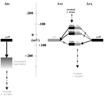
The Aio-type arsenite oxidase catalyzes electron transfer from AsIII to soluble electron carrier proteins in the cytoplasm which generally feature midpoint redox potentials in the range of +250 mV or higher (Fig. 2) [49]. Since the midpoint potential (Em) of the AsIII/AsV redox couple
is +60 mV at pH7 [50], electron transfer from the oxidation of arsenite to the electron shuttle proteins is strongly exergonic whereas the reverse redox reaction towards the reduction of arsenate would be substantially uphill. The electron transfer chain involving Aio and its soluble acceptor proteins thus appears specifically tuned to the oxidation of arsenite only.
The vast majority of Arr/Arx enzymes appears to connect redox conversions of arsenicals to the oxidoreduction of quinones. This conclusion is suggested by our analysis of the subunit composition of Arr/Arx as deduced from operon structures. Indeed, the majority of operons coding for Arr and all of those coding for Arx feature a third gene, arrC/arxC, in addition to the canonical dyad of arrA/arxA and arrB/arxB. The product of arrC/arxC is predicted by hydropathy analysis to be a membrane-integral protein with 10 transmembrane helices (data not shown). These ArrC/ArxC sequences show high similarities to those of PsrC and TtrC, the third, membrane-anchor subunits of polysulfide and tetrathionate reductase. In these latter enzymes, the C-subunits serve to connect intra-enzyme electron transfer chains to the pool of quinones [51-54]. Therefore, it is likely that the ArrC/ArxC-proteins also play the role of connecting the quinone pool to electron flow to/from arsenicals. However, the gene coding for a C-subunit is not found in several members of the Arr clade (Sup Table A.1). Among them, proteobacteria contain a cytochrome belonging to the group of CymA/NrfH-type and mediating electron transfer between the quinone-pool and the Arr-enzyme, as demonstrated in Shewanella species [55]. In the other cases, the protein responsible for this function remains to be identified but its host has been recognized to contain quinones (Sup Table A.1). In the case of Natranaerobius thermophilus, the absence of arrC is in line with the absence of a quinone pool (Sup Table B1). Overall, it can be thought that Arr/Arx enzymes redox-interact with quinones.
13
The fact that the vast majority of Arr and Arx appears to use quinones as redox partners renders considerations of thermodynamic constraints on individual reaction pathways more intricate. Indeed, a plethora of chemical types of pool-quinones with varying redox potentials have been identified so far and microbial species may either contain one or several quinones (for extensive inventories see [56-58]). Mena- (MK), methylmena- (MMK) and rhodo- (RQ) quinones feature redox midpoint potentials in the region of -60 mV and lower. Ubiquinone (UQ), by contrast, is much less reducing with an Em value at about +90 mV. Demethylmenaquinone (DMK)’s redox
potential is only slightly higher [59, 60] than that of MK. The other mentioned chemical species of quinones aren’t relevant to the questions addressed in this article since they do not occur in the repertoire of species containing Arr/Arx as listed in Sup Table B1. That these quinones feature dissimilar redox potentials while the midpoint potential of the AsIII/AsV couple obviously is universal has far-reaching thermodynamic consequences.
Figure 2: Schematic representation of the various redox couples playing a role in the bioenergetic
conversion of arsenic compounds by the Aio, Arr and Arx enzymes. Black color denotes the reduced forms while oxidized forms are shown in grey. Arrows represent thermodynamically favorable electron transfers whereas, broken arrows represent unfavorable electron transfers.
14
As illustrated in Fig. 2, the two electrochemically differing groups of quinones, i.e. those of the low-potential MKs, RQs and DMKs and of the higher-potential UQs, have redox potentials either lower (MK, RQ and DMK) or slightly higher (UQ) than the AsIII/AsV couple. This suggests a
correlation of the electrochemical property of the involved quinone and directionality of the reactions redox-converting arsenicals. It would make thermodynamic sense that, for building up a pmf needed for growth, Arr would have to oxidize low potential quinones to subsequently reduce AsV into AsIII, while Arx would need to reduce the high potential UQ with electrons originating from oxidation of AsIII to AsV. In the following we therefore tried to assess whether such a correlation indeed exists by analyzing the presence or absence of specific quinone-biosynthesis pathways in the species containing Arr/Arx by searching for the relevant genes in the respective genomes.
3.3. Genome analyses of quinone biosynthesis pathways correlate Arr and Arx to specific quinones
A single pathway has been identified for UQ/RQ biosynthesis, whereas two pathways – the so-called “men”- and “futalosine”-pathways - yield MK [21, 61]). The men pathway involves men genes and the futalosine-pathway, which was characterized in detail in S. coelicolor [20] and was shown to occur in many prokaryotes [62], involves mqn genes. The UQ biosynthesis route involves
ubi genes [63] and RQ is synthesized from UQ in one step via RquA [64, 65]. We searched for all
characterized men, mqn, ubi and rquA genes in genomes of species harboring arr/arx genes. Because several analogs to ubi genes (ubiC and ubiF) have been revealed, we also searched for these genes (coq7, rv2949c, xanB2). We synthetized the results of this genomic analysis of quinone biosynthetic pathways in the Sup Table B1. Altogether these results show a clear dichotomy between the Arr and the Arx clades:
- To the only exception of Sulfuritalea hydrogenivorans (S. hydrogenivorans), the Arr containing organisms fall into two categories: MK-only producers or, MK- and UQ-producers. S.
hydrogenivorans is the only species which exclusively contains UQ biosynthesis genes and we
confirmed the nature of the quinone pool by biochemical analysis of cells grown on AsV (data not shown).
- For the Arx-containing species, we also observed three categories of quinone producers: either these organisms rely exclusively on UQ or they can produce both MK and UQ or RQ and
15
UQ (harboring the rquA xanB2 or Rv2949 gene). In contrast to the Arr-group, no examples of MK-only organisms were observed in the Arx-group. Since a single step is sufficient for the production of RQ (a low redox-potential) from UQ (a high redox-potential quinone), we suspect that different enzymes unrelated or only remotely related to rquA, xanB2 or Rv2949 products might be able to catalyze this transition in Arx-containing species. We therefore biochemically analyzed the quinone content of the model organism for arsenite oxidation mediated by Arx, Alkalilimnicola
ehrlichii. Combining voltammetry and TLC/spectroscopy, the sole quinone identified in this strain
was UQ (Sup Fig. B2), thereby excluding the possibility that the low potential RQ might be produced in this species. This confirms that AsIII oxidation reduces the high potential UQ in A.
ehrlichii.
Except for the case of S. hydrogenivorans, the observed distribution of quinone-type and Arr/Arx is thus in line with our prediction based on thermodynamic arguments. Indeed, the reduction of arsenate by Arr seems correlated to the occurrence of the low potential MK whereas the oxidation of arsenite by Arx requires the high potential UQ as electron acceptor. The case of S.
hydrogenivorans has been examined in more detail below.
3.4. In vivo directionality of Arr/Arx fully correlates with thermodynamics
To further substantiate this scenario, we have singled out four model species in which we have characterized their respective reactivity in reducing arsenate or oxidizing arsenite. Three of them are single-quinone cases, i.e. the UQ-only and Arx-containing species A. ehrlichii, the MK-only and Arr-containing D. hafniense, and the UQ-MK-only and Arr-containing S. hydrogenivorans. The fourth organism, H. halophila, has been chosen to represent the more complicated case of a species containing an Arx-type enzyme with both UQ and MK.
16 Figure 3: Em diagram of UQ/UQH2, MK/MKH2, BV2+/BV0, DCPIP/DCPIPH2 and AsV/AsIII
couples versus pH values. Whereas H. halophila and A. ehrlichii oxidize AsIII and reduce UQ at pH values around 9, D. hafniense is not able to oxidize AsIII by using MK. H. halophila and S.
hydrogenivorans are able to reduce AsV using UQH2 at pH values around 7, but require the
consumption of pmf for that task.
The thermodynamic rationale for the experiments described below is the following: the arsenic oxyanion features two distinct pKa values in the circum-physiological region of pH, that is, a pKa of 6.97 on its oxidized form (AsV; H2AsO4-/HAsO42-) and a second one at 9.22 on its
reduced form (AsIII; H3AsO3/H2AsO3-) [50]. These two pKa values entail a pH-dependence of the
arsenic oxyanion’s redox midpoint potential of about -90 mV/pH unit below pH 6.97, of -120 mV/pH unit from pH 6.97 to pH 9.22 and again of -90 mV/pH unit at pH values beyond 9.22 (Fig. 3, continuous line). Since their physiological quinone/quinol partners are characterized by monotonous -60mV/pH dependences in this region of pH values, the relative redox driving forces for electron transfer between arsenicals and UQ/UQH2 or MK/MKH2 are strongly altered upon
changing ambient pH (see Fig. 3). The relative redox potentials depicted in Fig. 3 are midpoint potentials (Em) and the effective driving forces may vary slightly due to oxidized/reduced ratios
17
differing from unity in the redox compounds considered in this figure. However, this does not change the general trend that (a) arsenate reduction from MKH2 oxidation and arsenite oxidation
to UQ reduction are predicted to be thermodynamically favorable over the entire range considered, while arsenate reduction from UQH2 oxidation and arsenite oxidation towards MK reduction are
unfavorable and (b) arsenate reduction from MKH2 oxidation is thermodynamically more favorable
at low pH than at high pH while the opposite is true for arsenite oxidation towards UQ reduction. To assess the relevance of the outlined thermodynamic constraints on actual directionality, we have grown the four mentioned species at pH values in the vicinity of 7 and 9 and in the presence of arsenite, arsenate or in the absence of any arsenicals. We first evaluated the in vivo capacity of the Arr and Arx systems to convert arsenate or arsenite sustaining growth (therefore implying sufficient pmf generation) but also a potential capacity of these systems to convert arsenicals without allowing growth (Sup Table A.2 for a summary).
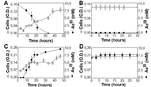
3.4.1. Arsenic redox conversion by A. ehrlichii
A. ehrlichii has been shown to grow efficiently oxidizing AsIII for the reduction of nitrate even under chemoautotrophic conditions at its physiological pH of 9.3 by using its arx genes [5, 13, 66]. Fig. 4Ashows that A. ehrlichii rapidly grows and oxidizes AsIII at pH 9.3. A. ehrlichii is unable to grow by reducing AsV even at pH 7.3. When A. ehrlichii pre-cultured at pH 9.3 in the
presence of AsIII, i.e. where high levels of the Arx-enzyme are expressed [16], is inoculated at
saturating cell density in pH 7.3 medium containing AsV, no reduction of AsV is detectable (Fig.
4B). This implies that A. ehrlichii is unable to perform this reaction at pH 7.3 and even as non-dividing cells. The directionality of the Arx enzyme in A. ehrlichii therefore cannot be reversed in
vivo over the examined pH range and the enzyme is only able to perform the thermodynamically
favorable reaction of reducing UQ by oxidizing AsIII.
3.4.2. Arsenic redox conversions by D. hafniense
D. hafniense is known to grow efficiently reducing AsV with electrons derived from acetate [28]. Genomic/phylogenetic analysis of arr genes detected in the D. hafniense genome (Sup Table A.1) suggests a bona fide ArrABC as most likely candidate for AsV reduction in this species. As shown in Fig. 4C, D. hafniense rapidly reduces AsV at pH 7.3 while it is unable to grow at pH 9.3 oxidizing AsIII. When D. hafniense pre-cultured at pH 7.3 in the presence of arsenate, i.e. where
18
high levels of the Arr-enzyme are thought to be expressed, is shifted to pH 9.3 in a medium containing AsIII at the end of growth, no oxidation of AsIII is detectable (Fig. 4D). This implies that D. hafniense is unable to perform this reaction at pH 9.3 and even as non-dividing cells. The
directionality of the Arr enzyme in D. hafniense therefore cannot be reversed over the examined pH range and the enzyme is only able to perform the thermodynamically favorable reduction of AsV by oxidizing MKH2.
Figure 4: Conversion of AsIII or AsV by A. ehrlichii and D. hafniense. A. ehrlichii was grown at pH 9.3 under AsIII 10 mM (A) and subsequently resuspended at pH7.3 under AsV 5 mM (B). D.
hafniense was grown at pH 7.3 under AsV 10 mM (C) and subsequently shifted to pH 9.3 under AsIII 10 mM (D). Open squares represent cell O.D while closed squares represent AsIII concentrations. Error bars indicate the standard deviations.
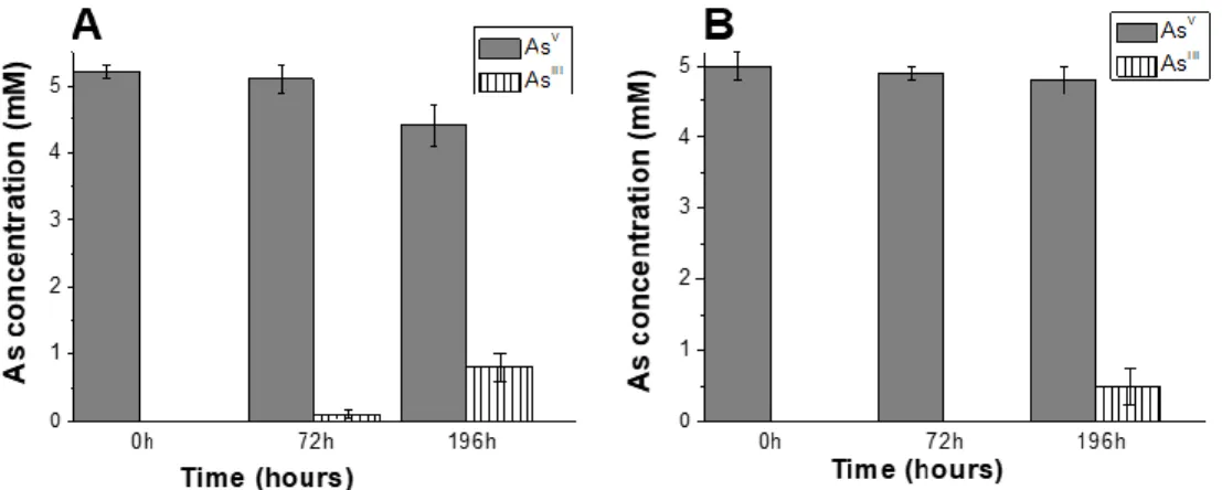
3.4.3. Arsenic redox conversions by S. hydrogenivorans
The capacity of S. hydrogenivorans to convert arsenic had never been examined. We grew the strain under different conditions were AsV was the electron acceptor or AsIII the electron donor, in presence or absence of thiosulfate and/or hydrogen [67]. Only AsV could be converted to AsIII (even without thiosulfate) but not the reverse (Figure 5A). When we repeated the experiment in the presence of gramicidin, a chemical compound able to abolish the pmf by forming prototypical ion channels specific for monovalent cations in the cytoplasmic membrane (Figure 5B), the AsV conversion level was significantly decreased. This result suggests that the implication of UQH2 in
19
the reduction of AsV to AsIII necessitates consumption of pmf to counterbalance the unfavorable
thermodynamics of the reaction.
Figure 5. AsV conversion by S. hydrogenivorans in absence (A) or presence (B) of gramicidin
at pH 7. AsV conversion by S. hydrogenivorans by using UQH2 is observable in the absence of
gramicidin but is decreased in its presence (at 10 M). Error bars indicate the standard deviations.
3.4.4. Arsenic redox conversions by H. halophila
H. halophila is a strict phototroph growing optimally at pH 8.8. Similar to Ectothiorhodospira sp. PHS-1 [14], it doesn’t grow in media containing AsIII concentrations higher than 2.5 mM whereas it grows well in media complemented with up to 10 mM AsV. 2 mM AsIII in a medium containing sulfide was found to be rapidly transformed to AsV[45]. In our hands and contrary to [45], even in absence of sulfide and thiosulfate, H. halophila grew, although significantly less, by converting AsIII to AsV (Figure 6 A and C). Judging from the relevant sequences retrieved from the H. halophila genome and from molecular phylogeny thereof ([45], Fig. 1) the species contains the genes coding for an Arx-type enzyme. Mass spectrometry performed on the single activity-destained band observed in native gels identified proteins coded by the genes HHAL_RS01810, HHAL_RS01815 and HHAL_RS01820 corresponding to the canonical triad ArxABC (data not shown) and quantitative PCR during growth under AsIII
20 Figure 6: Conversion of AsIII or AsV by H. halophila in absence of Na2S. A and C panels represent
growth at pH 8.8 under AsIII 2 mM, in presence (A) or in absence (C) of thiosulfate. B and D panels represent experiments at pH 7 under AsV 5 mM, after growth at pH 8.8 under AsIII 2 mM (such as described in (A)) and subsequent transfer to pH 7 at saturating density (B), or after sterilization of the culture (D). In all panels open squares represent cells (O.D.) while closed symbols represent AsIII concentrations. In B conversion of AsV is monitored in absence (closed squares) or in presence (closed triangles) of gramicidin (10 M). arxA gene expression was quantified by qPCR and presented relative to that measured in cells grown on Na2S used as inoculum of growth under AsIII.
Error bars indicate the standard deviations.
H. halophila contains the full set of genes required for MK-synthesis (Sup Table A.1) and
we previously have shown that the organism uses an MK/MKH2-pool when growing with electron
donors other than AsIII [32]. Nevertheless, precedence in other prokaryotes for differentially regulated expression of quinone types [68-71] raises the question whether MK/MKH2 is used in H. halophila for As conversion. We therefore have determined the amounts of MK/MKH2 and UQ/UQH2 present in cells grown under the conditions used in this work. As depicted in Sup Fig.
B2, MK/MKH2 is twice as abundant as UQ/UQH2 in cells grown in the presence of strongly
reducing substrates such as Na2S. When cells were grown with AsIII as electron donor, the amount
of MK/MKH2 substantially decreased. This suggests UQ to be the electron acceptor for the
21
When first grown at pH 8.8 under AsIII (to induce the expression of the enzyme) and then
transferred at saturating density at pH 7, H. halophila was able to convert AsV to AsIII (Figure 6
B). Sterilized cultures were no longer able to convert AsV, demonstrating that this conversion was
due to living cells (Figure 6 D). This showed that microbial reduction of AsV does occur under these conditions, although the redox reaction cannot sustain growth (data not shown). This raised the question of the quinol responsible for this AsV reduction since both MKH2 and UQH2 are
available (Sup Fig. B1).
We therefore repeated the experiment of AsV conversion at pH 7 in presence of gramicidin (Figure 6B). The experiment clearly shows that without consumption of pmf, H. halophila isn’t able any more to convert AsV to AsIII. This result suggests that UQH2 is also used for reduction of
AsV to AsIII but needs, in this case, consumption of pmf to counterbalance the unfavorable thermodynamics of the reaction.
Together (Sup Table B2), the experiments using cell cultures of the four selected species support the conclusion that directionality of As redox reactions is determined exclusively by thermodynamic constraints which are (1) imposed by the involved quinone/quinol and (2) correlated to the pH value of the physiological growth medium of the strain. This prompted us to assess the intrinsic capacities of the isolated Arr and Arx systems to perform AsIII and AsV
conversion over the whole pH range.
3.5. In-vitro directionalities of Arr and Arx also are controlled by thermodynamics
At first sight the observed inability of Arx and Arr to reverse directionality in A.ehrlichii and D. hafniense, respectively, may appear at odds with previously published results showing that both Arx and Arr can perform reduction of AsV as well as oxidation of AsIII in vitro [5, 16]. However, these in vitro experiments have been performed using the artificial electron donor benzylviologen (BV2+/BV0) and the acceptor di-chlorophenol-indophenol (DCPIP/DCPIPH2)
rather than the physiological quinones/quinols. Both BV2+/BV0 and DCPIP/DCPIPH2 are known
to react directly with the 1-electron cubane iron-sulfur clusters of the B-subunits. To better understand the influence of thermodynamic parameters, i.e. redox driving forces, on directionalities, we have tested the Arx system of H. halophila as well as the Arr model-system enzyme purified from Shewanella ANA-3 [31] using these artificial electron donors and acceptors
22
while also modifying redox driving force by varying pH. Again, as for the case of the quinones, the ΔEms for the respective oxido-reduction reactions are strongly pH-dependent as shown in Fig.
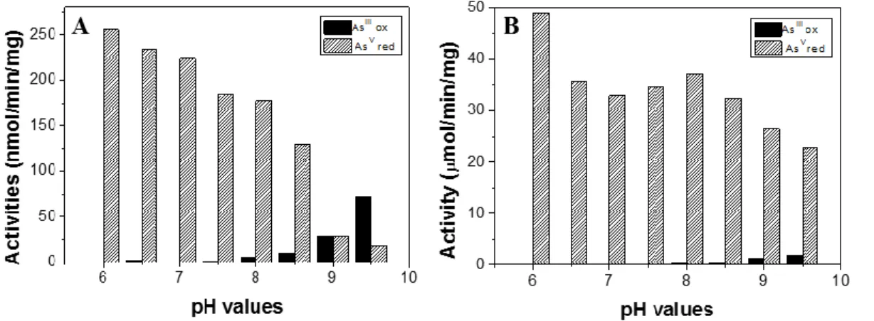
3. The obtained results are depicted in Fig. 7.
Figure 7: pH dependence of Arx and Arr-catalyzed reactions. In solution arsenite oxidation (AsIII
ox) and arsenate reduction (AsV red) were measured on membrane fragments from H. halophila (A) or on purified recombinant Arr from S. ANA-3 (B).
In line with the substantially higher redox driving force for arsenate reduction from BV0 as compared to arsenite oxidation by DCPIP at low pH, the activities of AsV reduction by the Arx- and the Arr-enzymes vastly surpass those of AsIII oxidation. At high pH, i.e. when the ΔEm for the
former reaction shrinks while that for the latter one grows, AsIII oxidation activities increase while
those for AsV reduction decrease. Above pH 9, AsIII oxidation activities in the Arx-enzyme of H. halophila surpass those for AsV reduction. In the Arr-enzyme from Shewanella AsV, reduction
activities decreased from low to high pH values but still remained above those for AsIII oxidation,
for which a substantial increase was observed. These results demonstrate that while there is some built-in bias (related to the GRY-T or the GRGWG motif for example as proposed by Glasser et al. [48]) favoring the physiological directionality of the Arr and Arx enzymes, both are reversible and the directionality is crucially controlled by the thermodynamic driving force of the Arsenicals: quinone/quinol oxido-reduction reaction imposed in vivo.
4. Discussion
Based on molecular phylogeny, we have previously proposed a scenario in which the oxidation of AsIII by the Aio enzyme was a bioenergetic process operating in the early Archaean
23
[9]. Furthermore, we proposed that the phylogenetically distant sister-enzyme dyad Arr/Arx emerged later in the domain Bacteria from a precursor enzyme performing sulfur-based redox chemistry [11]. Subsequently, this scenario was challenged by the hypothesis of the Arx-type enzyme being the ancestral AsIII oxidizing entity operating in the LUCA [14].
In this work we have updated the set of sequences used to reconstruct the corresponding phylogenetic trees. The obtained trees, while still suggesting the notion of the presence of Aio in Archaean geological eon, where not conclusive on the ancestrality of Arr and Arx. By exclusively using phylogenetics, we thus could not solve the controversy about the identity of the enzyme responsible for arsenite oxidation in the Archean geological eon. To enrich the debate on this evolutionary controversy, we have tried in this article to leave the phylogenetic box and to add arguments derived from both function and thermodynamic constraints.
The above described results demonstrate a striking correlation between directionality (assessed via phylogenetic clustering with either the Arx- or the Arr-clade) and quinone usage (identified through presence/absence of specific biosynthesis pathways as well as biochemical analyses). Low potential MK were found to be associated with the reduction of AsV by Arr while presence of the high potential UQ correlates perfectly with oxidation of AsIII by Arx. Our in-vivo measurements of arsenic redox conversions performed by four selected organisms containing either only UQ, or only MK or both UQ and MK, have confirmed the mandatory association of AsV
reduction to the presence of MK and of AsIII oxidation to the presence of UQ for building-up pmf,
an association which makes perfect thermodynamic sense (see Figs. 2 and 3).
A scenario of pre-LUCA Arx-based AsIII oxidation [14] would therefore require high
potential quinones to have been present in the common ancestor of Archaea and Bacteria. All available evidence suggests that this was not the case [35]. While the question whether LUCA contained quinones or not is still under debate [62, 72], it appears inevitable that the ancestral quinones were low potential MK [57, 72-74]. The emergence of the high potential UQ can be traced back to the common ancestor of Proteobacteria while no other prokaryotic phylum containing UQ are known (to the possible exception of cyanobacteria with their structurally related plastoquinones [75]). Consequently, and in line with thermodynamic reasoning, Arx genes have only been detected in Proteobacteria so far. Functional characterizations of a class of enzymes unrelated to those examined in this work in species containing low potential or high potential quinones strongly suggested that the appearance of high potential quinone-based bioenergetic chains was driven by
24
the need to adapt to the deleterious effects of rising O2-tensions [76] at and after the Grand
Oxygenation Event (GOE). High potential quinones therefore likely have entered the evolutionary scene only about 2.5 billion years ago. An Arx-type arsenite oxidase reducing quinones therefore became thermodynamically viable only after the redox upshift of energy-converting electron transfer chains. Arr-based reduction of arsenate, by contrast, is linked to the presence of MK and therefore is potentially older than Arx-type metabolism. However, due to its moderately high redox midpoint potential, AsV is unlikely to have been present in substantial amounts during the anoxic, mildly reducing conditions of the early Archaean. The bulk of arsenic was likely present as AsIII at the time.
In striking contrast, the enzyme Aio couples oxidation of AsIII to reduction of soluble electron acceptors which provide a considerably higher redox driving force (Fig. 2) for forward electron transfer. Aio thus may have operated in the early Archaean no matter whether low or high or no quinones were present and the thermodynamic viability of the reaction only depends on the availability of sufficiently oxidizing electron acceptor substrates. We have addressed the question of the likely acceptors in the past [3] and presently consider nitrogen oxyanions as the most likely candidates.
The scenario of an early AsIII oxidation via Aio and of an emergence of the corresponding reaction catalyzed by Arx only after the GOE is thus in line with all thermodynamic constraints, while the hypothesis of an Arx-mediated oxidation of AsIII severely clashes with basic principles
of bioenergetic electron transfer.
5. Acknowledgments and funding information
Our work is funded by the CNRS, CEA, Aix-Marseille Univ, ANR (ANR-12-BS08-0014-01 and ANR-15-CE11-0001-02).
6. Appendix A. Supplementary data
Supplementary data to this article can be found online at XXX.
25 [1] R. Mukhopadhyay, B.P. Rosen, L.T. Phung, S. Silver, Microbial arsenic: from geocycles to genes and enzymes, FEMS microbiology reviews, 26 (2002) 311-325.
[2] J.F. Stolz, P. Basu, J.M. Santini, R.S. Oremland, Arsenic and selenium in microbial metabolism, Annual review of microbiology, 60 (2006) 107-130.
[3] R. van Lis, W. Nitschke, S. Duval, B. Schoepp-Cothenet, Arsenics as bioenergetic substrates, Biochimica et biophysica acta, 1827 (2013) 176-188.
[4] N. Kumari, S. Jagadevan, Genetic identification of arsenate reductase and arsenite oxidase in redox transformations carried out by arsenic metabolising prokaryotes - A comprehensive review,
Chemosphere, 163 (2016) 400-412.
[5] C. Richey, P. Chovanec, S.E. Hoeft, R.S. Oremland, P. Basu, J.F. Stolz, Respiratory arsenate reductase as a bidirectional enzyme, Biochemical and biophysical research communications, 382 (2009) 298-302. [6] G.L. Anderson, J. Williams, R. Hille, The purification and characterization of arsenite oxidase from Alcaligenes faecalis, a molybdenum-containing hydroxylase, The Journal of biological chemistry, 267 (1992) 23674-23682.
[7] K. Zargar, A. Conrad, D.L. Bernick, T.M. Lowe, V. Stolc, S. Hoeft, R.S. Oremland, J. Stolz, C.W. Saltikov, ArxA, a new clade of arsenite oxidase within the DMSO reductase family of molybdenum oxidoreductases, Environmental microbiology, 14 (2012) 1635-1645.
[8] M.C. Sforna, P. Philippot, A. Somogyi, M.A. van Zuilen, K. Medjoubi, B. Schoepp-Cothenet, W. Nitschke, P.T. Visscher, Evidence for arsenic metabolism and cycling by microorganisms 2.7 billion years ago, Nature Geosci, 7 (2014) 811-815.
[9] E. Lebrun, M. Brugna, F. Baymann, D. Muller, D. Lievremont, M.C. Lett, W. Nitschke, Arsenite oxidase, an ancient bioenergetic enzyme, Molecular biology and evolution, 20 (2003) 686-693.
[10] E. Lebrun, J.M. Santini, M. Brugna, A.L. Ducluzeau, S. Ouchane, B. Schoepp-Cothenet, F. Baymann, W. Nitschke, The Rieske protein: a case study on the pitfalls of multiple sequence alignments and phylogenetic reconstruction, Molecular biology and evolution, 23 (2006) 1180-1191.
[11] S. Duval, A.L. Ducluzeau, W. Nitschke, B. Schoepp-Cothenet, Enzyme phylogenies as markers for the oxidation state of the environment: the case of respiratory arsenate reductase and related enzymes, BMC evolutionary biology, 8 (2008) 206.
[12] B. Schoepp-Cothenet, S. Duval, J.M. Santini, W. Nitschke, Comment on "Arsenic (III) fuels
anoxygenic photosynthesis in hot spring biofilms from Mono Lake, California", Science, 323 (2009) 583; author reply 583.
[13] S.E. Hoeft, J.S. Blum, J.F. Stolz, F.R. Tabita, B. Witte, G.M. King, J.M. Santini, R.S. Oremland, Alkalilimnicola ehrlichii sp. nov., a novel, arsenite-oxidizing haloalkaliphilic gammaproteobacterium capable of chemoautotrophic or heterotrophic growth with nitrate or oxygen as the electron acceptor, International journal of systematic and evolutionary microbiology, 57 (2007) 504-512.
[14] T.R. Kulp, S.E. Hoeft, M. Asao, M.T. Madigan, J.T. Hollibaugh, J.C. Fisher, J.F. Stolz, C.W. Culbertson, L.G. Miller, R.S. Oremland, Arsenic(III) fuels anoxygenic photosynthesis in hot spring biofilms from Mono Lake, California, Science, 321 (2008) 967-970.
[15] S.E. Hoeft, T.R. Kulp, S. Han, B. Lanoil, R.S. Oremland, Coupled arsenotrophy in a hot spring photosynthetic biofilm at Mono Lake, California, Applied and environmental microbiology, 76 (2010) 4633-4639.
[16] K. Zargar, S. Hoeft, R. Oremland, C.W. Saltikov, Identification of a novel arsenite oxidase gene, arxA, in the haloalkaliphilic, arsenite-oxidizing bacterium Alkalilimnicola ehrlichii strain MLHE-1, Journal of bacteriology, 192 (2010) 3755-3762.
[17] R.S. Oremland, C.W. Saltikov, J.F. Stolz, J.T. Hollibaugh, Autotrophic microbial arsenotrophy in arsenic-rich soda lakes, FEMS microbiology letters, 364 (2017).
26 [18] T. Krafft, R. Gross, A. Kroger, The function of Wolinella succinogenes psr genes in electron
transport with polysulphide as the terminal electron acceptor, European journal of biochemistry / FEBS, 230 (1995) 601-606.
[19] A.P. Hinsley, B.C. Berks, Specificity of respiratory pathways involved in the reduction of sulfur compounds by Salmonella enterica, Microbiology, 148 (2002) 3631-3638.
[20] T. Hiratsuka, K. Furihata, J. Ishikawa, H. Yamashita, N. Itoh, H. Seto, T. Dairi, An alternative menaquinone biosynthetic pathway operating in microorganisms, Science, 321 (2008) 1670-1673. [21] B. Nowicka, J. Kruk, Occurrence, biosynthesis and function of isoprenoid quinones, Biochimica et biophysica acta, 1797 (2010) 1587-1605.
[22] P. Stenmark, J. Grunler, J. Mattsson, P.J. Sindelar, P. Nordlund, D.A. Berthold, A new member of the family of di-iron carboxylate proteins. Coq7 (clk-1), a membrane-bound hydroxylase involved in ubiquinone biosynthesis, The Journal of biological chemistry, 276 (2001) 33297-33300.
[23] G. Stadthagen, J. Kordulakova, R. Griffin, P. Constant, I. Bottova, N. Barilone, B. Gicquel, M. Daffe, M. Jackson, p-Hydroxybenzoic acid synthesis in Mycobacterium tuberculosis, The Journal of biological chemistry, 280 (2005) 40699-40706.
[24] L. Zhou, J.Y. Wang, J. Wu, J. Wang, A. Poplawsky, S. Lin, B. Zhu, C. Chang, T. Zhou, L.H. Zhang, Y.W. He, The diffusible factor synthase XanB2 is a bifunctional chorismatase that links the shikimate
pathway to ubiquinone and xanthomonadins biosynthetic pathways, Molecular microbiology, 87 (2013) 80-93.
[25] J.D. Thompson, T.J. Gibson, F. Plewniak, F. Jeanmougin, D.G. Higgins, The CLUSTAL_X windows interface: flexible strategies for multiple sequence alignment aided by quality analysis tools, Nucleic acids research, 25 (1997) 4876-4882.
[26] P. Di Tommaso, S. Moretti, I. Xenarios, M. Orobitg, A. Montanyola, J.M. Chang, J.F. Taly, C.
Notredame, T-Coffee: a web server for the multiple sequence alignment of protein and RNA sequences using structural information and homology extension, Nucleic acids research, 39 (2011) W13-17. [27] N. Galtier, M. Gouy, C. Gautier, SEAVIEW and PHYLO_WIN: two graphic tools for sequence
alignment and molecular phylogeny, Computer applications in the biosciences : CABIOS, 12 (1996) 543-548.
[28] A. Niggemyer, S. Spring, E. Stackebrandt, R.F. Rosenzweig, Isolation and characterization of a novel As(V)-reducing bacterium: implications for arsenic mobilization and the genus
Desulfitobacterium, Applied and environmental microbiology, 67 (2001) 5568-5580.
[29] H. Kojima, M. Fukui, Sulfuritalea hydrogenivorans gen. nov., sp. nov., a facultative autotroph isolated from a freshwater lake, International journal of systematic and evolutionary microbiology, 61 (2011).
[30] B. Chardin, F. Chaspoul, P. Gallice, Heavy metals speciation by HPLC/ICP-MS - Application to the reduction of Cr(VI) and As(V) by sulphate-reducing bacteria Can J Anal Sci Spect, 48 (2003) 336-342. [31] D. Malasarn, J.R. Keeffe, D.K. Newman, Characterization of the arsenate respiratory reductase from Shewanella sp. strain ANA-3, Journal of bacteriology, 190 (2008) 135-142.
[32] B. Schoepp-Cothenet, C. Lieutaud, F. Baymann, A. Vermeglio, T. Friedrich, D.M. Kramer, W. Nitschke, Menaquinone as pool quinone in a purple bacterium, Proceedings of the National Academy of Sciences of the United States of America, 106 (2009) 8549-8554.
[33] U.K. Laemmli, Cleavage of structural proteins during the assembly of the head of bacteriophage T4, Nature, 227 (1970) 680-685.
[34] M. Hajj Chehade, L. Loiseau, M. Lombard, L. Pecqueur, A. Ismail, M. Smadja, B.
Golinelli-Pimpaneau, C. Mellot-Draznieks, O. Hamelin, L. Aussel, S. Kieffer-Jaquinod, N. Labessan, F. Barras, M. Fontecave, F. Pierrel, ubiI, a new gene in Escherichia coli coenzyme Q biosynthesis, is involved in aerobic C5-hydroxylation, The Journal of biological chemistry, 288 (2013) 20085-20092.
27 [35] L. Pelosi, A.L. Ducluzeau, L. Loiseau, F. Barras, D. Schneider, I. Junier, F. Pierrel, Evolution of
Ubiquinone Biosynthesis: Multiple Proteobacterial Enzymes with Various Regioselectivities To Catalyze Three Contiguous Aromatic Hydroxylation Reactions, mSystems, 1 (2016).
[36] R. Clement, S. Lignon, P. Mansuelle, E. Jensen, M. Pophillat, R. Lebrun, Y. Denis, C. Puppo, S.C. Maberly, B. Gontero, Responses of the marine diatom Thalassiosira pseudonana to changes in CO2 concentration: a proteomic approach, Scientific reports, 7 (2017) 42333.
[37] C.W. Saltikov, D.K. Newman, Genetic identification of a respiratory arsenate reductase,
Proceedings of the National Academy of Sciences of the United States of America, 100 (2003) 10983-10988.
[38] N. Rascovan, J. Maldonado, M.P. Vazquez, M. Eugenia Farias, Metagenomic study of red biofilms from Diamante Lake reveals ancient arsenic bioenergetics in haloarchaea, The ISME journal, 10 (2016) 299-309.
[39] R.S. Oremland, J.F. Stolz, Arsenic, microbes and contaminated aquifers, Trends in microbiology, 13 (2005) 45-49.
[40] D. Malasarn, C.W. Saltikov, K.M. Campbell, J.M. Santini, J.G. Hering, D.K. Newman, arrA is a reliable marker for As(V) respiration, Science, 306 (2004) 455.
[41] O.F. Ordonez, M.C. Rasuk, M.N. Soria, M. Contreras, M.E. Farias, Haloarchaea from the Andean Puna: Biological Role in the Energy Metabolism of Arsenic, Microbial ecology, (2018).
[42] S. Nelson-Sathi, T. Dagan, G. Landan, A. Janssen, M. Steel, J.O. McInerney, U. Deppenmeier, W.F. Martin, Acquisition of 1,000 eubacterial genes physiologically transformed a methanogen at the origin of Haloarchaea, Proceedings of the National Academy of Sciences of the United States of America, 109 (2012) 20537-20542.
[43] F. Baymann, B. Schoepp-Cothenet, E. Lebrun, R. van Lis, W. Nitschke, Phylogeny of Rieske/cytb complexes with a special focus on the Haloarchaeal enzymes, Genome biology and evolution, 4 (2012) 720-729.
[44] J. Hernandez-Maldonado, B. Sanchez-Sedillo, B. Stoneburner, A. Boren, L. Miller, S. McCann, M. Rosen, R.S. Oremland, C.W. Saltikov, The genetic basis of anoxygenic photosynthetic arsenite oxidation, Environmental microbiology, 19 (2017) 130-141.
[45] S. Hoeft McCann, A. Boren, J. Hernandez-Maldonado, B. Stoneburner, C.W. Saltikov, J.F. Stolz, R.S. Oremland, Arsenite as an Electron Donor for Anoxygenic Photosynthesis: Description of Three Strains of Ectothiorhodospira from Mono Lake, California and Big Soda Lake, Nevada, Life, 7 (2016).
[46] G. Durante-Rodríguez, H. Fernández-Llamosas, E. Alonso-Fernandes, M.N. Fernández-Muñiz, R. Muñoz-Olivas, E. Díaz, M. Carmona, ArxA From Azoarcus sp. CIB, an Anaerobic Arsenite Oxidase From an Obligate Heterotrophic and Mesophilic Bacterium, Frontiers in microbiology, 10 (2019).
[47] A. Arshad, D.R. Speth, R.M. de Graaf, H.J. Op den Camp, M.S. Jetten, C.U. Welte, A Metagenomics-Based Metabolic Model of Nitrate-Dependent Anaerobic Oxidation of Methane by Methanoperedens-Like Archaea, Frontiers in microbiology, 6 (2015) 1423.
[48] N.R. Glasser, P.H. Oyala, T.H. Osborne, J.M. Santini, D.K. Newman, Structural and mechanistic analysis of the arsenate respiratory reductase provides insight into environmental arsenic
transformations, Proceedings of the National Academy of Sciences of the United States of America, 115 (2018) E8614-E8623.
[49] A. Lieutaud, R. van Lis, S. Duval, L. Capowiez, D. Muller, R. Lebrun, S. Lignon, M.L. Fardeau, M.C. Lett, W. Nitschke, B. Schoepp-Cothenet, Arsenite oxidase from Ralstonia sp. 22: characterization of the enzyme and its interaction with soluble cytochromes, The Journal of biological chemistry, 285 (2010) 20433-20441.
[50] B.W. Vink, Stability relations of antimony and arsenic compounds in the light of revised and extended Eh-pH diagrams, Chemical Geology, 130 (1996) 9.

![Risiko- & [und] Schutzfaktoren der psychischen Gesundheit humanitärer Einsatzhelfer : eine systematische Literaturübersicht](data:image/gif;base64,R0lGODlhAQABAIAAAP///wAAACH5BAEAAAAALAAAAAABAAEAAAICRAEAOw==)