The Development and Application of an
InVitro Model of Coronary
Lesion Thrombosis
by
Kumaran Kolandaivelu
B.S. Mechanical Engineering
Massachusetts Institute of Technology, 1995 M.S. Mechanical Engineering
Massachusetts Institute of Technology, 2003
MASSACHUSES INSr
OF TECHNOLOGY
JUN 3 2005
LIBRARIES .;j .!
SUBMITTED TO THE DEPARTMENT OF HEALTH SCIENCES AND TECHOLOGY IN PARTIAL FULFILLMENT OF THE REQUIREMENTS FOR THE DEGREE OF DOCTOR OF PHILOSOPHY IN MEDICAL ENGINEERING MEDICAL PHYSICS
AT THE MASSACHUSETTS INSTITUTE OF TECHNOLOGY MAY 2005 [T : .
( 2005 Kumaran Kolandaivelu. All rights reserved.
The author hereby grants MIT permission to reproduce and distribute publicly paper and electronic copi/of this thesis document in whole or in part.
/
7/S
Signature of Author: . iI7I
I
Department of Health Sciences and TechnologyMay 16, 2005
Certified by: .. &d _
,J Elazer R. Edelman
Thomas D. and Virginia W. Cabot Professor Division of Health Sciences and Technology
Thesis Supervisor
Accepted by: It
i / Martha L. Gray, Ph.D.
Edward Hood Taplin Professor of Medi al and Electrical Engineering Director, Harvard-MIT Division of He lth Sciences and Technology
Of
L. i
The Development and Application of
an In Vitro Model of Coronary
Lesion Thrombosis
by Kumaran Kolandaivelu
Submitted to the Department of Mechanical Engineering on May 16, 2005 in Partial Fulfillment of the Requirements for the Degree of Doctor of Philosophy in Medical Engineering Medical Physics
Abstract
Thrombosis is an initiating response to vascular injury. Physiologically, this process aids in the repair and remodeling of the vessel wall. However, if left unchecked, luminal occlusion may rapidly occur. The coronary vascular bed is a life-sustaining environment in which pathological thrombosis can lead to devastating outcomes such as acute coronary syndromes or post-interventional thrombosis. In order to study these coronary thrombotic reactions, it is essential to consider the physical environment in which they occur. We have developed an in vitro method for creating pulsatile flows to mimic the coronary hernodynamic setting on a beat-to-beat basis. Furthermore, he have developed techniques and protocols to parametrically vary both the biological and physical aspects of thrombosis and in doing so, have investigated the effects of real-world temporal and spatial flow perturbations on local site platelet adhesion. Not only do such variations create quantitative differences in local reactions, but qualitative differences as well as various receptors must interact to create stable adhesions in a given hemodynamic environments. These findings have implications on the propensity for certain individuals to form clot under certain condtitions, as well as the environment-dependent efficacy of various clinically relevant anti-thrombotic strategies
Thesis Supervisor: Elazer R. Edelman
Thomas D. and Virginia W. Cabot Professor of Health Sciences and Technology
Acknowledgments
First and foremost, I would like to thank my mentor, Prof. Elazer R. Edelman who through a perfect balance of guidance, acceptance and support over the past six years has guided me over an improbable path towards an impossible goal. Thanks to his
tremendous foresight and insight, I have, in hindsight, enjoyed all that I have seen along the way.
I would also like to thank my thesis committee: Prof. Roger D. Kamm who has been a beacon on the often choppy waters of fluid mechanics, Dr. Fredrick D. Welt whose ability to mix the intricacies of cardiovascular biology with an all around good nature provide on inspiration for the years that lie ahead, and Dr. Robert D. Handin who helped me begin to appreciate the important distinctions between a scientific and engineering approach.
I give special thanks to Nick Houstis, Siraj Ali, two great friends with whom I have been stumbling with for the past seven years, and undoubtedly will stumble with for many more. Also, I would like to thank Mercedes for being a great friend and a portal to a Spanish sun-- to endless rays of light and laughter which have illuminated even my darkest days (and nights) as a researcher. Viva la Spain! And of course, my partner-in-crime, Bobbie Bushiae-- without whom I would have likely finished a year or two ago, but with whom I would gladly spend a lifetime more. Viva la Bobbie Bushaie!
Last, I dedicate this thesis to my mom and dad, who have taught me above all else to take them for granted. You are my foundation.
Table of Contents
Abstract
...
2
Acknowledgements
...
3
Table of Contents
...
4
List of Figures
...
7
List of Tables
...
11
CHAPTER 1 BACKGROUND AND SIGNIFICANCE
...
13
1.1 Coronary Thrombosis
...
13
1.1.1 Pathophysiology ... 13 1.1.2 Treatments ... 1... 15 1.2 The Platelet ... 17 1.2.1 Platelet Reactions ... 17 1.2.2 Platelet Interactions ... 201.3 The Local Hemodynamic Setting ... 24
1.3.1 Local Flow Interactions ... ...25
1.3.2 Local Geometric Environment ... ... 28
1.4 Methods of studying flow dependent thrombosis
...
32
1.4.1 In Vivo Models (Realistic) . ... 32
1.4.2 In Vitro Models (Perturbable) ... 34
1.4.3 Proposed Flow System ... 36
CHAPTER 2 FLOW SYSTEM DESIGN
...
38
2.1 Theory
...
38
2.1.1 Description ... 38
2.1.2 Analytical, Straight Tube Approximation ... 39
2.1.3 Curvature Effects ... 47
2.2 Embodiment ... 49
2.2.1 Prototype evolution ... 49
2.2.2 Fluid Loops ... 54 4
2.2.3 Rotors ... 56
2.2.4 Motor Selection ... 58
2.2.5 Control System ... 60
2.2.6 Measurement/Recording System ... 62
2.2.7 Design Summary ... 66
CHAPTER 3 FLOW SYSTEM TESTING
...
67
3.1 Mechanical validation
...
67
3.1.1 Flow profiles ... 67
3.1.2 Coronary-Flow ... 69
3.2
Biological validation
...
72
3.2.1 Materials ... 72
3.2.2 Systemic Noise Characterization ... 74
3.2.3 Signal to Noise ... 75
CHAPTER 4 PARAMETERIZING
THE
THROMBOTIC
RESPONSE
80
4.1 Platelet Function
...
82
4.1.1 Observing Platelet Function ... 82
4.1.2 Parameterizing Platelet Function ... 86
4.2
Dynamic Bio-lmplant Platelet Reactions
...
91
4.2.1 Methods ... 92
4.2.2 Decoupled Platelet/Coagulation Responses ... 95
4.2.3 Coupled Platelet Response ... 97
4.2.4 Richness in the Thrombotic Response ... 102
CHAPTER 5 THE CORONARY LESION ENVIRONMENT
108
5.1 Reactive Site Development ... 1085.1.1 Geometry ... 110
5.1.2 Surfaces...1...113
5.2 REACTIVE SITE CHARACTERIZATION
...
117
5.2.1 Methods ... 118
5.2.2 Functional Validation ... 121
CHAPTER 6 REAL-WORLD
HEMODYNAMIC
ENVIRONMENTS ...
127
6.1 Temporal Variations in Hemodynamic Environments
...
129
6.1.1 Methods ... 130
6.1.2 Platelet Adhesion in Pulsatile Environments ... 137
6.1.3 D iscussion . ... 143
6.2 Spatial Variations in Hemodynamic Environments
...
146
6.2.1 Methods ... 148
6.2.2 Platelet Accumulation in Stented Environments . ... 152
6.2.3 Discussion ... 156
6.3 Optimizing Platelet Inhibition to Hemodynamic Settings 162
6.3.1 Methods ... 1636.3.2 Platelet inhibition in complex hemodynamic environments ... 165
6.3.3 Discussion ... ... 169
CHAPTER 7 LIMITATIONS.
CONCLUSIONS.
AND
FUTURE DIRECTIONS
...
175
APPENDICES
...
178
A. IQ MASTER SAMPLE FILES ... 178
B. EXPERIMENTAL DATA ... 186
REFERENCES
...
192
6
---List of Figures
Figure 1.1: Schematic diagram if the heart and major coronary arteries ... 13
Figure 1.2: Healthy and stenotic coronary arteries ... 14
Figure 1.3: Clinical and silent thrombotic outcomes of vascular injury ... 16
Figure 1.4: Tunneling electron microscope image of internal platelet compartments .... 17
Figure 1.5: Sequence of platelet/vessel wall reactions ... 18
Figure 1.6: Classical coagulation reactions ... 20
Figure 1.7: Platelets entangled in fibrin mesh ... 21
Figure 1.8: Typical phasic coronary flow profile ... 24
Figure 1.9: Coronary angiogram depicting curved, branching geometries ... 28
Figure 1.10: Vessel architecture and resultant complex flow patterns ... ...29
Figure 2.1: Fluid filled torus schematic ... 38
Figure 2.2: Theoretical system assuming linear rather than angular motions ... 40
Figure 2.3: Relative radial velocity profiles given an impulse wall acceleration ... 42
Figure 2.4: Relative radial velocity profile given a constant wall acceleration ... 43
Figure 2.5: Relative radial velocity profile given a sinusoidal wall acceleration ... 45
Figure 2.6 Relative axial velocity profile f\given a square wall acceleration ... 46
Figure 2.7: Single fluid loop, infra-red sensing rotor stage ... ...49
Figure 2.8: Single fluid loop rotor stage with ultrasound flow probe ... ...50
Figure 2.9: Multi-rotor system ... ...51
Figure 2.10: Current multi-rotor, multi-axis embodiment ... ... ...52
Figure 2.11: General system schematic ... 53
Figure 2.13: Filling of fluid loop ... . ... 55
Figure 2.14: Rotor-stage design schematic . ... 56
Figure 2.15: Fluid loop positioned on rotor stage ... 57
Figure 2.16: Rotor shaft schematic ... 58
Figure 2.17: Single axis control schematic ... ... 60
Figure 2.18: Multi axis control schematic ... 61
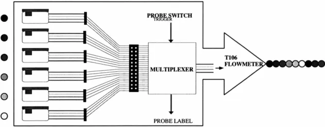
Figure 2.19: Measurement/Recording system schematic ... ... ...62
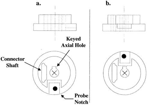
Figure 2.20: 24-pin connector design ... ... ... ...64
Figure 2.21: Multiplexing schematic ... ... ... 65
Figure 3.1: Relative fluid flow to impulsive wall acceleration ... ... 67
Figure 3.2: Various flow profiles (Square, Triangular, Sinusoidal) ... 68
Figure 3.3: Generating coronary type flow profiles (fluid and wall motions) ... 69
Figure 3.4: Changing amplitude of coronary-type flows ... 71
Figure 3.5: Stent placement within fluid loop ... ... ... 73
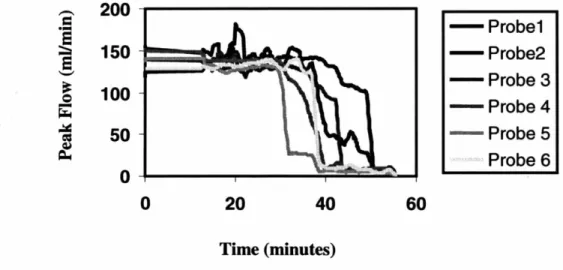
Figure 3.6: System precision. Six stent loop occlusion times ... 74
Figure 3.7: Background thrombotic noise levels. Stented vs. Non-stented loops ... 75
Figure 3.8: Inter-stent studies ... 77
Figure 4.1: Phenomenological representation of loop occlusion ... ... 80
Figure 4.2: Flow cytometric assessment of platelet responsiveness ... 84
Figure 4.3: Visual assessment of platelet responsiveness ... 86
Figure 4.4: Electronmicrograph depicting platelet adhesion w/ and w/out coagulation...88
Figure 4.5: Generated coronary-like flow profiles ... 92
Figure 4.6: Decoupled coagulation and platelet response to stent surface ... 96
Figure 4.7: Surface and Bulk platelet response to stent with increasing anticoagulant..98
Figure 4.8: Effect of flow and coagulation on stent platelet reactivity ... 100
Figure 4.9: Platelet reactivity surfaces as a function of flow and coagulation ... 103
Figure 4.10: Schematic representation of path to clinical development ... 105
Figure 5.1: Angiogram depicting coronary lesion ... 108
Figure 5.2: Schematic representation of localized reactive site ... 109
Figure 5.3: Embodied reactive site model ... 110
Figure 5.4: Variations in reactive site geometry ... 111
Figure 5.5: Stented reactive sites w/ and w/out wall apposition ... 112
Figure 5.6: Generated flow wave form with glass and silicone reactive sites ... 112
Figure 5.7: Schematic depiction of variable reactive site surfaces ... 114
Figure 5.8: Generated coronary-like flow profiles ... ...118118... Figure 5.9: P'latelet adhesion to glass and silicone reactive sites ... 121
Figure 5.10: Visualization of reactive site platelet adhesion ... ...122
Figure 5.11: Inhibition of reactive site platelet adhesion with albumin coating ... 123
Figure 5.12: Time course of platelet adhesion at varied flow rates ... 124
Figure 6.1: Imnpule-boosted square wave wall acceleration profile ... 131
Figure 6.2: Relative velocity profile given boosted square wave wall motion ... 132
Figure 6.3: Bulk flows for boosted and regular square wall accelerations ... 132
Figure 6.4: Wall shear rates for boosted and regular square wall accelerations ... 133
Figure 6.5: Impulse-train boosted square wave wall acceleration profile ... 134
Figure 6.6: Flow and shear for impulsive-train square wall accelerations ... 135
Figure 6.8: Full-range of triangular and square flow profiles .... ...136
Figure 6.9: Platelet adhesion w/ and w/out AK2 for triangular and square flows...138
Figure 6.10: Visualization of pulsatile flow platelet adhesion w/ and w/out AK2...139
Figure 6.11: Ratio of platelet adhesion under triangular to square flow conditions .... 140
Figure 6.12: Definition of Tlow ... ... ... ... 141
Figure 6.13: 1Hz Square flow profiles of altering duty cycle ... ...142
Figure 6.14: Platelet adhesion as function of TL ... 143
Figure 6.15: Schematic of coronary interventions ... 146
Figure 6.16: Generated coronary-like flow profiles ... 148
Figure 6.17: Stent induced spatial flow variations ... 150
Figure 6.18: Closed vs. Open-Cell stent geometries ... 151
Figure 6.19: Effect of stent presence on collagenized site platelet accumulation ... 153
Figure 6.20: Effect of stent presence on albuminized site platelet accumulation ... 154
Figure 6.21: Ratio of albumenized to collagenized platelet accumulation w/stent... 158
Figure 6.22: Generated triangular flow wave forms ... 163
Figure 6.23: Effect of GPIb, a2B 1, and IIbIIIa blockade on platelet adhesion ... 166
Figure 6.24: Modified duty cycle flows w/ extended low flow phase ... 167
Figure 6.25: Platelet Tadhesion w/ dual GPIb/a2B 1 blockade ... ... 168
Figure 6.26: Effect of stenting and flow pulsatility on methods of platelet inhibtion.. 169
List of Tables
Table 1.1: Comparison of prior in vitro and in vivo methods of studying thrombosis ....36
Table 2.1: List of dimensionless parameters considered and customary definitions ...39
Table 6.1: Platelet adhesion to reactive sites for open-cell stent design depicting robustness of findings . . ... ... ...155
Table 6.2: Computational results for flow over various stent condtions ... 159
Table 6.3: Platelet receptors and specific functional inhibitors ... ... 165
Table B. 1: Loop occlusion times for polished and unpolished stainless steel stents .... 186
Table B.2: Loop occlusion times for gold-coated, gold-coated+heat treated, and polished stainless steel stents .. ... 186
Table B.3: Comparison of coagulation activation for varied stent coating ... 187
Table B.4: Comparison of surface bound platelets for varied stent coating ... 187
Table B.5: Comparison of bulk platelet activation for varied stent soating ... 187
Table B.6: Platelet surface adhesion to stented loops w/increasing anti-coagulation .... 188
Table B.7: Bulk platelet activation in stented loops w/increasing anti-coagulation ... 188
Table B.8: Platelet surface accumulation data for stented loops at high anti-coagulant level and varied flows ... ... 188
Table B.9: Platelet surface accumulation data for stented loops at low anti-coagulant level and varied flows ... 188
Table B.10: Effect of flow pulsatility w/ and w/out AK2 (400ml/min mean flow) ... 189
Table B.11: Effect of flow pulsatility w/ and w/out AK2 (300ml/min mean flow) ... 189
'Table B. 13: Effect of flow pulsatility w/ and w/out AK2 (100ml/min mean flow) ... 190
Table B. 14: Effect of flow pulsatility w/ and w/out AK2 (50ml/min mean flow) ... 190
'Table B. 15: Platelet adhesion of a function of TL ... 190
Table B. 16: Effect of G19 on platelet adhesion to collagen reactive site as a function of
pulsatile triangular flow rate ... 191
Table B 17:Effect of G19 and AK2 on platelet adhesion to collagen reactive site as a
function of pulsatile, triangular flow rate ... 191
Table B18: Effect of Reopro on platelet adhesion to collagen reactive site as a function of
pulsatile, triangular flow rate ... ... 191
Table B 19: Effect of co-administration of G19 and AK2 or Reopro on platelet adhesion
to stented, collagenized reactive sites at 50 and 200 ml/min pulsatile flow
condition ... 191
CHAPTER 1
Background and Significance
1.1 Coronary Thrombosis
1.1.1 Pathophysiology
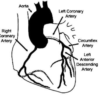
The vital coronary arteries transport freshly oxygenated blood to the heart (see Fig. 1.1). These vessels branch off of the aorta, curving over the epicardial surface while bifurcating and providing tributaries to the
ever-beating myocardial tissue. Such
complex geometries and the pulsing intramural pressures create a high degree of spatial and temporal diversity in the local hemodynamic environment, resulting in a unique vascular setting. Right Corona Arter) iry Circumflex Artery Left Anterior ascending Artery
Figure 1.1 Schematic diagram of the heart and major overlying coronary arteries.
In states of health, the coronary vessels, as all parts of the vasculature, are lined with a protective endothelial barrier that separates the highly reactive vessel wall and contained blood compartments. Traversing through these conduits, changes in the vascular environment are accompanied by changes in the phenotypes of these cells [1]. Gene expression patterns, surface receptor expression, and a slew of other cellular mechanisms are tightly linked to the environment and work to keep this living surface appropriately matched with local needs.
Within the first decades of life, nearly ubiquitous environmental and genetic stresses conspire to create loci of low-grade injury to the protective endothelial surface allowing the accumulation of subendothelial deposits of lipids and lipid-laden
macrophages [2-6]. These minor breaches instigate a complex cycle of chronic inflammation and immune recruitment through endothelial activation, smooth muscle cell proliferation and migration, extracellular matrix turnover, and the build up of cellular and . necrotic debris. Over time, such processes transform into full fledged atherosclerotic
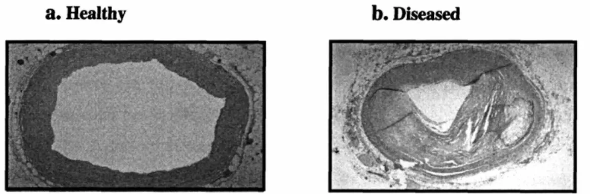
disease and luminal stenosis. Figure 1.2 a, b depicts a healthy vessel and one whose
lumen has been diminished by atherosclerotic plaque burden.
8.Healthy b.Diseased
Figure 1.2. a. Healthy coronary artery with patent lumen; b. Atherosclerotic coronary artery with luminal stenosis
Typically, the plaques are characterized by a lipid-laden, highly thrombogenic, necrotic core, covered by a fibrous cap region. As the plaques continue to grow, regulatory mechanisms are often able to compensate and uphold the balance between blood supply and myocardial demand. While such progressive, chronic, atherosclerotic processes can eventually lead to significant, uncompensatable disease (> 75% area reduction) characterize
a
by ischemic episodes and stable angina (exertional chest pain), a far more devastating and unpredictable outcome is plaque rupture or endothelial erosion (see Fig1.3) [2-4, 7, 8].
Under such circumstances, the protective endothelial layer is acutely disrupted, exposing highly reactive subendothelial, atheromatous components. A rapid, thrombotic process ensues which can either exacerbate disease progression or lead to acute coronary syndromes (ACS) such as unstable angina, acute myocardial infarction, or ischemic sudden death.
1.1.2 Treatments
Prevention is the preferred treatment option for coronary disease. Epidemiological studies have generated a great deal of information regarding the different genetic and environmental risk factors involved in coronary disease progression [9-11]. Accordingly, appropriate measures can be taken to minimize risk. Currently, our cellular and molecular based understanding of the etiology of atherosclerosis and thrombosis has also allowed the use of relatively safe, long-term drug strategies. Perhaps the most notable example is the widespread use of aspirin in treating populations at risk of coronary disease. By targeting platelet responsiveness (a key component of the arterial thrombotic response), aspirin has offered a 25% relative risk reduction of severe cardiovascular incidence (vascular death, MI, stroke) becoming a nearly ubiquitously used background therapy [12]. A battery of such agents (statins, etc) has shown great risk benefit, with novel drugs, either targeting new mechanisms for reducing the complications of pre-existing ones, constantly emerging [13, 14].
Unfortunately, many cases of coronary disease are brought on unexpectedly (ACS), or are simple too far progressed (severe luminal stenosis), necessitating more intensive strategies. In cases of thrombosis, highly potent anti-thrombotics and fibrinolytic agents (which prevent the build up and promote the dissolution of clots respectively) have proven highly effective if administered within critical windows [15].
Such powerful, systemic agents are not suitable for sustained therapy do to excessive complications, and while these provide immediate, life-saving benefit, the underlying problems of' atherosclerosis and plaque instability remain [16, 17]. The supply/demand mismatch accompanying severe stenosis can also often be managed medically with agents such as nitroglycerin which, up to a point, can aid innate autoregulatory mechanisms [18]. While such therapies may provide immediate relief, the chronic need for medication often indicates a need for more invasive revascularization procedures. Coronary bypass arterial grafting (CABG) has long been the standard revascularization procedure [19, 20]. However, the advent of minimally-invasive, ever-improving, catheter-based therapies has transformed the state of coronary disease management. Two wide spread approaches include angioplasty, which mechanically disrupts the atherosclerotic segment through balloon expansion, and stenting, which involves the
co-expanSIon of a permanent, rigid, foreign structure adding post-procedural, luminal support. Although the benefits offered by such treatments are indisputable, changes in the biological and physical environment accompanying mechanical disruption of the endothelial barrier and insertion of a foreign structure, even as minor as a intravascular wire, can lead to deleterious complications by iatrogenically creating an ACS-like state.
Endothelial Iniury
Embolization
SILENT CLINICAL
Figure 1.3. Clinical and silent (subclinical) thrombotic outcomes of vascular InJury.
Much effort has been placed in understanding and controlling the complex,
pathophysiological mechanisms of atherosclerotic disease and its devastating outcome, coronary thrombosis. Still, in westernized societies, such endovascular disease processes account for close to 50% of all deaths [6]. For continued improvement of safe, efficacious management of coronary events (pathological and iatrogenic), it is important to gain further understanding into the mechanisms and events leading up to acute thrombosis within the vital coronary arterial setting.
1.2 The Platelet
While a multitude of biological
effectors are involved in vascular
thrombosis, we will focus on the
platelet as an essential mediator of the biological response to vascular injury. Platelets are circulating cells (1.5-4.5
X 108/ml) which bud off from
megakaryocytes resident in the bone
marrow [21]. Although anucleate,
platelets are highly responsive to their environment. A multitude of surface receptor interactions, second messenger
Figure 1.4 Tunneling electron microscope
image of anucleate platelet depicting
numerous internal storage granules and canalicular system (image reproduced from Feng D, et aI. Blood. 99( 11), 2(02).
pathways, pre-packaged granule release
substances (Fig 1.4), and translatable proteins allow these cells to provide constant monitor of the vascular environment and playa key role in initiating biological responses. The importance of platelets in arterial thrombosis cannot be overstated as attested to by the clinical effectiveness of anti-platelet therapies in minimizing arterial thrombotic risk [22]. Upon binding to an injured wall, they alter their expression of surface molecules and release substances which both attract and adhere to flowing cells, as well as promote the enzymatically driven coagulative response. Accordingly, platelet wall reactions and subsequent component interactions are essential to understand when considering the thrombotic response to vascular injury.
1.2.1 Platelet Reactions
Platelet/wall reactions classically take place in a series of steps, beginning with initial contact, followed by a sequence of activation, firm adhesion, cell spreading, and aggregation (Fig. 1.5).
4. Aggregation (GPIIbIIIa, 1e , f -I I fibrinoLen. 1. Dlnlulng vWF molecules to Collagen
/ '/p 1
S a. . , ,4 Ar ZS z=_z Z= II S . z I me., o f 1 i .~tz~:"Z~zFigure 1.5. Sequence of platelet/vessel wall reactions.
Tethering/A ctivation
The platelet glycoprotein Ib (GPIb; in complex with GPIX, GPV) and von Willebrand's Factor (vWF) are important components in enabling initial platelet/surface contact in
arterial, high flow environments [23, 24]. Upon endothelial breach, platelets tether to sub-endothelial matrix vWF (either directly present or adsorbed to matrix collagen from the blood) via fast on-off rate GPIb bonds [25]. The speed of these bonds is an essential quality of this interaction in allowing the platelets to grab onto the surface under high
shear conditions [26-28]. Several studies have shown that these tethering interactions become increasingly important at shear rates above 300 s-1, becoming essential at shears above 1500 s' in reducing the platelet relative wall velocity in a start-stop type dynamic
[26, 29]. As the platelet slows and translocates, a multitude of other activating responses take place. The GPIB-vWF bond itself is known to be one of these activating pathways by interacting with internal, second messenger machinery [28, 30-32]. Concurrently, autocrine and paracrine substances, such as ADP, thrombin, thromboxane, and epinephrine, bind to specific receptors, further potentiating platelet activation [21, 33].
Phrst
a
'r ..
L ,
IlrJl
ClIMnIInn
nl
18
we alesl I.. LJaLn'yILuJu allu sJy cIuiiuICIal F 2. Platelets tether and translocate on surface (GPIb, vWF) 3a. Activation of platelets (GPVI, GPIb, ADP, etc.) 3b. Integrin
/Activation/Adhesion
Recently, the importance of the collagen activating receptor GPVI has also been shown to provide a physiologically essential activating interaction with exposed collagen and vWF surfaces [341. It is an integration of these coalescing, outside-in, activating pathways that dictate the final activation decision.
Firm adhesion/Spreading
An essential outcome of platelet activation in supporting shear dependent thrombosis is the modulation of platelet integrin receptors from their low to high affinity state. Integrins are cell surface molecules, composed of an a and subunit, and support adhesion to particular substrates [35]. The major platelet integrins are aIbffII, a2131, and aV33 [35, 36]. Upon activation, these receptors support the firm adhesion of platelets onto fibrinogen, collagen, and fibronectin surfaces respectively. However, the existence of other, less prevalent integrins and substrate cross reactivity create redundancy in the adhesion pathways and while providing a safety net given the importance of adequate platelet adhesion, dramatically complicating the mechanistic picture.
After firm adhesion, internal processes that are not fully characterized lead to filipodial extensions and cell spreading [37]. Such cytoskeletal rearrangement allows more complete surface contact, enhanced integrin binding, and increased binding strength of the adherent platelets.
Aggregation
Platelet shape change is accompanied by granular exocytosis which helps to transform the local biological milieu by the release of substances such as ADP and thromboxane [21]. Free-stream platelets are potentiated and activated in this pro-thrombotic environment. As induced during surface interactions, such activation enables high
affinity integrin conformations. Platelet-derived fibrinogen and vWF add to pre-existing, local plasma concentrations of these proteins, enhancing binding to activated aIIb3JII integrin receptors [21, 38]. These protein/receptor interactions mediate inter-platelet cross-linking and aggregation, forming what is commonly referred to as the primary hemostatic plug.
1.2.2 Platelet Interactions
Though platelets play an essential, initiating role in arterial thrombosis, they are far from the only participants. Numerous interactions with other blood-borne cellular and protein components have long been recognized as mediators of thrombotic disease.
Coagulation
When platelets are activated, negatively charged phospholipids, such as
phosphotidylcholine and phosphotidylserine, are externally presented, localizing various enzymatic reaction complexes of the coagulation cascade to the platelet surface [21, 33, 39]. This sequestration keeps the reactants in close proximity to one another, while protecting them from other free-stream anti-coagulative mechanisms. Upon association with the platelet membrane, conformational changes in enzyme/substrate complexes also dramatically accelerate the reactions [21, 39]. Such localization and catalysis are essential for the successful progression of coagulation under flow conditions where convective mass transport would otherwise wash away the formed products [21].
I INTRINSIC
ACTIVATION
Tissue
Factor
X -
-Charged Surface ACTIVATION
VII -VIIa
XIIa
+XII
1
|XIa
+
XI
XX+Xa a
FINAL COMMON PATHWAY Prothrombin 4 THROMBIN Fibrinogen - I FIBRINFig 1.6. Classical reactions of the coagulation cascade commensing with the exposure of tissue factor (Extrinsic activation) and/or an appropriate surface (Intrinsic activation), leading to the explosive, proteolytic production of thrombin and fibrin
- _, I , I. __ l _ _ x- _ __ _ 20
cnapter 7:: BacKground anda i-gnmicance
While platelets are necessary in supporting coagulative propagation, the products of the coagulation cascade in turn play a critical role in potentiating and stabilizing primary, platelet-driven hemostasis. The key reactions of this proteolytic pathway are shown in Figure 1.6 [21, 39]. Classically, coagulation is initiated via the extrinsic or intrinsic pathways, merging into the common pathway with the eventual production of thrombin which catalyzes the conversion of monomeric fibrinogen to fibrin. While numerous interactions exist, thrombin and fibrin are two of the most important and well characterized links between platelet function and coagulation.
Thrombin is known to be a potent activator of platelets through a variety of mechanisms. Protease activated receptors (PAR-I, 4) have been characterized on the surface of platelets [40-42]. Using a unique cleavage mechanism, thrombin causes receptor self-association and activation. Interestingly, the various PARs are functionally activated at different threshold concentrations of thrombin, while additional interactions with the GPIb"receptor have been recently recognized to affect platelet activation [42-45]. Such connections allude to the delicate control structures poised between the coagulative network and platelet function.
While the physiological relevance of thrombin-activation has been shown, its catalYtic conversion of fibrinogen to fibrin also plays a critical role in
platelet-driven thrombosis. Once formed,
monomeric fibrin quickly polymerizes into a
mesh-like network of filaments. Platelets can adhere to this fibrin mesh via
allbpm
receptor linkage, greatly strengthening the cohesiveness ofr:r-, I
the aggregating, hemostatic plug (Fig. 1.7) [46] .
.Though these classical platelet/coagulation interactions are key mediators of physiological thrombus formation, they are only representative
Figure 1.7 .Platelets 'entangled' in
fibrin mesh (surface electron
of a long list of interactions, many of which are characterized, and a great many of which undoubtedly remain to be found.
Leukocytes
Leukocytes are the cellular mediators of the inflammatory and immune responses. In blunt categorizations, these responses have often been considered as separate to the thrombotic reactions. However, upon closer examination, the great deal of interactions between these biological processes has grayed the distinction. As with coagulation, a variety of platelet processes are involved in initiating inflammatory responses and vice-versa. Upon vessel wall injury and platelet recruitment, P-selectin is rapidly presented on the surface of the activated platelet layer. Surface expression of this selectin is able to recruit various leukocytic cells. Neutrophils and monocytes initially adhere via P-selectin glycoprotein ligand-1 (PSGL-1), tethering in a manner analogous to the GPIb-vWF interactions of platelets [25, 47]. Again, outside-in signaling causes leukocytic activation and integrin modulation enabling firm adhesion. The importance of platelets in heralding such inflammatory processes is witnessed in the effectiveness of P-selectin blockade or powerful platelet inhibition on limiting the development of chronic vascular inflammatory processes such as atherosclerosis or the neointimal formation after interventional treatments [48].
Just as platelets play a critical role in inflammation, the importance of inflammatory mediators in thrombosis is now being recognized. Cytokines produced during inflammatory processes such as tumor necrosis factor (TNF), interlukin-1 (IL-1), monocytic chemoattractant protein-1 (MCP-1), and IL-6, have all been found to upregulate the expression of tissue factor (TF) on endothelial and monocytic surfaces. TF is the key initiating factor in extrinsic activation (see Fig. 1.6), and it is becoming recognized that such cytokine-mediated expression of TF acts as a primary initiator of thrombosis by shifting the vascular environment into a prothrombotic state [49].
Erythrocytes
Erythrocytes (red blood cells; RBCs) are also important in modulating arterial thrombotic events. Being the most numerous blood cell, RBCs dramatically alter the fluidic
properties of blood. Flow-dependent gradients in erythrocyte distribution marginalize the platelets to the flow periphery, dramatically increasing local platelet wall concentrations [50, 51]. Further, by increasing the viscous forces of blood, erythrocytes dramatically affect the shear force at a given shear rate which can have significant impact on biophysical processes such as thrombosis.
RBC's are also one of the primary, physiological sources of ADP, a potent
activator of platelets. Under conditions of shear, ADP pathways have been shown to greatly enhance platelet activation and adhesion [52, 53]. The relevance of this mechanism in arterial thrombus formation is attested to by the efficacy of ADP receptor blockade via thienopyridine derivatives (i.e. clopidogrel, ticlopidine) in minimizing post-interventional thrombotic risk [53, 54].
In this section, we have described the fundamental processes underlying platelet/wall adhesion, which serves an essential, initiating role in arterial thrombus formation. We have also introduced some of the classic interactions that platelets undergo with other blood-borne, vascular components. While these interactions only breach the surface of the thrombotic response, they point to the complexity of the biological issue and the need to develop controllable systems where parameters can be sufficiently isolated, allowing tractable, mechanistic studies to be performed.
1.3 The Local Hemodynamic Setting
It is important to recognize that biological interactions both occur in and react to their physical surroundings. In vascular environments, flow is one of the critical physical parameters that helps to maintain vascular diversity and dictate vascular responses. The coronary environment provides a distinctive setting where curving vessels, numerous branch points, geometric pathologies and the time-dependent nature of the driving pressure create highly unique, complex flow patterns (see Fig. 1.8) [1, 55-58].
LAD Phasic Coronary Blood Flow
-
c .- 100 E ::::80 E ... 60 ;: .2 40 lL "a 20 o .2 0 mo
0.2 0.4 0.6 DIASTOLE 0.8 1 Time (see)Figure 1.8. Typical phasic coronary flow profile in the left anterior descending coronary branch (LAD). In supplying blood to the beating myocardial wall, coronary flow principally occurs during diastolic relaxation, being driven to a halt with rising, systolic, intramural pressures.
These spatially and temporally diverse conditions must be appreciated when considering highly flow-sensitive events such as thrombosis. Here, we introduce some of the mechanisms whereby physical flow parameters may interact with biological factors and the manner by which the local hemodynamic environment can be altered in states of disease.
1.3.1 Local Flow Interactions
Flow can affect biological interactions on variety of levels, from molecular to microscopic to macroscopic scales. Often times these dependencies stem from basic physical concepts such as mass transport and physical forces. However, given the long time scales that these biological mechanisms have been interacting within relatively stable physical environments, selective pressures have also enabled biological systems to evolve sophisticated machinery, allowing them to respond and adapt to their surroundings in a more complex manner.
Molecular Interactions
The mass transport of molecular substances to and from an activating locus plays an important role in thrombus formation. In flowing environments, fleeting conditions are setup as diffusive and convective forces transport substances to and from the reactive site.
The mass flux, J, of a substance reacting at a surface has been shown to be:
J(x,t) C0
1 1
kw k (xt) (1)
for arteries of diameter > 0. Imm [59]. This relation shows that the flux is dependent on the mean bulk concentration Co, the wall reactivity of the substance, k, as well as the mass transport coefficient, kl. In cases where the wall reactivity is very low compared to the mass transport coefficient, the flux is said to be reaction limited. Alternatively, with low mass transport coefficients, transport dictates the surface flux.
Flow affects this relation implicitly through kl, which, in the case of the entrance region in large cylindrical arteries, is given by:
k =.5K
2j
where D is the diffusion constant, K is a proportionality constant dependent on geometry
and boundary conditions, x is the axial distance from the start of the region, and y is the wall shear rate [59]. Thus, when substances are transport limited (J-Cokl), flow modulates the surface flux. This becomes highly relevant in cases of static or recirculant flows such as those which are often established in the complex coronary vasculature (particularly given the autocatalytic nature of many blood-borne reactions). That the physiological reactions of thrombosis globally function in this flow dependent regime is revealed by the morphological differences in thrombus formation under venous (low) and arterial (high) flow systems and by observations such as the increased likelihood of post-interventional acute thrombotic events in poorly perfused vessels [21, 60-62].
Microscopic Interactions
Cellular Transport (km)
Just as reactive molecules are transported to and from the reactive surfaces, so are cellular components. However, due to special non-Newtonian properties of blood, the effective diffusion coefficient, D, of cells is itself a function of erythrocyte concentration and flow parameters. As blood flows through the vasculature, RBCs migrate towards the flow axis creating a flow-dependent, radial concentration gradient. As a result, platelets are marginalized to the flow periphery, increasing the shear dependence of platelet mass flux. [50]. Various studies of platelet wall interactions have shown surface accumulation to be largely dependent on flow parameters, implicating the importance of transport phenomena and the underlying diffusion limitations involved in this process [50].
Wall Reactivity (kw)
While cells and molecules must be transported to the wall, they must also be able to react when appropriate, and the wall reactivity, kw, can also be considered a complex function of fluid flow. Local shear stresses and pressure forces have been shown to greatly affect vessel wall morphology. Endothelial cells and wall smooth muscle cells are able to sense these physical forces and respond with directed genotypic and phenotypic changes. When activated, these cells alter their expression of surface adhesion molecules [1, 63]. These, along with numerous other changes (i.e. TF, vWF, tissue plasminogen activator,
platelet activating factor-1, thrombospondin, RANTES, monocytic chemoattractant protein-i), alter the local environment creating a generalized, prothrombotic,
proinflammatory state [1, 58, 64-66].
The paradigm of vascular cell adhesion to this reactive wall in flowing environments parallels the scheme outlined for platelet adhesion (recall Fig. 1.5), beginning with the presentation of a particular class of surface adhesion molecules called selectins (or vWF in the case of platelets). Various cell types express various types of selectins. These molecules interact with respective ligands on free-stream cells, thereby :recruiting them to the vessel wall. Selectin/ligand interactions are characterized by fast binding constants, and are thus able to catch flowing cells under high shear conditions [25]. While necessary in supporting the initial cell/wall contact under flow, they do not bind tightly, and instead produce a rolling motion as bonds are sequentially broken and formed. During this slowed, translocation, several other pathways cause vascular cell activation, resulting in integrin receptor activation and firm, shear-resistant adhesion to surface ligands [67]. These prototypical flow mediated interactions are representative of vascular cell/wall interactions, with the exact receptor/ligand interactions dependent on the particular cell type and substrate under consideration.
Macroscopic Interactions
On macroscopic scales, shear stresses can integrate to create high local forces which can have a significant impact on the pathogenic progression of vascular disease. Atherosclerotic plaques are characterized by atheromatous core regions, covered by a fibrous cap. Acute thrombotic events often occur when such plaques rupture by sudden fracture of the cap region and currently there is much interest in studying the strength of the cap as varied biological properties can affect plaque stability [68-71]. However, the final rupture event is predicated on the fact the cap strength is too weak in relation to local fluidic forces.
As cells and molecules aggregate on the reactive wall via the flow-dependent processes outlined above, larger mural thrombus develops and is increasingly subject to macroscopic embolizing forces. As in the case of plaque rupture, embolization is known to be highly dependent on shear rates, with embolic stresses varying linearly with wall
Figure 1.9. Angiogram of coronary arteries depicting highly branching, curving geometries overlying and penetrating the heart tissue (image reproduced from Dhond, MK, et. al. Clinical shear at thrombus heights less than 0.1 mm and quadratically at larger heights as a result of flow separation [72, 73]. In either situation, the rheological situation dictates the removal forces that help to determine detachment probability.
1.3.2 Local Geometric Environment
Local fluid flow plays an important role in directing the thrombotic response. By prescribing the flow boundary conditions, the local physical geometry is critical to determining the contained flow field, and thus also plays a crucial role. These geometries can be dramatically altered in states of disease or intervention, having great impact on the local,hemodynamic environment and the tightly dependent biological outcomes.
Physiological Geometries
Physiological vascular properties such as curvature, branching, and tapering are all highly relevant in establishing the local flow environment, particularly in the coronary bed where the arteries coarse over the curved epicardial surface, supplying numerous braches to the demanding myocardial wall (Fig. 1.9) [74].
In purely axial flows, radial and
circumferential sYmmetry ensure
purely axial pressure gradients and relatively simple flow profiles. However, in more complex, curved
flow streams, adverse axial and
orthogonal pressure gradients are established, creating secondary flow fields and zones of recirculation (Fig.
1.10a, b) [56, 75, 76]. These can
have ,significant impacts on local wall shear stresses and through the
biophysical connections outlined above, orchestrate local biological responses.
a. Vessel Branching
Flow recirculation //~~~~1b. Vessel Curvature
Secondary flow effectsFigure 1.10. Examples of vessel architecture leading to complex flow patterns. a. Vessel branching and axial flow, reversal and recirculation; b. Vessel curvature and transverse, secondary flows.
Such responses are essential in vascular remodeling and maintaining healthy vessels. However, when inappropriately harnessed, they can also lead to vascular disease progression. The effect of local, physiological geometries and the resultant flow fields in vascular pathology is well recognized, as indicated by the frequent initiation of atherosclerosis at sites of vessel branching or the increased atherosclerotic burden observed on the inner curvature of coronary vessels [77, 78].
Pathological Geometries
While physiological geometries correlate well with disease initiation, local geometries can be significantly altered with disease progression, often compounding the detrimental effects. As cellular and necrotic debris accumulate within the wall, volume constraints result in either inward (stenotic) or outward (aneurismal) remodeling of the vessel wall. In cases of compensatable stenosis, mass conservation implies increased flow velocities for a given total flow rate, with wall shear rates at times exceeding 10,000 s-' (typically < 600 s-1in the coronary arteries) [51, 52, 79]. Under such pathological conditions, free-stream vascular cell activation and erythrocyte hemolysis can significantly alter volumetric blood status leading to both local and far-reaching systemic effects [79, 80]. When the total flow rate can no longer be maintained due to excessive stenotic flow resistance, severe decompensation can conversely result in dramatically reduced flows. Indeed, when plaques rupture and mural thrombus occludes the lumen, increased resistance can totally eliminate blood flow, quickly potentiating further thrombus growth. Alternatively, under aneurismal conditions, flow rheology can also be significantly affected. As a result of reduced transport and embolic forces in recirculant zones, significant, venous-like mural thrombus formation is often observed, creating a nidus for thromboembolic disease [21, 59].
Interventional Geometries
Significant geometry-altering vascular disease is often treated with interventional revascularization techniques which can in themselves affect the local geometry and hemodynamic environment. Stenting involves the placement of an intravascular structure that props open the vessel lumen. The post-interventional luminal shape is determined by the overall profile of the implanted stent device, and has been shown to have an impact on long-term biological outcomes, likely due in part to alterations in fluid flow [81]. Furthermore, in order to provide sufficient radial support, stents are typically made of a mesh work of steel struts on the order of 100 igm thick. While this thickness is relatively small compared to the entire flow diameter (> 2 mm), their juxtaposition next to the vessel wall can significantly alter local wall shear stresses, with complex flow patterns developing in the inter-strut regions [82-85]. Berry et. al. have shown that
changes in stent geometry, can dramatically affect these flow fields and resultant mass transport parameters [82].
The effects of stent placement can also be more pronounced depending on
implantation technique. Significant over-expansion can lead to aneurismal-like
conditions, while insufficient expansion can results in significant flow abnormalities. Indeed, such improper expansions have been associated with the high acute thrombotic rates observed in the early clinical stenting experience [61, 86].
While the placement of intravascular objects can create altered local flow, a more subtle point is that revascularization itself alters the geometry and 'expected' flow field of the vessel due to the significant biological adaptations that occur during disease progression. Recently, Richter and Edelman (unpublished observations, 2003) have shown that such acute interventional revascularization can lead to adverse, unexpected outcomes.
We have briefly described some of the key mechanisms by which flow can interact with the thrombotic processes. Furthermore, we have discussed how the physical, geometric environment can have a significant impact on flow parameters under physiological, pathological and interventional conditions. Considering these significant interactions, an attempt to study thrombotic reactions in the coronary setting would hopefully take these physical parameters into account, allowing them to be manipulated and studied in a controlled fashion, along with the pertinent biological components.
1.4 Methods of studying flow dependent thrombosis
A variety of methods have been utilized to investigate thrombotic reactions under appropriate hemodynamic conditions. These methods include both in vivo and ex vivo models, as well as bench-top in vitro setups. Each of these methodologies has offered unique insights by virtue of inherent advantages, though often these same advantages limit the types of information that can be gleaned from their use. It has been a combined use of these techniques, along with an appreciation of their limitations that has contributed greatly to our current understanding of vascular biology. Here, we present various methodologies that have been and are currently being used to study flow-dependent thrombotic reactions and the types of advances they have provided.
1.4.1
In VivoModels (Realistic)
In vivo models offer a degree of realism and physiological relevance unattainable in
bench-top models, and when chosen appropriately, allow thrombotic outcomes to be assessed in relevant hemodynamic conditions. The two most basic types of in vivo study include clinical trials and animal studies.
Human Studies/Clinical Trials
Human studies and clinical trials generate immediately applicable, clinical information, inherently taking into account the complex biological interactions at work in the human body. Such studies allow integrated, relevant, systemic and long-term processes to be observed, offering clear cut advantages over other systems when considering actual disease characterization, patient risk stratification, and the efficacy of therapeutic options. Historically, mechanistic insights have been obtained from observing the human condition, often times becoming evident when systems overtly fail. Indeed, many important processes of the thrombotic response have been initially discovered through clinical presentation (GPVI deficiency, Glanzmann's thrombasthenia, Bernard-Soulier, GJPIb deficiency, vWF disease, etc.) [21]. While such accidental observations are predicated on patients and patience to search the space of biological processes, they are implicitly of great relevance.
Human studies and clinical trials are by definition the most relevant to the human condition. However, as the primary goal is to benefit the immediate patient, making clear scientific progress is difficult at best. The lack of ideal controls and patient variability (particularly given the optimizing nature of medical advance) causes such trials to be expensive and long, oftentimes yielding unclear conclusions. Further complicating the issue is that as trials are performed, the evolving ground of clinical experience often races by, altering the clinical relevance, therapeutic dogmas and potentially, for patient benefit, the clinical trials themselves.
Animal Studies
One step removed, animal models of physiology and pathology allow detailed observation of complex biological situations while offering a more controlled setting than human studies. Though ethical considerations and respect must always accompany the experimental use of living organisms, an accepted goal in animal use is the progress of scientific understanding for human benefit, thus allowing intensive, well-controlled, prospective studies to be carried out to completion.
Animal models of thrombosis span the evolutionary ladder, from the use of non-human primates to mice. Architectural similarity and flow conditions in large animals allow highly relevant studies of thrombotic processes under appropriate hemodynamic settings. Alternatively, the ease of manipulation and relatively low cost of small animal studies enable more fundamental questions to be epistatically probed, dramatically accelerating the pace of scientific discovery and understanding. However, the relevance to the human condition varies tremendously as a result of species-specific biological and physical differences and the often crude models used to approximate human vascular disease. Furthermore, while offering greater experimental possibilities than human trials, there are still limitations in the parameters that can be reasonably manipulated in living
animals, particularly when considering complex traits such as the hemodynamic environment [79, 87-89].
1.4.2 In Vitro Models (Perturbable)
To gain detailed mechanistic insight into the heological dependence of thrombosis, it is important to have parametric control over the hemodynamic and complex biological environments. In vitro models allow powerful manipulations to be performed on isolated biological components under highly consistent, prescribed physical conditions, thus offering a degree of versatility not attainable with in vivo settings. Such models can broadly be categorized into systems that maintain physical geometric properties and those that maintain flow conditions.
Maintaining Geometry
Geometrically relevant in vitro systems maintain realistic vascular dimensions and have been applied towards various issues, from endovascular device thrombogenicity to cellular adhesion in vascular deformities. One limitation in these models has been in developing suitable bench-top mechanisms of flow actuation and control. While some setups have employed gravity to provide a pressure head for generating blood flow, the need for sizable, static holding volumes in these one-pass systems significantly constrains the allowable flow rates and experimental run times [89]. Alternatively, loops partially filled with blood and mounted on a tilted turntable have been employed to create relative wall motion. This method allows the use of small blood volumes, though effects of blood recirculation and the required air-fluid interface must be considered when interpreting results [90-92]. Moreover, the constant flow potential of these gravity-driven systems results in restricted control of the flow environment. To establish more controllable flows, the most basic approach has been to use pumps to drive blood through a model flow circuit [93, 94]. However, even external peristaltic drives have generated excessive background levels of activation, proving too traumatic in setting up arterial-like flow conditions.
Maintaining Flow
Strategies that attempt to maintain geometry are typically constrained by the need to create high flow rates to generate arterial-like conditions. In order to study flow effects, laxness in the physical circuit architecture has enabled alternate schemes to be employed.
The use of small circuit geometries such as narrow, parallel-plate flow chambers, allows high shear rates to be developed under relatively low flow conditions [26, 83, 95-97]. Such in vitro systems allow great control of the biological environment while enabling microscopic visualization of surface-cellular interactions in real time. As a result of their controllability and observability, these studies have greatly enhanced our fundamental understanding of shear-dependent, thrombotic surface reactions.
Techniques such as cone-plate or annular ring devices create relative motion between two fluid-contacting surfaces, thus generating Cuvette-type flow. These models can establish well-described surface and volumetric shear profiles. Furthermore, while these Cuvette-systems dramatically change the physical flow shape, suitable scaling and experimental design have allowed certain geometrically relevant questions to be asked under appropriate flow settings [58, 98].
1.4.3 Proposed Flow System
'Table 1.1 compares prior model systems that have been employed to study the hemodynamics of thrombosis with respect to the human condition.
Table 1.1. Comparison of prior methods showing an incomplete niche for experimental strategies supporting the simultaneous parametric analysis of biological and physical systems. (Subjective scale, using clinical trials to reference extremes ratings: +++ ---; Excellent --> Poor).
Combining the information obtained from these systems has lead to great leaps in our understanding of acute thrombosis. Ex vivo hybrids, where external sections are placed in-line with a natural circulation, have helped to bridge the gap where in vivo and in vitro models do not meet [51]. Still, cracks remain and alternative models addressing new
%.iiaptai i.. L.a7cldyIl uIIu czIIU %7IYIIIIIUC*II ucp
Parametric arametric omplex Physiol. Physiol. Signal/
Analysis Analysis Cost Interactions Shear Geom. Noise
(Biological (physical) linical
+++ +++ +++ -- --
---rials
In Vivo arge Anima ++ ++ ++ .. . ..
tudies mall Anima + + - + _. tudies ravity
riven
-
-++
+ - + + ystems eristaltic In Vitro ystems aePn--
++--
++-
++ ++ Chambers uvette-uvette-- ++-
++-
+ ++ ystems x -Vivo Ex ivo-
++ ++-
+ +--Circuits
~~~+~r·
L~l/lr4.. -4~~;·;~C~~
36
issues are needed. One particular niche which we hope to address in this work is the need for systems that concurrently retain significant control of both the biological and physical axes, specifically with regards to the human coronary setting. The key selection criterion we considered when developing the system were:
1.) Generating and controlling physiologically relevant flows 2.) Creating versatile, geometrically relevant circuits
3.) Retaining control of the biological setting
4.) Maximizing the biological signal/circuit background noise ratio
After considering various strategies, we developed a novel, inertial mechanism that could produce highly controllable flows in relevant arterial geometries while maximizing the biological signal to background noise ratio by minimizing the circuit length and surface discontinuities. Briefly, fluid-filled loops are spun about their axis in a prescribed and controlled fashion to modulate relative fluid/wall motions through transmitted shear forces. This method further allows us to maintain low test volumes and cost, enabling reasonable parametric analysis of the biological and physical settings.
FIGURE 2.1. Fluid-filled torus. Clockwise motion of the torus causes relative counter-clockwise inertial motion of the contained fluid and vice versa.
_C_H_AP_T_E_R_2__
F_lo_w_System
Design
2.1 Theory
2.1.1 Description
In order to create the desired flow profiles, a fluid-filled torus (figure 4)
is rotated about its axis. When
impulsively started, there is inertial fluid motion relative to the toroid wall:
With time, the fluid is accelerated due Fluid to momentum transfer into the fluid
bulk via shear forces. Lyne has
previously analogized such fluid
motion to pressure driven flows where, moving in a reference frame with the
wall (WRF), the acceleration takes on the driving character of a body force [99]. While the absolute fluid velocity in an inertial reference frame (IRF) may change under pressure driven or accelerating wall conditions, it is the maintenance of the velocity gradients, or shear, that is important in the flow-dependent thrombotic reactions [21, 50, 59]. Rather than fluid being conducted to and from a reactive wall surface, the relevant reaction control volumes are conducted away from the lagging fluid.
Using such a technique, we hoped to create time-varying flows whose characteristics, as
defined by the dimensionless mean Reynolds (Re; based on an absolute velocity) and Womersley (a) parameters were typical of coronary flows, as well as estimate the secondary flow effects of curvature by considering the peak Dean number (K'peak) associated with our system (see Table 2.1).
Table 2.1. List of the dimensionless parameters
considered, along with
customary definitions and typical values found in the left anterior descending coronary branch (LAD) [13]. Vfluid is the mean cross-sectional axial velocity of the fluid, v is the kinematic viscosity, o is the flow oscillatory frequency, a is the vessel radius, and R is the radius of curvature of the loop. T
This value for Kpeak was calculated assuming a peak Re of twice the Re and a aspect ratio, R/a, of 10 as suggested by Chang and Tarbell [15].
Table 2.1 defines these conventional dimensionless parameters [100]. In each of these parameters, a Newtonian approximation for the kinematic viscosity, v, is used, as has been shown to be valid for high shear conditions (> 100 s-1) and often applied when considering coronary flow [76, 78, 101, 102].
2.1.2 Analytical, Straight Tube Approximation
Equations
Simplifying to a linearly accelerating, straight tube model (Fig. 2.2), streamline curvature effects can be neglected and only the axial (z) component of the Navier-Stokes equation for cylindrical coordinates need be considered:
Typical Coronary Values Dimensionless # Definition (LAD) Mean Reynolds # 153+64 2 aVfluid (Re) v Womersley # (a) a 1.5+0.27
Peak Dean # (peak) I 2aVfl F 95T Ma V-~,/
a Vz
+V
+V
aVz= _ aP d2Z a 2VzI(
Vjl( 1 (a21VZa
--
[-z
+
Vr--
-- Z + Vz - + -~-- - +--[-at
r
a
az)
p az
dt 2- r r r) ao2) aZ2 (3)where Vz, Vr, and Vo are the velocity components in the axial (z), radial (r) and tangential (0) directions respectively, t is time, p is the fluid density, 6P/6z is the axial pressure gradient, and d2Z/dt2 is the axial wall acceleration.
C
Figure 2.2. Theoretical system assuming linear rather than angular motions.
In the special case of flow in a circular pipe of constant cross sectional area, circumferential and axial symmetry allow several dependencies to be eliminated, leaving only an axial velocity component, Vz, dependent on r and t, and the driving pressure and acceleration terms. These considerations yield the equation:
-av 1 aP d2Z a(2v
av
)
__ _-+ v _ z + - (4)at p
az
dt2 ar2 r
ar
By prescribing periodic functions for the driving terms of the form,
1 aP(t) = , ina
pj az n=1
(5)
40
Chapter 2:: Flow System Design
NEEMMMONN-,
rJJFFaJVJ ';iS;T77TTI;FI-1
r
d2Z(t) = Ane (6)
dt2 n=1
analytical solutions with appropriate wall and centerline boundary conditions (Vz(R, t)=0; dV/dr (O,t)=0; where R is the tube radius) have been derived elsewhere [101, 103-105], yielding:
V(r,t)
=
-(G + A0)(R
2_r
2)+
(G
+
An)
1-4v n=1 mino
(7)
where the axial flow velocity, Vz(r, t) is a function of radius and time, and can be driven by pressure gradients (Eq. 5) or wall accelerations (Eq. 6). G and An represent the amplitudes for the Fourier components of the nth harmonic of co for the pressure gradient and wall acceleration respectively, while Go and Ao are the steady forcing terms. From this straight; tube model we see that identical relative flow profiles can be obtained through either pressure gradients or wall accelerations given the interchangeability of G and A.
Solutions
Using this linearly accelerating straight tube approximation, we can analytically study the nature of the contained flows under various prototypical forcing conditions. By applying an acceleration impulse of the form:
d 2Z(t) (8)
,'2 n.c.,., (8)