HAL Id: hal-01064085
https://hal.archives-ouvertes.fr/hal-01064085
Submitted on 28 May 2021
HAL is a multi-disciplinary open access
archive for the deposit and dissemination of
sci-entific research documents, whether they are
pub-lished or not. The documents may come from
teaching and research institutions in France or
abroad, or from public or private research centers.
L’archive ouverte pluridisciplinaire HAL, est
destinée au dépôt et à la diffusion de documents
scientifiques de niveau recherche, publiés ou non,
émanant des établissements d’enseignement et de
recherche français ou étrangers, des laboratoires
publics ou privés.
influence male germ cell differentiation in the foetal
mouse testis
B. Moniot, S. Ujjan, J. Champagne, H. Hirai, K. Aritake, K. Nagata, Emeric
Dubois, Sabine Nidelet, M. Nakamura, Y. Yrade, et al.
To cite this version:
B. Moniot, S. Ujjan, J. Champagne, H. Hirai, K. Aritake, et al.. Prostaglandin D2 acts through
the Dp2 receptor to influence male germ cell differentiation in the foetal mouse testis. Development
(Cambridge, England), Company of Biologists, 2014, 141 (18), pp.3561-3571. �10.1242/dev.103408�.
�hal-01064085�
RESEARCH ARTICLE
Prostaglandin D
2
acts through the Dp2 receptor to influence male
germ cell differentiation in the foetal mouse testis
Brigitte Moniot1, Safdar Ujjan1, Julien Champagne1, Hiroyuki Hirai2, Kosuke Aritake3, Kinya Nagata2,
Emeric Dubois4, Sabine Nidelet4, Masataka Nakamura5, Yoshihiro Urade3, Francis Poulat1,* and
Brigitte Boizet-Bonhoure1,*
ABSTRACT
Through intercellular signalling, the somatic compartment of the foetal testis is able to program primordial germ cells to undergo spermatogenesis. Fibroblast growth factor 9 and several members of the transforming growth factor β superfamily are involved in this process in the foetal testis, counteracting the induction of meiosis by retinoic acid and activating germinal mitotic arrest. Here, using in vitro and in vivo approaches, we show that prostaglandin D2 (PGD2), which is produced through both L-Pgds and H-Pgds enzymatic activities in the somatic and germ cell compartments of the foetal testis, plays a role in mitotic arrest in male germ cells by activating the expression and nuclear localization of the CDK inhibitor p21Cip1and by repressing pluripotency markers. We show that PGD2acts through its Dp2 receptor, at least in part through direct effects in germ cells, and contributes to the proper differentiation of male germ cells through the upregulation of the master gene Nanos2. Our data identify PGD2signalling as an early pathway that acts in both paracrine and autocrine manners, and contributes to the differentiation of germ cells in the foetal testis.
KEY WORDS: Prostaglandin D2, Germ cells, Mitotic arrest, Differentiation, Embryonic testis, Mouse
INTRODUCTION
In mammals, the formation of a functional testis involves two successive cellular determination processes that take place during embryonic and foetal life. The first of these occurs in somatic cells, and the second takes place in the germ cells; in both cases, the process involves a choice between male and female fates. In male mice, the somatic cell fate decision is effected by the Sry gene, which is expressed in the supporting cell lineage between embryonic stages E10.5 and E12.5. Sry gene expression leads to the upregulation of Sox9 expression and the subsequent differentiation of these cells into Sertoli cells, which then influence the germ cell lineage (McClelland et al., 2012). In both sexes, primordial germ cells (PGCs) colonize the genital ridges at around E10.5, and continue proliferating until E13.5. The
sexual fate of the germ cells becomes apparent between E12.5 and E15.5. In the developing ovary, germ cells stop undergoing mitosis and enter the prophase of the first meiotic division at E13.5. In the testicular environment, the proliferation of germ cells gradually slows down and the cells ultimately reach quiescence, also called ‘mitotic arrest’, which corresponds to a block in the G0/G1phase. Male germ cells remain quiescent until shortly after birth, at which time they resume mitosis and then initiate meiosis at around 8 dpp (days post partum) (for a review, see Ewen and Koopman, 2010).
This male-specific quiescence is a crucial event in the establishment of the male germ cell fate and is tightly associated with the expression of G1/S phase checkpoint regulators such as the CDK inhibitors p27Kip1(Cdkn1b – Mouse Genome Informatics)
and p21Cip1(Cdkn1a– Mouse Genome Informatics), cyclins E1, E2
and D3 (Spiller et al., 2009; Western et al., 2008), and the retinoblastoma 1 protein (Rb1) (Spiller et al., 2010). Concomitant with these events, male germ cell commitment is also associated with the repression of key regulators of pluripotency, including Oct4, Sox2 and Nanog; this repression is achieved by E15.5 (Western et al., 2010). Various factors are known to be involved in the regulation of these events in male germ cells. The transcription factor Dmrt1 influences cell cycle arrest by directly regulating the expression of the CDK inhibitor p19ink(Cdkn2a– Mouse Genome
Informatics) and the pluripotency marker Sox2 (Krentz et al., 2009). In addition, the RNA-binding protein Dnd1 (dead end homolog 1) permits p21Cip1 expression by protecting its mRNA from
degradation (Kedde et al., 2007); loss of Dnd1 expression in male germ cells has multiple effects, including (1) the prevention of cells from entering mitotic arrest at G0, (2) the strong downregulation of the male germ cell fate factor Nanos2, (3) the ectopic upregulation of meiotic markers and (4) the maintenance of pluripotency genes (Cook et al., 2011). Multiple Tgfβ superfamily members have also been associated with male germ cell differentiation, including Tgfβ2 (Miles et al., 2013; Moreno et al., 2010) and activinβ5 (Inhba– Mouse Genome Informatics) (Mendis et al., 2011). These factors repress germ cell proliferation, and participate in the entry into quiescence (Moreno et al., 2010) and probably also in its maintenance (Mendis et al., 2011; Moreno et al., 2010). Notch pathway members also appear to be involved in male-specific differentiation, as their overexpression in foetal Sertoli cells can induce gonocytes to prematurely exit the quiescent state and enter meiosis (Garcia et al., 2013). However, the early-acting mechanisms that regulate and trigger these processes remain poorly understood (Western, 2009).
The decision between male and female germ cell fates in germ cells is known to depend on environmental signals (Adams and McLaren, 2002) that control the expression of two master genes: Stra8 (stimulated by retinoic acid gene 8), which is required for
Received 10 September 2013; Accepted 23 July 2014
1
Genetic and Development department, Institute of Human Genetics, CNRS UPR1142, Montpellier 34094, Cedex 05, France.2Department of Advanced Technology and Development, BML, Matoba, Kawagoe, Saitama 350-1101, Japan.
3
Department of Molecular Behavioral Biology, Osaka Bioscience Institute, Osaka 565-0874, Japan.4Plateforme MGX, Functional Genomic Institute, CNRS UMR 5203– INSERM U 661, Montpellier 34094, Cedex 05, France.5Human Gene Sciences Center, Tokyo Medical and Dental University, Yushima, Bunkyo-ku, Tokyo 113-8510, Japan.
*Authors for correspondence (francis.poulat@igh.cnrs.fr; brigitte.boizet@igh.cnrs.fr)
DEVEL
O
the initiation of meiosis in females (Baltus et al., 2006), and Nanos2, which blocks Stra8 expression in males and thereby prevents meiosis (Suzuki et al., 2010, 2012; Suzuki and Saga, 2008). Null mutations of Nanos2 in males lead to germ cell death (Tsuda et al., 2003), to the transient upregulation of meiotic markers (Suzuki and Saga, 2008) and to defects in the upregulation of male-specific markers such as the DNA methylase Dnmt3l (Suzuki et al., 2012).
One important environmental factor known to play a role in female-specific development is retinoic acid (RA), which activates Stra8 in female germ cells (Bowles et al., 2006; Koubova et al., 2006; Kumar et al., 2011). In the male, germ cells are protected from exposure to RA by Cyp26b1, an RA-metabolizing enzyme of the cytochrome P450 family that is produced by the Sertoli cells (Bowles et al., 2006; Koubova et al., 2006; MacLean et al., 2007) and the Leydig cells (Kashimada et al., 2011). This degradation of RA in males results in the suppression of meiosis after E13.5, thereby allowing mitotic arrest (Trautmann et al., 2008). However, despite the importance of RA inhibition in males, multiple lines of evidence indicate that additional secreted factors also play crucial roles (Best et al., 2008; Guerquin et al., 2010; Ohta et al., 2012). One candidate that has been proposed for such a secreted male-specific factor is Fgf9, as its secretion by differentiating Sertoli cells promotes the survival of germ cells after E12.5 (DiNapoli et al., 2006). In addition, Fgf9 signalling maintains the expression of pluripotency-related genes, and actively suppresses entry into meiosis in male germ cells by activating Nanos2 expression (Barrios et al., 2010; Bowles et al., 2010) via the transient activation of expression of the Cripto/Nodal pathway (Spiller et al., 2012). Indeed, this latter pathway displays an autocrine role in the inhibition of the meiotic entry in foetal XY germ cells (Souquet et al., 2012), a role that has also been observed with Tgfβ2 signalling (Miles et al., 2013). However, in double mutants for Fgf9 and Wnt4, germ cells do not enter meiosis and the male marker Dnmt3l is still expressed (Jameson et al., 2012), suggesting that Fgf9 is not the only signalling molecule involved in inducing these effects in male germ cells.
Considered together, these studies indicate that the crucial decision of the germ line to commit to either a male or a female fate involves a complex regulatory network, and that the previously identified factors and pathways are insufficient to explain fully this decision in males. Here, we highlight the role of an additional factor, prostaglandin D2(PGD2), in this process. PGD2has been known to act during Sertoli cell differentiation to induce the nuclear translocation of Sox9 protein (Malki et al., 2005; Moniot et al., 2009, 2011), and to help maintain Sox9 gene expression (Moniot et al., 2009; Wilhelm et al., 2005); its role in the male germ line, however, has not previously been established.
PGD2 is produced in the developing mouse testes by two enzymes: lipocalin-type prostaglandin D2 synthase (L-Pgds or Ptgds), an enzyme that is expressed specifically in males at E12.5 by Sertoli cells and by differentiating germ cells (Adams and McLaren, 2002); and hematopoietic Pgds (H-Pgds or Ptgds2), which is expressed in both sexes (Moniot et al., 2011). In this study, using multiple approaches [in vivo analysis of double-knockout L/H-Pgds (L/H-Pgds−/−, i.e. depleted for all PGD2) and Dp2−/− gonads; ex vivo gain-of-function studies on isolated germ cells, mixed somatic and germ cell cultures; and transcriptome analysis of E13.5 wild type and L/H-Pgds−/−testes] we show that both somatic- and germ cell-produced PGD2, acting in both a paracrine and an autocrine manner, play a role in the regulation of male foetal germ cell differentiation.
RESULTS
Germ cells in PGD2-depleted foetal testes proliferate abnormally
As both of the prostaglandin D synthases are expressed in both the somatic and the germ cell lineages (Adams and McLaren, 2002; Moniot et al., 2009, 2011), we analysed gonads from double L- and H-Pgds (L/H-Pgds−/−) mutant embryos (Qu et al., 2006). At the somatic level, their phenotype was similar to that previously reported for L-Pgds−/− testes (Moniot et al., 2009). Both Sox9 action and testis cord organization were delayed in mutant gonads up to E13.5, but both were achieved by late E17.5 (supplementary material Fig. S1A). In addition, the level of Sox9 and Amh transcripts were significantly lower in the mutant E13.5 gonads than in wild type, with Sox9 expression remaining affected up to E17.5. By contrast, the expression of Fgf9 and Dmrt1 was not modified in the mutant gonads, although the expression of another somatic factor, Notch1, was significantly reduced in E13.5 mutant gonads (supplementary material Fig. S1B).
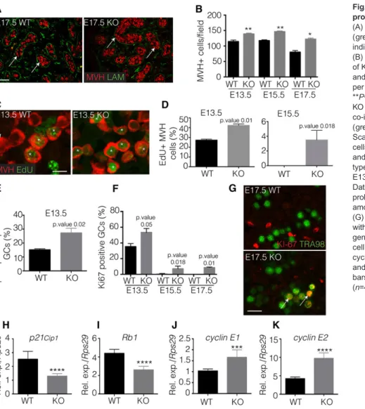
In the mutant testes, we also observed that the number of cells positive for Mvh (mouse vasa homolog, a germ cell marker; Ddx4– Mouse Genome Informatics) was significantly higher than it is in the wild-type gonads from stages E13.5 to E17.5 (Fig. 1A,B). This result was specific to the L/H-Pgds−/−germline, as the phenotype of the single mutant L-Pgds−/− or H-Pgds−/−gonads was similar to that of the wild type (supplementary material Fig. S2A,B). We thus hypothesized that the proliferation rate of germ cells might be modified in the double mutant gonads. Indeed, co-staining for Mvh together with EdU detection (S-phase) or phospho-Histone H3 (M-phase) ( pH3) in wild-type and L/H-Pgds−/− gonads showed that, at E13.5, the percentage of S-phase positive germ cells in the PGD2-depleted mutant gonads was increased to 42% compared with 27% in wild type (1.5 fold) (Fig. 1C,D); however, only a small number of S-phase-positive germ cells was detected in the mutant gonads at E15.5 (Fig. 1D). Immunofluorescence staining for pH3 also showed a twofold higher percentage of mutant germ cells in M-phase at E13.5 than in wild type (Fig. 1E), whereas no pH3 staining was detected in mutant E15.5 germ cells (not shown). In addition, single L-Pgds−/− or H-Pgds−/− mutant gonads had the same pH3 expression pattern as wild-type gonads (supplementary material Fig. S2C). This increased proliferation appeared to be limited to the germ cells, and the proliferation of the Sertoli cells at E13.5 was not modified in the L/H-Pgds−/−gonads (supplementary material Fig. S3A). Finally, an immunofluorescence experiment against the proliferation marker Ki-67, which is expressed in all phases of the cell cycle except for G0, detected 38% Ki-67-positive germ cells in wild-type gonads and 58% positive cells in the mutant E13.5 gonads (Fig. 1F). At E15.5 and even E17.5, 8-10% of the mutant germ cells were still Ki-67 positive (Fig. 1F,G), showing that a significant proportion of the mutant germ cells were not mitotically arrested and were still engaged in the cell cycle.
We thus surmised that PGD2 signalling might control the expression of cell cycle genes as several key regulators of the G1/S phase checkpoint are known to be transcriptionally regulated in the male germ line during mitotic arrest (Spiller et al., 2010; Western et al., 2008). Indeed, we observed a halving in the mRNA level of both p21Cip1and Rb1 in L/H-Pgds−/−testes (KO) at E13.5 compared
with wild type (Fig. 1H,I), as well as a significant increase in the expression of cyclin E1 (Ccne1) and cyclin E2 (Ccne2) at the same stage (Fig. 1J,K). These findings were consistent with the enhanced proliferation observed in the mutant germ cells at E13.5. Finally, we did not observe any differences in the number of apoptotic cells between wild-type and mutant gonads, as measured by TUNEL at
DEVEL
O
the E13.5 stage (data not shown). Taken together, our results suggested that PGD2, produced by germ cells and/or surrounding cells, is involved in the control of cell cycle genes in the foetal testis, acting to slow down germ cell proliferation within this tissue.
L/H-Pgds depletion leads to an altered transcriptional profile of germ cell-specific and cell cycle genes
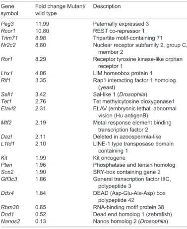
To identify genes regulated by PGD2, we performed an RNA sequencing analysis from wild-type and L/H-Pgds−/−E13.5 testes. We identified 2829 genes that were differentially expressed between the two types (P<0.01), including 1484 genes upregulated and 1345 genes downregulated in mutant compared with wild-type gonads (supplementary material Table S1). We identified genes involved in the cell cycle and cell proliferation regulation (Table 1), as well as genes involved in the regulation of germ cell differentiation and pluripotent marker expression (Table 2). We validated the expression of 29 differentially expressed genes by real time RT-qPCR, obtaining results similar to those observed with the RNA-seq experiments (supplementary material Fig. S4). In mutant gonads, we observed a significant decrease in the expression of the cell cycle inhibitors p21Cip1and
p57Kip1, and an increase in cell cycle activators such as the
retinoblastoma-like gene p130 (Rbl2) and in oncogenes such as Kit, Mybl1 and Erbb4. Furthermore, several regulators of p21Cip1
expression were either upregulated [Elavl2 (Wiszniak et al., 2011; Yoon et al., 2012), Trim71 (Chang et al., 2012), Pten (Luo et al.,
2013) and Cpeb4 (Novoa et al., 2010)] or downregulated [Rbm38 (Feldstein et al., 2012) and Dnd1 (Zhu et al., 2011) (supplementary material Fig. S4B)]. Significantly, the crucial male germ cell gene Nanos2 (Suzuki and Saga, 2008) was downregulated in the absence of PGD2. Moreover, numerous regulators of pluripotency, such as Sox2 (Takahashi and Yamanaka, 2006), Peg3 (Jiang et al., 2007), Nr2c2 (Wagner and Cooney, 2013), Trim71 (Chang et al., 2012), Lhx1 (Birk et al., 2000), Sall1 (Karantzali et al., 2011), Mtf2 (Zhang et al., 2011), L1td1 (Narva et al., 2012), Tet1 (Vincent et al., 2013) and Gtf3c3 (Luzzani et al., 2011), were upregulated in the absence of PGD2(Table 2; supplementary material Fig. S4A). These data have been deposited in the Gene Expression Omnibus database (Edgar et al., 2002) and are accessible through GEO Series accession number GSE55744 (http://www.ncbi.nlm.nih.gov/geo/ query/acc.cgi?acc=GSE55744). In view of these observations, we next investigated germline differentiation in L/H-Pgds−/−foetal testes in more detail.
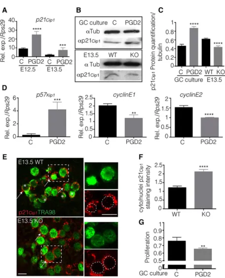
PGD2regulates the cell cycle inhibitor p21Cip1expression
As the CDK inhibitor p21Cip1, a potential regulator of the mitotic
arrest process (Western et al., 2008), was found to be downregulated in mutant E13.5 L/H-Pgds−/−gonads, we next evaluated the direct action of PGD2on p21Cip1expression in the male germ line. Germ
cells from E12.5 and E13.5 testes cultured in the presence of exogenous PGD2 revealed that the levels of p21Cip1 mRNA
increased 3- and 3.5-fold, respectively, at E12.5 and E13.5
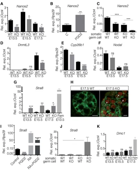
Fig. 1. PGD2signalling controls the germline proliferation in the embryonic testis.
(A) Co-immunofluorescence of Mvh (red) and laminin (green) on E17.5 KO and wild-type testes. Arrows indicate Mvh-positive germ cells. Scale bar: 200μm. (B) Quantification of germ cells within the testis cords of KO and wild-type testes at stages E13.5, E15.5 and E17.5 is represented as numbers of germ cells per field from independent gonads (n=10). *P<0.05; **P<0.01. (C) Germ cell proliferation in E13.5 KO and wild-type testes was evaluated by co-immunofluorescence using Mvh (red) and EdU (green). Asterisks indicate proliferative germ cells. Scale bars: 50μm. (D-F) Quantification of Mvh germ cells that are positive for EdU in E13.5 and E15.5 (D) and for phospho-histone H3 in E13.5 (E) KO and wild-type gonads; quantification of Tra98+KI-67+cells on E13.5, E15.5 and E17.5 KO and wild-type gonads (F). Data are represented as the percentage of
proliferating (EdU, pH3 or KI-67 positive) germ cells among Mvh- (D,E) or Tra98- (F) positive germ cells. (G) Co-immunofluorescence on E17.5 testis sections with KI-67 and Tra98. Arrows indicate proliferating germ cells. Scale bars: 100μm. (H-K) Expression of cell cycle genes p21Cip1(H), Rb1 (I), cyclin E1 (J) and cyclin E2 (K) is studied by RT-qPCR in E13.5 KO and wild-type gonads, and normalized to Rps29. Error bars indicate s.d. of assays carried out in triplicate (n=4). ***P<0.001; ****P<0.0001.
DEVEL
O
(Fig. 2A). In addition, PGD2 significantly increased the level of p21Cip1protein when applied to cultured E13.5 germ cells (Fig. 2B,C).
The effect of PGD2on p21Cip1protein expression was confirmed in
E13.5 whole gonads as p21Cip1protein expression was significantly
decreased in mutants compared with wild-type gonads (Fig. 2B,C). Also, we found that PGD2was able to directly regulate the expression of other cell cycle genes in cultured germ cells, such as p57Kip1and
Ccne1 and Ccne2 (Fig. 2D).
Depending on its subcellular localization, p21Cip1 displays
diverse activities with respect to proliferation (Romanov et al., 2012). Using co-immunofluorescence with the germ cell marker Tra98, we found that p21Cip1was present in both the cytoplasmic
and nuclear compartments with a cytoplasm:nuclei ratio of close to 1:1 (Fig. 2F) in the wild-type E13.5 germ cells, with nuclear localization occurring only in some germ cells (Fig. 2E). By contrast, p21Cip1 was mainly found in the cytoplasm, with a
cytoplasm:nuclei ratio of 2:1 (Fig. 2F) in L/H-Pgds−/−germ cells (Fig. 2E); this result indicated that, in the absence of PGD2, foetal XY germ cells display a proliferative pattern of p21Cip1subcellular
localization. Furthermore, after culturing purified E12.5 germ cells in the presence of PGD2, we found that their proliferation rate was significantly lower than that of the culture without PGD2, confirming the direct effect of PGD2 on the proliferation rate of the male germline (Fig. 2G).
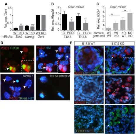
PGD2signalling regulates the expression of pluripotency factors
As the level of Sox2 mRNA was increased in mutants (Table 2), we next compared the level of pluripotent gene expression in L/H-Pgds−/− and wild-type testes at E17.5, a stage when these markers are normally fully repressed. First, we found that the level of Sox2 mRNA was increased by 2.5-fold in the mutant testes when compared with wild-type testes, whereas the levels of Nanog and Oct4 mRNAs remained unchanged (Fig. 3A). By culturing male germ cells at the E12.5 and E13.5 stages in the presence of PGD2, we observed a direct repressive effect of PGD2on Sox2 mRNA level in germ cells at both stages (Fig. 3B). Furthermore, using mixed cultures of somatic and germ cells from wild-type and mutant E13.5 gonads, we determined that PGD2produced by both the somatic and germ cell compartments contributes to the full repression of Sox2 expression (Fig. 3C). We also confirmed that PGD2 is produced by both the somatic and germ cell lineages, following chemical fixation of PGD2 on its production site and immunofluorescence experiments (Fig. 3D).
Using immunofluorescence we then observed the expression of the Sox2, Oct4 and Nanog proteins in mutant testes at E17.5 (Fig. 3E). Notably, for all three of the factors, their subcellular localization was mainly restricted to the cytoplasmic compartment at this stage, whereas under identical conditions the same three proteins were fully nuclear in wild-type and mutant E13.5 gonads (supplementary material Fig. S5). Furthermore, the L-Pgds and H-Pgds enzymatic activities were complementary for this phenotype as in either single mutant L- or H-Pgds gonads, Nanog (supplementary material Fig. S2D), Oct4 and Sox2 (data not shown) expression was normally downregulated. Our data therefore show that PGD2 is involved in the downregulation of the
Table 1. Cell cycle and cell proliferation genes up- and downregulated in E13.5L/H-Pgds mutants relative to wild-type testes
Gene symbol
Fold change
mutant/wild type Description
Cpeb4 12.29 Cytoplasmic polyadenylation
element-binding protein 4
Mybl1 11.12 Myeloblastosis oncogene-like 1
Rcor1 10.80 REST co-repressor 1
Pde3a 9.18 Phosphodiesterase 3A, cGMP inhibited
Erbb4 8.30 v-erb-a erythroblastic leukaemia viral
oncogene homolog 4
Crebbp 6.14 CREB-binding protein
Erbb3 5.72 v-erb-b2 erythroblastic leukaemia viral
oncogene homolog 3
Atm 2.14 Ataxia telangiectasia mutated homolog
Arid4a 2.08 AT rich interactive domain 4A (Rbp1 like)
Cdk17 2.02 Cyclin-dependent kinase 17
Cdkn2aip 1.94 CDKN2A interacting protein
Pak3 1.92 p21 protein (Cdc42/Rac)-activated
kinase 3
Rbl1 1.88 Retinoblastoma-like 1 (p107)
Rbl2 1.78 Retinoblastoma-like 2 (p130)
Cdk12 1.78 Cyclin-dependent kinase 12
Pkd2 1.78 Polycystic kidney disease 2
Ccnl1 1.74 Cyclin L1
Pkd1 1.69 Polycystic kidney disease 1 like 3
Cdk13 1.67 Cyclin-dependent kinase 13
Ccnd2 1.67 Cyclin D2
Ccng2 1.64 Cyclin G2
Cdk8 1.55 Cyclin-dependent kinase 8
Cdk4 0.65 Cyclin-dependent kinase 4
Cdk5 0.62 Cyclin-dependent kinase 5, regulatory
subunit 2 (p39)
Cdk2ap2 0.58 CDK2-associated protein 2
Camk1 0.56 Calcium/calmodulin-dependent protein
kinase
Cdc37 0.54 Cell division cycle 37 homolog (-like 1)
Cdkn1c 0.48 Cyclin-dependent kinase inhibitor 1C
(P57)
Cdkn1a 0.47 Cyclin-dependent kinase inhibitor 1A
(P21)
Table 2. Germ cells and pluripotency genes up- and downregulated in E13.5L/H-Pgds mutants relative to wild-type testes
Gene symbol
Fold change Mutant/ wild type
Description
Peg3 11.99 Paternally expressed 3
Rcor1 10.80 REST co-repressor 1
Trim71 8.98 Tripartite motif-containing 71
Nr2c2 8.80 Nuclear receptor subfamily 2, group C,
member 2
Ror1 8.29 Receptor tyrosine kinase-like orphan
receptor 1
Lhx1 4.06 LIM homeobox protein 1
Rif1 3.35 Rap1 interacting factor 1 homolog
(yeast)
Sall1 3.42 Sal-like 1 (Drosophila)
Tet1 2.76 Tet methylcytosine dioxygenase1
Elavl2 2.31 ELAV (embryonic lethal, abnormal
vision (Hu antigenB)
Mtf2 2.19 Metal response element binding
transcription factor 2
Dazl 2.11 Deleted in azoospermia-like
L1td1 2.10 LINE-1 type transposase domain
containing 1
Kit 1.99 Kit oncogene
Pten 1.96 Phosphatase and tensin homolog
Sox2 1.90 SRY-box containing gene 2
Gtf3c3 1.86 General transcription factor IIIC,
polypeptide 3
Ddx4 1.84 DEAD (Asp-Glu-Ala-Asp) box
polypeptide 42
Rbm38 0.65 RNA-binding motif protein 38
Dnd1 0.52 Dead end homolog 1 (zebrafish)
Nanos2 0.13 Nanos homolog 2 (Drosophila)
DEVEL
O
pluripotency factors Sox2, Oct4 and Nanog, and in the subcellular distribution of these proteins during male germ-line differentiation, acting through different transcriptional (Sox2) or translational (Nanog and Oct4) and putative post-translational mechanisms.
PGD2signalling contributes to the establishment of the male cell differentiation
We next asked whether PGD2signalling could have a role in the regulation of male germ cell differentiation. In our RNA-seq screen, we had observed changes in the expression of genes known to be important for male germ-cell development, such as Nanos2. First, we confirmed that the Nanos2 transcript level at E13.5, E15.5 and E17.5 was lower in mutant than in wild-type gonads (Fig. 4A). Using cultures of isolated male germ cells obtained from E13.5 gonads, we showed that this effect of PGD2on Nanos2 was direct, because upon PGD2treatment Nanos2 levels were 2.5-fold higher in treated than in non-treated cells (Fig. 4B). Then, using mixed cultures of somatic and germ cells from WT and mutant E13.5 gonads, we showed that PGD2produced by both the somatic and the germ cell compartments was necessary for the full activation of Nanos2 expression (Fig. 4C). Dnmt3l, another male-specific gene. was also expressed at lower levels in mutant gonads, but only at the E17.5 stage (Fig. 4D); Cyp26b1 was downregulated in mutant male gonads at E13.5 and E15.5 (Fig. 4E). However, Nodal expression level was similar in mutant and wild-type gonads (Fig. 4F).
As the expression of Nanos2 and Cyp26b1 is directly or indirectly linked to the repression of Stra8 in male foetal germ cells (MacLean
et al., 2007; Suzuki and Saga, 2008), we next tested whether their decreased expression in mutant male gonads led to the activation of meiotic markers. In E13.5, E15.5 and E17.5 testes, Stra8 mRNA levels were significantly higher in mutant gonads than in wild-type gonads (Fig. 4G), although their levels in the male mutant gonads were still 40-fold lower than in female gonads at E13.5 (Fig. 4G). Despite this low level of the Stra8 transcript, clear expression of the Stra8 protein was detected in mutant male gonads at E17.5 (Fig. 4H). Using cultures of germ cells from E12.5 XY gonads, we also observed that PGD2 treatment directly repressed the expression of the Stra8 transcript (Fig. 4I). Furthermore, PGD2was able to diminish the activation of Stra8 by RA by 50%, confirming the negative effect of PGD2on Stra8 expression (Fig. 4I). However, we cannot exclude the possibility that PGD2 acts by directly upregulating Nanos2. Finally, using mixed cultures of somatic and germ cells from wild-type and mutant E13.5 gonads, we showed that PGD2 produced by either the somatic or the germ cell compartments was sufficient to repress Stra8 expression (Fig. 4J). However, the mutant germ cells did not show a reversed sexual fate; they did not overcome the block to meiosis entry as the recombination marker Dmc1 was absent in L/H-Pgds−/− XY gonads from E13.5 to E17.5 (Fig. 4K).
PGD2signals through its Dp2/Crth2 receptor to control the male germline differentiation
As PGD2 can act on both the Dp1 and Dp2/Crth2 G-protein-associated receptors (Matsuoka et al., 2000; Nagata and Hirai,
Fig. 2. PGD2regulates the cell cycle p21Cip1inhibitor towards the mitotic arrest. (A) Purified XY E12.5 or E13.5 germ cells were cultured in the presence of PGD2(5 ng/ml) or not (C=control). RT-qPCR was used to evaluate the expression of p21Cip1normalized to Rps29 expression. Error bars represent s.d. of assays carried out in triplicate (n=3). (B,C) Protein levels of p21Cip1(α-p21Cip1) were analysed by western blot and compared with the ubiquitous tubulin (α-Tub) protein in E13.5 male germ cells cultured with PGD2 or not (C=control), and in E13.5 KO and WT gonads. (C) p21Cip1protein expression levels were quantified (n=3)
and are represented on the corresponding graph. (D) Purified XY E12.5 germ cells were cultured for 18 h in the presence of PGD2(5 ng/ml) or not (C=control). RT-qPCR evaluated the expression of p57Kip1, Ccne1 and Ccne2 normalized to Rps29 expression. Error bars represent s.d. of assays carried out in triplicate (n=3). (E) Immunofluorescence analysis of p21Cip1and Tra98 proteins in E13.5 wild-type and
L/H-Pgds−/−gonads (KO). Asterisks highlight nuclear p21Cip1 and arrows indicate cytoplasmic p21Cip1. Outlined areas are enlarged in the right-hand panels, in which dotted circles delineate nuclei. Scale bars: 25μm. (F) Cytoplasmic and nuclear signals were quantified on five different gonads (70 and 90 germ cells) for each genotype using the ImageJ software and were represented as the ratio of cytoplasmic/ nuclei staining. (G) Proliferation of E12.5 germ cells cultured in the presence of PGD2(PGD2) or not (C) for 24 h was measured using the cell proliferation assay. **P<0.01; ***P<0.001; ****P<0.0001.
DEVEL
O
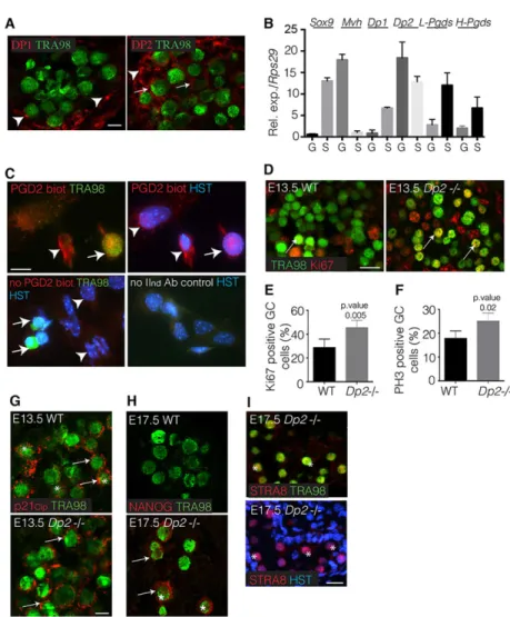
2003), we first determined the localization of the Dp1 (Ptgdr) and Dp2 (Ptgdr2) receptors in E13.5 male gonad. Using co-immunofluorescence experiments with Tra98 staining, we found that Dp2 was expressed in both germ cells and somatic compartments, whereas Dp1 was only expressed in somatic cells (Fig. 5A). These Dp1 and Dp2 patterns were confirmed by purifying somatic and germ cell fractions from male E13.5 gonads: the germ cell fraction only expressed Dp2 mRNA (Fig. 5B), whereas the somatic cell fraction expressed both Dp2 and Dp1 mRNAs (Fig. 5B). In the same experiments, we confirmed that both L-Pgds and H-Pgds mRNAs were expressed in both somatic and germ cell compartments, although the somatic expression was higher than that in the germ cells (Fig. 5B). Furthermore, using the PGD2-biotin tool followed by immunofluorescence, we were able to visualize the binding of PGD2 to Tra98-expressing germ cells (Fig. 5C). These data confirmed that germ cells expressing only Dp2 are effectively able to bind PGD2produced by both germ cells and somatic cells, as shown in Fig. 3D.
To evaluate the function of Dp2 within the gonad, we orally administered the specific Dp2 antagonist CAY10471 (Royer et al., 2007) to pregnant females and analysed the proliferation rate of the developing germ cells. At E13.5, 10% of the germ cells were positive for EdU in the control gonads, compared with 28% in the CAY10471-treated gonads (supplementary material Fig. S6A,B); at the same time, no modified proliferation of the Sox9-expressing Sertoli cells was detected upon CAY10471 treatment (supplementary material Fig. S6C,D). The different percentages of EdU-positive germ cells compared with those obtained with E13.5 L/H-Pgds−/− testes (Fig. 1D) were probably due to the different genetic backgrounds, namely CD1 (CAY experiments) and C57BL/6 (L/H-Pgds−/−) (Western et al., 2011). This effect on proliferation was specific to the Dp2 receptor, as oral administration of the Dp1 antagonist BWA868C did not significantly modify the number of proliferating germ cells,
whereas the Dp2 agonist 15R-PGD2 potentiated the effect of endogenous PGD2(supplementary material Fig. S6B). Furthermore, upon CAY10471 treatment, a decreased level of expression of p21Cip1
and Nanos2 was found (supplementary material Fig. S6E). Together, these data showed that the administration of the Dp2 antagonist mimicked the loss of PGD2signalling at E13.5.
To confirm these data, we next analysed E13.5 Dp2−/− testes (Satoh et al., 2006). We observed that the numbers of Ki-67 proliferating (Fig. 5D,E) and pH3 mitotic (Fig. 5F) germ cells were significantly higher in Dp2 mutant gonads than in wild type (45%/ 28% and 24%/17%, respectively). These differences were in the same range as those described above for the L/H-Pgds−/− testes. Furthermore, E13.5 Dp2−/− testes expressed the p21Cip1 protein
within the cytoplasm of germ cells (Fig. 5G), and at E17.5 the germ cells ectopically expressed the pluripotent marker Nanog (Fig. 5H) and the premeiotic marker Stra8 (Fig. 5I), similarly to what was observed in L/H-Pgds−/−mutant germ cells.
The function of Dp2 was confirmed by culturing purified E12.5 germ cells in the presence of PGD2with either the Dp1antagonist BWA868C or the Dp2 antagonist BAY-U3450: the repression of Stra8 and Sox2 expression and the activation of Nanos2 and p21Cip1expression observed in the presence of PGD
2was partially
reversed only in the presence of BAY-U3450 (supplementary material Fig. S6F-I). These data confirmed that PGD2 signalling indeed acts on male foetal germ cells through the Dp2 receptor.
DISCUSSION
In this study, we have investigated the role of PGD2 in the differentiation of male foetal germ cells, significantly extending earlier findings on the role of this signalling molecule in the biology of the foetal testis (Moniot et al., 2009, 2011; Wilhelm et al., 2007). Our analysis of gonads from double L- and H-Pgds (L/H-Pgds−/−) and Dp2−/−mutant embryos has shown that PGD2acting through its
Fig. 3. PGD2regulates the expression of pluripotency genes. (A,B) RT-qPCR analysis of the expression of the pluripotent genes Sox2 (A,B), Nanog and Oct4 (A) in KO and wild-type gonads at E17.5 (A) or in E13.5 germ cells cultured with PGD2(5 ng/ml) or not (C=control) (B) using Oct4 (A) or Rps29 (B) as the normalization control gene. Error bars indicate s.d. of assays carried out in triplicate (n=5). (C) Somatic and germ cells from wild-type and mutant E13.5 gonads were mixed and cultured for 24 h; Sox2 expression levels were quantified by RT-qPCR relative to Oct4 expression. Error bars indicate s.d. of assays carried out in triplicate (n=2). *P<0.05; **P<0.01. (D) Dissociated somatic/germ cells from E13.5 wild-type testes were cultured for 24 h; PGD2was fixed using the EicosaCell protocol (upper panels and control 2) or cells were fixed in 4% PFA (control 1) and co-immunofluorescence with anti-PGD2(red) and anti-Tra98 (green) antibodies was performed (upper panels and control 1); control 2 was only incubated with secondary antibodies. HST, Hoechst dye. Arrows and arrowheads show germ cells and somatic cells expressing PGD2, respectively. Asterisks show somatic cells that weakly produce PGD2. Germ cells producing PGD2are outlined. Scale bar: 25μm. (E) Immunofluorescence with anti-Sox2 (red), anti-Nanog (red) and anti-Oct4 (green) antibodies, and Hoechst dye (HST, blue) on sections of E17.5 KO and wild-type testes. Arrows indicate Sox2, Nanog and Oct4 staining (enlarged in the insets). Scale bars: 50μm.
DEVEL
O
Dp2 receptor, is involved in processes required for proper male germ cell differentiation: slowing down proliferation to reach the mitotic arrest, inhibiting the expression of pluripotency master genes and upregulating male germ cell genes.
Consistent with previous studies (Adams and McLaren, 2002; Moniot et al., 2009, 2011), we have confirmed that L-Pgds and H-Pgds are both expressed at early stages in the male germline and have found that both enzymatic activities act in a complementary manner with respect to germ cell differentiation, as L-Pgds and H-Pgds single mutants have a normal phenotype. Furthermore, we have demonstrated that PGD2is indeed synthesized by both the somatic and germ cell populations and provide evidence that both sources of PGD2 work in concert to effect germ cell differentiation. This effect might be the result of a direct action on germ cells to activate expression of the male germ cell marker Nanos2 and of p21Cip1and to downregulate pluripotency markers;
this might also result from indirect effects by reinforcing the male phenotype in somatic cells through the activation of Notch signalling and Cyp26b1 expression. Further work will decipher the respective contribution of both cell types in the PGD2-mediated germ cell differentiation using germ cell- and Sertoli cell- specific Dp2 receptor knockout strains. Adding to our knowledge of Fgf9 (Bowles et al., 2010; Spiller et al., 2012), Notch (Garcia et al., 2013) and Tgfβ signalling (Miles et al., 2013) in Sertoli cells and of Dmrt1 (Krentz et al., 2009) and Dnd1 (Cook et al., 2011) in germ cells, we have thus identified the PGD2signalling pathway, expressed in both
compartments and active in the germline in both a paracrine and an autocrine manner, as a new factor contributing to the male germ cell differentiation process. In contrast to Fgf9 (Bowles et al., 2010) and Nodal signalling (Souquet et al., 2012; Spiller et al., 2012), however, suppressing PGD2 signalling alone is not sufficient to allow progression into meiosis; this is presumably because these two other pathways, and also Cyp26b1 and Nanos2, continue to exert their strong repressive influences even in the absence of PGD2.
In the absence of PGD2, we observed an increase in the proliferation rate at E13.5; even at E17.5, 10% of the mutant gonocytes remained in the cell cycle, strongly suggesting that the PGD2signalling pathway contributes to mitotic arrest in male foetal germ cells. This impaired mitotic arrest in the L/H-Pgds−/−testes was similar to that observed in mutant gonads for Dmrt1 (Krentz et al., 2009) and for Dnd1 (Cook et al., 2011), where persistent Ki-67-positive and pH3-negative cells remained at E17.5. Our experiments showed that the gene coding for p21Cip1, a key
regulator of the G1-S phase checkpoint (Fotedar et al., 2004) that is specifically expressed in the male germ line at the time of mitotic arrest (Western et al., 2008), is directly upregulated by PGD2. Furthermore, in L/H-Pgds−/−germ cells, we detected the p21Cip1
protein mainly within the cytoplasm, suggesting a role for PGD2in the post-transcriptional regulation of p21Cip1 and potentially
explaining the reduced cell-cycle inhibitory activity of p21Cip1
observed in the absence of PGD2(Starostina et al., 2010; Wu et al., 2011). As Dnd1 expression is downregulated in L/H-Pgds−/−
Fig. 4. PGD2contributes to the male germ cell
differentiation. (A,D-G,K) RT-qPCR analysis of Nanos2 (A), DnmtL3 (D), Cyp26b1 (E), Nodal (F) Stra8 (G) and Dmc1 (K) expression levels in E13.5, E15.5 (A,D-G,K) and E17.5 (A,D,F,G,K) KO and type male gonads and in E13.5 wild-type ovaries (Stra8, G; Dmc1, K). Error bars indicate s.d. of assays carried out in triplicate (n=4). (B) Germ cells were isolated from E12.5 testes and cultured with PGD2(5 ng/ml) or not (C=control). RT-qPCR analysis of Nanos2 expression is represented using Rps29 as the normalization gene; error bars represent s.d. of assays carried out in triplicate (n=4). (C) Somatic and germ cells from wild-type and mutant E13.5 gonads were mixed and cultured for 24 h; Nanos2 expression levels were quantified by RT-qPCR relative to Oct4 expression. Error bars indicate s.d. of assays carried out in triplicate (n=2). (H) Co-immunofluorescence of pre-meiotic marker Stra8 (red) with Amh (green) on E17.5 KO and wild-type testes. Arrows highlight the expression of Stra8. Scale bars: 50μm (wild type) and 25 μm (KO). (I) Isolated germ cells from E12.5 testes were cultured with PGD2(5 ng/ml) and/or retinoic acid (RA, 10 nM) (C=control). RT-qPCR analysis of Stra8 expression is represented using Rps29 as the normalization gene. Error bars indicate s.d. of assays carried out in triplicate on three independent samples. (J) Somatic and germ cells from wild-type and mutant E13.5 gonads were mixed and cultured for 24 h; Stra8 expression levels were quantified by RT-qPCR relative to Oct4 expression. Error bars indicate s.d. of assays carried out in triplicate (n=2). *P<0.05; **P<0.01; ***P<0.001; ****P<0.0001.
DEVEL
O
gonads, PGD2signalling might affect p21Cip1expression, indirectly
by activating Dnd1 expression (Cook et al., 2011; Kedde et al., 2007). Furthermore, the RNA-seq analysis highlighted differences in a number of genes known to be involved in the complex p21Cip1
regulation (Jung et al., 2010), both at the transcriptional (Hdac4, Pde3 and Pten) and the post-transcriptional levels (Elavl2, Dnd1, Rbm38, Ddx4 and Trim71). Thus, these data show that PGD2might be a pathway acting early at multiple levels of p21Cip1regulation
during the mitotic arrest within male germ cells, through still unknown mechanisms.
Concomitant with the impaired mitotic arrest observed in mutant germ cells at E17.5, the pluripotent proteins Oct4, Sox2 and Nanog were still present, even though only Sox2 was upregulated at the mRNA level. The regulation of Sox2 by PGD2 appears to be independent of Dmrt1 (Krentz et al., 2009) as Dmrt1 expression was not modified by PGD2. After E12.5, the downregulation of the Fgf9 (Bowles et al., 2010) and Cripto/Nodal (Spiller et al., 2012) pathways, together with the upregulation of L-Pgds (Moniot et al., 2009), can explain the slow decrease in Sox2 mRNA that is observed in the male germ line (Western et al., 2010). Here, we also observed that PGD2 could post-transcriptionally regulate the expression of Oct4 and Nanog at the level of mRNA translation or protein stability (or both). Interestingly, numerous RNA-binding proteins such as L1td1 (Iwabuchi et al., 2011) are upregulated in the absence of PGD2and might participate in this regulation. In the absence of PGD2, Nanog, Oct4 and Sox2 are ectopically expressed in mutant foetal testes and display cytoplasmic localisation,
suggesting that PGD2 might also participates in the nuclear translocation of these transcription factors as has been previously observed for Sox9 (Malki et al., 2005). This phenotype might reflect a transient and incomplete differentiation of the germline, as described in embryonic stem cells (da Cunha et al., 2013; Elatmani et al., 2011) and in a variety of cancer cells (Gu et al., 2012; Guo et al., 2011).
Our ablation of the PGD2 pathway in the teratoma-resistant C57Bl/6 (B6) background induced phenotypes that were similar to those that have been observed in the teratoma-susceptible strains 129/SvJ or 129-Chr19MOLF/Ei(Heaney et al., 2012), likely related to
the impaired mitotic arrest. Along these lines, questions related to germ cell differentiation and mitotic arrest can have clear implications for human health, as germ cells that are not controlled appropriately during foetal life can later transform into carcinoma in situ (CIS), the precursor for testicular germ cell tumours (Kristensen et al., 2008). Furthermore, testicular cancers commonly include molecular abnormalities such as mutations in cell cycle regulators (Bartkova et al., 2000), and the PGD2target gene L1td1 is highly expressed in seminomas and testicular germ cell tumours (Narva et al., 2012). Further work will determine whether the double L/H-Pgds mutation can lead to a high incidence of germ-line tumours in the 129sv background, as has been described for Dmrt1 (Krentz et al., 2009, 2013), Pten (Kimura et al., 2003) and Dnd1 (Cook et al., 2011) mutants.
In summary, the present study identifies the PGD2pathway as one of the earliest signalling pathway involved in the male germ cell
Fig. 5. The Dp2 receptor is expressed in the male germ line and functions to control proliferation and differentiation of the germ line.
(A) Co-immunofluorescence of Dp1 or Dp2 (red) and germ cells stained with Tra98 (green). Arrows and arrowheads indicate germ and somatic cells, respectively. Scale bar: 25μm. (B) RT-qPCR analysis of Dp1, Dp2, L-Pgds and H-Pgds expression in E13.5 germ cells (G) and somatic (S) cell fractions. Mvh and Sox9 expression is shown to assess the purity of both somatic and germ cell fractions.
(C) Dissociated somatic/germ cells from E13.5 wild-type testes were cultured for 24 h and were treated by PGD2-biotin (5 ng/ml) for 10 min. Germ cells able to bind PGD2-biotin were visualised by immunofluorescence (IF) using streptavidin-Texas Red antibody (red) together with anti-Tra98 (green) (upper panels). Two controls (lower panels) were performed: one was not treated with PGD2-biotin but was submitted to immunofluorescence and one was treated by PGD2-biotin but submitted to immunofluorescence without secondary antibodies. HST, Hoechst dye. Scale bar: 50μm. Arrow and arrowheads indicate germ cell and somatic cells, respectively. (D) Co-immunofluorescence with Ki-67 (red) and Tra98 (green) on E13.5 wild-type and Dp2 mutant (Dp2−/−) testes. Arrows indicate proliferating germ cells. Scale bar: 50μm. (E,F) Quantifications of KI-67 proliferating germ cells (E) and pH3 mitotic germ cells (F) are represented as percentages of total Tra98+ germ cells in the E13.5 Dp2−/− and wild-type testes. (G-I) Co-immunofluorescence of p21Cip1(G), Nanog (H), Stra8 (I) (red) and Tra98 (green) on E13.5 (G) or E17.5 (H-I) wild-type (WT) and Dp2−/−testes. Asterisks indicate nuclear staining of p21Cip1(G), Nanog (H) or Stra8 (I); arrows indicate cytoplasmic localisation of p21Cip1 (G) and Nanog (H). Scale bars: 25μm in G,H; 50 μm in I.
DEVEL
O
differentiation, showing that PGD2is a male fate-promoting factor. As PGD2 is a potential target for endocrine disruptors (ED) (Kristensen et al., 2011), our findings thus open new perspectives for future investigations into how germ cell development can be perturbed by the external environment.
MATERIALS AND METHODS
Mice
L-Pgds KO (Eguchi et al., 1999) and H-Pgds KO (Trivedi et al., 2006) mice were generated at Osaka Bioscience Institute (Osaka, Japan) using the C57BL/6 strain. They were cross-bred to generate the L/H-Pgds double KO mice that were used in this work. L/H-Pgds double KO animals were kept and bred at the IGH animal care facility under controlled environmental conditions. Dp2/Crth2 mice were generated at BioMedical Laboratories (Saitama, Japan) (Satoh et al., 2006) and were transferred into the C57BL/6 genetic background. For the pharmaceutical experiments, wild-type CD1 E10.5 females were purchased from Charles River Laboratories. All animal uses were conducted according to procedures approved by the Réseau des Animaleries de Montpellier (RAM) (agreement number 34-366 for B.B.-B.) and by the Regional Ethics committee.
In vivo EdU incorporation and treatments
EdU (5-ethynyl-2′-deoxyuridine) was intraperitoneally injected into pregnant females, 2 h before dissecting the embryos. Detection of the EdU-positive cells on testis sections was performed using the Click-iT EdU Assay, according to the supplier’s instructions (Invitrogen). The Dp2 antagonists CAY10471 and BAY-U3450, the Dp2 agonist 15(R)-PGD2), and the Dp1 antagonist BW A868C were administered as previously described (Woodward et al., 2011). See methods in the supplementary material for further details.
Immunofluorescence
Dissected gonads from staged embryos were processed into cryosections and immunofluorescence was performed using the primary antibodies that are listed in supplementary material Table S2, as previously described (Malki et al., 2005; Moniot et al., 2009). The appropriate secondary antibodies (Alexa-Ig, Molecular Probe) were used. Histology images were captured with a Leica DM6000 fluorescent microscope or with a Leica SP8-UV Confocal microscope.
RNA isolation and real-time PCR analysis
Embryonic gonads were dissected and separated from mesonephros and were then pooled by sex within each litter (between four and seven pairs of gonads). RNA extraction was performed using the TRIZOL technique (Invitrogen). Real-time RT-PCR was performed as previously described (Moniot et al., 2009, 2011) using primers listed in supplementary material Table S3. Oct4 or Rps29 were used as the normalization gene in experiments with whole gonads, and Rps29 was used with isolated germ cells.
Immunomagnetic germ cell and somatic cell isolation and culture
Germ cell isolation using the Ssea-1 antigen was performed as previously described (Moniot et al., 2011), using magnetic sorting. In vitro cultures of germ cells and mixed somatic and germ cells were performed using previously described techniques (Bowles et al., 2010; Munger et al., 2013). Dissociated gonadal cells (250,000 cells) were also cultured on glass coverslips in 24-well plates for 24 h (Munger et al., 2013) to detect intracellular production of PGD2 using the EicosaCell technique (Bandeira-Melo et al., 2011) and to analyse the PGD2 binding on embryonic cells using the PGD2-biotin tool. For details, see methods in the supplementary material.
Protein extracts and western blots
Protein extracts from E13.5 testes or from cultured germ cells were prepared in Tris buffer ( pH 8) with 25 U benzonase (Sigma-Aldrich) and protein contents were quantified using the micro BCA protein assay kit (Thermo Scientific). Proteins (20μg) were electrophoresed in SDS/PAGE gels and
then electroblotted onto nitrocellulose membranes. Membranes were incubated with primary antibodies (see supplementary material Table S1 for concentration), followed by HRP-conjugated secondary antibodies. Signal was detected using the Chemiluminescent Substrate detection kit (Thermo Scientific).
Statistical analysis
Statistical analysis with PRISM 6 software (GraphPad Software) was performed using the Student’s t-test to compare two groups in a independent experiments or the ANOVA test with the Geisser-Greenhouse correction for multiple comparisons (qPCR experiments) and using the Fisher’s exact test (cell counting experiments), and the results were considered statistically significant at P<0.05. Asterisks indicate the level of statistical significance: *P<0.05; **P<0.01; ***P<0.001; ****P<0.0001 (Student’s or ANOVA tests) or P-values are indicated on each graph (Fisher’s exact test); ns indicates not significant. For details of the analysis, see methods in the supplementary material.
mRNA expression profiling and analysis
Whole-transcriptome analysis of E13.5 wild-type and mutant male gonads was performed using RNA-seq experiments, as described in detail in the methods in the supplementary material.
Acknowledgements
We thank the staff of the IGH animal care facility, particularly Elodie Gavois, Frédéric Gallardo and Florence Arnal. We thank Dr Julien Cau and Amélie Sarrazin from the Imagery platform of IGH (MRI Montpellier) for their help in imagery and quantification analysis with ImageJ. We are grateful to Prof. Gabriel Livera, Dr Peter Follette and Dr Rosemary Kiernan for critical reading of the manuscript.
Competing interests
The authors declare no competing financial interests.
Author contributions
B.M., S.U., J.C., F.P. and B.B.-B. performed experiments. E.D. and S.N. performed RNA-seq experiments and analysis. K.A. and Y.U. provided L/H-Pgds KO mice. H.H., K.N. and M.N. provided Dp2 knockout mice. F.P. and B.B.-B. designed the experiments and wrote the paper.
Funding
This work was supported by the CNRS and by the Agence Nationale pour la Recherche (ANR blanc MolMechMeiosis programme). S.U. is the recipient of a PhD fellowship from the Shah Abdul Latif University, Pakistan.
Supplementary material
Supplementary material available online at
http://dev.biologists.org/lookup/suppl/doi:10.1242/dev.103408/-/DC1
References
Adams, I. R. and McLaren, A. (2002). Sexually dimorphic development of mouse primordial germ cells: switching from oogenesis to spermatogenesis. Development 129, 1155-1164.
Baltus, A. E., Menke, D. B., Hu, Y.-C., Goodheart, M. L., Carpenter, A. E., de Rooij, D. G. and Page, D. C. (2006). In germ cells of mouse embryonic ovaries, the decision to enter meiosis precedes premeiotic DNA replication. Nat. Genet. 38, 1430-1434.
Bandeira-Melo, C., Weller, P. F. and Bozza, P. T. (2011). EicosaCell - an immunofluorescent-based assay to localize newly synthesized eicosanoid lipid mediators at intracellular sites. Methods Mol. Biol. 689, 163-181.
Barrios, F., Filipponi, D., Pellegrini, M., Paronetto, M. P., Di Siena, S., Geremia, R., Rossi, P., De Felici, M., Jannini, E. A. and Dolci, S. (2010). Opposing effects of retinoic acid and FGF9 on Nanos2 expression and meiotic entry of mouse germ cells. J. Cell Sci. 123, 871-880.
Bartkova, J., Thullberg, M., Rajpert-De Meyts, E., Skakkebaek, N. E. and Bartek, J. (2000). Cell cycle regulators in testicular cancer: loss of p18INK4C marks progression from carcinoma in situ to invasive germ cell tumours. Int. J. Cancer 85, 370-375.
Best, D., Sahlender, D. A., Walther, N., Peden, A. A. and Adams, I. R. (2008). Sdmg1 is a conserved transmembrane protein associated with germ cell sex determination and germline-soma interactions in mice. Development 135, 1415-1425.
Birk, O. S., Casiano, D. E., Wassif, C. A., Cogliati, T., Zhao, L., Zhao, Y., Grinberg, A., Huang, S., Kreidberg, J. A., Parker, K. L. et al. (2000). The LIM homeobox gene Lhx9 is essential for mouse gonad formation. Nature 403,
909-913.
DEVEL
O
Bowles, J., Knight, D., Smith, C., Wilhelm, D., Richman, J., Mamiya, S., Yashiro, K., Chawengsaksophak, K., Wilson, M. J., Rossant, J. et al. (2006). Retinoid signaling determines germ cell fate in mice. Science 312, 596-600.
Bowles, J., Feng, C.-W., Spiller, C., Davidson, T.-L., Jackson, A. and Koopman, P. (2010). FGF9 suppresses meiosis and promotes male germ cell fate in mice. Dev. Cell 19, 440-449.
Chang, H.-M., Martinez, N. J., Thornton, J. E., Hagan, J. P., Nguyen, K. D. and Gregory, R. I. (2012). Trim71 cooperates with microRNAs to repress Cdkn1a expression and promote embryonic stem cell proliferation. Nat. Commun. 3, 923. Cook, M. S., Munger, S. C., Nadeau, J. H. and Capel, B. (2011). Regulation of male germ cell cycle arrest and differentiation by DND1 is modulated by genetic background. Development 138, 23-32.
da Cunha, J. M., da Costa-Neves, A., Kerkis, I. and da Silva, M. C. P. (2013). Pluripotent stem cell transcription factors during human odontogenesis. Cell Tissue Res. 353, 435-441.
DiNapoli, L., Batchvarov, J. and Capel, B. (2006). FGF9 promotes survival of germ cells in the fetal testis. Development 133, 1519-1527.
Edgar, R., Domrachev, M. and Lash, A. E. (2002). Gene Expression Omnibus: NCBI gene expression and hybridization array data repository. Nucleic Acids Res. 30, 207-210.
Eguchi, N., Minami, T., Shirafuji, N., Kanaoka, Y., Tanaka, T., Nagata, A., Yoshida, N., Urade, Y., Ito, S. and Hayaishi, O. (1999). Lack of tactile pain (allodynia) in lipocalin-type prostaglandin D synthase-deficient mice. Proc. Natl. Acad. Sci. USA 96, 726-730.
Elatmani, H., Dormoy-Raclet, V., Dubus, P., Dautry, F., Chazaud, C. and Jacquemin-Sablon, H. (2011). The RNA-binding protein Unr prevents mouse embryonic stem cells differentiation toward the primitive endoderm lineage. Stem Cells 29, 1504-1516.
Ewen, K. A. and Koopman, P. (2010). Mouse germ cell development: from specification to sex determination. Mol. Cell. Endocrinol. 323, 76-93.
Feldstein, O., Ben-Hamo, R., Bashari, D., Efroni, S. and Ginsberg, D. (2012). RBM38 is a direct transcriptional target of E2F1 that limits E2F1-induced proliferation. Mol. Cancer Res. 10, 1169-1177.
Fotedar, R., Bendjennat, M. and Fotedar, A. (2004). Functional analysis of CDK inhibitor p21WAF1. Methods Mol. Biol. 281, 55-71.
Garcia, T. X., Defalco, T., Capel, B. and Hofmann, M.-C. (2013). Constitutive activation of NOTCH1 signaling in Sertoli cells causes gonocyte exit from quiescence. Dev. Biol. 377, 188-201.
Gu, T.-T., Liu, S.-Y. and Zheng, P.-S. (2012). Cytoplasmic NANOG-positive stromal cells promote human cervical cancer progression. Am. J. Pathol. 181, 652-661. Guerquin, M.-J., Duquenne, C., Lahaye, J.-B., Tourpin, S., Habert, R. and
Livera, G. (2010). New testicular mechanisms involved in the prevention of fetal meiotic initiation in mice. Dev. Biol. 346, 320-330.
Guo, Y., Liu, S., Wang, P., Zhao, S., Wang, F., Bing, L., Zhang, Y., Ling, E.-A., Gao, J. and Hao, A. (2011). Expression profile of embryonic stem cell-associated genes Oct4, Sox2 and Nanog in human gliomas. Histopathology 59, 763-775. Heaney, J. D., Anderson, E. L., Michelson, M. V., Zechel, J. L., Conrad, P. A.,
Page, D. C. and Nadeau, J. H. (2012). Germ cell pluripotency, premature differentiation and susceptibility to testicular teratomas in mice. Development 139, 1577-1586.
Iwabuchi, K. A., Yamakawa, T., Sato, Y., Ichisaka, T., Takahashi, K., Okita, K. and Yamanaka, S. (2011). ECAT11/L1td1 is enriched in ESCs and rapidly activated during iPSC generation, but it is dispensable for the maintenance and induction of pluripotency. PLoS ONE 6, e20461.
Jameson, S. A., Lin, Y.-T. and Capel, B. (2012). Testis development requires the repression of Wnt4 by Fgf signaling. Dev. Biol. 370, 24-32.
Jiang, H., Sun, B., Wang, W., Zhang, Z., Gao, F., Shi, G., Cui, B., Kong, X., He, Z., Ding, X. et al. (2007). Activation of paternally expressed imprinted genes in newly derived germline-competent mouse parthenogenetic embryonic stem cell lines. Cell Res. 17, 792-803.
Jung, Y.-S., Qian, Y. and Chen, X. (2010). Examination of the expanding pathways for the regulation of p21 expression and activity. Cell. Signal. 22, 1003-1012. Karantzali, E., Lekakis, V., Ioannou, M., Hadjimichael, C., Papamatheakis, J.
and Kretsovali, A. (2011). Sall1 regulates embryonic stem cell differentiation in association with nanog. J. Biol. Chem. 286, 1037-1045.
Kashimada, K., Svingen, T., Feng, C. W., Pelosi, E., Bagheri-Fam, S., Harley, V. R., Schlessinger, D., Bowles, J. and Koopman, P. (2011). Antagonistic regulation of Cyp26b1 by transcription factors SOX9/SF1 and FOXL2 during gonadal development in mice. FASEB J. 25, 3561-3569.
Kedde, M., Strasser, M. J., Boldajipour, B., Oude Vrielink, J. A. F., Slanchev, K., le Sage, C., Nagel, R., Voorhoeve, P. M., van Duijse, J., Ørom, U. A. et al. (2007). RNA-binding protein Dnd1 inhibits microRNA access to target mRNA. Cell 131, 1273-1286.
Kimura, T., Suzuki, A., Fujita, Y., Yomogida, K., Lomeli, H., Asada, N., Ikeuchi, M., Nagy, A., Mak, T. W. and Nakano, T. (2003). Conditional loss of PTEN leads to testicular teratoma and enhances embryonic germ cell production. Development 130, 1691-1700.
Koubova, J., Menke, D. B., Zhou, Q., Capel, B., Griswold, M. D. and Page, D. C. (2006). Retinoic acid regulates sex-specific timing of meiotic initiation in mice. Proc. Natl. Acad. Sci. USA 103, 2474-2479.
Krentz, A. D., Murphy, M. W., Kim, S., Cook, M. S., Capel, B., Zhu, R., Matin, A., Sarver, A. L., Parker, K. L., Griswold, M. D. et al. (2009). The DM domain protein DMRT1 is a dose-sensitive regulator of fetal germ cell proliferation and pluripotency. Proc. Natl. Acad. Sci. USA 106, 22323-22328.
Krentz, A. D., Murphy, M. W., Zhang, T., Sarver, A. L., Jain, S., Griswold, M. D., Bardwell, V. J. and Zarkower, D. (2013). Interaction between DMRT1 function and genetic background modulates signaling and pluripotency to control tumor susceptibility in the fetal germ line. Dev. Biol. 377, 67-78.
Kristensen, D. M., Sonne, S. B., Ottesen, A. M., Perrett, R. M., Nielsen, J. E., Almstrup, K., Skakkebaek, N. E., Leffers, H. and Meyts, E. R.-D. (2008). Origin of pluripotent germ cell tumours: the role of microenvironment during embryonic development. Mol. Cell. Endocrinol. 288, 111-118.
Kristensen, D. M., Skalkam, M. L., Audouze, K., Lesné, L., Desdoits-Lethimonier, C., Frederiksen, H., Brunak, S., Skakkebæk, N. E., Jégou, B., Hansen, J. B. et al. (2011). Many putative endocrine disruptors inhibit prostaglandin synthesis. Environ. Health Perspect. 119, 534-541.
Kumar, S., Chatzi, C., Brade, T., Cunningham, T. J., Zhao, X. and Duester, G. (2011). Sex-specific timing of meiotic initiation is regulated by Cyp26b1 independent of retinoic acid signalling. Nat. Commun. 2, 151.
Luo, L., Gong, Y. Q., Qi, X., Lai, W., Lan, H. and Luo, Y. (2013). Effect of tumor suppressor PTEN gene on apoptosis and cell cycle of human airway smooth muscle cells. Mol. Cell. Biochem. 375, 1-9.
Luzzani, C., Solari, C., Losino, N., Ariel, W., Romorini, L., Bluguermann, C., Sevlever, G., Barañao, L., Miriuka, S. and Guberman, A. (2011). Modulation of chromatin modifying factors’ gene expression in embryonic and induced pluripotent stem cells. Biochem. Biophys. Res. Commun. 410, 816-822. MacLean, G., Li, H., Metzger, D., Chambon, P. and Petkovich, M. (2007).
Apoptotic extinction of germ cells in testes of Cyp26b1 knockout mice. Endocrinology 148, 4560-4567.
Malki, S., Nef, S., Notarnicola, C., Thevenet, L., Gasca, S., Méjean, C., Berta, P., Poulat, F. and Boizet-Bonhoure, B. (2005). Prostaglandin D2 induces nuclear import of the sex-determining factor SOX9 via its cAMP-PKA phosphorylation. EMBO J. 24, 1798-1809.
Matsuoka, T., Hirata, M., Tanaka, H., Takahashi, Y., Murata, T., Kabashima, K., Sugimoto, Y., Kobayashi, T., Ushikubi, F., Aze, Y. et al. (2000). Prostaglandin D2 as a mediator of allergic asthma. Science 287, 2013-2017.
McClelland, K., Bowles, J. and Koopman, P. (2012). Male sex determination: insights into molecular mechanisms. Asian J. Androl. 14, 164-171.
Mendis, S. H., Meachem, S. J., Sarraj, M. A. and Loveland, K. L. (2011). Activin A balances Sertoli and germ cell proliferation in the fetal mouse testis. Biol. Reprod. 84, 379-391.
Miles, D. C., Wakeling, S. I., Stringer, J. M., van den Bergen, J. A., Wilhelm, D., Sinclair, A. H. and Western, P. S. (2013). Signaling through the TGF beta-activin receptors ALK4/5/7 regulates testis formation and male germ cell development. PLoS ONE 8, e54606.
Moniot, B., Declosmenil, F., Barrionuevo, F., Scherer, G., Aritake, K., Malki, S., Marzi, L., Cohen-Solal, A., Georg, I., Klattig, J. et al. (2009). The PGD2 pathway, independently of FGF9, amplifies SOX9 activity in Sertoli cells during male sexual differentiation. Development 136, 1813-1821.
Moniot, B., Farhat, A., Aritake, K., Declosmenil, F., Nef, S., Eguchi, N., Urade, Y., Poulat, F. and Boizet-Bonhoure, B. (2011). Hematopoietic prostaglandin D synthase (H-Pgds) is expressed in the early embryonic gonad and participates to the initial nuclear translocation of the SOX9 protein. Dev. Dyn. 240, 2335-2343.
Moreno, S. G., Attali, M., Allemand, I., Messiaen, S., Fouchet, P., Coffigny, H., Romeo, P.-H. and Habert, R. (2010). TGFbeta signaling in male germ cells regulates gonocyte quiescence and fertility in mice. Dev. Biol. 342, 74-84. Munger, S. C., Natarajan, A., Looger, L. L., Ohler, U. and Capel, B. (2013). Fine
time course expression analysis identifies cascades of activation and repression and maps a putative regulator of Mammalian sex determination. PLoS Genet. 9, e1003630.
Nagata, K. and Hirai, H. (2003). The second PGD(2) receptor CRTH2: structure, properties, and functions in leukocytes. Prostaglandins Leukot. Essent. Fatty Acids 69, 169-177.
Nä rvä, E., Rahkonen, N., Emani, M. R., Lund, R., Pursiheimo, J.-P., Nästi, J., Autio, R., Rasool, O., Denessiouk, K., Lä hdesmäki, H. et al. (2012). RNA-binding protein L1TD1 interacts with LIN28 via RNA and is required for human embryonic stem cell self-renewal and cancer cell proliferation. Stem Cells 30, 452-460.
Novoa, I., Gallego, J., Ferreira, P. G. and Mendez, R. (2010). Mitotic cell-cycle progression is regulated by CPEB1 and CPEB4-dependent translational control. Nat. Cell Biol. 12, 447-456.
Ohta, K., Yamamoto, M., Lin, Y., Hogg, N., Akiyama, H., Behringer, R. R. and Yamazaki, Y. (2012). Male differentiation of germ cells induced by embryonic age-specific Sertoli cells in mice. Biol. Reprod. 86, 112.
Qu, W.-M., Huang, Z.-L., Xu, X.-H., Aritake, K., Eguchi, N., Nambu, F., Narumiya, S., Urade, Y. and Hayaishi, O. (2006). Lipocalin-type prostaglandin D synthase produces prostaglandin D2 involved in regulation of physiological sleep. Proc. Natl. Acad. Sci. USA 103, 17949-17954.
DEVEL
O
Romanov, V. S., Pospelov, V. A. and Pospelova, T. V. (2012). Cyclin-dependent kinase inhibitor p21(Waf1): contemporary view on its role in senescence and oncogenesis. Biochemistry (Mosc.) 77, 575-584.
Royer, J. F., Schratl, P., Lorenz, S., Kostenis, E., Ulven, T., Schuligoi, R., Peskar, B. A. and Heinemann, A. (2007). A novel antagonist of CRTH2 blocks eosinophil release from bone marrow, chemotaxis and respiratory burst. Allergy 62, 1401-1409.
Satoh, T., Moroi, R., Aritake, K., Urade, Y., Kanai, Y., Sumi, K., Yokozeki, H., Hirai, H., Nagata, K., Hara, T. et al. (2006). Prostaglandin D2 plays an essential role in chronic allergic inflammation of the skin via CRTH2 receptor. J. Immunol. 177, 2621-2629.
Souquet, B., Tourpin, S., Messiaen, S., Moison, D., Habert, R. and Livera, G. (2012). Nodal signaling regulates the entry into meiosis in fetal germ cells. Endocrinology 153, 2466-2473.
Spiller, C., Wilhelm, D. and Koopman, P. (2009). Cell cycle analysis of fetal germ cells during sex differentiation in mice. Biol. Cell 101, 587-598.
Spiller, C. M., Wilhelm, D. and Koopman, P. (2010). Retinoblastoma 1 protein modulates XY germ cell entry into G1/G0 arrest during fetal development in mice. Biol. Reprod. 82, 433-443.
Spiller, C. M., Feng, C.-W., Jackson, A., Gillis, A. J. M., Rolland, A. D., Looijenga, L. H. J., Koopman, P. and Bowles, J. (2012). Endogenous Nodal signaling regulates germ cell potency during mammalian testis development. Development 139, 4123-4132.
Starostina, N. G., Simpliciano, J. M., McGuirk, M. A. and Kipreos, E. T. (2010). CRL2(LRR-1) targets a CDK inhibitor for cell cycle control in C. elegans and actin-based motility regulation in human cells. Dev. Cell 19, 753-764.
Suzuki, A. and Saga, Y. (2008). Nanos2 suppresses meiosis and promotes male germ cell differentiation. Genes Dev. 22, 430-435.
Suzuki, A., Igarashi, K., Aisaki, K.-I., Kanno, J. and Saga, Y. (2010). NANOS2 interacts with the CCR4-NOT deadenylation complex and leads to suppression of specific RNAs. Proc. Natl. Acad. Sci. USA 107, 3594-3599.
Suzuki, A., Saba, R., Miyoshi, K., Morita, Y. and Saga, Y. (2012). Interaction between NANOS2 and the CCR4-NOT deadenylation complex is essential for male germ cell development in mouse. PLoS ONE 7, e33558.
Takahashi, K. and Yamanaka, S. (2006). Induction of pluripotent stem cells from mouse embryonic and adult fibroblast cultures by defined factors. Cell 126, 663-676.
Trautmann, E., Guerquin, M.-J., Duquenne, C., Lahaye, J.-B., Habert, R. and Livera, G. (2008). Retinoic acid prevents germ cell mitotic arrest in mouse fetal testes. Cell Cycle 7, 656-664.
Trivedi, S. G., Newson, J., Rajakariar, R., Jacques, T. S., Hannon, R., Kanaoka, Y., Eguchi, N., Colville-Nash, P. and Gilroy, D. W. (2006). Essential role for hematopoietic prostaglandin D2 synthase in the control of delayed type hypersensitivity. Proc. Natl. Acad. Sci. USA 103, 5179-5184.
Tsuda, M., Sasaoka, Y., Kiso, M., Abe, K., Haraguchi, S., Kobayashi, S. and Saga, Y. (2003). Conserved role of nanos proteins in germ cell development. Science 301, 1239-1241.
Vincent, J. J., Huang, Y., Chen, P.-Y., Feng, S., Calvopiña, J. H., Nee, K., Lee, S. A., Le, T., Yoon, A. J., Faull, K. et al. (2013). Stage-specific roles for tet1 and tet2 in DNA demethylation in primordial germ cells. Cell Stem Cell 12, 470-478. Wagner, R. T. and Cooney, A. J. (2013). Minireview: the diverse roles of nuclear
receptors in the regulation of embryonic stem cell pluripotency. Mol. Endocrinol. 27, 864-878.
Western, P. (2009). Foetal germ cells: striking the balance between pluripotency and differentiation. Int. J. Dev. Biol. 53, 393-409.
Western, P. S., Miles, D. C., van den Bergen, J. A., Burton, M. and Sinclair, A. H. (2008). Dynamic regulation of mitotic arrest in fetal male germ cells. Stem Cells 26, 339-347.
Western, P. S., van den Bergen, J. A., Miles, D. C. and Sinclair, A. H. (2010). Male fetal germ cell differentiation involves complex repression of the regulatory network controlling pluripotency. FASEB J. 24, 3026-3035.
Western, P. S., Ralli, R. A., Wakeling, S. I., Lo, C., van den Bergen, J. A., Miles, D. C. and Sinclair, A. H. (2011). Mitotic arrest in teratoma susceptible fetal male germ cells. PLoS ONE 6, e20736.
Wilhelm, D., Martinson, F., Bradford, S., Wilson, M. J., Combes, A. N., Beverdam, A., Bowles, J., Mizusaki, H. and Koopman, P. (2005). Sertoli cell differentiation is induced both cell-autonomously and through prostaglandin signaling during mammalian sex determination. Dev. Biol. 287, 111-124. Wilhelm, D., Hiramatsu, R., Mizusaki, H., Widjaja, L., Combes, A. N., Kanai, Y.
and Koopman, P. (2007). SOX9 regulates prostaglandin D synthase gene transcription in vivo to ensure testis development. J. Biol. Chem. 282, 10553-10560.
Wiszniak, S. E., Dredge, B. K. and Jensen, K. B. (2011). HuB (elavl2) mRNA is restricted to the germ cells by post-transcriptional mechanisms including stabilisation of the message by DAZL. PLoS ONE 6, e20773.
Woodward, D. F., Jones, R. L. and Narumiya, S. (2011). International Union of Basic and Clinical Pharmacology. LXXXIII: classification of prostanoid receptors, updating 15 years of progress. Pharmacol. Rev. 63, 471-538.
Wu, D.-D., Feng, C., Xu, X.-Y., Xiao, J.-Y., Liu, C., Meng, J., Wang, E.-H. and Yu, B.-Z. (2011). Protein kinase B/Akt may regulate G2/M transition in the fertilized mouse egg by changing the localization of p21(Cip1/WAF1). Cell Biochem. Funct. 29, 265-271.
Yoon, J.-H., Abdelmohsen, K., Srikantan, S., Yang, X., Martindale, J. L., De, S., Huarte, M., Zhan, M., Becker, K. G. and Gorospe, M. (2012). LincRNA-p21 suppresses target mRNA translation. Mol. Cell 47, 648-655.
Zhang, Z., Jones, A., Sun, C.-W., Li, C., Chang, C.-W., Joo, H.-Y., Dai, Q., Mysliwiec, M. R., Wu, L.-C., Guo, Y. et al. (2011). PRC2 complexes with JARID2, MTF2, and esPRC2p48 in ES cells to modulate ES cell pluripotency and somatic cell reprograming. Stem Cells 29, 229-240.
Zhu, R., Iacovino, M., Mahen, E., Kyba, M. and Matin, A. (2011). Transcripts that associate with the RNA binding protein, DEAD-END (DND1), in embryonic stem (ES) cells. BMC Mol. Biol. 12, 37.