HAL Id: hal-02548559
https://hal.archives-ouvertes.fr/hal-02548559
Submitted on 2 Mar 2021
HAL is a multi-disciplinary open access
archive for the deposit and dissemination of
sci-entific research documents, whether they are
pub-lished or not. The documents may come from
teaching and research institutions in France or
abroad, or from public or private research centers.
L’archive ouverte pluridisciplinaire HAL, est
destinée au dépôt et à la diffusion de documents
scientifiques de niveau recherche, publiés ou non,
émanant des établissements d’enseignement et de
recherche français ou étrangers, des laboratoires
publics ou privés.
Distributed under a Creative Commons Attribution| 4.0 International License
Melampsora larici-populina reveals two candidate
effector proteins adopting cystine knot and NTF2-like
protein folds
Karine de Guillen, Cécile Lorrain, Pascale Tsan, Philippe Barthe, Benjamin
Petre, Natalya Saveleva, Nicolas Rouhier, Sébastien Duplessis, André Padilla,
Arnaud Hecker
To cite this version:
Karine de Guillen, Cécile Lorrain, Pascale Tsan, Philippe Barthe, Benjamin Petre, et al.. Structural
genomics applied to the rust fungus Melampsora larici-populina reveals two candidate effector proteins
adopting cystine knot and NTF2-like protein folds. Scientific Reports, Nature Publishing Group, 2019,
9 (1), pp.18084. �10.1038/s41598-019-53816-9�. �hal-02548559�
Structural genomics applied to
the rust fungus Melampsora
larici-populina reveals two candidate
effector proteins adopting cystine
knot and NTF2-like protein folds
Karine de Guillen
1, cécile Lorrain
2, pascale tsan
3, philippe Barthe
1, Benjamin petre
2,
Natalya Saveleva
2, nicolas Rouhier
2, Sébastien Duplessis
2, André padilla
1& Arnaud Hecker
2* Rust fungi are plant pathogens that secrete an arsenal of effector proteins interfering with plant functions and promoting parasitic infection. Effectors are often species-specific, evolve rapidly, and display low sequence similarities with known proteins. How rust fungal effectors function in host cells remains elusive, and biochemical and structural approaches have been scarcely used to tackle this question. In this study, we produced recombinant proteins of eleven candidate effectors of the leaf rust fungus Melampsora larici-populina in Escherichia coli. We successfully purified and solved the three-dimensional structure of two proteins, MLP124266 and MLP124017, using NMR spectroscopy. Although both MLP124266 and MLP124017 show no sequence similarity with known proteins, they exhibit structural similarities to knottins, which are disulfide-rich small proteins characterized by intricate disulfide bridges, and to nuclear transport factor 2-like proteins, which are molecular containers involved in a wide range of functions, respectively. Interestingly, such structural folds have not been reported so far in pathogen effectors, indicating that MLP124266 and MLP124017 may bear novel functions related to pathogenicity. Our findings show that sequence-unrelated effectors can adopt folds similar to known proteins, and encourage the use of biochemical and structural approaches to functionally characterize effector candidates.To infect their host, filamentous pathogens secrete effector proteins that interfere with plant physiology and immunity to promote parasitic growth1. Although progresses have been made in the past decade, how effectors
act in host cells remains a central question in molecular plant pathology. Effectors of filamentous pathogens are secreted and either stay in the apoplast or penetrate inside the cell through specialized infection structures such as haustoria2. Effectors are detected by the host plant by two layers of immune receptors at the cell surface or inside
the cell, which trigger plant defence response3.
To evade recognition by the host immune system, pathogen effector genes evolve rapidly, notably through the diversification of the amino acid sequence of the encoded proteins4. Such diversification impairs the
iden-tification of amino acid motifs or sequences similar to known proteins, which could give insights on effector functions inside the host cell5. Several superfamilies of effector proteins, such as the fungal MAX or the oomycete
WY-domain families, have members showing similar fold but divergent primary sequences6,7, the fold being
con-served probably due to the strong link between protein structure and function8. Research efforts have been set in
this direction and applied in order to determine the structure of several effector proteins6,9–11.
Rust fungi (Pucciniales, Basidiomycetes) constitute the largest group of obligate biotrophic pathogens, that collectively infect almost all plant families, causing serious damages to cultures12,13. In the past decade, genomics
and transcriptomics pushed forward rust fungal effector biology research, unravelling hundreds to thousands of candidate effectors with common but not exclusive features14 such as: protein size, presence of a predicted
1Centre de Biochimie Structurale (CBS), INSERM U1054, CNRS UMR 5048, Univ Montpellier, F-34090, Montpellier,
France. 2Université de Lorraine, INRA, IAM, F-54000, Nancy, France. 3Université de Lorraine, CNRS, CRM2, F-54000,
Nancy, France. *email: arnaud.hecker@univ-lorraine.fr
secretion signal, absence of functional information, richness in cysteines, transcriptional regulation during infec-tion, and/or presence of signatures of rapid evolution. Such features have been used as a basis for identifying candidate effectors15–18. Due to the difficulty to genetically manipulate rust fungi and their host plants, only a
handful of rust effectors have been reported so far19. Apart from their avirulence properties (i.e. recognition by
plant immune receptors inside the cell), the functions of these rust effectors remain unknown or need to be clar-ified13. Effectoromic pipelines based on heterologous systems have been recently established to get insights about
the plant cellular and molecular targets of candidate effectors, and thus to prioritize candidate effectors for further research20–24. But so far, only one study has set up a small-scale effort using production of candidate effectors in
bacterial system to unravel their structure and function25. The identification of plant targets of effectors
associ-ated with structure/function analyses of recombinant effectors can reveal how they interact with plant partners and how co-evolution with the host plants promotes the diversification of surface-exposed amino acids1,11,26–29.
The avirulence proteins AvrL567, AvrM, and AvrP of the flax rust fungus Melampsora lini are the three effector structures described in rust fungi so far10,11,30.
The poplar rust fungus Melampsora larici-populina is the causal agent of the poplar leaf rust disease. It causes important damages in poplar plantations across Europe31. It is also a model pathosystem to study tree-pathogen
interaction. As such, recent research efforts have identified and initiated the characterization of M. larici-populina candidate effectors using transcriptomics and functional screens in heterologous plant systems such as Nicotiana benthamiana and Arabidopsis thaliana21,22,24,32. These studies highlighted that M. larici-populina candidate
effec-tors target multiple cell compartments and plant proteins; similar effectoromic screens set with other rust fungi have drawn the same conclusions20.
In this study, we combined biochemical and structural approaches to explore further M. larici-populina candi-date effector proteins. To this end, we used Escherichia coli as an heterologous system to express eleven candicandi-date effectors that were previously described to target particular cell compartments and/or to interact with specific plant proteins and/or that are homologues of known rust avirulence effectors21,22,24,32. Among the eleven selected
proteins, three were successfully produced and purified from E. coli as recombinant proteins. We could deter-mine the nuclear magnetic resonance (NMR) structures of two of them, highlighting structural similarities with Knottins and with Nuclear Transport Factor 2-like proteins.
Results
Selection of M. larici-populina candidate effector proteins.
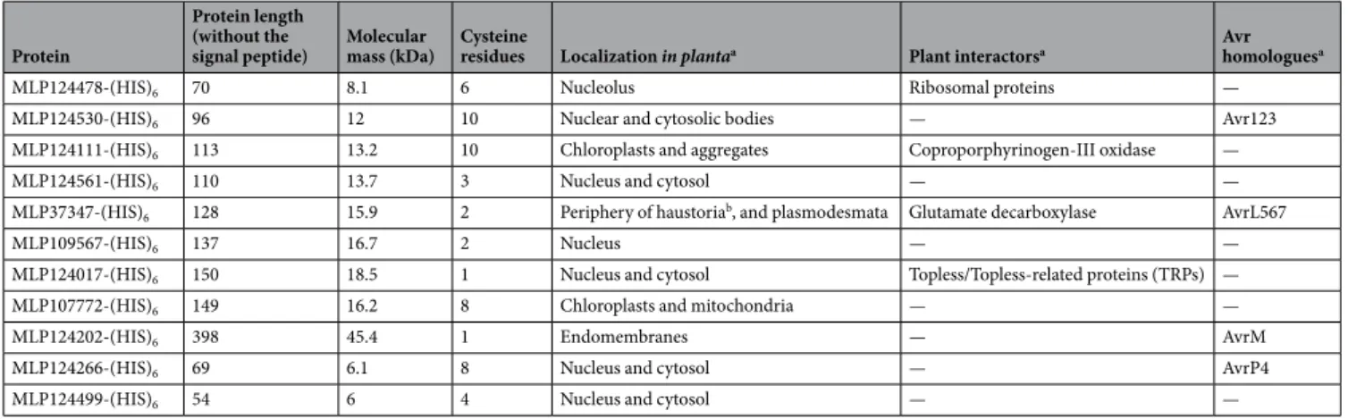
We selected 11 candidate effector pro-teins of M. larici-populina (Table 1), out of a catalogue of 24 candidate effectors previously screened in planta for their subcellular localization and plant protein partners22. Notably, we retained proteins showing (i) aspe-cific and informative subcellular localization, such as nucleus (MLP109567), nucleolus (MLP124478), nuclear bodies (MLP124530), chloroplasts and mitochondria (MLP107772, aka CTP1), chloroplasts and aggregates (MLP124111), endomembranes (MLP124202), and plamodesmata (MLP37347), (ii) specific plant pro-tein partners (MLP124017, MLP37347, MLP124448, MLP124111), (iii) similarities with M. lini Avr effectors (MLP124530, MLP37347, MLP124202, MLP124266), or iv) proteins belonging to large families of small-secreted proteins (MLP124499, MLP124561).
Successful production and purification of three candidate effectors in E. coli.
To inves-tigate the structural properties of the 11 selected candidate effectors (Fig. 1), we first aimed at obtaining the recombinant proteins. To this end, the sequence encoding mature proteins (i.e. without signal peptide) were cloned into pET-26b (for Mlp124111, Mlp124478, Mlp124530, Mlp124561, Mlp37347, Mlp109567, Mlp107772, Mlp124202, Mlp124266, and Mlp124499) or pET-28a (for Mlp124017) expression vectors, in order to incorpo-rate a C-terminal 6-histidine tag (Table S1). Small-scale expression assays achieved into E. coli Rosetta2 (DE3) pLysS strain indicated that nine out of the eleven proteins accumulated using a standard induction protocol (i.e. ProteinProtein length (without the
signal peptide) Molecular mass (kDa) Cysteine residues Localization in plantaa Plant interactorsa Avr homologuesa
MLP124478-(HIS)6 70 8.1 6 Nucleolus Ribosomal proteins —
MLP124530-(HIS)6 96 12 10 Nuclear and cytosolic bodies — Avr123
MLP124111-(HIS)6 113 13.2 10 Chloroplasts and aggregates Coproporphyrinogen-III oxidase —
MLP124561-(HIS)6 110 13.7 3 Nucleus and cytosol — —
MLP37347-(HIS)6 128 15.9 2 Periphery of haustoriab, and plasmodesmata Glutamate decarboxylase AvrL567
MLP109567-(HIS)6 137 16.7 2 Nucleus — —
MLP124017-(HIS)6 150 18.5 1 Nucleus and cytosol Topless/Topless-related proteins (TRPs) —
MLP107772-(HIS)6 149 16.2 8 Chloroplasts and mitochondria — —
MLP124202-(HIS)6 398 45.4 1 Endomembranes — AvrM
MLP124266-(HIS)6 69 6.1 8 Nucleus and cytosol — AvrP4
MLP124499-(HIS)6 54 6 4 Nucleus and cytosol — —
Table 1. Features of the eleven M. larici-populina candidate effector proteins investigated in this study. aAs
described in Petre et al.22 and/or Germain et al.24. bImmunolocalization performed on infected poplar leaves by
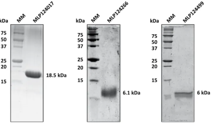
addition of 100 µM IPTG in mid-exponential growth phase and further growing for 3 to 4 hours at 37 °C). Among those, five (MLP124111, MLP124561, MLP37347, MLP107772 and MLP124202) accumulated in the insoluble protein fraction, and one (MLP109567) was not expressed (Fig. 2), despite the use of other E. coli expression strains (SoluBL21 (DE3), Origami2 (DE3) pLysS, Rosetta-Gami2 (DE3) pLysS) and modification of the culture conditions (induction time, temperature, and osmolarity). Among the five remaining soluble proteins we choose MLP124017, MLP124266, and MLP124499, the most stable along the purification procedure, for further analyses. We thus purified the His-tagged recombinant proteins in native conditions using a two-step protocol including immobilized-metal affinity chromatography (IMAC), then size exclusion chromatography (Fig. 3). The purified proteins, yielding respectively 50 mg/L (cell culture), 0.5 mg/L, and 0.5 mg/L for MLP124017, MLP124266 and MLP124499, respectively, eluted in size exclusion chromatography as a single peak corresponding to an estimated apparent molecular mass compatible with a monomeric organization data not shown.
MLP124266 is a thermostable protein that exhibits a cystine knot.
From a previous study, we reported that the Mlp124266 and Mlp124499 genes are strongly expressed and induced during poplar leaf col-onization by M. larici-populina, and belong to large multigene families of 13 and 31 members, respectively, in M. larici-populina16. Mlp124266 and Mlp124499 encode mature proteins of 69 and 50 amino acids, respectively(Fig. S1). MLP124266 has an N-terminal part enriched in charged residues and a C-terminal region that possesses six conserved cysteines predicted to form a cystine knot structure (Fig. S1A). This typical protein organization is shared by all members of the family as well as by alleles of M. lini AvrP433,34. In MLP124499, several acidic
resi-dues are found in the N-terminal part whereas the C-terminal part contains three conserved cysteines (Fig. S1B). Prediction programs indicate that all members of both protein families exhibit highly conserved N-terminal signal peptides for protein secretion. Following the production and the purification of both MLP124266 and MLP124499, we undertook a structural characterization of each recombinant protein using a NMR spectroscopy approach.
Standard homonuclear 2D experiments and 15N-edited TOCSY-HSQC and NOESY-HSQC experiments
car-ried out on MLP124499 allowed the assignment of 1H and 15N resonances except for the four N-terminal residues
(Fig. S2A). However, several minor peaks were observed, especially for Ala14-Glu16, Gly25-Gln26, Glu30, Asp49
residues, suggesting the presence of multiple forms or conformations. Changing the temperature and the ionic strength, or adding dithiothreitol failed to improve the quality of the MLP124499 NMR spectra. Consequently, very few experimental restraints could be derived and structure calculations led to very ill-defined models.
For MLP124266, the assignment of 1H, 15N and 13C resonances has been obtained for all residues except for
the five N-terminal amino acids (Fig. S2B) and its 3D structure could further be modelled by the NMR derived constraints (Table S2; Figs. 4 and S3). This analysis showed that the Cys36-Leu69 C-terminal region exhibits a
typ-ical cystine knot structure involving three disulfide bonds (Cys39-Cys55, Cys44-Cys58, and Cys50-Cys64), a β-sheet
composed of anti-parallel strands between Thr42-Cys44, Gly57-Ser59 and Val63-Val65, and a short α-helix formed
by the Gln49-Ala52 segment. In contrast, the Met1-Asp35 N-terminal region displays large structural disorder, as
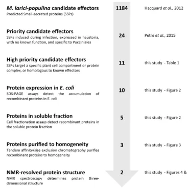
M. larici-populina candidate effectors
Predicted Small-secreted proteins (SSPs)
Priority candidate effectors
SSPs induced during infec on, expressed in haustoria, with no known func on, and specific to Pucciniales
High priority candidate effectors
SSPs target a specific plant cell compartment or protein complex, or homologous to known effectors
Protein expression in E. coli
SDS-PAGE assays detect the accumula on of recombinant proteins in E. coli
Proteins in soluble frac on
Cell frac ona on assays detect recombinant proteins in the soluble protein frac on
Proteins purified to homogeneity
Tandem affinity/size exclusion chromatography purifies recombinant proteins to homogeneity
NMR-resolved protein structure
NMR spectroscopy determines protein three-dimensional structure 1184 24 11 10 5 3 2 Hacquard et al., 2012 Petre et al., 2015
this study - Table 1
this study - Figure 2
this study - Figure 2
this study - Figure 3
this study - Figures 4 & 5
Figure 1. Overview of the effectoromics pipeline. A total of eleven M. larici-populina candidate effectors were
selected from the previous study of Petre et al.22 (i.e. particular localization and/or specific plant interactors and/
or homologies to M. lini Avr effectors). Effector candidates were expressed in E. coli SoluBL21 (DE3) pRARE2, Rosetta2 (DE3) pLysS or RosettaGami2 (DE3) pLysS strains. Soluble recombinant proteins were purified and their structure solved by NMR spectroscopy.
shown by the superposition of the 20 NMR models (Fig. S3). Very few NOE correlations were indeed observed for residues 1 to 35. A few sequential and medium-range NOE correlations characteristic of transient helical conformations can however be noticed (Fig. S3) and explain the presence of short secondary structures in some of these 20 models. Indeed, residues 8–17 and 28–30 exhibit helical structures in 30 to 50% and around 25% of
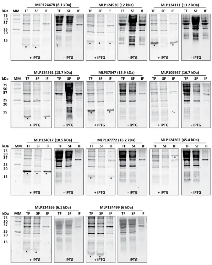
* * * * kDa 75 50 37 25 20 15 TF SF IF TF SF IF TF SF IF TF SF IF MM MLP124266 (6.1 kDa) MLP124499 (6 kDa)
+ IPTG - IPTG + IPTG - IPTG
* * kDa 75 50 37 25 20 15 TF SF IF TF SF IF TF SF IF
MLP124561 (13.7 kDa) MLP37347 (15.9 kDa) MLP109567 (16.7 kDa) TF SF IF
MM TF SF IF TF SF IF
+ IPTG - IPTG + IPTG - IPTG + IPTG - IPTG
* * * * * * * TF SF IF TF SF IF MLP124478 (8.1 kDa) kDa 75 50 37 25 20 15 TF SF IF TF SF IF TF SF IF MLP124530 (12 kDa) MLP124111 (13.2 kDa) + IPTG TF SF IF MM + IPTG
- IPTG - IPTG + IPTG - IPTG
* * * * * * * kDa 75 50 37 25 20 15 TF SF IF TF SF IF TF SF IF TF SF IF MM TF SF IF TF SF IF
MLP124017 (18.5 kDa) MLP107772 (16.2 kDa) MLP124202 (45.4 kDa)
+ IPTG - IPTG + IPTG - IPTG + IPTG - IPTG
*
Figure 2. Small-scale expression test of selected candidate effector proteins carried out in Escherichia coli
Rosetta2 (DE3) pLysS expression strain. Coomassie blue-stained sodium dodecylsulfate-polyacrylamide gel electrophoresis (SDS-PAGE) analysis of total (TF), soluble (SF) and (IF) insoluble protein fractions of E. coli Rosetta2 pLysS expression strain grown in presence (+) or in absence (−) of 0.1 mM isopropyl β-D-thiogalactopyranoside (IPTG). Asterisks indicate the expected migration of overexpressed proteins. MM: molecular mass marker.
the models, respectively. Backbone dynamic properties of MLP124266 have been investigated by 15N relaxation
measurements. Heteronuclear 1H-15N NOE values showed a contrasted profile with low values for N-terminal
res-idues (indicative of a flexible structure) and high values for C-terminal resres-idues (indicative of a rigid structure). Indeed, amino acids Asp6 to Gly38 and Cys39 to Leu69 presented heteronuclear NOE averaged values of 0.26 ± 0.05
and 0.66 ± 0.10, respectively. Several secondary structure prediction softwares predict a helix between residues 11 and 18 (data not shown) and a Consurf analysis shows that the proportion of conserved amino acids in the N-terminal region is much higher than in the C-terminal part (Fig. S4). Interestingly 6 out of the 8 cysteines are gathered in the C-terminal region between Cys39 and Cys69, following a spacing (Cys-X2–7-Cys-X3–10-Cys-
X0–7-Cys-X1–17-Cys-X4–19-Cys) typical of cystine knot structures, (i.e., three intricate disulfide bridges that confer
very high stability to proteins; Fig. 4)35. Hence, it is likely that rigidity originates from the structure formed by
these cysteines that are highly conserved in the protein family, as indicated by the Consurf analysis (Figs. S1 and S4). Thus, we sought to determine whether these disulfides are formed and whether they influence the stability and/or the oligomerization state of the protein by covalent bonds. A single peak corresponding to the theoret-ical mass of MLP124266 monomer was obtained by mass spectrometry (data not shown). The titration of free thiol groups in an untreated recombinant MLP124266 gave an averaged value of 1 mole SH per mole of protein. Considering the presence of 8 cysteines in the protein, these results are consistent with the existence of three intramolecular disulfide bridges (Fig. S3). The thermostability of recombinant protein was estimated by heating the protein for 10 min at 95 °C. The observation that the protein remained in solution (i.e. no precipitation was observed) indicates that it is thermosoluble. In order to investigate the role of the disulfides for such property, we should compare the results obtained with an oxidized and a reduced protein. However, as assessed by thiol titration experiments, we failed to obtain a complete reduction of these disulfides despite extensive incubation of the protein at high temperatures, in denaturing and reducing environments. Altogether, these results indicate that recombinant MLP124266 is properly folded by E. coli, and that the disulfide bridges, which are partially resistant to reduction, confer a high rigidity and stability to the protein.
MLP124017 is part of the nuclear transport factor 2-like protein superfamily.
MLP124017 is a small-secreted protein (167 amino acids with its signal peptide; 150 amino acids in its mature form, with a molecular mass of 18 kDa) of unknown function, highly expressed during infection of poplar leaves by M. larici-populina16. MLP124017 shares sequence similarity with neither other M. larici-populina nor other rustfungal proteins. In a previous study, we demonstrated the nucleocytoplasmic localization of MLP124107 in N. benthamiana and its interaction with poplar TOPLESS-related 4 protein22. To further investigate MLP124017
structure and to get insights into its function, we first attempted to solve its 3D structure by crystallization cou-pled to X-ray diffraction. We were unable to obtain exploitable diffracting crystals despite the use of different versions (untagged or N- or C-terminal His-tagged) of MLP124017 protein and therefore switched to NMR. The recombinant 15N and 13C-labelled MLP124017 protein was used for structure determination by two- and
three-dimensional NMR experiments (Table S3). The assigned 1H, 15N-HSQC spectra were well dispersed but
the peaks for residues from the N-terminal 1–14 and 86–95 segments were missing (Fig. S5). From preliminary structures, the production of a truncated recombinant protein for the first eight N-terminal residues that could mask residues 86–95 did not improve the data. The solution structure of MLP124017 was determined based on 1727 NOE-derived distance restraints, 214 dihedral angle restraints and 102 hydrogen bond restraints. All proline residues have been determined to be in a trans-conformation according to the 13Cβ chemical shift at 32.21; 32.46;
31.02 and 32.40 ppm for Pro36; Pro51; Pro54 and Pro146 respectively. The best conformers with the lowest energies,
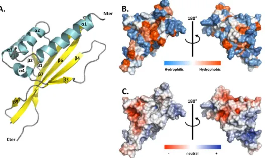
which exhibited no obvious NOE violations and no dihedral violations >5° were selected for final analysis. The Ramachandran plot produced shows that 99.6% of the residues are in favoured regions (Table S4). MLP124017 structure is composed of a α + β barrel with seven β-strands forming one mixed β-sheet, four β-hairpins, four β-bulges, and four α-helices (Fig. 5A). Residues 1–14 and 150–151 having missing assignments are not defined in the final models. This arrangement of secondary structure produces a cone-shaped fold for the protein, which generates a distinctive hydrophobic cavity (Fig. 5B, Fig. 5C).
75 50 37 25 20 15 kDa 6 kDa 75 50 37 25 20 15 18.5 kDa kDa 75 50 37 25 20 15 6.1 kDa kDa
Figure 3. Candidate effectors purified as soluble proteins. Ten micrograms of recombinant
MLP124017-(His)6; MLP124266-(His)6, MLP124499-(His)6 have been separated by SDS-PAGE (17%). Molecular mass
To identify potential structural homologs of MLP124017, we performed structural similarity searches using the Dali server36. Queries identified SBAL_0622 (PDB code 3BLZ) and SPO1084 (PDB code 3FKA) as the closest
structural homologs with the highest Z-score of 6.0 and 6.3, respectively, and a RMSD of 4.1 and 3.5 Å, respec-tively (Fig. S6). These two proteins, which are from the bacteria Shewanella baltica and Ruegeria pomeroyi, have no known function, but share a common Nuclear Transport Factor 2-like (NTF2) fold. The NTF2 superfamily comprises a large group of proteins that share a common fold and that are widespread in both prokaryotes and eukaryotes37. Taken together these results show that although MLP124017 do not share sequence similarities
or domain with other proteins in sequence databases, its structure is similar to proteins of the NTF2 folding superfamily.
Discussion
In this study, we have set up a small-throughput effectoromics pipeline based on recombinant protein produc-tion and structural characterizaproduc-tion to get insights on 11 candidate effectors of the poplar leaf rust fungus M. larici-populina.
Although the production of recombinant proteins in E. coli is a valuable approach to perform biophysical and biochemical analyses of candidate effectors5,25, we have faced issues for the production of soluble small-cysteine
rich proteins in this system. Indeed, among the eleven candidate effectors screened for expression, only three were found in the soluble protein fraction and stable enough to tolerate the purification procedure. Although we tested different E. coli strains and protein expression induction protocols, the other candidate effectors were either not expressed or expressed as inclusion bodies. It is possible to purify recombinant proteins from inclusion bodies by using denaturing extraction conditions and further refolding proteins38. However, this approach is not
recom-mended for structural analysis as the refolding of the proteins in vitro may alter folding. Another limit of prokar-yote systems to produce eukarprokar-yote proteins is the lack of post-translational modifications such as methylations.
A.
B.
C.
Nter Cter Cys39(I) Cys55(IV) Cys44(II) Cys58(V) Cys50(III) Cys64(VI) β1 β2 β3 α1 Nter Cys39(I) (IV) Cys55 (III) Cys50 Cys64(VI) β3 β1 β2 Cys58 (V) Cys44 (II) α1 Cter α1 β1 β2 β3 Nter Cter 310 310Figure 4. NMR spectroscopy solution structure of MLP124266. (A) The structure of MLP124266 is represented
as cartoon and consists of one α-helix in cyan and a mixed β-sheet composed by three β-strands coloured in yellow. (B) The C-terminal small disulfide-rich domain is related to the structural family of knottins, which contain at least 3 disulfide bridges and 6 cysteines and implies a disulfide between cysteines III and VI going through disulfides I-IV and II-V. In MLP124266, a disulfide bridge between Cys50 (III) and Cys64 (VI), in blue,
goes through disulfide bridges between Cys39 (I) and Cys55 (IV) as well as Cys44 (II) and Cys58 (V), in green.
(C) Schematic representation of disulfide bridge connectivity. Disulfide bridge Cys50 (III)-Cys64 (VI) is
represented in blue and disulfide bridges between Cys39 (I)-Cys55 (IV) and Cys44 (II)-Cys58 (V) are represented
An alternative to this is to use the yeast Pichia pastoris, which has proven useful for several fungal effectors such as Leptospheria maculans AvrLm4–7 or Cladosporium fulvum Avr2 and Avr439,40. We have tried this system to
produce the candidate effector (MLP107772), but without success (data not shown). Nevertheless, this system may be useful and deserves to be considered as an alternative to assay other rust effectors for which we were not able to obtain production in E. coli.
Out of the three effectors successfully purified as recombinant proteins, two were structurally characterized by NMR spectroscopy. MLP124266 is a homolog of the M. lini AvrP4 effector protein41, and we showed that it
exhibits a cystine knot (or knottin) structural motif commonly encountered in small disulfide-rich proteins. MLP124017 is an orphan protein in M. larici-populina with no known ortholog in Pucciniales. MLP124017 phys-ically associates with the poplar TOPLESS-related protein 4 (TRP4)22, and we showed that it exhibits a fold similar
to two bacterial proteins that belong to the Nuclear-Transport factor 2-like protein superfamily.
We showed that MLP124266 possesses two distinct regions with contrasted structural properties. The C-terminal part is rigidified by a cystine knot motif whereas the N-terminal part is globally flexible. The knottin folded proteins display a variety of functions such as venoms and spider toxins42,43 but also antimicrobial
prop-erties such as the cyclotides44. Some are also found to interact with protease inhibitors found in plants, insects
and plant parasites45. The three disulfide bridges within the C-terminal part of MLP124266 confer its rigidity and
probably contribute to the high protein stability35. MLP124266 presents a β-sheet structure typical of knottins,
but interestingly it also has an additional helix between β2 and β3 strands. To our knowledge, the presence of such a helix in knottins has been reported in cyclotides only, and more precisely in bracelet cyclotides contain-ing six or seven residues in the loop between Cys(III) and Cys(IV)46. This loop often contains an alanine, which
favours the formation of the helix as well as a highly conserved glycine allowing its connection to the cystine knot47. Interestingly, the loop in MLP124266 has such residues, i.e. Ala
52 and Gly54 Ala and Gly, but consists of
four residues only. In Viola odorata cycloviolacin O2 (cO2), the additional helix is located in a hydrophobic loop that interacts with the membrane-mimicking micelles48. Therefore, it might help disrupting membranes and thus
contribute to the cytotoxicity activity of cO2 and play a role in plant defence. In MLP124266, the helical turn is not particularly hydrophobic (Fig. S4B) and may not have these properties. To our knowledge, MLP124266 is the first fungal protein to present a knottin-like structure49. It would be interesting to collect structural data from
other potential fungal knottins to find out whether the additional helix is always present and to clarify its role. The intrinsic disorder of the N-terminus of MLP124266 also deserve to be pointed out. This region, approximately extending up to residue 35 and thus representing half of the primary sequence, globally presents a high flexibility, as demonstrated by the NMR dynamic results. Nevertheless, a few residues exhibit a propensity to form helical struc-tures, which may support a biochemical role that remains to be elucidated. Interestingly, other effectors possess a predicted disordered N-terminal region29. For instance, M. lini effectors AvrL567 and AvrM have predicted disordered
N-terminal regions that are susceptible to protease degradation11,50. Flexible folds are known to be adaptable linkers
that favour the ability to bind partners51. As the N-terminus of many cytoplasmic effectors is anticipated to mediate
protein entry into host cells2, it is tempting to speculate that this flexible part may bind a target important for cell entry.
Nter Cter β1 α1 α2 α3 α4 β2 β3 β4 β5 β6 β7
A.
180° 180°B.
C.
Hydrophilic Hydrophobic + - neutralFigure 5. NMR spectroscopy solution structure of MLP124017. (A) The structure of MLP124017 is represented
as cartoon and consists of four α-helices in cyan (α1: residues 16–32; α2: 41–48; α3: 65–73; α4: 80–82) and a mixed β-sheet composed by seven β-strands coloured in yellow (β1: residues 55–58; β2: 61–63; β3: 84–86; β4: 98–104; β5: 108–109; β6: 115–129; β7: 132–144). (B) Front and rear views on the surface of MLP124017 illustrating the surface hydrophobic potential. The hydrophobic and hydrophilic patches are shown in red and in blue respectively. (C) Front and rear views on the surface of MLP124017 illustrating the surface electrostatic Coulomb potential at pH 7.0 using APBS plugin from Pymol 2.3 software with a contour of −10 kT/e to 10 kT/e. The positive-charge and negative-charge densities are coloured in blue and red respectively.
The structure of MLP124017 solved by NMR spectroscopy showed a fold similar to members of the NTF2-like superfamily. The NTF2-like superfamily is a group of protein domains sharing a common fold, but showing no sequence similarity. MLP124017 is structurally similar to two bacterial proteins, despite the lack of sequence similarity. The structures of these two bacterial proteins consist of a β-sheet surrounding a binding pocket and α-helices acting as a lid52. The NTF2 family regroups catalytic and non-catalytic proteins that contain cone-like
structured proteins with a cavity that often acts as a molecular container involved in a wide range of cellular functions37. Interestingly, the cone-shaped structure of MLP124017 is widespread across both prokaryotes and
eukaryotes. The first proteins of the NTF2 family were reported to play a role in the transport of molecules from the cytoplasm to the nucleus. Arabidopsis NTF2 protein is required to import nuclear proteins via the recognition of a nuclear localization signal (NLS). This protein also plays a role in the nuclear import of the small-GTPase Ran-GDP that is a central protein in various signal transduction pathways (e.g. mitotic spindle formation, nuclear envelope assembly, or responses to biotic stresses)53–56. In bacteria, some NTF2-like proteins play a role in
bacte-rial conjugation as part of the type IV secretion system57, whereas non-catalytic NTF2-like domains act as
immu-nity proteins58. In fungi, Saccharomyces cerevisiae NTF2 mutant is defective for nuclear import59. Although NTF2
folded proteins are widespread across kingdoms, very few is known about their role. A recent study presented that the silencing of NTF2 in wheat decreased the resistance against avirulent isolates of the wheat stripe rust fungus P. striiformis f. sp. tritici60. Since MLP124017 has been shown to interact with TOPLESS and TOPLESS-related
proteins22, it is tempting to speculate that the cavity formed by the β-sheet could be involved in the association
with these plant partners.
Although MLP124266 and MLP124017 show no primary sequence similarity to known proteins, they adopt a three dimensional fold similar to knottins and NTF2 family members, respectively. Thus, knowing the struc-ture of both candidate effectors allowed us to classify them as members of large superfamilies of proteins. The concept of structural families whose members share no, or very limited, primary sequence homology emerges in effector biology61. This concept promises to revolution the way we predict and categorize effector proteins6,7. For
instance, members of the MAX effector family share a common β-sandwich fold, but show no primary sequence similarity6. Similarly, members of the WY superfamily of RXLR effectors in oomycetes share a common three- to
four-helix bundle29. Such features are now used to search and categorize fungal and oomycete effector proteins
into structural superfamilies1,6. To our knowledge, MLP124266 and MLP124017 are the first effector proteins to
adopt knottins and NTF2 folds. Whether other effector proteins adopt similar folds remains to be identified to determine if knottins and NTF2 folds define structural superfamilies in fungi.
Experimental Procedures
Sequence analyses and names.
Alignment and phylogenetic analyses were performed on the phylogeny website (www.phylogeny.fr) with default parameters. Alignments were corrected and edited manually, and phy-logenetic trees were generated with FigTree v1.4.3 (http://tree.bio.ed.ac.uk/software/figtree/). Physical and chem-ical parameters of proteins were estimated using protparam tool (http://web.expasy.org/protparam/). Common names and JGI ID of genes described in this study are as follow: Mlp124266, Mlp124499, Mlp124111, Mlp124478, Mlp124530, Mlp124561, Mlp37347, Mlp109567, Mlp124017, Mlp107772, and Mlp124202. The mapping of the family-wide conservation pattern of amino acids onto MLP124266 structure was performed with Consurf (http:// consurf.tau.ac.il/2016/).Cloning of selected effector encoding sequences.
Open reading frames coding for the mature forms (i.e. devoid of the sequence encoding N-terminal secretion peptide) of MLP124266 and MLP124499 of M. larici-populina isolate 98AG31 were ordered as synthetic genes cloned in pBSK(+) vectors (Genecust). Coding sequence of the mature forms of the nine other candidate effectors (Mlp124111, Mlp124478, Mlp124530, Mlp124561, Mlp37347, Mlp109567, Mlp124017, Mlp107772, and Mlp124202) were amplified by polymerase chain reaction (PCR) using cDNAs from leaves of the poplar hybrid Beaupré infected by M. larici-populina (isolate 98AG31) and further cloned into pICSL01005 vector as described previously22. The sequences encoding themature form of each effector were subsequently cloned by PCR in either pET26b or pET28a vector between NdeI and XhoI (or NotI) or NcoI and XhoI restriction sites, respectively, using primers shown in Table S1.
Expression and purification of recombinant proteins in Escherichia coli.
Expression of recombinants proteins was performed at 37 °C using the E. coli SoluBL21 (DE3) pRARE2 (Amsbio Abington, UK), Rosetta2 (DE3) pLysS, Origami2 (DE3) pLysS or RosettaGami2 (DE3) pLysS strains (Novagen) containing the adequate pET expres-sion vector coding for the selected candidate effector (Table S1) in LB medium supplemented with kanamycin (50 μg/ ml) and chloramphenicol (34 μg/ml). When the cell culture reached an OD600nm of 0.7, protein expression was inducedwith 0.1 mM isopropyl-β-D-1-thiogalactopyranoside (IPTG) and cells were grown for a further 4 h. To improve the solubility of some recombinant candidate effectors, other protocols were used as follows. First, we added 0.5% (v/v) of ethanol in the medium when culture reached an OD600nm of 0.7. The cells were cooled to 4 °C for 3 h, recombinant
protein expression induced with 0.1 mM IPTG and cells further grown for 18 h at 20 °C. We also tested a combination of an osmotic and a thermal shock62. When the culture reached an OD
600nm of 0.5–0.6, 500 mM NaCl and 2 mM of betaine
were added to the culture medium and the culture incubated at 47 °C for 1 hour under stirring. Cells were then cooled to 20 °C and the expression of recombinant proteins induced with 0.1 mM IPTG. After induction, cells were harvested by centrifugation, suspended in a 30 mM Tris-HCl pH 8.0 and 200 mM NaCl lysis buffer, and stored at −20 °C. Cell lysis was completed by sonication (three times for 1 min with intervals of 1 min). The cell extract was then centrifuged at 35 000 g for 25 min at 4 °C to remove cellular debris and aggregated proteins. After the addition of 10 mM imidazole, soluble fraction containing C-terminal His-tagged recombinant proteins were then purified by gravity-flow chroma-tography on a nickel nitrilotriacetate (Ni-NTA) agarose resin (Qiagen). After a washing step with lysis buffer supple-mented with 20 mM imidazole, the proteins were eluted using lysis buffer containing 250 mM imidazole. The fractions
of interest were pooled, concentrated by ultrafiltration then injected onto a gel filtration HiLoad 16/600 Superdex 75 prep grade (GE Healthcare) column connected to an ÄKTA PurifierTM (GE Healthcare) and eluted with lysis buffer.
The fractions containing recombinant MLP124017 were pooled, concentrated, and stored at −20 °C as such, whereas for MLP124266 and MLP124499 fractions were pooled, dialyzed against 30 mM Tris-HCl, 1 mM EDTA (TE) pH 8.0 buffer, and stored at 4 °C until further use.
For the NMR spectroscopy analyses, MLP124266-(His6) and MLP124499-(His6) recombinant proteins were
15N-labelled in M9 minimal synthetic medium containing 15NH
4Cl (1 g/L). MLP124017 was single 15N or double 15N
and 13C labelled in M9 minimal medium containing 1 g/l NH
4Cl (15N) and 2 g/l glucose (13C) supplemented with 2,5%
thiamine (m/v), 1 mg/ml biotin, 50 mM FeCl3, 10 mM MnCl2, 10 mM ZnSO4, 2 mM CoCl2, 2 mM NiCl2, 2 mM NaSeO3,
and 2 mM H3BO3. After purification as described above, labelled MLP124266, MLP124499, and MLP124017 were
dia-lyzed against a 50 mM phosphate pH 6.0 or a 20 mM phosphate pH 6.8 buffer supplemented with 200 mM NaCl. The homogeneity of purified proteins was checked by SDS-PAGE and protein concentration determined by measuring the absorbance at 280 nm and using theoretical molar absorption coefficients of 500 M−1.cm−1, 3 105 M−1.cm−1, 29
450 M−1.cm−1 deduced from the amino acid sequences of mature MLP124266, MLP124499, and MLP124017 proteins
respectively. For MLP124266, protein concentration was also verified using a colorimetric assay (BC assay, Interchim).
Protein sample preparation for NMR spectroscopy.
Uniformly labelled 15N MLP124017 (1 mM in20 mM phosphate pH 6.8, 200 mM NaCl) was supplemented with 5 mM 4,4-dimethyl-4-silapentane-1-sulfonic acid (DSS) in D2O as a lock/reference. For the D2O experiments, the sample was lyophilized and dissolved in
200 µL D2O. For the 3D heteronuclear experiments, a 13C/15N labelled sample was diluted at a final concentration
of 0.6 mM in 200 μL of the previous phosphate buffer supplemented with 10% D2O and 0.5 mM DSS as a
refer-ence. MLP124266 and MLP124499 samples were dissolved in 50 mM phosphate pH 6.0 buffer with 10% D2O and
0.02% sodium azide. The concentration of unlabelled MLP124266 and MLP124499 was 0.85 and 0.65 mM, and the one for uniformly 15N labelled MLP124266 and MLP124499 was 1.8 and 0.135 mM, respectively.
Nuclear magnetic resonance spectroscopy.
For MLP124017, spectra were acquired on 800 and 700 MHz Avance Bruker spectrometers equipped with triple-resonance (1H, 15N, 13C) z-gradient cryo-probe at298 K. Experiments were recorded using the TOPSPIN pulse sequence library (v. 2.1) (Table S2). All spectra are referenced to the internal reference DSS for the 1H dimension and indirectly referenced for the 15N and 13C
dimensions63. Sequential assignment was performed using 3D 15N-NOESY-HSQC, 15N-TOCSY-HSQC, HNCO,
HNCACO, HNCA, HNCOCACB, and HNCACB. Side chain 1H assignments were carried out using combined
analysis with 3D 15N-NOESY-HSQC, 15N-TOCSY-HSQC, and 2D NOESY and TOCSY with D
2O samples. A
series of three HSQC spectra was performed after lyophilisation and dilution of the first sample in D2O to
deter-mine amide protons in slow exchange (Table S2).
For MLP124266 and MLP1124499, NMR spectra were acquired on a Bruker DRX 600 MHz spectrometer equipped with a TCI cryoprobe. For MLP124266 and MLP124499, COSY, TOCSY (mixing time of 60 ms) and NOESY (mixing time of 150 ms) experiments were run at 298 K, respectively. For MLP124266, HNHA, HNHB, R1 and R2 15N relaxation rates, 1H-15N heteronuclear NOE, HNCA (with 24 (15N) × 28 (13C) complex points and
192 transients per increment) standard experiments were recorded. Spectra were processed using Topspin
®
3.0 software (Bruker) and analysed with NmrViewJ64, CcpNmr65 and ARIA266.Structure calculation.
For MLP124017 structure calculation, NOE peaks identified in 3D 15N-NOESY-HSQCand 2D NOESY experiments were automatically assigned during structure calculations performed by the pro-gram CYANA 2.167. The 15N, H
N, 13C’, 13Cα, Hα, and 13Cβ chemical shifts were converted into ϕ/Ψ dihedral angle
constraints using TALOS + (v. 1.2)68. Hydrogen bond constraints were determined according to 1H/2H exchange
experiments of backbone amide protons (HN). Each hydrogen bond was forced using following constraints:
1.8–2.0 Å for HN,O distance and 2.7–3.0 Å for NH,O distance. Final structure calculations were performed with
CYANA (v. 2.1) using all distance and angle restraints (Table S3). 600 structures were calculated with CYANA 2.1, of which the 20 conformers with the lowest target function were refined by CNS (v. 1.2) using the refinement in water of RECOORD69 and validated using PROCHECK70.
For MLP124266 structure calculation, 783 NOE peaks identified in 3D 15N-NOESY-HSQC or 2D NOESY
spectra, 75 φ/Ψ dihedral angles generated by DANGLE71 and 10 hydrogen bonds were input as unambiguous
restraints in ARIA2. Covalent disulfide bonds between Cys39-Cys55, Cys44-Cys58 and Cys50-Cys64 were also
intro-duced. Among the 400 structures generated by ARIA2, 20 models of lowest energy were refined in water (Table S2). NMR assignment and structure coordinates have been deposited in the Biological Magnetic Resonance Data Bank (BMRB code 34423 and 34298) and in the RCSB Protein Data Bank (PDB code 6SGO and 6H0I), respectively.
Data availability
The data that support the findings of this study are openly available in the Biological Magnetic Resonance Data Bank (http://www.bmrb.wisc.edu/) and in the RCSB Protein Data Bank (http://www.rcsb.org/).
Received: 4 September 2019; Accepted: 31 October 2019; Published: xx xx xxxx
References
1. Win, J., Chaparro-Garcia, A., Belhaj, K. et al. Effector Biology of Plant-Associated Organisms: Concepts and Perspectives. Cold
Spring Harb Symp Quant Biol. 77, 235–247 (2012).
3. Jones, J. D. G. & Dangl, J. L. The plant immune system. Nature 444, 323–329 (2006).
4. Persoons, A. et al. The escalatory Red Queen: Population extinction and replacement following arms race dynamics in poplar rust.
Mol. Ecol. 26, 1902–1918 (2017).
5. Franceschetti, M. et al. Effectors of Filamentous Plant Pathogens: Commonalities amid Diversity. Microbiol. Mol. Biol. Rev. MMBR 81 (2017).
6. de Guillen, K. et al. Structure Analysis Uncovers a Highly Diverse but Structurally Conserved Effector Family in Phytopathogenic Fungi. PLoS Pathog. 11, e1005228 (2015).
7. Joe, W. et al. Sequence Divergent RXLR Effectors Share a Structural Fold Conserved across Plant Pathogenic Oomycete Species.
PLoS Pathogens 8(1), e1002400 (2012).
8. Illergård, K., Ardell, D. H. & Elofsson, A. Structure is three to ten times more conserved than sequence–a study of structural response in protein cores. Proteins 77, 499–508 (2009).
9. Wirthmueller, L., Maqbool, A. & Banfield, M. J. On the front line: structural insights into plant-pathogen interactions. Nat. Rev.
Microbiol. 11, 761–776 (2013).
10. Ve, T. et al. Structures of the flax-rust effector AvrM reveal insights into the molecular basis of plant-cell entry and effector-triggered immunity. Proc. Natl. Acad. Sci. USA 110, 17594–17599 (2013).
11. Wang, C.-I. A. et al. Crystal structures of flax rust avirulence proteins AvrL567-A and -D reveal details of the structural basis for flax disease resistance specificity. Plant Cell 19, 2898–2912 (2007).
12. Dean, R. et al. The Top 10 fungal pathogens in molecular plant pathology. Mol. Plant Pathol. 13, 414–430 (2012).
13. Lorrain, C., Gonçalves dos Santos, K. C., Germain, H., Hecker, A. & Duplessis, S. Advances in understanding obligate biotrophy in rust fungi. New Phytol. 222, 1190–1206 (2019).
14. Duplessis, S., Bakkeren, G. & Hamelin, R. Advancing Knowledge on Biology of Rust Fungi Through Genomics. in Advances in Botanical Research vol. 70 173–209 (Elsevier, 2014).
15. Saunders, D. G. O. et al. Using hierarchical clustering of secreted protein families to classify and rank candidate effectors of rust fungi. PloS One 7, e29847 (2012).
16. Hacquard, S. et al. A comprehensive analysis of genes encoding small secreted proteins identifies candidate effectors in Melampsora larici-populina (poplar leaf rust). Mol. Plant-Microbe Interact. MPMI 25, 279–293 (2012).
17. Nemri, A. et al. The genome sequence and effector complement of the flax rust pathogen Melampsora lini. Front. Plant Sci. 5, 98 (2014).
18. Cantu, D. et al. Genome analyses of the wheat yellow (stripe) rust pathogen Puccinia striiformis f. sp. tritici reveal polymorphic and haustorial expressed secreted proteins as candidate effectors. BMC Genomics 14, 270 (2013).
19. Petre, B., Joly, D.L. & Duplessis, S. Effector proteins of rust fungi. Frontiers in Plant Science 5,416, https://doi.org/10.3389/ fpls.2014.00416 (2014).
20. Lorrain, C., Petre, B. & Duplessis, S. Show me the way: rust effector targets in heterologous plant systems. Curr. Opin. Microbiol. 46, 19–25 (2018).
21. Petre, B. et al. Heterologous Expression Screens in Nicotiana benthamiana Identify a Candidate Effector of the Wheat Yellow Rust Pathogen that Associates with Processing Bodies. PLOS ONE 11, e0149035 (2016).
22. Petre, B. et al. Candidate Effector Proteins of the Rust Pathogen Melampsora larici-populina Target Diverse Plant Cell Compartments. Mol. Plant. Microbe Interact. 28, 689–700 (2015).
23. Qi, M. et al. Suppression or Activation of Immune Responses by Predicted Secreted Proteins of the Soybean Rust Pathogen Phakopsora pachyrhizi. Mol. Plant-Microbe Interact. MPMI 31, 163–174 (2018).
24. Germain, H. et al. Infection assays in Arabidopsis reveal candidate effectors from the poplar rust fungus that promote susceptibility to bacteria and oomycete pathogens. Mol. Plant Pathol. 19, 191–200 (2018).
25. Zhang, X. et al. Production of small cysteine-rich effector proteins in Escherichia coli for structural and functional studies. Mol.
Plant Pathol. 18, 141–151 (2017).
26. Yaeno, T. et al. Phosphatidylinositol monophosphate-binding interface in the oomycete RXLR effector AVR3a is required for its stability in host cells to modulate plant immunity. Proc. Natl. Acad. Sci. USA 108, 14682–14687 (2011).
27. Chou, S. et al. Hyaloperonospora arabidopsidis ATR1 effector is a repeat protein with distributed recognition surfaces. Proc. Natl.
Acad. Sci. USA 108, 13323–13328 (2011).
28. Leonelli, L. et al. Structural elucidation and functional characterization of the Hyaloperonospora arabidopsidis effector protein ATR13. PLoS Pathog. 7, e1002428 (2011).
29. Boutemy, L. S. et al. Structures of Phytophthora RXLR effector proteins: a conserved but adaptable fold underpins functional diversity. J. Biol. Chem. 286, 35834–35842 (2011).
30. Zhang, X. et al. Crystal structure of the Melampsora lini effector AvrP reveals insights into a possible nuclear function and recognition by the flax disease resistance protein P. Mol. Plant Pathol. 19, 1196–1209 (2018).
31. Pinon, J. & Frey, P. Interactions between poplar clones and Melampsora populations and their implications for breeding for durable resistance. In Rust diseases of willow and poplar (eds Pei, M. H. & McCracken, A. R.) 139–154 (CABI), https://doi. org/10.1079/9780851999999.0139 (2005).
32. Gaouar, O., Morency, M.-J., Letanneur, C., Séguin, A. & Germain, H. The 124202 candidate effector of Melampsora larici-populina interacts with membranes in Nicotiana and Arabidopsis. Can. J. Plant Pathol. 38, 197–208 (2016).
33. Barrett, L. G. et al. Diversity and Evolution of Effector Loci in Natural Populations of the Plant Pathogen Melampsora lini. Mol. Biol.
Evol. 26, 2499–2513 (2009).
34. Persoons, A. et al. Patterns of genomic variation in the poplar rust fungus Melampsora larici-populina identify pathogenesis-related factors. Front. Plant Sci. 5 (2014).
35. Daly, N. L., Gruber, C. W., Göransson, U. & Craik, D. J. Cystine Knot Folding in Cyclotides. In Folding of Disulfide Proteins 43–61 (Chang, R. J. Y & Ventura, S., 2011).
36. Holm, L. & Rosenström, P. Dali server: conservation mapping in 3D. Nucleic Acids Res. 38, W545–549 (2010). 37. Eberhardt, R. Y. et al. Filling out the structural map of the NTF2-like superfamily. BMC Bioinformatics 14, 327 (2013).
38. Palmer, I. & Wingfield, P. T. Preparation and Extraction of Insoluble (Inclusion-Body) Proteins from Escherichia coli. In Current
Protocols in Protein Science (eds Coligan, J. E., Dunn, B. M., Speicher, D. W. & Wingfield, P. T.) 6.3.1–6.3.20 (John Wiley & Sons,
Inc.), https://doi.org/10.1002/0471140864.ps0603s70 (2012).
39. Blondeau, K. et al. Crystal structure of the effector AvrLm4-7 of Leptosphaeria maculans reveals insights into its translocation into plant cells and recognition by resistance proteins. Plant. J. Cell Mol. Biol. 83, 610–624 (2015).
40. Rooney, H. C. E. et al. Cladosporium Avr2 inhibits tomato Rcr3 protease required for Cf-2-dependent disease resistance. Science 308, 1783–1786 (2005).
41. Catanzariti, A.-M., Dodds, P. N., Lawrence, G. J., Ayliffe, M. A. & Ellis, J. G. Haustorially expressed secreted proteins from flax rust are highly enriched for avirulence elicitors. Plant Cell 18, 243–256 (2006).
42. Lee, S.-Y. & MacKinnon, R. A membrane-access mechanism of ion channel inhibition by voltage sensor toxins from spider venom.
Nature 430, 232–235 (2004).
43. Garcia, M. L. Ion channels: gate expectations. Nature 430, 153–155 (2004).
44. Tam, J. P., Lu, Y. A., Yang, J. L. & Chiu, K. W. An unusual structural motif of antimicrobial peptides containing end-to-end macrocycle and cystine knot disulfides. Proc. Natl. Acad. Sci. USA 96, 8913–8918 (1999).
45. Kim, J.-Y. et al. Protease inhibitors from plants with antimicrobial activity. Int. J. Mol. Sci. 10, 2860–2872 (2009).
46. Chen, B. et al. Isolation and characterization of novel cyclotides from Viola hederaceae: solution structure and anti-HIV activity of vhl-1, a leaf-specific expressed cyclotide. J. Biol. Chem. 280, 22395–22405 (2005).
47. Rosengren, K. J., Daly, N. L., Plan, M. R., Waine, C. & Craik, D. J. Twists, knots, and rings in proteins. Structural definition of the cyclotide framework. J. Biol. Chem. 278, 8606–8616 (2003).
48. Wang, C. K., Colgrave, M. L., Ireland, D. C., Kaas, Q. & Craik, D. J. Despite a conserved cystine knot motif, different cyclotides have different membrane binding modes. Biophys. J. 97, 1471–1481 (2009).
49. Postic, G., Gracy, J., Périn, C., Chiche, L. & Gelly, J.-C. KNOTTIN: the database of inhibitor cystine knot scaffold after 10 years, toward a systematic structure modeling. Nucleic Acids Res. 46, D454–D458 (2018).
50. Catanzariti, A.-M. et al. The AvrM effector from flax rust has a structured C-terminal domain and interacts directly with the M resistance protein. Mol. Plant-Microbe Interact. MPMI 23, 49–57 (2010).
51. Chouard, T. Structural biology: Breaking the protein rules. Nature 471, 151–153 (2011).
52. Marcos, E. et al. Principles for designing proteins with cavities formed by curved β sheets. Science 355, 201–206 (2017).
53. Avis, J. M. & Clarke, P. R. Ran, a GTPase involved in nuclear processes: its regulators and effectors. J. Cell Sci. 109(Pt 10), 2423–2427 (1996).
54. Carazo-Salas, R. E., Gruss, O. J., Mattaj, I. W. & Karsenti, E. Ran-GTP coordinates regulation of microtubule nucleation and dynamics during mitotic-spindle assembly. Nat. Cell Biol. 3, 228–234 (2001).
55. Zhang, C. & Clarke, P. R. Chromatin-independent nuclear envelope assembly induced by Ran GTPase in Xenopus egg extracts.
Science 288, 1429–1432 (2000).
56. Hetzer, M., Bilbao-Cortés, D., Walther, T. C., Gruss, O. J. & Mattaj, I. W. GTP hydrolysis by Ran is required for nuclear envelope assembly. Mol. Cell 5, 1013–1024 (2000).
57. Goessweiner-Mohr, N., Arends, K., Keller, W. & Grohmann, E. Conjugative type IV secretion systems in Gram-positive bacteria.
Plasmid 70, 289–302 (2013).
58. Zhang, D., De Souza, R.F., Anantharaman, V., Iyer, L.M. & Aravind, L. Polymorphic toxin systems: Comprehensive characterization of trafficking modes, processing, mechanisms of action, immunity and ecology using comparative genomics. Biol. Direct 14, 18,
https://doi.org/10.1186/1745-6150-7-18 (2012).
59. Corbett, A. H. & Silver, P. A. The NTF2 Gene Encodes an Essential, Highly Conserved Protein That Functions in Nuclear Transport in Vivo. J. Biol. Chem. 271, 18477–18484 (1996).
60. Zhang, Q. et al. TaNTF2, a contributor for wheat resistance to the stripe rust pathogen. Plant Physiol. Biochem. PPB 123, 260–267 (2018).
61. Białas, A. et al. Lessons in Effector and NLR Biology of Plant-Microbe Systems. Mol. Plant-Microbe Interact. MPMI 31, 34–45 (2018). 62. Oganesyan, N., Ankoudinova, I., Kim, S.-H. & Kim, R. Effect of osmotic stress and heat shock in recombinant protein overexpression
and crystallization. Protein Expr. Purif. 52, 280–285 (2007).
63. Wishart, D. S., Bigam, C. G., Holm, A., Hodges, R. S. & Sykes, B. D. 1H, 13C and 15N random coil NMR chemical shifts of the common amino acids. I. Investigations of nearest-neighbor effects. J. Biomol. NMR 5, (1995).
64. Johnson, B. A. & Blevins, R. A. NMR View: A computer program for the visualization and analysis of NMR data. J. Biomol. NMR 4, 603–614 (1994).
65. Vranken, W. F. et al. The CCPN data model for NMR spectroscopy: development of a software pipeline. Proteins 59, 687–696 (2005). 66. Rieping, W. et al. ARIA2: automated NOE assignment and data integration in NMR structure calculation. Bioinforma. Oxf. Engl. 23,
381–382 (2007).
67. Güntert, P. Automated NMR structure calculation with CYANA. Methods Mol. Biol. Clifton NJ 278, 353–378 (2004).
68. Shen, Y., Delaglio, F., Cornilescu, G. & Bax, A. TALOS+: a hybrid method for predicting protein backbone torsion angles from NMR chemical shifts. J. Biomol. NMR 44, 213–223 (2009).
69. Nederveen, A. J. et al. RECOORD: a recalculated coordinate database of 500+ proteins from the PDB using restraints from the BioMagResBank. Proteins 59, 662–672 (2005).
70. Laskowski, R. A., MacArthur, M. W., Moss, D. S. & Thornton, J. M. PROCHECK: a program to check the stereochemical quality of protein structures. J. Appl. Crystallogr. 26, 283–291 (1993).
71. Cheung, M.-S., Maguire, M. L., Stevens, T. J. & Broadhurst, R. W. DANGLE: A Bayesian inferential method for predicting protein backbone dihedral angles and secondary structure. J. Magn. Reson. San Diego Calif 1997 202, 223–233 (2010).
Acknowledgements
The authors would like to thank Prof. David L. Joly (Université de Moncton, Nouveau Brunswick, Canada) and Prof. Hugo Germain (Université du Québec à Trois-Rivières, Québec, Canada) for early discussions about candidate effector selection and continuous collaborations and discussions on the topic through the years. The authors would also like to acknowledge the French EFFECTOME network financed by INRA Plant Health and Environment and Ecology of Forest, Grasslands and Freshwater Divisions, as a fruitful platform for sharing ideas and creating collaborations. Access to the Bruker DRX 600 of the UMS2008 IBSLor Biophysics and Structural Biology Core Facility (Université de Lorraine-CNRS-INSERM) was appreciated. The UMR1136 is supported by a grant overseen by the French National Research Agency (ANR) as part of the “Investissements d’Avenir” program (ANR-11-LABX-0002-01, Lab of Excellence ARBRE). This work was supported by the French Infrastructure for Integrated Structural Biology (ANR-10-INBS-0005). The authors declare that the research was conducted in the absence of any commercial or financial relationships that could be construed as a potential conflict of interest.
Author contributions
K.G., C.L., P.T., P.B., B.P., N.S., N.R., S.D., A.P. and A.H. conceived, designed experiments and analysed the data. All the authors wrote and revised the manuscript.
competing interests
The authors declare no competing interests.
Additional information
Supplementary information is available for this paper at https://doi.org/10.1038/s41598-019-53816-9.
Correspondence and requests for materials should be addressed to A.H. Reprints and permissions information is available at www.nature.com/reprints.
Publisher’s note Springer Nature remains neutral with regard to jurisdictional claims in published maps and
institutional affiliations.
Open Access This article is licensed under a Creative Commons Attribution 4.0 International
License, which permits use, sharing, adaptation, distribution and reproduction in any medium or format, as long as you give appropriate credit to the original author(s) and the source, provide a link to the Cre-ative Commons license, and indicate if changes were made. The images or other third party material in this article are included in the article’s Creative Commons license, unless indicated otherwise in a credit line to the material. If material is not included in the article’s Creative Commons license and your intended use is not per-mitted by statutory regulation or exceeds the perper-mitted use, you will need to obtain permission directly from the copyright holder. To view a copy of this license, visit http://creativecommons.org/licenses/by/4.0/.