HAL Id: hal-02996611
https://hal.archives-ouvertes.fr/hal-02996611
Submitted on 9 Nov 2020HAL is a multi-disciplinary open access archive for the deposit and dissemination of sci-entific research documents, whether they are pub-lished or not. The documents may come from teaching and research institutions in France or abroad, or from public or private research centers.
L’archive ouverte pluridisciplinaire HAL, est destinée au dépôt et à la diffusion de documents scientifiques de niveau recherche, publiés ou non, émanant des établissements d’enseignement et de recherche français ou étrangers, des laboratoires publics ou privés.
with type 2 diabetes activate platelets
Marie Michèle Boulet, D. Cheillan, M. Di Filippo, C. Buisson, Marie-Caroline
Michalski, P. Moulin, Catherine Calzada
To cite this version:
Marie Michèle Boulet, D. Cheillan, M. Di Filippo, C. Buisson, Marie-Caroline Michalski, et al.. Large triglyceride-rich lipoproteins from fasting patients with type 2 diabetes activate platelets. Journal of Diabetes & Metabolism, OMICS International, 2020, 46 (1), pp.54-60. �10.1016/j.diabet.2019.03.002�. �hal-02996611�
Large Triglyceride-Rich Lipoproteins from fasting
1
patients with Type 2 Diabetes activate platelets
2 3
Boulet Marie Michèle1, Cheillan David1,2, Di Filippo Mathilde1,2, Buisson Charline1, 4
Michalski Marie-Caroline1, Moulin Philippe1,3, Calzada Catherine1. 5
6 7
1
Univ-Lyon, CarMeN Laboratory, INSERM U1060, INRA U1397, INSA Lyon, Université 8
Claude Bernard Lyon 1, IMBL, 69621 Villeurbanne, France. 9
2
Laboratoire de Biochimie et de Biologie Moléculaire Grand Est, Centre de Biologie et de 10
Pathologie Est, Hospices Civils de Lyon, 69677 Bron, France. 11
3
Fédération d'endocrinologie, Maladies Métaboliques, Diabète et Nutrition, Hôpital Louis 12
Pradel, Hospices Civils de Lyon, 69677, Bron, France. 13
14 15
Address for correspondence and reprint requests:
16
Catherine Calzada, Ph.D. 17
Inserm U.1060, Université de Lyon 18
CarMeN Laboratory 19
INSA Lyon-Institut Multidisciplinaire de Biochimie des Lipides 20
11 avenue Jean Capelle 69621 Villeurbanne, France 21 Tel: (33) 4 72 43 84 79 22 Email: catherine.calzada@insa-lyon.fr 23 24 25 26
ABSTRACT
27
Aim. Type 2 diabetic (T2D) patients present risk factors for atherothrombosis such as fasting
28
hypertriglyceridemia and platelet hyperactivity. Our objective was to determine the effect of 29
large triglyceride-rich lipoproteins (large TGRL) from fasting T2D patients on platelet 30
aggregation and, if any, to identify the signaling pathway involved. Methods. Large TGRL 31
were isolated from the plasma of 25 T2D patients by ultra-centrifugation (d<1.000). Platelets 32
were isolated from healthy blood donors and suspended in buffer. Platelets were pre-33
incubated in the presence or absence of TGRL and stimulated with either collagen or 34
thrombin. Platelet aggregation and the arachidonic acid (AA) signaling pathway were studied. 35
Results. Fasting T2D large TGRL were mostly of hepatic origin (ApoB-100/ApoB-48 ratio of
36
42 ± 7) and were enriched in TG (TG/total ApoB ratio of 4.2 ± 0.5). They potentiated agonist-37
stimulated platelet aggregation (collagen: +68%, p<0.05, thrombin: +771%, p<0.05). It 38
should be mentioned that TGRL from the plasma of healthy blood donors (n=7) had no effect 39
on platelet aggregation. T2D large TGRL increased thromboxane B2 (TxB2) concentration in
40
platelets stimulated with collagen (+34%, p<0.05) or thrombin (+37%, p<0.05) compared to 41
platelets stimulated with one or the other agonists alone. Phosphorylation of p38 MAPK and 42
cytosolic phospholipase A2 (cPLA2) was enhanced after incubation of platelets with T2D
43
TGRL and thrombin (+87% and +32%, respectively, p<0.05) compared to platelets incubated 44
with thrombin. Conclusion. Large TGRL from fasting T2D patients may play a role in the 45
development of atherothrombosis by increasing platelet aggregation and activating platelet 46
AA signaling pathway. 47
Keywords: Triglyceride rich lipoproteins, platelets, type 2 diabetes, cardiovascular risk
1. INTRODUCTION
49
Type 2 diabetes (T2D) and ischemic cardiovascular risk are highly associated and diabetic 50
patients have a risk of ischemic coronary artery event increased by two to four fold [1]. The 51
presence of T2D is associated with increased atherosclerotic plaque formation and eventually 52
atherothrombosis when platelets that are already hyperactive are triggered following plaque 53
rupture, leading to stroke or myocardial infarction [2]. It is well documented that risk factors 54
for atherothrombosis in T2D patients include elevation of fasting plasma triglycerides, which 55
are mostly carried by triglyceride rich lipoproteins (TGRL) including remnants from 56
chylomicrons (CM) and very large density lipoproteins (VLDL) [3]. There is growing 57
evidence that the increased production of large triglyceride-enriched VLDL1 and remnants 58
particles in insulin resistant states and their prolonged presence in circulation may play a 59
significant role in the atherothrombosis process [4]. While there are many studies about 60
functional effects of modifications of LDL and HDL particles size, concentration and 61
composition in diabetic patients [5-7], a direct involvement of the large triglyceride-rich 62
lipoproteins subfraction in T2D-associated atherothrombotic events remains unclear. A direct 63
role of TGRL has already been demonstrated in the inflammatory response of endothelial 64
cells to cytokine stimulation and in the recruitment of monocytes on inflamed endothelial 65
cells under shear flow [8]. However, there is currently very little data about the functional 66
properties of TGRL on platelets compared to the numerous data regarding atherogenic 67
functions of other particles such as LDL.1 68
Hyperglycemia, glycemic variability and insulin resistance are also associated with altered 69
blood platelet activation, implying a change in platelet composition and function in diabetic 70
1Abbreviations: AA: arachidonic acid, BMI: body mass index, CM: chylomicrons, cPLA
2: cytosolic
phospholipase A2, HBD: healthy blood donors, HDL: high density lipoproteins, HTG: hypertriglyceridemia,
LDL: low density lipoproteins, PRP: platelet-rich plasma, VLDL: very low density lipoproteins, TGRL: triglyceride rich lipoproteins, TG: triglycerides, T2D: type 2 diabetes, TxA2: thromboxane A2, TxB2:
individuals. High plasma lipids or glucose concentrations often observed in T2D patients can 71
cause modifications of the lipid composition of platelets, reduce their membrane fluidity and 72
lead to protein glycation [9, 10]. Consequently, platelets from T2D patients exhibit an 73
increased reactivity/activation [9, 11], and this even in the absence of previous vascular 74
complications [12], as well as increased adhesion and aggregation [10]. 75
Many studies focused on interactions between platelets and lipoproteins, especially LDL, 76
HDL and their oxidized forms. Our group, as well as others, showed an inhibitory effect of 77
HDL particles on platelet aggregation and a stimulating effect of oxidized LDL [13-16]. 78
Binding of lipoproteins to the platelet receptors can have an impact on multiple agonist 79
dependent signaling pathways essentials for platelet activation and aggregation. Among them, 80
activation of p38 MAPK results in phosphorylation and activation of cytosolic phospholipase 81
A2 (cPLA2) that catalyzes the release of arachidonic acid from membrane phospholipids. This
82
leads to the formation of thromboxane A2 (TxA2) that is released and amplifies the activation
83
of adjacent platelets promoting thrombus formation [16, 17]. To our knowledge, there are no 84
studies on the potential effect of large TGRL from fasting T2D patients on platelet function 85
and the mechanisms implied. 86
Thus, the aims of our study were to determine if large TGRL from fasting T2D patients have 87
an effect on platelets and if so, to identify the platelet signaling pathway involved. 88
89
2. SUBJECTS AND METHODS
90
2.1. T2D patients characteristics
91
T2D patients were recruited at the Endocrinology, Metabolic Disease, Diabetes and Nutrition 92
Department at the Louis Pradel Hospital (Hospices Civils de Lyon) in Bron, France and 93
written informed consent was obtained for biobanking prior to their inclusion into the study. 94
Blood was collected after a 12 hours overnight fast. Exclusion criteria were presence of any 95
metabolic disease other than T2D and record of any type of cardiovascular complications. 96
Patients were treated with either one or a combination of oral anti-diabetic medication:
97
92% had metformin, 40% had sulfonylurea, 32% had gliptin and 24% had GLP-1
98
inhibitor) and 28% had insulin therapy. A total of 72% of the patients had lipid
99
lowering therapy: 64% had statin therapy and 8% had a combined therapy of statin and
100
ezetimibe. Subjects in the control group were non-diabetic blood donors for which general
101
good health status was determined by a confidential medical assessment at the Etablissement 102
Français du Sang prior to blood donation. The study was conducted in accordance with the 103
principles of the Helsinki declaration and medication was not discontinued for ethical reasons 104
in the T2D patients group prior to venipuncture. Table 1 shows study participants 105
characteristics (n=32). The mean age was of 59.8 ± 2 years old for T2D patients and 27 ± 4 106
years old for controls. T2D patients mean BMI was in the obese category (31.9 ± 1.4) and 107
lipid profiles and blood glucose variables were in the normal range since patients had an 108 ongoing follow-up. 109 110 2.2. Plasma biochemistry 111
Measurements of ApoB-100 and ApoB-48 were performed by ELISA specific for each 112
apolipoprotein (MABTECH, Nacka Strand, Sweden and SHIBAYAGI, Shibukawa, Gunma 113
Prefecture, Japan, respectively). Total cholesterol (TC) and triglycerides (TG) were 114
enzymatically determined using commercial kits (ABBOTT Diagnostics) on an Architect 115
C16000 autoanalyser (ABBOTT Diagnostics, Illinois, USA). HbA1c was determined by high-116
performance liquid chromatography using a Variant II Hemoglobin testing system (Bio-Rad). 117
118 119
120
2.3. Isolation and characterization of TGRL fractions
121
Blood was collected into EDTA tubes and plasma was immediately isolated by centrifugation 122
(1500 × g, 10 min, at 4 °C). Plasma was stored in aliquots at −20 °C. Collection of the TGRL 123
fraction was done by careful deposition of plasma under a layer of distilled water followed by 124
ultracentrifugation at 9777 × g for 1 hour at 12°C using a Beckman Coulter Optima TLX 125
ultracentrifuge. Top 100 µL of the floating layer corresponding to TGRL with density <1.000 126
g/ml was collected and stored at 4°C for no longer than 2 days. Protein concentration of the 127
fractions was measured using a modified Lowry method [18]. 128
TGRL ApoB-48, ApoB-100, cholesterol and triglycerides concentrations were measured 129
using the same methods as plasma samples. The hydrodynamic diameter of TGRL was 130
measured by dynamic light scattering at 25°C with a ZetaSizer using 1.0658 as viscosity and 131
1.445 as refractive index and 1.330 as refractive index of the aqueous phase. 132
133
2.4. Platelet isolation
134
Venous blood was obtained from the local blood bank (Etablissement Français du Sang) on 135
citrate-phosphate-dextrose anticoagulant (19.6 mmol/L citric acid, 89.4 mmol/L sodium 136
citrate, 16.1 mmol/L NaH2PO4, 128.7 mmol/L dextrose; pH 5.6) from healthy volunteers who
137
had not ingested any aspirin or anti-inflammatory drugs in the previous 10 days. Platelet-rich 138
plasma (PRP) was prepared by centrifugation of the blood at 200 × g for 17 minutes at 20°C. 139
PRP was collected, acidified to pH 6.4 with 0.15 M citric acid and centrifuged at 900 × g for 140
12 minutes at 20°C. Platelet-poor plasma (PPP) was removed and platelets were suspended in 141
a Tyrode-HEPES buffer (pH 7.35). 142
144
2.5. Platelet aggregation experiments
145
Aggregation was measured in isolated platelets in a Chrono-log dual-channel aggregometer 146
(Coulter, Margency, France) according to the method of Born [19]. A volume of 400 µL of 147
platelet suspensions was preincubated for 5 minutes at 37°C in the presence or absence of 148
different preparations of TGRL (25µg proteins/mL) and then stimulated with subthreshold 149
concentrations of collagen (Diagnostica Stago, Asnières sur Seine, France) or thrombin 150
(Sigma-Aldrich, L'Isle d'Abeau Chesnes, France) with continuous stirring at 1000 rpm. The 151
subthreshold concentrations of collagen or thrombin were defined as the concentrations of 152
agonists that induced approximately less than a 20-30% increase in light transmission. The 153
extent of platelet aggregation was expressed as percentage of change in light transmission 4 154
minutes after the addition of either agonist. 155
156
2.6. Platelet thromboxane B2 measurement
157
Freshly isolated platelet suspensions were incubated at 37°C for 30 minutes in the absence or 158
presence of TGRL (25µg proteins/mL) while being slowly agitated. Platelets were then 159
incubated at 37°C for an hour with either collagen (0.5µg/mL) or thrombin (0.1 U/mL) while 160
being slowly agitated before being immediately stored at -80°C until measurement. Following 161
3 cycles of thawing and freezing of samples for platelets lysis, TxB2 concentrations were
162
determined by enzyme immunoassays according to the manufacturer’s recommendations 163
(Enzo Life Sciences Inc.,Villeurbanne, France). 164
165
2.7. Western blotting analysis
Following platelet incubation as described above and platelet lysis in the presence of anti-167
phosphatases and anti-proteases, proteins were denatured, electrophoresed in 12% bis-Tris or 168
3-8% Tris-Acetate (Bio-Rad Laboratories, Marnes-la-Coquette, France) and transferred to 169
nitrocellulose membranes. The membranes were blocked with 5% skimmed milk and 170
incubated with either 1:2500 anti-phospho-p38 MAPK or 1:1000 anti-phospho-cPLA2
171
polyclonal antibodies (Cell Signaling Technology, Beverly, MA, USA), washed, and 172
incubated with 1:5000 or 1:2000 goat anti-rabbit horseradish peroxidase conjugate. Phospho-173
p38 MAPK (38 kDa) and phospho-cPLA2 (85 kDa) were visualized by enhanced
174
chemiluminescence (GE Healthcare, Little Chalfont Buckinghmashire, UK ), and bands were 175
quantified by densitometry using Quantity One (Bio-Rad). Results were normalized for β-176
actin protein levels. 177
178
2.8. Statistical analyses
179
Data were expressed as means ± S.E.M. Normality of data distribution was assessed using 180
d'Agostino-Pearson normality test. Paired Student’s t-test was used to assess differences 181
between platelets incubated with large TGRL and stimulated with an agonist compared to 182
platelets stimulated with an agonist. Differences between platelets stimulated with an agonist 183
compared to platelets alone were performed to ensure platelet functionality in response to 184
agonists using Student’s t-test as well. Differences were considered significant at p<0.05. All 185
statistical analyses were performed using GraphPad Prism 7.0 (Graphpad Software, San 186 Diego, CA). 187 188 3. RESULTS 189 3.1. TGRL fractions characterization 190
Lipoprotein particles recovered in the top aqueous fractions of ultracentrifuged plasma had a 191
density lower than 1.000 g/ml. Both apoB-48 and apoB-100 were detected in the isolated 192
fractions, indicative of the presence of CM and CM remnants containing apoB-48, as well as 193
VLDL and their remnants containing apoB-100. Collected particles had a mean 194
hydrodynamic diameter of 205 nm confirming that they were large. TG/total apoB and 195
TG/total cholesterol ratios were 4-fold higher in TGRL than in plasma showing an enrichment 196
of the particles in TG (Table 2). 197
198
3.2. Effect of TGRL isolated from T2D patients on collagen-stimulated platelet
199
aggregation and TxB2 concentration
200
As shown in Figure 1A, there was a significant increase of 68% (168 ± 22%) of collagen-201
stimulated platelet aggregation when platelets were preincubated with 25µg/mL T2D patients 202
large TGRL compared with 100% for platelets incubated with subthreshold concentrations of 203
collagen (n=25). We also determined the effect of large TGRL isolated from healthy blood 204
donors (HBD) on platelets (n=7). There was no impact of the incubation of platelets with 205
large TGRL from HBD prior to collagen stimulation: 102 ± 31% aggregation in platelets pre-206
incubated with 25µg/mL HBD TGRL and stimulated with 0.13 ± 0.02 µg/mL collagen vs 207
100% for platelets incubated with the same collagen concentration (Figure 1A). Addition of 208
large TGRL from T2D patients or HBD without agonist stimulation did not trigger platelet 209
aggregation on their own (not shown). To determine whether stimulating effects of large T2D 210
TGRL on platelet aggregation were associated with an increased biosynthesis of TxA2
211
produced from arachidonic acid by the cyclooxygenase pathway, concentrations of TxB2, its
212
stable catabolite, were measured in platelets pre-incubated with or without 25µg/mL T2D 213
large TGRL and stimulated with 0.37 ± 0.04 µg/mL collagen (n=22). As expected, there was 214
first a 55% increase in TxB2 concentration when platelets where incubated with collagen
(p<0.05), and the increase was even greater (+108% vs control platelets) when platelets were 216
pre-incubated with T2D large TGRL and with the same collagen concentration (p<0.05) 217
(Figure 1B). 218
3.3. Effect of large TGRL isolated from T2D patients on thrombin-stimulated platelet
219
aggregation and thromboxane B2 concentration
220
We also performed aggregation measurements on platelets pre-incubated with 25µg/mL large 221
TGRL from T2D patients and subthreshold concentrations of thrombin compared with 222
platelets incubated with thrombin alone. There was a 771% increase of aggregation when 223
platelets were pre-incubated with T2D large TGRL compared to 100% for platelets stimulated 224
with the same thrombin concentration as shown in Figure 2A. Platelets pre-incubated with 225
25µg/mL T2D large TGRL and further stimulated with thrombin (0.1 U/ml) showed higher 226
concentrations of TxB2 than platelets only stimulated with thrombin (p<0.05) (Figure 2B).
227
228
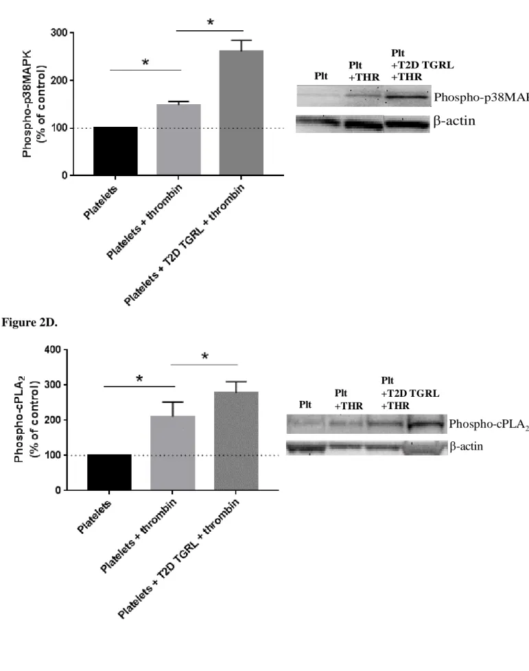
3.4. Effect of T2D large TGRL on platelet p38MAPK and cPLA2
229
Because TxA2 biosynthesis requires p38 MAPK-dependent activation of cPLA2, which
230
hydrolyzes membrane phospholipids to release arachidonic acid, we measured the 231
phosphorylation of p38 MAPK and cPLA2, in platelets pre-incubated with or without
232
25µg/mL T2D large TGRL and thrombin (0.09 ± 0.01 U/ml). As shown in Figure 2C and 2D, 233
there was an increase of phosphorylated amounts of both enzymes with the addition of 234
thrombin (phospho-p38MAPK: +48%, p<0.05, phospho-cPLA2: +110%, p<0.05). An
235
additional increase in phosphorylation of both enzymes was observed when platelets were 236
pre-incubated with T2D large TGRL and thrombin compared to platelets stimulated with 237
thrombin (phospho-p38MAPK: +87%, p<0.05, phospho-cPLA2: +32%, p<0.05).
238
240
241
4. DISCUSSION
242
The presence of large TGRL in the fasting state is known as a major lipid abnormality in T2D 243
patients; however, the potential implication of these specific particles in the development of 244
atherothrombosis is unknown. The present study supports the presence of remaining large 245
TGRL containing ApoB-48 and B-100 in the plasma of fasting T2D patients. Our data show 246
for the first time that large TGRL from fasting T2D patients potentiate activation of isolated 247
platelets when incubated with non-aggregating subthreshold concentrations of collagen or 248
thrombin. This platelet activation is triggered through the activation of the AA signaling 249
pathway. 250
Our lipoprotein fractions corresponded to ultracentrifuged fractions of the lightest density 251
(d<1.000) and were a mixture of large lipoproteins and their remnants enriched in 252
triglycerides. They had a mean diameter of 205 nm, which was higher than the reported 253
values in healthy individuals of 30-80 nm for VLDL and CM remnants [20].We were able to 254
detect and quantify ApoB-48 and ApoB-100 in plasma from fasting T2D patients, also 255
indicative of the presence of large CM remnants and large light VLDL. These findings are in 256
agreement with data showing that T2D patients present a higher production of large VLDL1 257
particles as well as a reduction of their hepatic catabolism [21, 22]. Since each triglyceride-258
rich particle contains only one apolipoprotein B, the ratio of TG/Total ApoB of 4.2 ± 0.5 259
shows the particles isolated from fasting T2D patients’ plasma display an enrichment in TG, 260
characteristic of large TGRL. Concentrations of ApoB-48 and ApoB-100 similar to ours have 261
been reported in plasma, CM and large VLDL fractions of T2D patients [23]. The 262
centrifugation method used for large TGRL isolation ensured that there was no contamination 263
with smaller particles like IDL, and most probably caused a loss of the small VLDL2 264
subclass, allowing the collection of the largest particles. Altogether, our results regarding the 265
presence of large TG-rich particles in fasting T2D patients are in accordance with the 266
literature [24-27]. 267
In this study, we chose to focus on large TGRL interactions with platelets in T2D patients 268
since this population presents an increased risk of developing cardiovascular complications 269
compared to non-diabetic. Indeed, in fasting healthy individuals, CM and their remnants are 270
rapidly cleared from circulation and negligible concentrations of ApoB-48 are reported in 271
their plasma [28-30]. These particles are unlikely to play a role in atherothrombotic events in 272
healthy subjects [31] therefor, the relevance of studying the effect of fasting large TGRL from 273
healthy subjects on platelets is weak. By contrast, previous studies have shown that large 274
TGRL particles from T2D and/or HTG patients display increased residence time in the 275
circulation, which may increase their atherogenicity and contribute to cardiovascular 276
complications [32]. We tested also the effect of large TGRL from fasting T2D patients on 277
platelets isolated from healthy donors and not on platelets from T2D patients. Since it was 278
already shown that platelets from T2D patients are prone to activation in a basal state, it 279
would have been uncertain if the effects on activation and aggregation were really caused by 280
large TGRL rather than by the dysfunction of the platelets itself. 281
282
Regarding the interaction of large TGRL with platelets, only a few studies have reported an 283
effect of large TGRL on platelet function in various populations in the postprandial state [33-284
37]. It is difficult to compare these post-prandial results with ours from fasting T2D patients, 285
since we cannot rule out that the large TGRL composition could be modified after a meal and 286
therefore have a different impact on platelet aggregation. There are also many methodological 287
differences between the studies that limit their comparison and the conclusions drawing. 288
Indeed, isolation method of the large TGRL, measurement of platelet aggregation performed 289
in whole blood, platelet-rich plasma (PRP) or washed platelets as well as the concentrations of 290
large TGRL and agonists used to assess platelet aggregation can all have a major impact on 291
the results. Our experiments were carried out in plasma-free platelet suspensions, which avoid 292
the interference of plasma proteins present in PRP (such as albumin and other lipoproteins), 293
and of other blood cells (such as erythrocytes) present in whole blood. Indeed, Yamazaki et 294
al. showed that post-prandial remnant-like particles in non-diabetic individuals enhanced 295
shear induced platelet aggregation and P-selectin expression in whole blood and suggested 296
that ADP released from erythrocytes might have activated platelets [38]. In addition, our large 297
TGRL fractions were isolated from fasting type 2 diabetic patients, which excludes the effect 298
of other fatty compounds released in the post-prandial state that could potentially stimulate 299
platelets. The T2D large TGRL fractions we tested were a mixture of apoB-48 and apoB-100 300
containing particles, thus it is not possible to conclude which class of lipoproteins in particular 301
drives the stimulating effect observed on platelet activation. However, our fractions are 302
representative of those of the large TGRL found in-vivo in plasma from fasting T2D patients. 303
Our results demonstrate for the first time that the aggregation response of platelets from 304
healthy donors to physiological agonists is enhanced by large TGRL from fasting T2D 305
patients. 306
307
This study also showed that the increased platelet activation caused by large TGRL from 308
fasting T2D patients involved the activation of the platelet AA signaling pathway. Indeed, 309
TxB2 concentration as well as phosphorylated amounts of p38MAPK and cPLA2 were higher
310
following platelet incubation with the particles and stimulation by an agonist compared to 311
agonist-stimulated platelets. Beitz et al. showed that VLDL from fasting patients with 312
coronary heart disease increased platelet generation of TxA2 in whole blood from healthy
volunteers [39]. Later, Englyst et al. showed that incubation of PRP with high concentrations 314
of VLDL from healthy volunteers in a post-prandial state increased collagen-stimulated 315
platelet TxB2 concentration when compared with platelets stimulated with collagen [35].
316
In conclusion, we showed that large TGRL from fasting T2D patients, containing both apoB-317
48 and apoB-100, have an enhancing effect on platelet aggregation via activation of the AA 318
signaling pathway and might contribute to the increased atherothrombotic risk of T2D 319
patients. Further studies about these fasting lipoproteins specific to T2D patients would be 320
required to have a better understanding of their implication in the development of 321
cardiovascular complications in this population. It would also be relevant to determine 322
whether postprandial large TGRL from T2D patients or from healthy subjects activate 323
platelets to the same extent as large TGRL from fasting T2D patients. 324
CONFLICT OF INTEREST
326
The authors have no conflict of interest related to this work to disclose. 327
328
FINANCIAL SUPPORT
329
This work was supported by Inserm. We would like to thank Ministère de l'Enseignement 330
supérieur, de la Recherche et de l'Innovation for granting a doctoral contract to Marie Michèle 331 Boulet. 332 333 AUTHOR CONTRIBUTIONS 334
MMB, MCM, DC and CC designed the study. MMB, MDF and CB performed the 335
experiments and collected the data. MMB and CC analyzed the data. MMB, DC, MCM, PM 336
and CC interpreted the data. MMB and CC wrote the manuscript. DC, MCM and PM 337
critically revised the manuscript. All co-authors read, commented and approved the 338 manuscript. 339 340 ACKNOWLEDGEMENTS 341
We would like to thank Harout Iliozier for carrying out plasma sampling and collecting 342
biological data from recruited T2D patients’ data bank at Louis Pradel Hospital. We also 343
thank the nurse team of the department of Endocrinology, Metabolic Disease, Diabetes and 344
Nutrition at Louis Pradel hospital for their essential collaboration with the patients’ blood 345
collection. 346
REFERENCES
348
1. Vazzana N, Ranalli P, Cuccurullo C, and Davi G (2012) Diabetes mellitus and 349
thrombosis. Thromb Res 129 : 371-377 350
2. Einarson TR, Acs A, Ludwig C, and Panton UH (2018) Prevalence of cardiovascular 351
disease in type 2 diabetes: a systematic literature review of scientific evidence from 352
across the world in 2007-2017. Cardiovasc Diabetol 17 : 83 353
3. Howard BV, Cowan LD, Go O, Welty TK, Robbins DC, and Lee ET (1998) Adverse 354
effects of diabetes on multiple cardiovascular disease risk factors in women. The 355
Strong Heart Study. Diabetes Care 21 : 1258-1265 356
4. Taskinen MR, and Boren J (2015) New insights into the pathophysiology of 357
dyslipidemia in type 2 diabetes. Atherosclerosis 239 : 483-495 358
5. Rivas-Urbina A, Benitez S, Perez A, and Sanchez-Quesada JL (2018) Modified low-359
density lipoproteins as biomarkers in diabetes and metabolic syndrome. Front Biosci 360
(Landmark Ed) 23 : 1220-1240 361
6. Gomez Rosso L, Lhomme M, Merono T, Dellepiane A, Sorroche P, Hedjazi L, Zakiev 362
E, Sukhorukov V, Orekhov A, Gasparri J, Chapman MJ, Brites F, and Kontush A 363
(2017) Poor glycemic control in type 2 diabetes enhances functional and 364
compositional alterations of small, dense HDL3c. Biochim Biophys Acta 1862 : 188-365
195 366
7. Morgantini C, Meriwether D, Baldi S, Venturi E, Pinnola S, Wagner AC, Fogelman 367
AM, Ferrannini E, Natali A, and Reddy ST (2014) HDL lipid composition is 368
profoundly altered in patients with type 2 diabetes and atherosclerotic vascular 369
disease. Nutr Metab Cardiovasc Dis 24 : 594-599 370
8. Ting HJ, Stice JP, Schaff UY, Hui DY, Rutledge JC, Knowlton AA, Passerini AG, 371
and Simon SI (2007) Triglyceride-rich lipoproteins prime aortic endothelium for an 372
enhanced inflammatory response to tumor necrosis factor-alpha. Circulation research 373
100 : 381-390 374
9. Santilli F, Simeone P, Liani R, and Davi G (2015) Platelets and diabetes mellitus. 375
Prostaglandins Other Lipid Mediat 120 : 28-39 376
10. Gaiz A, Mosawy S, Colson N, and Singh I (2017) Thrombotic and cardiovascular 377
risks in type two diabetes; Role of platelet hyperactivity. Biomed Pharmacother 94 : 378
679-686 379
11. D'Angelo A, Micossi P, Mannucci PM, Garimberti B, Franchi F, and Pozza G (1984) 380
Increased production of platelet thromboxane B2 in non-insulin-dependent diabetes. 381
Relationship to vascular complications. Eur J Clin Invest 14 : 83-86 382
12. Vericel E, Januel C, Carreras M, Moulin P, and Lagarde M (2004) Diabetic patients 383
without vascular complications display enhanced basal platelet activation and 384
decreased antioxidant status. Diabetes 53 : 1046-1051 385
13. Le QH, El Alaoui M, Vericel E, Segrestin B, Soulere L, Guichardant M, Lagarde M, 386
Moulin P, and Calzada C (2015) Glycoxidized HDL, HDL enriched with oxidized 387
phospholipids and HDL from diabetic patients inhibit platelet function. J Clin 388
Endocrinol Metab 100 : 2006-2014 389
14. Calzada C, Vericel E, Colas R, Guillot N, El Khoury G, Drai J, Sassolas A, Peretti N, 390
Ponsin G, Lagarde M, and Moulin P (2013) Inhibitory effects of in vivo oxidized 391
high-density lipoproteins on platelet aggregation: evidence from patients with 392
abetalipoproteinemia. FASEB J 27 : 2855-2861 393
15. Korporaal SJ, Relou IA, van Eck M, Strasser V, Bezemer M, Gorter G, van Berkel TJ, 394
Nimpf J, Akkerman JW, and Lenting PJ (2004) Binding of low density lipoprotein to 395
platelet apolipoprotein E receptor 2' results in phosphorylation of p38MAPK. J Biol 396
Chem 279 : 52526-52534 397
16. Colas R, Sassolas A, Guichardant M, Cugnet-Anceau C, Moret M, Moulin P, Lagarde 398
M, and Calzada C (2011) LDL from obese patients with the metabolic syndrome show 399
increased lipid peroxidation and activate platelets. Diabetologia 54 : 2931-2940 400
17. Hackeng CM, Relou IA, Pladet MW, Gorter G, van Rijn HJ, and Akkerman JW 401
(1999) Early platelet activation by low density lipoprotein via p38MAP kinase. 402
Thromb Haemost 82 : 1749-1756 403
18. Lowry OH, Rosebrough NJ, Farr AL, and Randall RJ (1951) Protein measurement 404
with the Folin phenol reagent. J Biol Chem 193 : 265-275 405
19. Born GV (1962) Aggregation of blood platelets by adenosine diphosphate and its 406
reversal. Nature 194 : 927-929 407
20. Feingold KR, and Grunfeld C (2000) Introduction to Lipids and Lipoproteins. In: De 408
Groot LJ, Chrousos G, Dungan K, Feingold KR, Grossman A, Hershman JM, Koch C, 409
Korbonits M, McLachlan R, New M, Purnell J, Rebar R, Singer F, and Vinik A (eds) 410
Endotext: South Dartmouth (MA) 411
21. Verges B (2015) Pathophysiology of diabetic dyslipidaemia: where are we? 412
Diabetologia 58 : 886-899 413
22. Krauss RM (2004) Lipids and lipoproteins in patients with type 2 diabetes. Diabetes 414
Care 27 : 1496-1504 415
23. Bozzetto L, Annuzzi G, Corte GD, Patti L, Cipriano P, Mangione A, Riccardi G, and 416
Rivellese AA (2011) Ezetimibe beneficially influences fasting and postprandial 417
triglyceride-rich lipoproteins in type 2 diabetes. Atherosclerosis 217 : 142-148 418
24. Garvey WT, Kwon S, Zheng D, Shaughnessy S, Wallace P, Hutto A, Pugh K, Jenkins 419
AJ, Klein RL, and Liao Y (2003) Effects of insulin resistance and type 2 diabetes on 420
lipoprotein subclass particle size and concentration determined by nuclear magnetic 421
resonance. Diabetes 52 : 453-462 422
25. Carmena R (2008) High Risk of Lipoprotein Dysfunction in Type 2 Diabetes Mellitus. 423
Revista Espanola de Cardiologia 8 : 18-24 424
26. Hirano T (2018) Pathophysiology of Diabetic Dyslipidemia. J Atheroscler Thromb 25 425
: 771-782 426
27. Adiels M, Boren J, Caslake MJ, Stewart P, Soro A, Westerbacka J, Wennberg B, 427
Olofsson SO, Packard C, and Taskinen MR (2005) Overproduction of VLDL1 driven 428
by hyperglycemia is a dominant feature of diabetic dyslipidemia. Arterioscler Thromb 429
Vasc Biol 25 : 1697-1703 430
28. Otokozawa S, Ai M, Diffenderfer MR, Asztalos BF, Tanaka A, Lamon-Fava S, and 431
Schaefer EJ (2009) Fasting and postprandial apolipoprotein B-48 levels in healthy, 432
obese, and hyperlipidemic subjects. Metabolism 58 : 1536-1542 433
29. Sakai N, Uchida Y, Ohashi K, Hibuse T, Saika Y, Tomari Y, Kihara S, Hiraoka H, 434
Nakamura T, Ito S, Yamashita S, and Matsuzawa Y (2003) Measurement of fasting 435
serum apoB-48 levels in normolipidemic and hyperlipidemic subjects by ELISA. J 436
Lipid Res 44 : 1256-1262 437
30. Bae JC, Han JM, Kwon S, Jee JH, Yu TY, Lee MK, and Kim JH (2016) LDL-C/apoB 438
and HDL-C/apoA-1 ratios predict incident chronic kidney disease in a large apparently 439
healthy cohort. Atherosclerosis 251 : 170-176 440
31. Karpe F, Olivecrona T, Hamsten A, and Hultin M (1997) Chylomicron/chylomicron 441
remnant turnover in humans: evidence for margination of chylomicrons and poor 442
conversion of larger to smaller chylomicron remnants. J Lipid Res 38 : 949-961 443
32. Tomkin GH, and Owens D (2012) The chylomicron: relationship to atherosclerosis. 444
International journal of vascular medicine 2012 : 784536 445
33. Olufadi R, and Byrne CD (2006) Effects of VLDL and remnant particles on platelets. 446
Pathophysiol Haemost Thromb 35 : 281-291 447
34. Orth M, Luley C, and Wieland H (1995) Effects of VLDL, chylomicrons, and 448
chylomicron remnants on platelet aggregability. Thromb Res 79 : 297-305 449
35. Englyst NA, Taube JM, Aitman TJ, Baglin TP, and Byrne CD (2003) A novel role for 450
CD36 in VLDL-enhanced platelet activation. Diabetes 52 : 1248-1255 451
36. Knofler R, Nakano T, Nakajima K, Takada Y, and Takada A (1995) Remnant-like 452
lipoproteins stimulate whole blood platelet aggregation in vitro. Thromb Res 78 : 161-453
171 454
37. Pedreno J, Hurt-Camejo E, Wiklund O, Badimon L, and Masana L (2000) Platelet 455
function in patients with familial hypertriglyceridemia: evidence that platelet reactivity 456
is modulated by apolipoprotein E content of very-low-density lipoprotein particles. 457
Metabolism 49 : 942-949 458
38. Yamazaki M, Uchiyama S, Xiong Y, Nakano T, Nakamura T, and Iwata M (2005) 459
Effect of remnant-like particle on shear-induced platelet activation and its inhibition 460
by antiplatelet agents. Thromb Res 115 : 211-218 461
39. Beitz A, Nikitina NA, Giessler C, Beitz J, Masaev VP, Perova NA, and Mest HJ 462
(1990) Modulation of TXA2 generation of platelets by human lipoproteins. 463
Prostaglandins Leukot Essent Fatty Acids 40 : 57-61 464
TABLE 1. Clinical and biological parameters of the study participants 466
Characteristics
T2D
HBD
n 25 7 Age (years) 59.8 ± 2 27 ± 4b BMI (kg/m2) 31.9 ± 1.4 ND TG (mmol/L) 1.63 ± 0.20 0.79 ± 0.14b HDL-C (mmol/L) 1.06 ± 0.05 1.04 ± 0.07b LDL-C (mmol/L) 2.22 ± 0.20 2.25 ± 0.27b Glycemia (mmol/L) 9.0 ± 0.6 ND HbA1c (%) 7.8 ± 0.3 ND T2D duration (years) 13.8 ± 2.0a NA a: n=24, b: n=6 467Data are presented as means ± SEM 468
469
TABLE 2. Characteristics of plasma and corresponding large TGRL fractions from T2D
470
patients 471
Characteristics
Plasma
Large TGRL
fractions
Hydrodynamic diameter (nm) - 205 ± 15
ApoB-48 (mg/L) 15.2 ± 3.7a 0.94 ± 0.40b
ApoB-100 (mg/L) 1327.2 ± 126.8a 28.1 ± 5.2b
TG (mmol/L) 1.37 ± 0.16 0.16 ± 0.04
Total cholesterol (mmol/L) 3.89 ± 0.32 0.09 ± 0.01
Ratios (mg/L) ApoB-100/apoB-48 153.9 ± 42.0 42.4 ± 6.5 TG/total cholesterol 0.4 ± 0.04 4.0 ± 0.4 TG/total apoB 0.9 ± 0.1 4.2 ± 0.5 n=10, a: n=9, b: n=7 472
Data are presented as means ± SEM 473
FIGURE LEGENDS
474
FIGURE 1. Collagen-stimulated platelet aggregation and thromboxane B2 concentration
475
are increased by large TGRL from type 2 diabetic patients.
476
(A): (i) Platelets from healthy blood donors were incubated in the absence or presence of
477
TGRL from healthy blood donors followed by stimulation with a concentration of collagen of 478
0.13 µg/ml ± 0.01 and agitated at 1000 rpm for 4 minutes at 37°C. Results are expressed as 479
mean percentage of control determined by platelets stimulated with collagen and are means ± 480
SEM of 7 healthy blood donors: NS. (ii) Platelets from healthy donors were incubated in the 481
absence or presence of T2D patients large TGRL followed by stimulation with a 482
concentration of collagen of 0.17 µg/ml ± 0.02 and agitated at 1000 rpm for 4 minutes at 483
37°C. Results are expressed as mean percentage of control determined by platelets stimulated 484
with collagen and are means ± SEM of 25 patients: *p<0.05. 485
(B): Platelets from healthy donors were incubated at 37°C for 30 minutes in the absence or
486
presence of T2D large TGRL (25 µg prot/ml) and stimulated with collagen (0.37 ± 0.04 487
µg/ml) for 1 hour at 37°C while undergoing slow agitation. Results are expressed as mean 488
TxB2 concentrations in pmol/109 platelets ± SEM of 25 patients. Platelets + collagen vs
489
platelet with vehicle: +36%, *p<0.05. Platelets + T2D large TGRL + collagen vs platelets + 490
collagen: +34%, *p<0.05. 491
FIGURE 2. Large TGRL from type 2 diabetic patients increase thrombin-stimulated
492
platelet aggregation and thromboxane B2 concentration via activation of the arachidonic
493
acid signaling pathway.
494
(A): Platelets from healthy donors were incubated in the absence or presence of T2D large
495
TGRL (25 µg prot/ml) followed by stimulation with thrombin (0.013 U/ml ± 0.001) and 496
agitated at 1000 rpm for 4 minutes Results are expressed as mean percentage of control 497
determined by platelets stimulated with thrombin and are means ± SEM of 6 patients: 498
*p<0.05. 499
(B): Platelets from healthy donors were incubated at 37°C for 30 minutes in the absence or
500
presence of T2D large TGRL (25 µg prot/ml) and stimulated with thrombin (0.1 U/ml) for 1 501
hour at 37°C while undergoing slow agitation (n=13). Results are expressed as mean TxB2
502
concentrations in pmol/109 platelets ± SEM. Platelets + thrombin vs platelets alone: +1453%, 503
*p<0.05. Platelets + T2D large TGRL + thrombin vs platelets + thrombin: +37%, *p<0.05. 504
(C and D): Platelets from healthy donors were incubated at 37°C with or without thrombin
505
(0.09 ± 0.01 U/ml) while undergoing slow agitation for controls. Platelets were incubated 506
with 25 µg prot/ml T2D large TGRL and thrombin under the same conditions for testing. 507
Phosphorylation of p38MAPK and cPLA2 was measured after SDS-PAGE separation and
508
immunoblotting with anti-phospho p38 MAPK or anti-phospho cPLA2 antibodies, followed
509
by reprobing with beta-actin antibody. (n=6 and n=3, respectively). Results are expressed as 510
mean percentage of control determined by platelets stimulated with thrombin (C) phospho-511
p38MAPK: Platelets + thrombin vs platelets alone: *p<0.05. Platelets + T2D large TGRL +
512
thrombin vs platelets + thrombin: *p<0.05 and the representative immunoblots. (D) phospho-513
cPLA2: Platelets + thrombin vs platelets alone: *p<0.05. Platelets + T2D large TGRL +
514
thrombin vs platelets + thrombin: *p<0.05 and the representative immunoblots. 515
516
Figure 1A. 518 (i) 519 520 521 522 523 524 525 526 527 528 529 530 531 532 (ii) 533 534 535 536 537 538 539 540 541 542 543 544 545 546 547
Figure 1B. 548 549 Figure 2A. 550 551
Figure 2B.
552
Figure 2C. 554 555 556 557 558 559 560 561 562 563 564 565 566 567 Figure 2D. 568 569 570 Phospho-cPLA2 β-actin Plt +T2D TGRL +THR Plt Plt +THR 1 2 3 4 5 6 Phospho-p38MAPK