Pulsatile stress in diabetes
Pulsatile stress in middle-aged patients with Type 1 or Type 2 diabetes
compared to nondiabetic controls
Short title: Pulsatile stress in diabetes
Jean-Christophe Philips, MD Monique Marchand André J. Scheen, MD, PhD
University of Liège, Division of Diabetes, Nutrition and Metabolic Disorders, Department of Medicine, CHU Sart Tilman, B-4000 Liège, Belgium
Corresponding author:
André J. Scheen, MD, PhD Email: [email protected]
Submitted 16 February 2010 and accepted 24 July 2010.
This is an uncopyedited electronic version of an article accepted for publication in Diabetes Care. The American Diabetes Association, publisher of Diabetes Care, is not responsible for any errors or omissions in this version of the manuscript or any version derived from it by third parties. The definitive publisher-authenticated version will be available in a future issue of Diabetes Care in print and online at http://care.diabetesjournals.org.
Background: Arterial pulse pressure (PP) is considered as an independent cardiovascular risk factor. We compared PP during an active orthostatic test in middle-aged patients with type 1 diabetes and with type 2 diabetes, and corresponding nondiabetic controls.
Methods: 40 patients with type 1 diabetes (mean age 50 years, diabetes duration 23 years, BMI 23.0 kg/m²) were compared to 40 non hypertensive patients with type 2 diabetes (respectively, 50 years, 8 years, 29.7 kg/m²). Patients taking antihypertensive agents or with renal insufficiency were excluded. All patients were evaluated with a continuous noninvasive arterial blood pressure monitoring (Finapres®) in standing (1 min), squatting (1 min) and again standing position (1 min). Patients with type 1 or type 2 diabetes were compared with two groups of 40 age-, sex- and BMI-matched healthy subjects.
Results: Patients with type 1 diabetes and patients with type 2 diabetes showed significantly higher PP, heart rate (HR) and PPxHR double product (type 1 : 5263 vs 4121 mmHg/min, p=0.0004; type 2 : 5359 vs 4321 mmHg, p=0.0023) levels than corresponding controls. There were no significant differences between patients with type 1 diabetes and type 2 diabetes regarding PP (59 vs 58 mmHg), HR (89 vs 88/min), and PPxHR product (5263 vs 5359 mmHg/min).
Conclusion: Patients with type 1 diabetes have comparable increased levels of peripheral PP, an indirect marker of arterial stiffness, and PPxHR, an index of pulsatile stress, as non-hypertensive patients with type 2 diabetes at similar mean age of 50 years.
rterial pulse pressure (PP), a surrogate marker of large artery stiffness, was shown to be an independent cardiovascular disease (CVD) risk factor in several large longitudinal studies in patients with type 2 diabetes mellitus (1,2). In patients with type 1 diabetes of the Finnish Diabetic Nephropathy (FinnDiane) study (3), a higher systolic blood pressure (BP) and an earlier decrease in diastolic BP resulted in a higher and more rapidly increasing PP as compared to non-diabetic control subjects. In the EURODIAB study (4,5), PP was also an independent risk factor for CVD and total mortality in patients with type 1 patients.
Middle-aged patients with type 1 diabetes are characterized by a long duration of the disease and therefore a sustained exposure to chronic hyperglycaemia, leading
to accelerated progression of arterial stiffness and increased PP (6). In contrast, middle-aged patients with type 2 diabetes have a much shorter duration of diabetes, but have other CVD risk factors such as abdominal obesity, insulin resistance and metabolic syndrome, which could accelerate arterial stiffness (1,2). To our knowledge, no study compared PP in patients with type 1 diabetes and in patients with type 2 diabetes at similar age. The primary aim of the present study was to investigate pulsatile stress in patients with type 1 and patients with type 2 diabetes at a similar mean age of 50 years. Each group of diabetic patients was compared with a group of nondiabetic controls, matched for age, sex, and body mass index (BMI). BP and PP were monitored during an active postural test, the so-called squatting test, which has been shown by our group to amplify PP increase
A
according to diabetes duration in patients with type 1 diabetes (7,8).
RESEARCH DESIGN AND METHODS Patients. Forty patients (20 men and 20
women) with type 1 diabetes and 40 patients (20 men and 20 women) with type 2 diabetes were recruited among the patients followed in our department. Patients with arterial hypertension, renal insufficiency or CVD or taking medications interfering with vascular reactivity (including any type of antihypertensive agents) were excluded from the study. All patients with type 1 diabetes received intensified insulin therapy with multiple daily insulin injections (n = 36) or continuous subcutaneous insulin infusion via a portable pump (n = 4). Patients with type 2 diabetes received various types of oral glucose-lowering therapies (metformin alone, sulfonylurera alone or metformin-sulfonylurea combination) (n = 25) or insulin alone (n = 5) or combined with metformin (n = 10). Two groups of healthy subjects were used as controls and matched for BMI with either type 1 diabetes patients or type 2 diabetes patients (Table 1). The study was accepted by the ethical committee of our institution.
Orthostatic test. The squatting test
(successively 1 min standing, 1 min squatting, 1 min standing) is an original active orthostatic manoeuvre that leads to the most important and fast variations of the hydrostatic level with posture (9). Squatting produces a prompt increase in cardiac output and arterial BP, essentially attributed to augmented venous return from compression of leg veins. These changes result in a significant increase in mean arterial BP and PP (7,8), which is accompanied by an immediate decrease in heart rate (HR) and forearm vascular resistance, probably due to activation of cardiopulmonary and arterial baroreflexes implicating autonomic nervous system. Later on, the active transition from
squatting to standing results in a profound initial BP decrease inducing a reflex tachycardia, which can be used to detect diabetic cardiac autonomic neuropathy (CAN) (10,11) and assess baroreflex sensitivity (12).
Measurements. Changes in systolic BP
(SBP), diastolic BP and HR were measured continuously with a FinapresR (from FINger Arterial PRESsure) instrument (Ohmeda, USA) that allows to carefully study cardiovascular reflexes, especially during an orthostatic manoeuvre (13). The FinapresR is based on servoplethysmomanometry, employing the volume clamp technique at the finger level. A good concordance was reported between Finapres® BP measurements and direct intra-arterial measurements (13). PP, i.e. SBP minus diastolic BP, was automatically calculated throughout the test. Mean arterial blood pressure (MBP) was calculated by the formula (SBP + 2 x diastolic BP)/3. To quantify the relative magnitude of the pulsatile to mean artery pressure (“pulsatility index”), we normalised the PP to the MBP and referred to this value as fractional PP (PPf) (14). “Pulsatile stress” was defined as the double product of PP and HR; it has been shown to be largely regulated by arterial stiffness and by sympathetic nerve activity and to be associated with a higher risk of (micro)albminuria (15). We also calculated the “SBP x HR” double product, an index of cardiac load that has been shown to be associated with an increased CVD risk (16). For each variable or parameter, mean levels were calculated in each subject during the whole period of the test, during the initial standing position and during the squatting position, after exclusion of the initial transition phase, as previously described (7,8).
During the transition from squatting to standing, there is an abrupt drop in BP associated with a reflex tachycardia, and that is followed by a rapid return to baseline
values of both parameters (BP increase and HR decrease). The mirror changes in HR and SBP allows the calculation of a baroreflex gain, by plotting the pulse intervals (R-R) against SBP values, and the slope of this relation represents the baroreflex sensitivity (17). We also calculated both a vagal index (SqTv : ratio between the baseline cardiac R-R interval and the longest R-R-R-R interval in the first 15 sec of squatting) and a sympathetic index (SqTs : ratio between the baseline cardiac R-R interval and the shortest R-R interval in the first 10–20 seconds of standing after squatting), as previously described (10,11). These indices, based on HR reduction during squatting and reflex tachycardia during standing, were considered as markers of cardiovascular autonomic neuropathy (CAN) : a higher SqtV value indicates a parasympathetic neuropathy while a lower SqTs is an indicator of sympathetic neuropathy (10-12).
Concomitant glycated hemoglobin (HbA1c) levels (normal values 4-6 %) were measured in order to assess recent blood glucose control in diabetic patients; for each patient, corresponding HbA1c mean level corresponded to the average of 1 to 3 measurements. Lipid profiles were also collected in diabetic patients and the prevalence of the metabolic syndrome (National Cholesterol Education Program – Adult Treatment Panel 3 criteria) was calculated in patients with type 1 diabetes and in patients with type 2 diabetes.
Statistical analysis. The required sample size
to have an 80% chance of detecting as significant (at the two sided 5% level) a 10 mm Hg difference in PP between two different subgroups, with an assumed standard deviation of PP of 14 mmHg, was 32 individuals. A difference of 10 mm Hg was chosen as clinically significant because it has been shown to be associated with increased cardiovascular mortality in type 2 diabetes (1) and total mortality in the large EURODIAB
cohort of patients with type 1 diabetes (5). Between-group differences were analyzed using unpaired t tests. The relationship between two variables, i.e. between pulsatile stress and baroreflex gain as a marker of CAN, was assessed with the Spearman correlation coefficient. Results were expressed as mean ± SD values for all continuous variables. A p value of less than 0.05 was considered significant.
RESULTS
Patients with type 1 diabetes versus nondiabetic lean subjects. As compared to
controls, patients with type 1 diabetes had similar MBP, but were characterized throughout the test by significantly higher PP, HR, PP/MBP, PPxHR and SBPxHR levels (Figure 1A, Table 1). When squatting was compared with initial standing position, a trend for higher increases in PP, PP/MBP and PPxHR was observed in patients with type 1 diabetes than in controls, with a significantly higher increase in SBPxHR double product ( Table 2). The baroreflex gain calculated during the transition from squatting to standing was markedly decreased in patients with type 1 diabetes compared to controls. SqTv and SqTs indices were also significantly different in patients with type 1 diabetes compared with lean controls (Table 2). There was a significant inverse correlation between pulsatile stress (PPxHR) and baroreflex gain in patients with type 1 diabetes (r = -0.383 ; p=0.023), but not in lean controls (r = -0.178; NS).
Patients with type 2 diabetes versus non-diabetic overweight/obese patients. As
compared to overweight/obese nondiabetic controls, patients with type 2 diabetes had similar MBP (hypertension was considered as an exclusion criterion in the present study). However, they showed higher PP, HR, PP/MBP, PPxHR and SBPxHR levels throughout the test (Figure 1B, Table 1). Increases in PP, PP/MBP, PPxHR and
SBPxHR when moving from standing to squatting were not significantly different in patients with type 2 diabetes and in overweight/obese non diabetic controls ( Table 2). The baroreflex gain was significantly decreased in patients with type 2 diabetes compared to controls. SqTs index (reflecting post-squatting tachycardia), but not SqTv index (a marker of bradycardia during squatting), was significantly lower in patients with type 2 diabetes compared with overweight/obese nondiabetic controls (Table 2). There was a highly significant inverse correlation between pulsatile stress and baroreflex gain in patients with type 2 diabetes (r = - 0.719 ; p=0.0001), but not in overweight/obese controls (r = - 0.272; NS). No significant differences in pulsatile markers and CAN indices were noticed between the patients with type 2 diabetes treated with insulin and those not treated with insulin.
Patients with type 1 diabetes versus patients with type 2 diabetes. On average,
MBP, PP, HR, PP/MBP, PPxHR and SBPxHR levels were comparable in middle-aged patients with type 1 and type 2 diabetes (Figure 1C, Table 1). The transition from standing to squatting resulted in similar increases in MBP, PP, PP/MBP, PPxHR and SBPxHR in the two groups of diabetic patients ( Table 2). Careful analysis of the two PP curves showed different kinetics in PP increases, with a second phase increase in PP in patients with type 1 diabetes that was not observed in patients with type 2 diabetes; however, the between-group difference during the second part of squatting did not reach statistical significance (Figure 1C). The baroreflex gain was similar in patients with type 1 and type 2 diabetes. Accordingly, SqTv and SqTs indices were not significantly different between the two diabetic groups (Table 2). Patients with type 1 diabetes had a much longer known disease duration (23 vs 8 years; p<0.0001), but a much lower prevalence of metabolic syndrome (3% vs
42%; p<0.01) than patients with type 2 diabetes
Overweight/obese versus lean subjects without diabetes. On average, MBP, PP, HR,
PP/MBP, PPxHR and SBPxHR levels were comparable in obese and lean non diabetic individuals of the present study (Table 1). The postural change from standing to squatting resulted in similar increases in MBP, PP, PP/MBP, PPxHR in overweight/obese and lean subjects, with only a trend for higher increase in SBPxHR double product (+ 963 ± 1178 vs + 601 ± 698 mmHg*min-1, p=0.0991) in presence of obesity (Table 2). The baroreflex gain was significantly lower in overweight/obese subjects compared with lean individuals (2.97 ± 2.18 vs 4.11 ± 2.26 mmHg*min-1, p=0.0332), even in absence of diabetes. SqTv index was higher in obese subjects than in lean controls (p = 0.0011), whereas SqTs index was almost similar in the two nondiabetic groups (Table 2).
CONCLUSION
The main findings of the present study are: 1) higher PP, PP/MBP, PPxHR and SBPxHR levels in middle-aged patients with type 1 diabetes compared with lean controls, in agreement wit higher pulsatile stress and cardiac workload in patients with long-standing type 1 diabetes ; 2) similarly, higher PP, PP/MBP, PPxHR and SBPxHR levels in middle-aged non hypertensive patients with type 2 diabetes compared with overweight/obese nondiabetic controls; 3) the absence of significant differences in PP, pulsatile index, pulsatile stress, and double product between patients with type 1 diabetes and with type 2 diabetes matched for age (50 years on average); and 4) indices of CAN as shown by lower baroreflex gain and altered SqT indices during squatting in both patients with type 1 diabetes and type 2 diabetes as compared to non-diabetic controls. Therefore, middle-aged patients with type 1 diabetes or with type 2 diabetes are exposed to a
comparable pulsatile stress, a known cardiovascular and renal risk marker (1-5, 15, 16). In patients with type 1 diabetes, the negative influence of a much longer diabetes duration (23 years on average in the present study) might be at least partially compensated for by the positive influence of lower BMI, less insulin resistance and a much lower prevalence of metabolic syndrome as compared with patients with type 2 diabetes. On the contrary, middle-age patients with type 2 diabetes are exposed to a high pulsatile stress despite a shorter known duration of diabetes (8 years on average in our population), presumably because the presence of other concomitant cardiovascular risk factors (even if hypertension was excluded in the present study), as shown by a much higher prevalence of metabolic syndrome compared to patients with type 1 diabetes.
The observation of higher PP levels in patients with type 1 diabetes as compared to controls in the age range 40-60 years is in agreement with previous studies from our group demonstrating an earlier PP increase with age in this population (7,8) and with the observational data of the large cross-sectional, case-control FinnDiane study (3). As PP is considered as an indirect marker of arterial stiffness, these higher PP results are in agreement with accelerated vascular aging in the population with type 1 diabetes (6), especially patients with chronic poor glucose control (18). In the FinnDiane, the ambient level of glucose was not associated with increased PP, but the time of exposure to hyperglycemia appeared to play a fundamental role in the process of premature arterial stiffening in patients with type 1 diabetes (3). In the EURODIAB Prospective Complications Study, PP was significantly associated with all-cause mortality and a mean 12 mm Hg higher PP was observed in patients with type 1 diabetes who died as compared to that of those who survived (5).
Decreased baroreflex gain was observed in our patients with type 1 diabetes, reflecting the presence of CAN after > 20 years of diabetes (19). This was confirmed by altered SqTs and SqTv indices during the squatting test, markers of parasympathetic and sympathetic dysfunction, respectively (10). CAN exposes diabetic patients to an increased mortality risk (19). There may be some connection between PP and CAN (8), between aortic stiffness and CAN (20) and between arterial stiffness, cardiovagal baroreflex sensitivity and postural blood pressure changes (21). Increased SBP was identified as a factor associated with an increased risk of developing CAN in the cohort of patients with type 1 diabetes of the EURODIAB Prospective Complications study (22). The pathophysiological mechanism linking CAN to arterial stiffness in patients with type 1 diabetes remains unknown, but this association persisted after adjustment for potential confounders such as baseline HbA1c, HDL cholesterol, and smoking history (23). In the present study, we found a significant relationship between pulsatile stress and baroreflex gain as a marker of CAN in patients with type 1 diabetes. In patients with type 2 diabetes, markers of CAN are also present (11), although less marked than in patients with prolonged type 1 diabetes (7). The relationship between PP and CAN is less well known in patients with type 2 diabetes even if associations between autonomic neuropathy, vascular dysfunction and hyperinsulinemia have been demonstrated (24). Interestingly, a remarkable significant inverse correlation was noticed between pulsatile stress and baroreflex gain in the group of patients with type 2 diabetes of our study. Several mechanisms may underlie the association between arterial stiffness and impaired cardiovagal baroreflex sensitivity. The stiffness of the carotid arteries and the aorta, where the arterial baroreceptors are located, may affect the stretch-sensitive
receptors and hence baroreflex sensitivity. In addition to structural vascular changes, functional mechanisms associated to endothelial dysfunction may also contribute to the impairment of baroreflex sensitivity associated with arterial stiffness (21).
Patients with type 2 diabetes also showed increased PP, pulsatility index and pulsatile stress as compared to overweight/obese non diabetic individuals matched for BMI, age and gender. This was observed despite the absence of hypertension and a much shorter duration of diabetes when compared to the population with type 1 diabetes analyzed in the present study. It is well known, however, that type 2 diabetes remains silent during an average of 10 years before diagnosis and initiation of treatment in most cases. Thus, selected patients may have a longer duration of type 2 diabetes than the average 8-year known duration noticed in the present population. In order to avoid the potential bias of hypertension and the interferences of antihypertensive agents, we deliberately selected type 2 diabetes patients without hypertension. Despite normal MBP, middle-aged patients with type 2 diabetes had higher PP and pulsatile stress and higher SBPxHR double product, two CVD risk markers (16). Increased PP levels have been repeatedly demonstrated in large longitudinal studies in patients with type 2 diabetes mellitus and shown to be associated with a higher incidence of cardiovascular events (1,2).
Some limitations of the present study should be discussed. Several studies have demonstrated that absolute brachial and finger PP measurements are not identical with larger differences in SBP. However, the differences were generally small and not considered of clinical relevance (13). Furthermore, some studies have shown a good concordance between periphery (finger, as in the present study) and central (aortic, now recognized as the most important risk factor) BP
measurements (25). Nevertheless, PP measured at the finger site may not necessarily reflect central PP because of the amplification phenomenon. Second, glucose control of patients with type 1 diabetes evaluated in the present study was far from optimal, despite intensified insulin therapy. Therefore, our results could not be necessarily extrapolated to patients with near normoglycemia for many years as chronic hyperglycemia seems to play a major role in accelerating arterial stiffness (18). Third, patients with type 2 diabetes selected in the present study did not have hypertension. Therefore, the similar results in markers of pulsatile stress in middle-aged patients with type 1 and type 2 diabetes should be interpreted in this context. We cannot exclude that overweight/obese patients with type 2 diabetes and hypertension may be exposed to a higher vascular stress than lean normotensive patients with type 1 diabetes at the same age. This would be certainly the case for the SBPxHR double product, but might also be true for the various pulsatility markers. Fourth, very few patients had positive microalbuminuria in the two diabetic cohorts analyzed in the present study, because we excluded patients with hypertension or antihypertensive agents. Therefore, we were not in a position to study the possible relationship between pulsatile stress and early renal alterations as shown in previous studies (15).
In conclusion, middle-aged patients with a long duration of type 1 diabetes have similarly increased pulsatile stress as compared to age-matched patients with type 2 diabetes characterized by a shorter duration of the disease but the presence of other vascular risk factor such as obesity and insulin resistance, but no hypertension. In addition, both diabetic groups have markers of CAN with a reduced baroreflex gain as compared to non diabetic controls. The combination of these risk factors may contribute to increase
the CVD risk in type 1 diabetic patients with a long exposition to chronic hyperglycemia in a similar fashion as patients with type 2 diabetes whose high CVD risk is well known.
Author contributions. JCP, MM and AJS
made substantial contributions to the concept and design of the study and outlined the structure of the report. JCP and MM were involved in the acquisition, analysis and interpretation of the data. AJS has had full
access to all the data, wrote the report and had final responsibility for the decision to submit for publication. All authors critically reviewed the reports and approved the final version of the report for submission.
ACKNOWLEDGEMENT
This work was supported by an unrestricted research grant from NovoNordisk Belgium
Disclosure. We declare that we have no
conflicts of interest
REFERENCES
1. Schram MT, Kostense PJ, Van Dijk RA, Dekker JM, Nijpels G, Bouter LM, Westerhof N, Stehouwer CD: Diabetes, pulse pressure and cardiovascular mortality the Hoorn Study. J Hypertens 20: 1743–1751, 2002
2. Nilsson PM, Cederholm J, Eeg-Olofsson K, Eliasson B, Zethelius B, Gudbjörnsdóttir S; Swedish National Diabetes Register (NDR): Pulse pressure strongly predicts cardiovascular disease risk in patients with type 2 diabetes from the Swedish National Diabetes Register (NDR). Diabetes Metab 35: 439-446, 2009
3. Ronnback M, Fagerudd J, Forsblom C, Pettersson-Fernholm K, Reunanen A, Groop PH; Finnish Diabetic Nephropathy (FinnDiane) Study Group: Altered age-related blood pressure pattern in type 1 diabetes. Circulation 110: 1076-1082, 2004
4. Schram MT, Chaturvedi N, Fuller JH, Stehouwer CD; EURODIAB Prospective Complications Study Group: Pulse pressure is associated with age and cardiovascular disease in type 1 diabetes: the Eurodiab Prospective Complications Study. J Hypertens 21: 2035-2044, 2003
5. Soedamah-Muthu SS, Chaturvedi N, Witte DR, Stevens LK, Porta M, Fuller JH, for the EURODIAB Prospective Complications Study Group: Relationship between risk factors and mortality in type 1 diabetic patients in Europe. The EURODIAB Prospective Complications Study (PCS). Diabetes Care 31: 1360-1366, 2008
6. Benetos A : Pulse pressure and arterial stiffness in type 1 diabetic patients. J Hypertens 21: 2005-2007, 2003
7. Philips JC, Marchand M, Scheen AJ: Pulse pressure and cardiovascular autonomic neuropathy according to duration of type 1 diabetes. Diabetes Metab Res Rev 25: 442-451, 2009 8. Philips JC, Marchand M, Scheen AJ: Squatting amplifies pulse pressure increase according to duration of type 1 diabetes. Diabetes Care 31: 322-324, 2008
9. Rickards CA, Newman DG: A comparative assessment of two techniques for investigating initial cardiovascular reflexes under acute orthostatic stress. Eur J Appl Physiol 90: 449-457, 2003
10. Marfella R, Giugliano D, Di Maro G, Acampora R, Giunta R, D'Onofrio F: The squatting test : a useful tool to assess both parasympathetic and sympathetic involvement of the cardiovascular autonomic neuropathy in diabetes. Diabetes 43: 607-612, 1994
11. Marfella R, Salvatore T, Giugliano D, Di Maro G, Giunta R, Torella R, Juchmes J, Scheen A, Lefèbvre PJ: Detection of early sympathetic cardiovascular neuropathy by squatting test in NIDDM. Diabetes Care 17: 149-151, 1994
12. Nakagawa M, Shinohara T, Anan F, Yufu K, Takahashi N, Okada N, Hara M, Yoshimatsu H, Saikawa T: New squatting test indices are useful for assessing baroreflex sensitivity in diabetes mellitus. Diabetic Med 25: 1309-1315, 2008
13. Imholz BP, Wieling W, van Montfrans GA, Wesseling KH: Fifteen years experience with finger arterial pressure monitoring: assessment of the technology. Cardiovasc Res 38: 605-616, 1998
14. NakayamaY, UedaH, TsumuraK, YoshimaruK, HayashiT:Ascending fractional pulse pressure closely relating to large artery function. J Hum Hypertens 16: 243-247, 2002
15. Baumann M, Pan CR, Roos M, von Eynatten M, Sollinger D, Lutz J, Heemann U: Pulsatile stress correlates with (micro-)albuminuria in renal transplant recipients. Transpl Int 23: 292-298, 2010
16. Thomas F, Bean K, Provost JC, Guize L, Benetos A: Combined effects of heart rate and pulse pressure on cardiovascular mortality according to age. J Hypertens 19: 863-869, 2001 17. Zhang R, Claassen JA, Shibata S, Kilic S, Martin-Cook K, Diaz-Arrastia R, Levine BD: Arterial-cardiac baroreflex function: insights from repeated squat-stand maneuvers. Am J Physiol Regul Integr Comput Physiol 297: R116-123, 2009
18. Schram MT, Schalkwijk CG, Bootsma AH, Fuller JH, Chaturvedi N, Stehouwer CD; EURODIAB Prospective Complications Study Group: Advanced glycation end products are associated with pulse pressure in type 1 diabetes: the EURODIAB Prospective Complications Study. Hypertension 46: 232-237, 2005
19. Vinik AI, Maser RE, Mitchell BD, Freeman R: Diabetic autonomic neuropathy. Diabetes Care 26: 1553–1579, 2003
20. Ahlgren AR, Sundkvist G, Wollmer P, Sonesson B, Länne T : Increased aortic stiffness in women with type 1 diabetes mellitus is associated with diabetes duration and autonomic nerve function. Diabetic Med 16: 291-297, 1999
21. Mattace-Raso FU, van den Meiracker AH, Bos WJ, van der Cammen TJ, Westerhof BE, Elias-Smale S, Reneman RS, Hoeks AP, Hofman A, Witteman JC. Arterial stiffness, cardiovagal baroreflex sensitivity and postural blood pressure changes in older adults: the Rotterdam Study. J Hypertens 25:1421-1426, 2007
22. Witte DR, Tesfaye S, Chaturvedi N, Eaton SE, Kempler P, Fuller JH ; EURODIAB Prospective Complications Study Group: Risk factors for cardiac autonomic neuropathy in type 1 diabetes mellitus. Diabetologia 48: 164-171, 2005
23. Prince CT, Secrest AM, Mackey RH, Arena VC, Kingsley LA, Orchard TJ. Cardiovascular autonomic neuropathy, HDL cholesterol, and smoking correlate with arterial stiffness markers determined 18 years later in type 1 diabetes. Diabetes Care 33: 652-657, 2010 24. Meyer C, Milat F, McGrath BP, Cameron J, Kotsopoulos D, Teede HJ. Vascular dysfunction and autonomic neuropathy in Type 2 diabetes. Diabet Med 21:746-751, 2004
25. Eckert S, Horstkotte D: Comparison of Portapres non-invasive blood pressure measurement in the finger with intra-aortic pressure measurement during incremental bicycle exercise. Blood Press Monit 7: 179-183, 2002
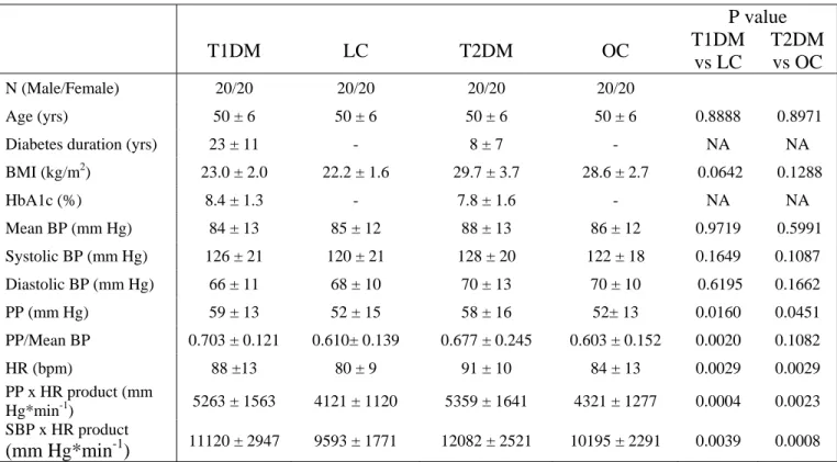
Table 1: Characteristics of middle-aged diabetic patients with type 1 diabetes (T1DM), patients
with type 2 diabetes (T2DM), non-diabetic lean controls (LC) and non-diabetic overweight/obese controls (OC) (20 men and 20 women in each group), and average values of mean blood pressure (BP), systolic BP, diastolic BP, pulse pressure (PP), pulsatility index (PP/mean BP), heart rate (HR), pulsatile stress (PPxHR), systolic BP x HR double product (SBPxHR) recorded during the whole 3-min squatting test. Results are expressed as mean ± SD. NA : not applicable. NS : non significant. BMI : body mass index
P value T1DM LC T2DM OC T1DM vs LC T2DM vs OC N (Male/Female) 20/20 20/20 20/20 20/20 Age (yrs) 50 ± 6 50 ± 6 50 ± 6 50 ± 6 0.8888 0.8971 Diabetes duration (yrs) 23 ± 11 - 8 ± 7 - NA NA BMI (kg/m2) 23.0 ± 2.0 22.2 ± 1.6 29.7 ± 3.7 28.6 ± 2.7 0.0642 0.1288 HbA1c (%) 8.4 ± 1.3 - 7.8 ± 1.6 - NA NA Mean BP (mm Hg) 84 ± 13 85 ± 12 88 ± 13 86 ± 12 0.9719 0.5991 Systolic BP (mm Hg) 126 ± 21 120 ± 21 128 ± 20 122 ± 18 0.1649 0.1087 Diastolic BP (mm Hg) 66 ± 11 68 ± 10 70 ± 13 70 ± 10 0.6195 0.1662 PP (mm Hg) 59 ± 13 52 ± 15 58 ± 16 52± 13 0.0160 0.0451 PP/Mean BP 0.703 ± 0.121 0.610± 0.139 0.677 ± 0.245 0.603 ± 0.152 0.0020 0.1082 HR (bpm) 88 ±13 80 ± 9 91 ± 10 84 ± 13 0.0029 0.0029 PP x HR product (mm Hg*min-1) 5263 ± 1563 4121 ± 1120 5359 ± 1641 4321 ± 1277 0.0004 0.0023 SBP x HR product (mm Hg*min-1) 11120 ± 2947 9593 ± 1771 12082 ± 2521 10195 ± 2291 0.0039 0.0008
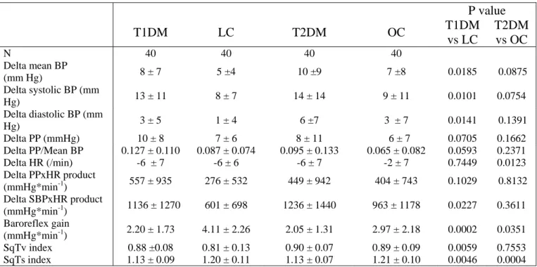
Table 2: Changes (delta) in mean blood pressure (BP), systolic BP, diastolic BP, pulse pressure
(PP), pulsatility index (PP/mean BP), heart rate (HR), pulsatile stress (PPxHR), systolic BP x HR double product (SBPxHR) occurring during the transition from the initial standing position to the squatting position in middle-aged diabetic patients with type 1 diabetes (T1DM), patients with type 2 diabetes (T2DM), non-diabetic lean controls (LC) and non-diabetic overweight/obese controls (OC) (20 men and 20 women in each group). Mean values of baroreflex gain as well as SqTv and SqTs indices of cardiac autonomic neuropathy are also presented in the four groups. Results are expressed as mean ± SD. NS : non significant. NA : not applicable.
P value T1DM LC T2DM OC T1DM vs LC T2DM vs OC N 40 40 40 40 Delta mean BP (mm Hg) 8 ± 7 5 ±4 10 ±9 7 ±8 0.0185 0.0875 Delta systolic BP (mm Hg) 13 ± 11 8 ± 7 14 ± 14 9 ± 11 0.0101 0.0754 Delta diastolic BP (mm Hg) 3 ± 5 1 ± 4 6 ±7 3 ± 7 0.0141 0.1391 Delta PP (mmHg) 10 ± 8 7 ± 6 8 ± 11 6 ± 7 0.0705 0.1662 Delta PP/Mean BP 0.127 ± 0.110 0.087 ± 0.074 0.095 ± 0.133 0.065 ± 0.082 0.0593 0.2371 Delta HR (/min) -6 ± 7 -6 ± 6 -6 ± 7 -2 ± 7 0.7449 0.0123 Delta PPxHR product (mmHg*min-1) 557 ± 935 276 ± 532 449 ± 942 404 ± 743 0.1029 0.8132 Delta SBPxHR product (mmHg*min-1) 1136 ± 1270 601 ± 698 1236 ± 1440 963 ± 1178 0.0227 0.3611 Baroreflex gain (mmHg*min-1) 2.20 ± 1.73 4.11 ± 2.26 2.05 ± 1.31 2.97 ± 2.18 0.0002 0.0351 SqTv index 0.88 ±0.08 0.81 ± 0.13 0.90 ± 0.07 0.89 ± 0.09 0.0059 0.7553 SqTs index 1.13 ± 0.09 1.20 ± 0.11 1.13 ± 0.07 1.21 ± 0.10 0.0046 0.0004
Figure 1: Changes in mean arterial blood pressure (MBP), pulse pressure (PP) and heart rate
(HR) during a posture test [1 min standing – 1 min squatting (grey zone) – 1 min standing]. (A) : 40 patients with type 1 diabetes (open circles) versus 40 non-diabetic (full circles) subjects
matched for age, sex and BMI; (B): 40 patients with type 2 diabetes (open triangles) versus 40 non-diabetic (full triangles) subjects matched for age, sex and BMI; and (C) 40 patients with type 1 diabetes (open circles) versus 40 patients with type 2 diabetes (open triangles) subjects
Figure