CD45 up-regulation during lymphocyte
maturation
Jorg Kirberg and Thomas Brocker
Basel Institute for Immunology, Grenzacherstrasse 487, Postfach, 4005 Basel, Switzerland Keywords: CD45 up-regulation, mouse, positive selection, signaling molecules, T lymphocytes
Abstract
CD4+CD8+ double-positive thymocytes differentiate into CD4+ and CD8+ single-positive T cells
during thymic positive selection. This process requires the interaction between the TCR and self MHC molecules. In this context we have analyzed the expression of CD45, an abundant
transmembrane protein tyrosine phosphatase, and describe here its differential surface expression during T cell maturation. Using four-color FACS analysis of thymocytes from normal as well as TCR-transgenic mice we demonstrate that CD45 is up-regulated only during positive selection concomitantly with the TCR-CD3 complex and the transient early activation marker CD69, but that this regulation precedes heat stable antigen down-regulation. The tight linkage of the up-regulation of the TCR-CD3 complex and CD45 may be required because the CD45 tyrosine phosphatase plays a role in modulating signal transduction by the TCR-CD3 complex during positive selection. In addition, our findings argue for a regulation mechanism that adapts the CD45 levels to increasing antigen receptor levels on mature T cells and B cells.
Introduction
CD45 is a transmembrane protein tyrosine phosphatase abundantly present on the cell surface of all nucleated hematopoietic cells. This protein exists in several isoforms due to alternative splicing of four of its exons (for reviews see 1,2). Studies with CD45" mutant cell lines that were antigen receptor "signaling deficient showed that expression of CD45 is a prerequisite for transmembrane signal transduction upon triggering of the TCR (3,4) as well as the B cell antigen receptor (5). It was further demonstrated that CD45 dephosphorylates specifically negative regulatory tyrosine residues of src family protein tyrosine kinases, an event that results in increased kinase activity in T cells (6-9) and B cells (5,10,11). The disruption of the CD45 gene underlined clearly the essential role of this phosphatase in T lymphocyte development and B cell function, since it resulted in a drastic reduction of mature single-positive thymocyte numbers and mature B lymphocytes with signaling-deficient antigen receptors (12).
Protein phosphorylation is an important mechanism of trans-ducing signals in eukaryotic cells. It has been proposed that a resting cell is maintained below a threshold for signaling through a balance between opposing dephosphorylation and phosphorylation reactions (13). In fact, chemical inactivation of phosphatases in Jurkat cells leads to IL-2 production without TCR triggering (14), which indicates that the cell has
to regulate both opposing activities tightly to keep them in balance.
Little is yet known about the regulation of CD45-phosphat-ase activity. It has been suggested that the different isoforms of CD45 could interact with different ligands and become thereby differentially activated (for review see 2). When mouse thymocytes were previously investigated for expression of certain isoforms that might correlate to positive or negative selection events, not all analyzed thymocytes (only 1-3%) expressed specific isoforms (15).
Therefore, in the present study we used three different CD45 pan-isoform-specific reagents together with CD4, CD8 and CD3, heat stable antigen (HSA) or CD69 as thymocyte markers as well as IgM, IgD, CD19 or B220 as B lymphocyte markers and extensively analyzed CD45 expression (regard-less of isoforms) in various thymocyte subsets as well as during B cell development.
In clear contrast to earlier reports (16), we demonstrate in this study that there is an extremely tight correlation between CD45 expression levels and positive selection events in the thymus. More than 90% of all positively selected thymocytes showed a CD45hiQh phenotype, whereas basically all
non-selected thymocytes stained CD45|OW. Similarly, we could
detect a drastic CD45 up-regulation during B cell development that paralleled B cell antigen receptor up-regulation and B
Correspondence to: T. Brocker
Q O
5 4
i - . . ' . . . .- \ 3 2 1 8 9 CD8 COo
o
21 29 9 6 41 95 83 1 4 T—,fc "I*—r.t
o
o
54 32 49 44CD45(Ly5
a)
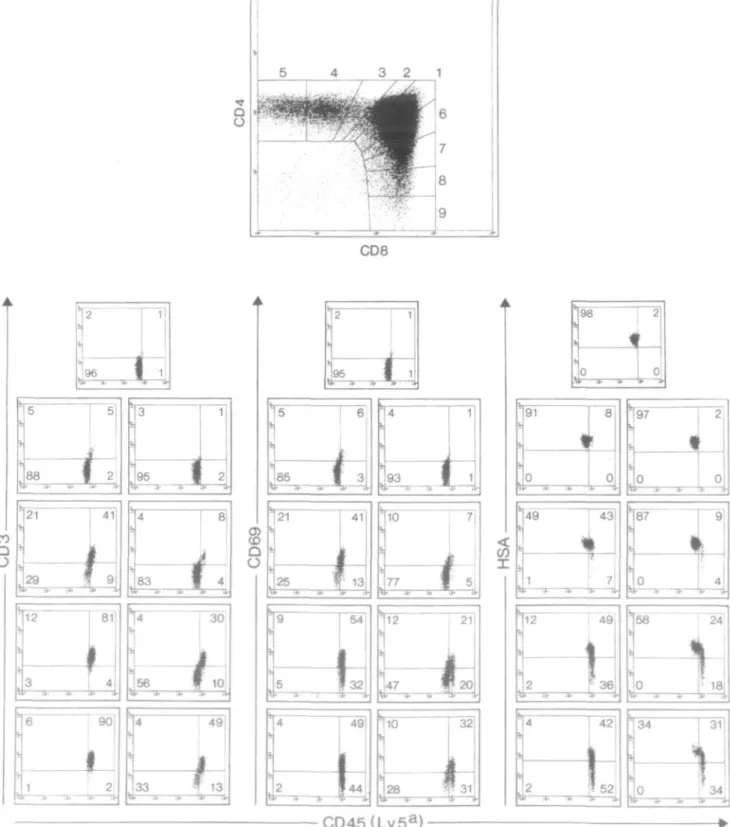
91 49 98 43 42 52 34 31 34Fig. 1. Four-color FACS analysis of B6.SJL (Ly-5a) thymocytes: CD4 and CD8 expression was analyzed with a gate set on forward versus side
scatter parameters to exclude dead cells and debris. Subsequently, nine gates, corresponding to different developmental stages between CD4+CD8+ DP and CD4+ SP and CD8+ SP thymocytes, were introduced. The lower three panels show for each of these nine gates the analysis with the following pairs of mAb: lower left, center and right panels show CD45 (clone 104.2.1) versus CD3, CD69 or HSA respectively. In each panel, gate 1 (CD4+CD8+ DP cells) corresponds to the top middle position, while gates 2-5 descend to the left (towards CD4+ SP cells) and gates 6-9 descend to the right (towards CD8+ SP cells). A total of 3X10⁵ cells were analyzed. All cells within the forward versus side scatter parameters are displayed in the top panel, whereas 2000 cells of the gated subpopulations are shown in the small panels below.
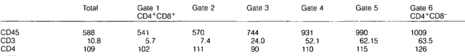
Table 1. CD45 and TCR up-regulation during thymocyte maturation CD45 CD3 CD4 Total 588 10.8 109 Gate 1 CD4+CD8+ 541 5.7 102 Gate 2 570 7.4 111 Gate 3 744 24.0 90 Gate 4 931 52.1 110 Gate 5 990 62.15 115 Gate 6 CD4+CD8-1009 63.5 126
Data are derived from the same analysis as shown in Fig. 1. Similar to Fig. 1, six gates were set to cover the developmental transition between immature CD4+CD8+ and mature CD4+CD8~ T cells. The mean relative fluorescence intensity of CD45, CD3 and CD4 of the various subpopulations is shown.
cell maturation. We discuss the implications of these findings with respect to TCR and B cell receptor expression levels during lymphocyte development.
Methods
Mice
B6.SJL (Ly-5a) and C56BL/6 mice were purchased from
Jackson Laboratories (Bar Harbor, ME) or IFFA Credo (Orleans, France) respectively. TCR transgenic mice, with a hemagglutinin-specific, l-Ed-restricted TCR, were as
described before and crossed to a recombination activating gene negative (RAG-2^-) background (17). Since RAG-2"'" mice were a mixture of Ly-5a and Ly-5b mice, we selected
for these markers to obtain Ly-5a or Ly-5b homozygotes.
Phenotyping was done by FACS staining of peripheral blood lymphocytes with mAb specific for TCR Vp8, TCR clonotype, Ly-5 allele and Dd or Kb. Mice were bred in the animal colony
of the Basel Institute for Immunology and were analyzed at 6-10 weeks of age.
mAb and FACS analysis
Hybridoma supernatants containing mAb were purified by Protein G (Pharmacia, San Diego, CA)-affinity chromato-graphy. mAb were labeled using biotin- or fluorescein-succinimidyl-ester (FLUOS) according to the manufacturer's instructions. The following mAb were used: T19.191 (anti-Dd) (17), AF6-88.5.3 (anti-Kb) (18), M1/69 (anti-HSA) (19),
KT-3 (anti-CD3) (20), F23.1 (anti-TCR Vp8) (21), A20-1.7
(anti-Ly-5b) (22), 104.2.1 (anti-Ly-5a) (22) and 6.5
(anti-clono-typic TCR) (23). Anti-CD4-phycoerythrin (Becton Dickinson, Plymouth, UK), anti-CD8-Red613 (Gibco/lmmunoselect/BRL, Grand Island, NY), anti-lgD (clone 217-170; PharMingen, San Diego, CA), anti-IgM (clone AF6-78; PharMingen), RA3-6B2 (anti-CD45, B220 isoform; PharMingen) and anti-CD45-FITC (clone 30F11.1, specific for all CD45 isoforms; PharMingen) conjugates were obtained commercially. With these and the second step reagent streptavidin-allophycocyanin (APC; Molecular Probes, Eugene, OR) four color flow-cytometry was performed on a FACStar"1" (Becton Dickinson) instrument
equipped appropriately.
Single cell preparation, staining and FACS analysis were done according to standard procedures.
Results
In order to investigate the expression of CD45 on thymocytes of B6.SJL mice that were homozygous for one of the CD45
alleles (Ly-5a), we performed four-color flow cytometric
ana-lysis with mAb specific for CD4, CD8, CD45 and a fourth marker (CD3, CD69 or HSA respectively) (Fig. 1). We first analyzed the thymocytes according to their CD4 and CD8 expression and gated them in distinct developmental stages between double-positive (DP) and single-positive (SP) thymo-cytes (see Fig. 1, gates 1-9). We further analyzed the cells in gates 1-9 according to their expression of CD45 versus either CD3, CD69 or HSA. Figure 1 shows that the expression levels of CD45 increase during maturation of thymocytes (Fig. 1, lower part, with the DP fraction of gate 1 on the top of the two columns and the maturing CD4+ SP cells shown in the
dot-plots descending to the left, the CD8+ SP cells to the
right). In the lower left panel of Fig. 1, CD45 expression is correlated with TCR-CD3 complex expression as measured by a mAb specific for CD3e. While all DP thymocytes are dull for CD3 and positive for CD45, >90% of the most mature CD4+ SP cells are expressing higher levels of CD3 and CD45.
The same shift can be observed for CD8+ SP thymocytes
with the remaining CD3dullCD45low cells being immature CD8+
SP (24,25).
When we compared CD69 and CD45 (Fig. 1) we observed the expected transient up-regulation of CD69 (26) by thymo-cytes on their way to mature CD4+ SP or CD8+ SP cells. This
transient up-regulation of CD69 takes place at the same stage as CD45 up-regulation. With further maturation CD69 is down-regulated again, while CD45 expression levels remain high (Fig. 1, gates 5 and 9). Immature CD8+ SP thymocytes remain
CD45 low (see lower right dot-plot corresponding to gate 9).
We then analyzed the expression of CD45 in the context of HSA. As described previously, immature thymocytes express high levels of HSA and lose this marker on their way to mature SP cells (27). According to Fig. 1 (right lower panel) practically all of the more mature thymocytes became CD45h'9h before
they started to lose HSA expression.
Quantitative analysis of three surface markers shows that CD4 levels do not change, CD3 is up-regulated - 1 2 times and CD45 levels are increased 2-fold during thymocyte maturation as shown in Table 1.
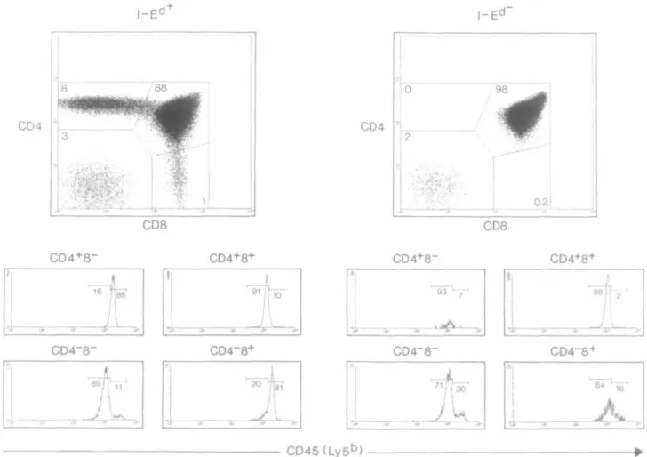
In order to see whether this event is dependent on positive selection, we repeated the experiments in RAG-2~'~ TCR-transgenic mice that can or cannot positively select immature thymocytes. As previously described (17), in the I-Ed+
TCR-transgenic RAG-2"'" mice, positive selection leads to the presence of CD4+ SP and CD8+ SP mature thymocytes (Fig.
D O 1-2 "
-•IF
3 CD8a
o
2 . > . - ' • ' * lfc '* ! 3 CD8 i i 0^91 1 i|
*"* '*' *
lfc>'•"
: I "
- • 0
98 I
' 2 4
I 01
!76 1 0
tt> 1*' i f I** 1* "0*99 i
0 . 1t
I
0
*• ir "0 s"99 1
4 . .•• .4. 0 -"11 V 1 47'1 I
1 J
r
I 36
93 0 173i J
6 L 1 0 i- "A-17 1u
95 1
: .
*4 0
f f
' 2 0
a- .»• it -i.—i97 I 1
' 2 0
t#* <•' !«• !»•:*-CD45(Ly5
a)
be detected (Fig. 2). We gated on CD4+CD8+ DP, CD4+ SP
or CD8+ SP thymocytes (gates 1, 2 and 3), and analyzed
them for CD45 versus CD3, CD69 and HSA. In case of I-Ed+ TCR-transgenic RAG-2"'"" mice we observed the same
correlation between these markers as described above: CD45 is up-regulated during positive selection on thymocytes (Fig. 2). In contrast, thymocytes from I-Ed"TCR-transgenic RAG-2^"
mice that cannot positively select with this TCR remain CD45l0W (Fig. 2). Thus CD45 up-regulation is dependent on
positive selection events.
To test whether CD45 up-regulation is allele-specific we compared I-Ed+ and I-Ed" TCR-transgenic RAG-2~'~ mice that
were homozygous for the CD45 allele Ly-5b. As shown in Fig.
3, we obtained similar results as in Ly-5a mice. Apparently CD45 up-regulation upon positive selection is a general phenomenon, not restricted to a particular allele.
While the majority of thymocytes and activated T cells
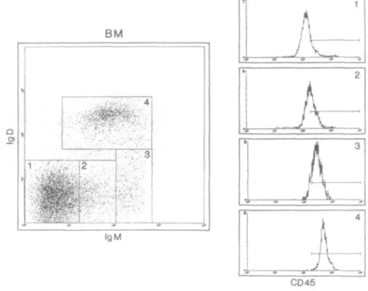
express different low mol. wt isoforms of CD45, B220, a high molecular isoform of CD45, has been routinely used as a marker for all B lineage cells (1,28). We were interested if the observed CD45 up-regulation during T cell maturation could similarly be detected with a pan-isoform CD45 specific mAb on developing B cells in the bone marrow. To this end we analyzed bone marrow cells as shown in Fig. 4. To concentrate on B lineage cells, we first gated on B220+ cells (data not
shown). According to their surface IgM and IgD expression we divided these into four stages corresponding to gates 1-4: IgM/lgD double negative (gate 1), lgMl0W SP (gate 2),
igMhigh S P a n d |gMhigh/|gD'°w DP (gate3), and IgM/lgD DP
(gate 4), reflecting the normal B cell developmental pathway. When these four populations were analyzed for CD45 expres-sion levels, we found an up-regulation of CD45 (see histo-grams in Fig. 4) tightly linked to surface Ig up-regulation and B cell maturation. CD4 • j . j 8 __ / 3 8 8 •<**• i CD4 CD8 CD8
CD4+8-
CD4+8-*CD4S+
>, 1
CD4+8~ CD4+8+ 98 J vCD4~8
+CD45(Ly5
b)
Fig. 3. Three-color FACS analysis of I-E+ and I-E" RAG-2~'~ Ly-5b TCR-transgenic thymocytes. Analysis was performed as described for Fig.
2, but with a pan-specific CD45 mAb (clone 30F11.1).
Fig. 2. Four-color FACS analysis of I-Ed+ and I-Ed~ RAG-2^" Ly-5a TCR-transgenic thymocytes. Analysis and data display was performed as
described for Fig. 1. CD4+CD8+ DP, CD4+ SP or CD8+ SP thymocytes were analyzed using gates 1, 2 and 3 as shown. In each panel, CD4+ SP, CD4+CD8+ DP and CD8+ SP cells correspond to the upper left, upper right or lower right dot-plot respectively.
BM
QTT>3
IgM i a. •••-' -j. ^-f a-CD45Fig. 4. Four-color FACS analysis of bone marrow. Bone marrow cells were isolated from C57BL/6 mice. Dead cells and debris was excluded
with a gate set on forward versus side scatter parameters. For further analysis, only B220+ cells were included. These were divided into four populations based on IgM and IgD expression. For each of these four populations, the histograms show the expression of CD45 (clone 30F11.1).
Discussion
Earlier studies have shown that it is difficult to define rules for the regulation of CD45 isoform expression on thymocytes, since only minimal populations (1-3%) express specific iso-forms (CD45RA and CD45RBh'9h) that occur during positive
or negative selection in the thymus (15). Since a much higher percentage of thymocytes are positively selected in normal and TCR-transgenic mice but do not express or up-regulate certain CD45 isoforms, we investigated the overall expression levels of CD45 on thymocytes with pan-CD45 antibodies to see if isoform-independent regulation might occur. Using four-color flow cytometry we analyzed thymocytes from normal and TCR-transgenic mice and compared CD45 expression levels with other more commonly used thymocyte markers like CD3, CD69 and HSA. As measured with three different mAb specific for all isoforms of CD45, we describe here that CD45 expression levels are lower on CD4+CD8+ DP
thymocytes, but increase when these cells mature to the CD4+ SP or CD8+ SP stage. The CD45 up-regulation occurs
simultaneously with an increase of TCR-CD3 levels and one might argue that the CD45 phosphatase levels are increased because of an ongoing increase of TCR-CD3 levels in order to regulate signals received during thymocyte positive selection. Nearly all (>90%) thymocytes followed this rule.
Interestingly, during B cell development, the CD45 isoform B220 shows a similar shift in surface expression (29). However, as yet undetected was the general, isoform-independent
up-regulation of CD45 expression when B cells start to express Ig, as shown in this study.
These findings indicate that the total CD45 levels (regard-less of its isoforms) may be important in developing lympho-cytes to regulate signal transduction activity during lymphocyte maturation.
Acknowledgements
The authors would like to thank Mark Dessing for expert technical assistance with FACS analysis, Hanspeter Stahlberger for graphic art work, and Drs Kerry Campbell, Hans-Reimer Rodewald, Klaus Karjalainen and Harald von Boehmer for reading the manuscript. The mAb A20-1.7 and 104.2.1 were a kind gift of Dr Hans-Reimer Rodewald. The Basel Institute for Immunology was founded and is supported by Hoffmann-La Roche Ltd, Basel, Switzerland.
Abbreviations
APC allophycocyanin DP double positive HSA heat shock antigen SP single positive
References
1 Thomas, M. L. 1989. The leukocyte common antigen family. Annu. Rev. Immunol. 7:339.
role as a protein tyrosine phosphatase required for lymphocyte activation and development. Annu. Rev. Immunol. 12:85. 3 Pingel, J. T. and Thomas, M. L. 1989. Evidence that the
leukocyte-common antigen is required for antigen-induced T lymphocyte proliferation. Cell 58:1055.
4 Weaver, C. T., Pingel, J. T., Nelson, J. O. and Thomas, M. L. 1991. CD8+ T-cell clones deficient in the expression of the CD45 protein tyrosine phosphatase have impaired responses to T-cell receptor stimuli. Mol. Cell. Biol. 11:4415.
5 Justement, L. B., Campbell, K. S., Chien, N. C. and Cambier, J. C. 1991. Regulation of B cell antigen receptor signal transduction and phosphorylation by CD45. Science 252:1839.
6 Ostergaard, H. L, Shackelford, D. A., Hurley, T. R., Johnson, P., Hyman, R., Sefton, B. M. and Trowbridge, I. S. 1989. Expression of CD45 alters phosphorylation of the Ick-encoded tyrosine protein kinase in murine lymphoma T-cell lines. Proc. Natl Acad. Sci. USA 86:8959.
7 Mustelin, T. and Altman, A. 1990. Dephosphorylation and activation of the T cell tyrosine kinase pbQlck by the leukocyte common antigen (CD45). Oncogene 5:809.
8 Shiroo, M., Goff, L, Biffen, M., Shivnan, E. and Alexander, D. 1992. CD45 tyrosine phosphatase-activated p59^n couples the T cell antigen receptor to pathways of diacylglycerol production, protein kinase C activation and calcium influx. EMBO J. 11:4887. 9 McFarland, E. D., Hurley, T. R., Pingel, J. T., Sefton, B. M., Shaw, A. and Thomas, M. L. 1993. Correlation between Src family member regulation by the protein-tyrosine-phosphatase CD45 and transmembrane signaling through the T-cell receptor. Proc. Natl Acad. Sci. USA 90:1402.
10 Ales-Martinez, J. E., Cuende, E., Martinez, C , Parkhouse, R. M., Pezzi, L. and Scott, D. W. 1991. Signaling in B cells. Immunol
Today 12:201.
11 Reth, M. 1992. Antigen receptors on B lymphocytes. Annu. Rev. Immunol. 10:97.
12 Kishihara, K., Penninger, J., Wallace, V. A., Kundig, T. M., Kawai, K., Wakeham, A., Timms, E., Pfeffer, K., Ohashi, P. S., Thomas, M. L, etal. 1993. Normal B lymphocyte development but impaired T cell maturation in CD45-exon6 protein tyrosine phosphatase-deficient mice. Cell 74:143.
13 Brautigan, D. L. 1994. Protein phosphatases. Recent Progr. Horm. Res. 49:197.
14 Secrist, J. P., Burns, L. A., Karnitz, L, Koretzky, G. A. and Abraham, R. T. 1993. Stimulatory effects of the protein tyrosine phosphatase inhibitor, pervanadate, on T-cell activation events. J. Biol. Chem. 268:5886.
15 Wallace, V. A., Fung Leung, W. P., Timms, E., Gray, D., Kishihara, K., Loh, D. Y., Penninger, J. and Mak, T. W. 1992. CD45RA and CD45RBhigh expression induced by thymic selection events. J. Exp.Med. 176:1657.
16 Trowbridge, I. S. 1991. CD45. A prototype for transmembrane protein tyrosine phosphatases. J. Biol. Chem. 266:23517. 17 Kirberg, J., Baron, A., Jakob, S., Rolink, A., Karjalainen, K. and
von Boehmer, H. 1994. Thymic selection of CD8+ single positive cells with a class II major histocompatibility complex-restricted receptor. J. Exp. Med. 180:25.
18 Loken, M. R. and Stall, A. M. 1982. Flow cytometry as an analytical and preparative tool in immunology. J. Immunol. Methods 50:R85. 19 Springer, T., Galfre, G., Secher, D. S. and Milstein, C. 1978. Monoclonal xenogeneic antibodies to murine cell surface antigens: identification of novel leukocyte differentiation antigens. Eur. J. Immunol. 8:539.
20 Portoles, P., Rojo, J., Golby, A., Bonneville, M., Gromkowski, S., Greenbaum, L, Janeway, C. A., Jr, Murphy, D. B. and Bottomly, K. 1989. Monoclonal antibodies to murine CD3 epsilon define distinct epitopes, one of which may interact with CD4 during T cell activation. J. Immunol. 142:4169.
21 Staerz, U. D., Rammensee, H. G., Benedetto, J. D. and Bevan, M. J. 1985. Characterization of a murine monoclonal antibody specific for an allotypic determinant on T cell antigen receptor. J. Immunol. 134:3994.
22 Shen, F. W. 1981. Monoclonal antibodies to mouse lymphocyte differentiation antigens. In Haemmerling, G., Haemmerling, U. and Kearney, J. F., eds, Monoclonal Antibodies and T Cell Hybridomas, p. 25. Elsevier, Amsterdam.
23 Kirberg, J., Baron, A., Jakob, S., Rolink, A., Karjalainen, K. and von-Boehmer, H. 1994. Thymic selection of CD8+ single positive cells with a class II major histocompatibility complex-restricted receptor. J. Exp. Med. 180:25.
24 Nikolic-Zugic, J. and Bevan, M. J. 1988. Thymocytes expressing CD8 differentiate into CD4+ cells following intrathymic injection.
Proc. Natl Acad. Sci. USA 85:8633.
25 Guidos, C. J., Weissman, I. L. and Adkins, B. 1989. Intrathymic maturation of murine T lymphocytes from CD8+ precursors. Proc.
Natl Acad. Sci. USA 86:7542.
26 Swat, W., Dessing, M., von Boehmer, H. and Kisielow, P. 1993. CD69 expression during selection and maturation of CD4+CD8+ thymocytes. Eur. J. Immunol. 23:739.
27 Wilson, A., Day, L. M., Scollay, R. and Shortman, K. 1988. Subpopulations of mature murine thymocytes: properties of CD4~ CD8+ and CD4+CD8" thymocytes lacking the heat-stable antigen.
Cell. Immunol. 117:312.
28 Coffman, R. L. 1982. Surface antigen expression and immunoglobulin gene rearrangement during mouse pre-B cell development. Immunol. Rev. 69:5.
29 Hardy, R. R., Carmack, C. E., Shinton, S. A., Kemp, J. D. and Hayakawa, K. 1991. Resolution and characterization of pro-B and pre-pro-B cell stages in normal mouse bone marrow. J. Exp. Med. 173:1213.