HAL Id: hal-01692779
https://hal-amu.archives-ouvertes.fr/hal-01692779
Submitted on 25 Jan 2018HAL is a multi-disciplinary open access archive for the deposit and dissemination of sci-entific research documents, whether they are pub-lished or not. The documents may come from teaching and research institutions in France or abroad, or from public or private research centers.
L’archive ouverte pluridisciplinaire HAL, est destinée au dépôt et à la diffusion de documents scientifiques de niveau recherche, publiés ou non, émanant des établissements d’enseignement et de recherche français ou étrangers, des laboratoires publics ou privés.
Endothiovibrio diazotrophicus gen. nov., sp. nov., a
novel nitrogen-fixing, sulfur-oxidizing
gammaproteobacterium isolated from a salt marsh
Dennis Bazylinski, Viviana Morillo, Christopher Lefèvre, Nathan Viloria,
Bradley Dubbels, Timothy Williams
To cite this version:
Dennis Bazylinski, Viviana Morillo, Christopher Lefèvre, Nathan Viloria, Bradley Dubbels, et al.. Endothiovibrio diazotrophicus gen. nov., sp. nov., a novel nitrogen-fixing, sulfur-oxidizing gammapro-teobacterium isolated from a salt marsh. International Journal of Systematic and Evolutionary Micro-biology, Microbiology Society, 2017, 67 (5), pp.1491-1498. �10.1099/ijsem.0.001743�. �hal-01692779�
Endothiovibrio diazotrophicus gen. nov., sp. nov., a novel nitrogen-fixing, sulfur-oxidizing
gammaproteobacterium isolated from a salt marsh
Dennis A Bazylinski1, Viviana Morillo1, Christopher T Lefèvre2, Nathan Viloria1, Bradley L Dubbels3, Timothy J Williams4
1 School of Life Sciences, University of Nevada at Las Vegas, Las Vegas, Nevada 89154-4004, USA
2 CEA Cadarache/CNRS/Université Aix-Marseille, UMR7265 Biosciences and Biotechnologies Institute, Laboratoire de Bioénergétique Cellulaire, Saint Paul lez Durance 13108, France
3 Novozymes North America Inc., 9000 Development Drive, Morrisville, North Carolina 27560, USA
4 School of Biotechnology and Biomolecular Sciences, The University of New South Wales, Sydney NSW 2052, Australia
*Correspondence: Dennis A. Bazylinski, dennis.bazylinski@unlv.edu
ABSTRACT
A novel non-phototrophic, marine, sulfur-oxidizing bacterium, strain S-1T, was isolated from a coastal salt marsh in Massachusetts, USA. Cells are Gram-stain-negative vibrios motile by means of a single polar unsheathed flagellum. S-1T is an obligate microaerophile with limited metabolic capacity. It grows chemolithoautotrophically utilizing sulfide and thiosulfate as electron donors, converting these compounds to sulfate, and the Calvin–Benson–Bassham cycle for carbon fixation. Cells of S-1T did not grow on any of a large number of organic carbon sources and there was no evidence for chemoorganoheterotrophic growth. Cells produced internal sulfur globules during growth on sulfide and thiosulfate. S-1T is strongly diazotrophic, as demonstrated by 15N2 fixation and
acetylene reduction activity by cells when a fixed nitrogen source is absent from the growth medium. The marine nature of this organism is evident from its ability to grow in 10 to 100 % artificial
seawater but not at lower concentrations and NaCl alone cannot substitute for sea salts. The major cellular fatty acids are C16 : 1ω7c, C16 : 0, and C18 : 1ω7c. Phosphatidylethanolamine and
phosphatidylglycerol are the major polar lipids. Q8 is the only respiratory quinone. S-1T genomic DNA has a G+C content of 67.6 mol%. Based on its 16S rRNA gene sequence, S-1T shows the closest phylogenetic relationship to non-phototrophic species within the
family Thioalkalispiraceae of the class Gammaproteobacteria . The name Endothiovibrio
diazotrophicus is proposed for this organism, with S-1T as the type strain (ATCC BAA-1439T=JCM 17961T).
Keyword(s): sulfur deposits, sulfur oxidization, Thioalkalispiraceae, S-1, nitrogen fixation, Endothiovibrio diazotrophicus
The Gammaproteobacteria class of the phylum Proteobacteria is one of the most genera-rich taxa among the bacteria [ 1 ] and includes a relatively large number of chemolithoauthotrophic and photolithoautotrophic sulfur-oxidizing bacteria. Most of these belong to one of two orders: the Chromatiales (the ‘purple sulfur bacteria’) and the Thiotrichales . Although the ability to store and metabolize elemental sulfur is widespread among these bacteria, only a relatively small subset is known to store sulfur intracellularly [ 2 ]. In these cells, sulfur inclusions are stored within the confines of the cell wall, and have an extracytoplasmic/periplasmic location [ 2, 3 ]. In many chemotrophic and phototrophic sulfur-oxidizers, the deposition of elemental sulfur appears to be an obligatory intermediate step in the complete oxidation of sulfide and thiosulfate to sulfate, probably for kinetic reasons [ 2, 4 ]. The formation of these so-called sulfur ‘globules’ is prevalent among
members of the order Chromatiales of the class Gammaproteobacteria , in which the oxidation of reduced sulfur compounds fuels the autotrophic assimilation of CO 2 (thioautotrophy). The
order Chromatiales can be classified into seven families
: Chromatiaceae , Ectothiorhodospiraceae ,Halothiobacillaceae , Thioalkalispiraceae , Wenzhouxi angellaceae , Woeseiaceae and Granulosicoccaceae [ 5–8 ]. Here we describe strain S-1 T, a marine, non-phototrophic, sulfur-oxidizing member of the family Thioalkalispiraceae , a family of strict chemolithoautotrophs that are mesophilic/moderately thermophilic and moderately halophilic [ 6 ]. S-1 T exhibits intracellular sulfur globule deposition associated with the oxidation of sulfide and thiosulfate.
Mud and water was collected from the brackish School Street Marsh at Woods Hole, (Massachusetts, USA) and used to inoculate sulfide-O 2concentration gradient media, prepared following the recipe of Nelson and Jannasch [ 9 ] but modified by using a diluted artificial seawater (ASW) solution to replace natural seawater and by the addition of 20 mM ferric quinate and 200 µl 0.2 % aqueous resazurin per litre. The ferric quinate was originally included in the growth medium because we were attempting to culture magnetotactic bacteria and later removed because the organism described here did not require this high concentration of iron for growth. The salinity of the water at the site was 10 p.p.t. (=1 % w/v) as determined with a Palm Abbe PA203 hand-held refractometer (MISCO Refractometer). The ASW used in routine culturing of this organism was adjusted to this salinity and consisted of (g l −1): NaCl, 7.82; MgCl 2·6H 2O, 1.66; Na 2SO 4, 1.30; KCl, 0.22; and CaCl 2·2H2O, 0.18. Cells initially grew at the oxic–anoxic interface (OAI), corresponding to the pink–colorless interface as a microaerobic band of cells. Although the culture was mixed, the dominant micro-organism was a vibrio that appeared to produce intracellular sulfur globules.
For isolation of the strain, cells from the enrichment gradient culture were inoculated in a dilution series of solid agar [13 g l −1 Agar Noble (Difco Laboratories)] in shake tubes of an O 2 concentration gradient medium containing: 5 ml modified Wolfe’s mineral elixir [ 10 ]; 0.5 ml vitamin solution [ 10 ]; 10 mM Na 2S 2O 3·5H 2O as the electron donor; 0.2 g NH 4Cl as the nitrogen source; 2 ml freshly prepared, filter-sterilized, neutralized 0.43 M cysteine· HCl·H 2O as the reducing agent; 1.8 ml 0.5 M KHPO 4 buffer pH 6.9 and 2.67 ml 0.8 M NaHCO 3 as the carbon source, per litre diluted ASW. The thiosulfate, cysteine and NaHCO 3 were added after autoclaving and the pH adjusted to 7.1–7.2. The medium was dispensed under air as 10 ml aliquots into sterile 15×125 mm test tubes which were kept at 44 °C, inoculated, inverted several times and then quickly cooled on ice. Cells of S-1 grew as lens-shaped colonies at the OAI of this medium after 7–10 days, and several were aseptically extracted and serially diluted in shake tubes two more times.
After isolation, S-1 T was routinely grown in a semi-solid growth medium containing (per l of ASW): 0.2 ml 0.2 % aqueous resazurin, 10.0 ml 25 % Na 2S 2O 3·5H 2O, 0.25 g NH 4Cl, 2.0 ml neutralized 0.43 M cysteine·HCl·H 2O, 2.67 ml 0.8 M NaHCO 3, 5 ml modified Wolfe’s mineral elixir [10 ], 0.5 ml vitamin solution [ 10 ] and 2.0 g Agar Noble (Difco Laboratories). The final pH was adjusted to 7.0–7.1. Cells grew as a microaerobic band of cells within the OAI below the surface of the medium ( Fig. 1a ). Although cysteine is a potential carbon and/or energy source, there was no observable growth of S-1 T on the aforementioned medium lacking sulfide or thiosulfate, either with or without NaHCO 3. Thus S-1 Tdoes not appear to be able to use cysteine as a carbon or energy source.
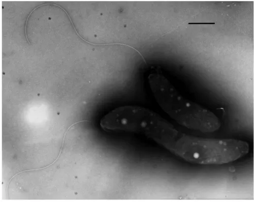
Fig. 1. (a) Growth of S-1 T in semi-solid, thiosulfate, oxygen-gradient medium containing phenol red (two tubes at left) as a pH indicator or resazurin (two tubes at right) as an indicator of oxygen. Note that cells grow in inoculated tubes (2 and 4) as a microaerophilic band of cells below the meniscus. The color of tube 2 near the band of cells is yellow, indicating the production of acid, specifically sulfuric acid. In tube 4 cells grew within the oxic–anoxic transition zone as indicated by the resazurin which is colorless when reduced, in this case when oxygen is not present. The uninoculated tubes remained unchanged. (b) Differential interference contrast microscopic image of S-1 cells grown with thiosulfate as electron donor showing the presence of intracellular sulfur globules (c and d). Scanning transmission electron microscope (STEM) image of unstained cells of S-1 T and corresponding O, P, S and Mg elemental maps of the same cells. One sulfur-rich and one phosphorus-rich inclusion is designated by labeled arrows in the STEM image. The map shows locations of numerous sulfur-rich globules and phosphorus-rich bodies that probably consist of polyphosphate. The lower cell appears to have lysed. Bars, 1 µm.
To scale up liquid cultures for DNA extraction, S-1 T was grown in liquid thiosulfate medium as described above, except that the volume of cysteine·HCl·H 2O solution was decreased to 0.5 ml l −1, the concentration of NaHCO 3 was increased to 15 mM, and the medium was rendered anaerobic by sparging the medium with O 2-free 7.5 % CO 2 in N 2. Oxygen was added to these bottles after autoclaving to a headspace concentration of 0.7 % to initiate growth. Once turbidity (growth) became apparent O 2 was added in increments of 1 % of the headspace when O 2 from the previous injection was utilized (when medium became colorless).
Determination of DNA G+C content was performed using HPLC by the Deutche Sammlung von Mikroorganismen und Zellkulturen (DSMZ) according to the methods described by Cashion et al. [ 11 ], Tamaoka and Komagata [ 12 ], and Mesbah et al. [ 13 ]. The G+C content of genomic DNA of S-1 T, as determined by HPLC, was 67.6±2.0 mol% ( n=6 replicates). The 16S rRNA gene sequence of S-1 T was amplified from genomic DNA, using primers fD1 and rD1 according to the method of Weisburg et al. [ 14 ]. Using blastn, the 16S rRNA gene sequence of strain S-1 T (GenBank accession number HQ379738) was shown to have the highest sequence identity to that of the rRNA gene from Thioalkalivibrio sulfidiphilus HL-EbGr7 T (93 % sequence identity; GenBank accession number CP001339), a member of the family Ectothiorhodospiraceae , among characterized strains. Phylogenetic analysis (Neighbor-Joining, based on 1078 aligned bp of 16S rRNA) recovered strain S-1 T as nested within the family Thioalkalispiraceae of the
class Gammaproteobacteria , although Thioalkalivibrio sulfidiphilus was found not to be the closest relative of S-1 T ( Fig. 2 ). This family contains obligate chemolithoautotrophs [ 6, 15–17 ], which accords with the chemolithoautotrophic metabolism of S-1 T. S-1 T was demonstrated only to be able to use sulfide and thiosulfate as electron donors for chemolithoautotrophic growth; other species of the family Thioalkalispiraceae have been shown to be able to use tetrathionate as an electron donor, including Thioprofundum lithotrophicum [ 17 ], Thioprofundum hispidum [ 6 ] and Thioalkalispira microaerophila [ 15 ], or to use thiocyanate, as in Thiohalophilus thiocyanoxidans [ 16 ]. S-1 T only grew aerobically; other members of the Thioalkalispiraceae can respire using nitrate [ 6, 17 ] or nitrite [ 16 ], although (like S-1 T) Thioalkalispira microaerophila only grows microaerobically [ 15 ]. As S-1 T did not utilize any of a large number of organic compounds for growth, it appears to be an obligate chemolithoautotroph like other known members of the family Thioalkalispiraceae [ 6, 15–17 ].
Fig. 2. Phylogenetic relationships of S-1 T within the order Chromatiales of the class Gammaproteobacteria based on 16S rRNA gene sequences. S-1 T was recovered within the family Thioalkalispiraceae . The phylogenetic tree was reconstructed using the Neighbor-Joining algorithm based on 1078 aligned bp of 16S rRNA gene sequences with sequences of Chromatium okeneii and Allochromatium vinosum (familyChromatiaceae ) used as outgroups. Bootstrap values (from 1000 replicates) higher than 50 % are shown at nodes. Bar, 1 replacement per 100 nucleotide positions.
Analytical electron microscopy was performed on cells using a VG Microscopes model HB-5 scanning transmission electron microscope (STEM; Fisons Instrument Surface Systems) operating at 100 kV linked to a field-emission electron gun, a Link LZ-5 X-ray detector (Link Analytical) and an AN10000 X-ray analysis system. Motility of S-1 T was determined using a Zeiss AxioImager M1 light microscope (Carl Zeiss MicroImaging) equipped with phase-contrast and differential interference contrast capabilities.
Cells of S-1 T are Gram-stain-negative rods that are straight to curved in morphology ( Figs 1b, 3 ). Cells have an average length of 4.9±1.1 µm and a width of 1.2±0.2 µm ( n=57). Cells are motile by means of a single, polar, unsheathed flagellum ( Fig. 3 ). The average swimming speed for cells of S-1 T in fresh culture was 40.3±8.2 µm s −1 ( n=61). Cells grew chemolithoautotrophically on sulfide and thiosulfate as electron donors, and produced large internal sulfur globules and phosphate-rich
deposits, presumably consisting of polyphosphate ( Fig. 1b–d ). These intracellular bodies were observed to be larger when cells were grown on thiosulfate than when they were grown on sulfide (data not shown). Such internal sulfur deposits are observed in some other members of the family Thioalkalispiraceae , and their presence is variable within this family [ 6, 18 ].
Fig. 3. Transmission electron microscope (TEM) image of cells of S-1 T grown with sulfide as the electron donor, negatively-stained with 0.5 % uranyl actetate. Note the presence of a single, polar, unsheathed flagellum. Bar, 1 µm.
During growth of S-1 T in thiosulfate medium, internal sulfur globules disappeared as thiosulfate became depleted in the growth medium and the pH dropped to about 5.4.
Sulfate concentrations were measured turbidometrically using the barium gel method of Tabatabai [ 19 ]. Sulfate was released by cells of S-1 Tonly during growth on thiosulfate ( Fig. 4 ). The evidence is consistent with elemental sulfur storage being an obligate intermediate in the oxidation of sulfide and thiosulfate by cells of S-1 T during thioautotrophic growth. The release of sulfate into the medium is consistent with the sulfane group of thiosulfate being transformed into storage sulfur, whereas the sulfone group is immediately converted to sulfate and excreted [ 2, 20, 21 ]. As exogenous thiosulfate appeared to be consumed by S-1 T cultures, the cells utilized internal stored sulfur, which accordingly resulted in the depletion of intracellular sulfur globules. Moreover, when resazurin was replaced with phenol red in thiosulfate-containing growth medium, the medium turned from pink to yellow, particularly around the band of cells, indicating that the strains oxidize thiosulfate to sulfuric acid (sulfate; Fig. 1a ).
Fig. 4. Growth (■) and sulfate production (⚫) from thiosulfate in a liquid culture of S-1 T grown microaerobically. Initial O 2 concentration 1.3 % of the headspace (20 ml) and increments of 2 % of
the headspace was added to the culture at times indicated by the arrows. Sulfate was never formed in the absence of thiosulfate.
S-1 T appears to be well adapted to the marine environment and grew in 10–100 % ASW using the ASW composition of Lyman and Fleming [22 ]. Sodium chloride alone did not substitute for the marine salts present in ASW in concentrations of 1.7, 2.6 and 3.5 % ( Table 1 ). Cells grew in media with a pH range of 5.2–8.0 with an apparent optimum growth pH of about 7.2 (the pH of the sample from which it was isolated) (Table 1 ). Catalase activity was determined by generation of O 2 gas bubbles when cell suspensions were added to aqueous 3 % H 2O 2solution. Oxidase activity was tested with the N, N, N′, N′-tetramethyl- p-phenylenediamine dihydrochloride assay using oxidase strips (Sigma-Aldrich). S-1 T was shown to be catalase-negative and oxidase-negative. For carbon and energy source testing, thiosulfate (as Na 2S 2O 3·5H 2O) was omitted from the semi-solid growth medium and a carbon source was substituted at a concentration of 0.1 % (w/v or v/v; sodium salts used for all acids, neutralized when necessary; l-enantiomers were used for amino acids, d-enantiomers for sugars). The following were tested as carbon sources for S-1 T: citrate, 2-oxoglutarate, malate, pyruvate, succinate, acetate, butyrate, glycolate, glyoxylate, lactate, malonate, propionate, quinate, salicylate, tartrate, valerate, urea, alanine, sodium glutamate, isoleucine, leucine, lycine, methionine, phenylalanine, proline, serine, threonine, tryptophan, valine, butanol, ethanol, glycerol, isopropanol, methanol, acetaldehyde, formaldehyde, adonitol (ribitol), aesculin, amygdalin, arabinose, cellobiose, dulcitol, fructose, fucose, galactose, gluconate, glucose, glycogen, hippurate, inulin, lactose, lyxose, maltose, mannitol, mannose, melezitose, melibiose, raffinose, rhamnose, ribose, salicin, sedoheptulose, sorbitol, sorbose, sucrose, trehalose, xylose, casamino acids, peptone, tryptone, yeast extract and gelatin. Cells for inoculation into carbon testing medium were aseptically centrifuged at 10 000 g at 4 °C for 20 min and washed once with sterile 20 mM Tris·HCl in ASW, recentrifuged and resuspended in the same buffer to a cell concentration of 1.0×10 8 cells ml −1. Each tube containing 10 ml medium was inoculated with 0.1 ml of this cell suspension (final cell concentration=1.0×10 6 cells ml −1) which was distributed evenly throughout the tube by inverting the tubes several times at 44 °C prior to placing the tubes quickly in an ice bath for solidification. Anaerobic growth was determined similarly except that cultures were sparged with O 2-free N 2. All cultures were incubated at 28 °C. Growth was determined by measuring cell numbers as direct cell counts. Tubes containing semi-solid agar where cells grew as a microaerophilic band of cells at the OAI were inverted quickly at least 20 times to evenly disperse the cells before a sample was removed for counting cells as described above. Cells of S-1 T did not grow with any of the carbon sources
tested and thus we infer that S-1 T is either not capable of chemoorganoheterotrophic growth or has an extremely limited capability of doing so.
*Cells did not grow when NaCl was the only salt present in cultures at 0.29, 0.44 and 0.60 mol l1 . They did grow, however, in concentrations of an artificial seawater (composition from [22] from 10 to 100 % but not below 10 %. †Growth temperature only, not optimum temperature or temperature range, was reported. ‡Thioprofundum lithotrophicum 106T was able to use sulfite as an electron donor in the analysis of Takai et al. [17], but was not able to use sulfite as an electron donor in the analysis of Mori et al. [6]. §Concentration values originally reported as % (w.v), and converted here to mol l1
Table 1. Characteristics that differentiate Endothiovibrio diazotrophicus S-1 T from other members of the family Thioalkalispiraceae .
Reference data are from [ 15, 16 ], Takai et al. [ 17 ] and Mori et al. [ 6 ]. +, positive; −, negative; nd, not determined.
Anaerobic growth (using thiosulfate as the electron donor) was tested in semi-solid medium using the following as terminal electron acceptors (sodium salts, where appropriate): NO 3− (5 and 10 mM); NO 2− (2 mM); N 2O (1 atm); fumarate (20 mM); SO 42− (5 and 10 mM); trimethylamine oxide (15 mM); and dimethylsulfoxide (15 mM). Cells only grew when oxygen was present and did not grow anaerobically using any of the tested electron acceptors aside from oxygen. Consequently, it appears that S-1 T is an obligate aerobe.
Alternative electron donors for S-1 T were tested microaerobically using semi-solid medium as described above, except that elemental sulfur (5 and 10 mM), tetrathionate (10 mM) and thiocyanate (10 mM) were used instead of thiosulfate. No growth was observed on any of these compounds. Thus, sulfide and thiosulfate are the only compounds that we could demonstrate supported growth of S-1 T.
The contribution of CO 2 to total cellular carbon was measured as 14C-bicarbonate incorporation into cell material. Briefly, sterile, molten semi-solid medium containing 10 mM thiosulfate as the electron source was supplemented with 14C-bicarbonate (typically 1.0 mCi ml −1, averaging 50 mCi mmol −1; New England Nuclear). Samples were removed for measurement of: (1) total counts per ml and (2) total inorganic carbon which was determined by acidification of an aliquot of growth medium and measuring the CO 2 evolved using a LIRA infrared gas analyzer. The final specific activity in the
growth medium was typically between 4.5 and 6.0×10 4 counts min −1 µg inorganic C −1. Treatment and fractionation of cells, protein determinations, controls and kinetic isotope correction were as previously described by Nelson and Jannasch [ 9 ]. Liquid scintillation counting was accomplished using a model LS 6500 (Beckman Instruments) liquid scintillation counter. S-1 Tcells clearly incorporated 14C from H 14CO 3−/ 14CO 2 in growth experiments in which radiolabelled H 14CO 3−/ 14CO 2 was the sole carbon source (Table S1, available in the online Supplementary Material). However, because heterotrophs assimilate CO 2 anaplerotically, to prove autotrophy in an organism it must be shown that most if not all the cellular carbon is derived from CO 2. To do this, we measured the amount of protein in the separate experimental cultures and then calculated the amount of protein carbon using an average C content of protein of 54 % [ 23, 24 ]. For each experiment, the amount of 14C from H 14CO 3−/ 14CO 2 incorporated into protein was compared with the calculated amount of protein carbon (Table S1). The results indicated that the average percentage of protein carbon derived from H 14CO 3−/ 14CO 2 was 97.6±3.2 % for S-1 T. Our results indicate that essentially all cellular carbon in S-1 T comes from H 14CO 3−/ 14CO 2, a result consistent with autotrophic growth.
Ribulose-1,5-bisphosphate carboxylase/oxygenase (RubisCO) activity of cell-free extracts was determined as described by Beudeker et al. [ 25] except that the dithiothreitol concentration was changed to 5 mM and the pH adjusted to either 7.2 or 8.2. Cell-free extracts were prepared by harvesting cells by centrifugation as described above, suspending cells in 20 mM Tris HCl, pH 7.2 or 8.2, and passing this suspension through a French pressure cell. Supernatant was obtained from the crude extracts by centrifugation at 10 000 g at 4 °C for 20 min. Cell-free extracts from S-1 T grown microaerobically under autotrophic conditions with thiosulfate as the electron donor and HCO 3− as the sole C source showed RubisCO activity (9.6±2.3 and 5.4±2.1 nmol CO 2 fixed min −1 mg protein −1 at pH 7.2 and 8.2, respectively; (Table S2). Fixation of CO 2 in the assays was always ribulose bisphosphate (substrate)-dependent (data not shown).
The partial sequences of the RubisCO forms I ‘green’ ( cbbL) and II ( cbbM) genes were amplified using the degenerate primer pairs RubIgF–RubIgR and RuIIF1–RuIIF3 [ 26 ], respectively. Primer synthesis and DNA sequencing was performed at Iowa State University’s DNA Synthesis and Sequencing Facility. Sequences of genes for the RubisCO subunits CbbL and CbbM were obtained (GenBank accession numbers JN676153 and JN676154), which confirms that S-1 T employs the Calvin–Benson–Bassham cycle for autotrophic growth.
For the determination of 15N 2 fixation, cells were grown microaerobically in 155 ml serum vials containing 55 ml nitrogen-free growth medium. The headspace gas was Ar with 1 % O 2. Additions to the growth medium were 4 mM NH 4Cl and/or 10 ml 14N 2 or 10 ml 15N 2 (99 atomic % 15N; Isotec). Cells were harvested by centrifugation (10 000 g , 4 °C, 20 min) after turbidity became apparent, then washed twice in cold 20 mM Tris buffer in diluted artificial sea water (pH 7.0), and freeze dried. The atomic percentage 15N of freeze dried cells was determined using a Model NA-1500 elemental analyzer (Fisons Instruments) linked to a Finnegan MAT Delta S isotope ratio mass spectrometer (ThermoQuest). Nitrogenase activity of whole cells was determined as acetylene (C 2H 2) reduction to ethylene (C 2H 4) in O 2/S2O 32− semi-solid gradient cultures as previously described [ 27 ]. Cells of S-1 T fixed 15N 2 when a fixed nitrogen source was absent from the growth medium; 15N 2 fixation was inhibited when 4 mM NH 4Cl was included in the growth medium (Table S3). Cells of S-1 T also showed relatively high nitrogenase activities (Table S4) as measured by acetylene (C 2H 2) reduction to C 2H 4 in O 2/S 2O 32− semi-solid gradient cultures. C 2H 2 reduction was inhibited by the addition of 4 mM NH 4Cl but not by 4 mM NaNO 3.
Fatty acid analyses were carried out by the Identification Service of the DSMZ GmbH (Braunschweig, Germany). Fatty acids were extracted, purified, methylated and quantified by gas chromatography using the standard Microbial Identification System [ 28, 29 ]. Liquid cultures grown on thiosulfate under microaerobic conditions were used, with the same composition as ASW-based semi-solid medium (see above), except that agar was withheld. Although it is difficult to absolutely
determine what stage of growth cells of S-1 T are in when grown in liquid, based on the observed growth, cells for these analyses were harvested about 24 h after a large injection of O 2 into the culture and the culture was therefore probably in late exponential phase. The major cellular fatty acids were C 16 : 1ω7 c (37 % of the total fatty acids), C 16 : 0 (30 %), and C18 : 1 ω7 c (22 %). The high proportions of the fatty acids C 16 : 1ω7 and C 16 : 0 are consistent with the cellular fatty acid composition of other species of the family Thioalkalispiraceae [ 6, 16, 17 ], with the caveat that for the piezophilic species Thioprofundum lithotrophicum the unsaturated fatty acid C 16 : 1ω7 exhibited an increased proportion of total fatty acids with increased hydrostatic pressure [ 17 ]. S-1 T also contained C 18 : 0 (5.0 %), C 18 : 2ω6,9 c (1.3 %), C 18 : 1ω5 c (1.2 %), C 17 : 0 (1.0 %) and C 16 : 1ω5 c (0.5 %), C 14 : 0 (0.4 %), C 17 : 1ω6 c (0.4 %), and C 15 : 0 (0.3 %) as minor fatty acid components.
Analysis of respiratory quinones and polar lipids were carried out by the Identification Service and Dr Brian Tindall, DSMZ (Braunschweig, Germany). Cells for these analyses were grown in liquid culture as described earlier. Q8 was found to be the only respiratory quinone. Major polar lipids were phosphatidylethanolamine and phosphatidylglycerol, as well as an unknown phospholipid (Fig. S1).
DESCRIPTION OF ENDOTHIOVIBRIO GEN. NOV.
Endothiovibrio [En.do.thi.o.vi′bri.o. Gr. pref. endo within; Gr. n. theion (Latin transliteration thium) sulfur; N.L. masc. n. Vibrio a bacterial genus; N.L. masc. n. Endothiovibrio vibrio with internal sulfur].
Members of this genus belong to the family Thioalkalispiraceae within the class Gammaproteobacteria . Cells are Gram-stain-negative rods that are straight to curved in morphology and motile by means of a single polar unsheathed flagellum. The Calvin–Benson– Bassham pathway is used for CO 2 fixation. The type species is Endothiovibrio diazotrophicus .
DESCRIPTION OF ENDOTHIOVIBRIO DIAZOTROPHICUS SP. NOV.
Endothiovibrio diazotrophicus (di.a.zo.tro′phi.cus. Gr. pref. di two, double; N. L. n. azotum nitrogen; Gr. adj. trophikos tending or feeding; N. L. masc. adj. diazotrophicus, one that feeds on dinitrogen). Description as for genus, with the following additional characteristics. Cells have an average length of 4.9±1.1 µm and a width of 1.2±0.2 µm. Doubling time approximately 27 h. Cells grow chemolithoautotrophically, possibly obligately, on sulfide and thiosulfate as the electron donors, only under microaerobic conditions. When grown on thiosulfate or sulfide, cells produced large internal sulfur globules and phosphate-rich (putative polyphosphate) deposits. Diazotrophic, with demonstrable nitrogenase activity. Nitrogen fixation is completely inhibited by NH 4Cl. Mesophilic, with a growth temperature range of 5–37 °C, and optimal growth occurring at around 28 °C. Can grow in medium containing 10–100 % artificial seawater, but not below 10 %, and NaCl alone cannot substitute for sea salts. Major cellular fatty acids are C 16 : 1 ω7c, C 16 : 0, and C 18 : 1 ω7 c. Major respiratory quinone is Q8. Phosphatidylethanolamine and phosphatidylglycerol are the dominant polar lipids. Catalase-negative. Oxidase-negative.
The type strain is S-1 T (=ATCC BAA-1439 T=JCM 17961 T), originally isolated from School Street Marsh at Woods Hole, Massachusetts, USA. The DNA G + C content of the type strain is 67.6±2.0 mol%.
TheGenBank accession numbers for the 16S rRNA, cbbL and cbbM gene sequences of Endothiovibriodiazotrophicus S-1T are HQ379738, JN676153, and JN676154, respectively.
FUNDING INFORMATIONS
This work was supported by US National Science Foundation (NSF) Grant EAR-1423939. ABBREVIATIONS
ASW, artificial seawater; OAI, oxic–anoxic interface; RubisCO, ribulose bisphosphate 30 carboxylase/oxygenase; STEM, scanning transmission electron microscope; TEM, transmission electron microscope.
REFERENCES
1. Garrity GM, Bell JA, Lilburn TG. Class III. Gammaproteobacteria class. nov. In Brenner DJ, Krieg NR, Staley JT, Garrity GM (editors) Bergey’s Manual of Systematic Bacteriology (2nd ed) 2nd ed. vol. 2New York: Springer; 2005; pp.:1–850
2. Dahl C, Prange A. Bacterial sulfur globules: occurrence, structure and metabolism. In Shively JM (editor) Inclusions in Prokaryotes Berlin: Springer Verlag; 2006 pp.:21–51.
3. Frigaard NU, Dahl C. Sulfur metabolism in phototrophic sulfur bacteria. Adv Microb Physiol 2009;54:103–200. doi:http://dx.doi.org.inee.bib.cnrs.fr/10.1016/S0065-2911(08)00002-7.
4. Pott AS, Dahl C. Sirohaem sulfite reductase and other proteins encoded by genes at the dsr locus of Chromatium vinosum are involved in the oxidation of intracellular
sulfur. Microbiol 1998;144:18811894. doi: http://dx.doi.org.inee.bib.cnrs.fr/10.1099/002212 87-144-7-1881.
5. Lee K, Lee HK, Choi TH, Kim KM, Cho JC. Granulosicoccaceae fam. nov., to
include Granulosicoccus antarcticus gen. nov., sp. nov., a non-phototrophic, obligately aerobic chemoheterotroph in the order Chromatiales, isolated from Antarctic seawater. J Microbiol Biotechnol 2007;17:1483–1490.
6. Mori K, Suzuki K, Urabe T, Sugihara M, Tanaka K, et al. Thioprofundum hispidum sp. nov., an obligately chemolithoautotrophic sulfur-oxidizing gammaproteobacterium isolated from the hydrothermal field on Suiyo Seamount, and proposal of Thioalkalispiraceae fam. nov. in the order Chromatiales. Int J Syst Evol Microbiol 2011;61:2412–
2418. doi: http://dx.doi.org.inee.bib.cnrs.fr/10.1099/ijs.0.026963-0.
7. Wang G, Tang M, Li T, Dai S, Wu H, et al. Wenzhouxiangella marina gen. nov, sp. nov, a marine bacterium from the culture broth ofPicochlorum sp. 122, and proposal
of Wenzhouxiangellaceae fam. nov. in the order Chromatiales. Antonie Van
Leeuwenhoek 2015;107:1625–1632. doi: http://dx.doi.org.inee.bib.cnrs.fr/10.1007/s10482-015-0458-7.
8. Du ZJ, Wang ZJ, Zhao J X, Chen GJ. Woeseia oceani gen. nov., sp. nov., a
chemoheterotrophic member of the order Chromatiales, and proposal of Woeseiaceae fam. nov. Int J Syst Evol
Microbiol 2016;66:107112. doi:http://dx.doi.org.inee.bib.cnrs.fr/10.1099/ijsem.0.000683. 9. Nelson DC, Jannasch HW. Chemoautotrophic growth of a marine Beggiatoa in
sulfide-gradient cultures. Arch Microbiol 1983; 136:
262-269. doi: http://dx.doi.org.inee.bib.cnrs.fr/10.1007/BF00425214.
10.Frankel RB, Bazylinski DA, Johnson MS, Taylor BL. Magneto-aerotaxis in marine coccoid bacteria. Biophys J 1997;73:994–1000. doi:http://dx.doi.org.inee.bib.cnrs.fr/10.1016/S0006-3495(97)78132-3.
11.Cashion P, Holder-Franklin MA, Mccully J, Franklin M. A rapid method for the base ratio determination of bacterial DNA. Anal Biochem 1977; 81:
12.Tamaoka J, Komagata K. Determination of base composition by reversed-phase high-performance liquid chromatography. FEMS Microbiol Lett 1984;25:125–
128. doi: http://dx.doi.org.inee.bib.cnrs.fr/10.1111/j.1574-6968.1984.tb01388.x.
13.Mesbah M, Premachandran U, Whitman W. Precise measurement of the G+C content of deoxyribonucleic acid by high performance liquid chromatography. Int J Syst
Bacteriol 1989;39:159–167. doi: http://dx.doi.org.inee.bib.cnrs.fr/10.1099/00207713-39-2-159.
14.Weisburg WG, Barns SM, Lane DJ. 16S ribosomal DNA amplification for phylogenetic study. J Bacteriol 1991;173:697–
703. doi:http://dx.doi.org.inee.bib.cnrs.fr/10.1128/jb.173.2.697-703.1991..
15.Sorokin DY, Tourova TP, Kolganova T V, Sjollema KA, Kuenen JG. Thioalkalispira
microaerophila gen. nov., sp. nov., a novel lithoautotrophic, sulfur-oxidizing bacterium from a soda lake. Int J Syst Evol Microbiol 2002;52:2175–
2182. doi:http://dx.doi.org.inee.bib.cnrs.fr/10.1099/00207713-52-6-2175.
16.Sorokin DY, Tourova TP, Bezsoudnova EY, Pol A, Muyzer G. Denitrification in a binary culture and thiocyanate metabolism in Thiohalophilus thiocyanoxidans gen. nov. sp. nov. – a moderately halophilic chemolithoautotrophic sulfur oxidizing gammaproteobacterium from hypersaline lakes. Arch Microbiol 2007;187:441–
450. doi: http://dx.doi.org.inee.bib.cnrs.fr/10.1007/s00203-006-0208-3.
17.Takai K, Miyazaki M, Hirayama H, Nakagawa S, Querellou J, et al. Isolation and
physiological characterization of two novel, piezophilic, thermophilic chemolithoautotrophs from a deep-sea hydrothermal vent chimney. Environ Microbiol 2009;11:1983–
1997. doi:http://dx.doi.org.inee.bib.cnrs.fr/10.1111/j.1462-2920.2009.01921.x. 18.Sorokin DY, Lysenko AM, Mityushina LL, Tourova TP, Jones BE, et
al. Thioalkalimicrobium aerophilum gen. nov., sp. nov. andThioalkalimicrobium sibericum sp. nov., and Thioalkalivibrio versutus gen. nov., sp. nov., Thioalkalivibrio
nitratis sp.nov., novel andThioalkalivibrio denitrificans sp. nov., novel obligately alkaliphilic and obligately chemolithoautotrophic sulfur-oxidizing bacteria from soda lakes. Int J Syst Evol Microbiol 2001;51:565–580. doi: http://dx.doi.org.inee.bib.cnrs.fr/10.1099/00207713-51-2-565.
19.Tabatabai MA. A rapid method for determination of sulfate in water samples. Environ Lett 1974;7:237–243. doi:http://dx.doi.org.inee.bib.cnrs.fr/10.1080/00139307409437403. 20.Hensen D, Sperling D, Trüper HG, Brune DC, Dahl C. Thiosulfate oxidation in the
phototrophic sulfur bacterium Allochromatium vinosum.Mol Microbiol 2006;62:794– 810. doi: http://dx.doi.org.inee.bib.cnrs.fr/10.1111/j.1365-2958.2006.05408.x..
21.Franz B, Gehrke T, Lichtenberg H, Hormes J, Dahl C, et al. Unexpected extracellular and intracellular sulfur species during growth ofAllochromatium vinosum with reduced sulfur compounds. Microbiol 2009;155:2766–
2774. doi:http://dx.doi.org.inee.bib.cnrs.fr/10.1099/mic.0.027904-0.
22.Lyman J, Fleming RH. Composition of seawater. J Mar Res 1940;3:134–146.
23.Jukes TH, Homquist R, Moise H. Amino acid composition of proteins: selection against the genetic code. Science 1975;189:50–
51. doi:http://dx.doi.org.inee.bib.cnrs.fr/10.1126/science.237322. 24.Doolittle RF. Similar amino acid sequences: chance or common
ancestry? Science 1981;214:149_159. doi:http://dx.doi.org.inee.bib.cnrs.fr/10.1126/science.7 280687.
25.Beudeker RF, Cannon GC, Kuenen JG, Shively JM. Relations between d-ribulose-1,5-bisphosphate carboxylase, carboxysomes and CO 2fixing capacity in the obligate chemolithotroph Thiobacillus neapolitanus grown under different limitations in the
chemostat. ArchMicrobiol 1980;124:185_189. doi: http://dx.doi.org.inee.bib.cnrs.fr/10.1007/ BF00427725.
26.Spiridonova EM, Berg IA, Kolganova TV, Ivanovsky RN, Kuznetsov BB, et al. An Oligonucleotide primer System for amplification of the ribulose-1,5-bisphosphate carboxylase/oxygenase genes of bacteria of various Taxonomic groups.
Microbiol-USSR 2004;73:316_325. doi:http://dx.doi.org.inee.bib.cnrs.fr/10.1023/B:MICI.0000032243.9 3917.30.
27.Bazylinski DA, Dean AJ, Schüler D, Phillips EJP, Lovley DR. N 2-dependent growth and nitrogenase activity in the metal-metabolizing
bacteria, Geobacter and Magnetospirillum species. Environ Microbiol 2000;2:266– 273. doi:http://dx.doi.org.inee.bib.cnrs.fr/10.1046/j.1462-2920.2000.00096.x.
28.Sasser M. Identification of Bacteria by Gas Chromatography of Cellular Fatty Acids (MIDI Technical Note 101). MIDI Technical Note 101. Newark, DE: MIDI Inc; 1990.
29.Kämpfer P, Kroppenstedt RM. Numerical analysis of fatty acid patterns of coryneform bacteria and related taxa. Can J Microbiol 1996;42:989–