HAL Id: hal-01844782
https://hal.sorbonne-universite.fr/hal-01844782
Submitted on 19 Jul 2018HAL is a multi-disciplinary open access
archive for the deposit and dissemination of sci-entific research documents, whether they are pub-lished or not. The documents may come from teaching and research institutions in France or abroad, or from public or private research centers.
L’archive ouverte pluridisciplinaire HAL, est destinée au dépôt et à la diffusion de documents scientifiques de niveau recherche, publiés ou non, émanant des établissements d’enseignement et de recherche français ou étrangers, des laboratoires publics ou privés.
A new promoter allows optogenetic vision restoration
with enhanced sensitivity in macaque retina
Antoine Chaffiol, Romain Caplette, Céline Jaillard, Elena Brazhnikova,
Mélissa Desrosiers, Elisabeth Dubus, Laëtitia Duhamel, Emilie Macé, Olivier
Marre, Patrick Benoit, et al.
To cite this version:
Antoine Chaffiol, Romain Caplette, Céline Jaillard, Elena Brazhnikova, Mélissa Desrosiers, et al.. A new promoter allows optogenetic vision restoration with enhanced sensitivity in macaque retina. Molecular Therapy, Nature Publishing Group, 2017, 25 (11), pp.2546-2560. �10.1016/j.ymthe.2017.07.011�. �hal-01844782�
Title: A new promoter allows optogenetic vision restoration with enhanced sensitivity
in macaque retina
Short title: Optogenetic vision restoration in macaque retina
Author names and affiliations: Antoine Chaffiol1,2,3*, Romain Caplette1,2,3*, Céline
Jaillard1,2,3*, Elena Brazhnikova1,2,3, Mélissa Desrosiers1,2,3, Elisabeth Dubus1,2,3, Laëtitia
Duhamel1,2,3, Emilie Macé1,2,3, Olivier Marre1,2,3, Patrick Benoit4, Philippe Hantraye5,6,
Alexis-Pierre Bemelmans5,6, Ernst Bamberg7, Jens Duebel1,2,3#, José-Alain Sahel1,2,3,8,9#,
Serge Picaud1,2,3#, Deniz Dalkara1,2,3#
1INSERM, U968, Institut de la Vision, Paris, F-75012, France, 2Sorbonne Universités, UPMC Univ Paris 06,
UMR_S968, Institut de la Vision, Paris, F-75012, France, 3CNRS UMR7210, Institut de la Vision, Paris,
75012, France, 4Sanofi Ophthalmology Unit, 17, rue Moreau, 75012, Paris, France 5Commissariat à
l’Energie Atomique et aux Energies Alternatives (CEA), Département des Sciences du Vivant (DSV), Institut d’Imagerie Biomédicale (I2BM), MIRCen, F-92260 Fontenay-aux-Roses, France. 6Centre National
de la Recherche Scientifique (CNRS), Université Paris-Sud, Université Paris-Saclay, UMR 9199, Neurodegenerative Diseases Laboratory, F-92260 Fontenay-aux-Roses, France. 7Department of
Biophysical Chemistry, 5Max Planck Institute of Biophysics, Frankfurt am Main, Germany 8CHNO des
Quinze-Vingts, DHU Sight Restore, INSERM-DHOS CIC, 28 rue de Charenton, 75012 Paris, France.
9Fondation Ophtalmologique Adolphe de Rothschild, Paris, France * Equal First Author Contribution # Equal Last Author contribution Corresponding authors: Jens Duebel, [email protected], Tel: 01.53.46.25.20, Institut de la Vision, 17 rue Moreau, 75012, Paris France José-Alain Sahel, [email protected], Tel: 01.53.46.25.01, Institut de la Vision, 17 rue Moreau, 75012, Paris France Serge Picaud, [email protected], Tel: 01.53.46.25.92, Institut de la Vision, 17 rue Moreau, 75012, Paris France Deniz Dalkara, [email protected], Tel: 01.53.46.25.32, Institut de la Vision, 17 rue Moreau, 75012, Paris France
Abstract:
The majority of inherited retinal degenerations converge on the phenotype of photoreceptor cell death. In these diseases second and third order neurons are spared making it possible to restore retinal light responses using optogenetics. Viral expression of channelrhodopsin in the third order neurons under ubiquitous promoters was previously shown to restore visual function, albeit at light intensities above illumination safety thresholds. Here, we report for the first time activation of macaque retinas, up to 6 months post-injection, using channelrhodopsin – CatCh at safe light intensities. High-level Catch expression was achieved thanks to a new promoter based on the regulatory region of the gamma synuclein gene (SNCG) allowing strong expression in ganglion cells across species. Our promoter, in combination with clinically proven AAV2, provides CatCh expression in peri-foveolar ganglion cells responding robustly to light under the illumination safety thresholds for the human eye. On the contrary, the threshold of activation and the proportion of unresponsive cells were much higher when a ubiquitous promoter (CMV) was used to express CatCh. The results of our study suggest that the inclusion of optimized promoters is key in the path to clinical translation of optogenetics.
Introduction: Inherited retinal degenerations (IRD) affect around 1 in 3000 people 1. IRDs are a diverse group of conditions that result from mutations in any one of over two hundred and fifty different genes with the most common form being Retinitis Pigmentosa (RP) 2. Despite the great diversity of mutations, RP converges on a phenotype of photoreceptor cell loss in later stages of the disease. Studies of post-mortem retinas from RP patients have shown that a large percentage of inner retinal neurons remain present even after photoreceptor degeneration 3. In absence of functional photoreceptors, electrical
stimulation of these inner retinal neurons was shown to enable patients to recover some visual perception, even perform some reading tasks 4. These results collectively
demonstrated that RGCs remain able to transmit information to the brain in late-stage RP 5. However, prosthetic approaches do not yet allow sufficient resolution for face
recognition or locomotion in an unknown environment 6.
As an alternative to retinal prosthesis, optogenetics can be used to restore vision by expressing optical neuromodulators such as channelrhodopsins or photochemically modified mammalian ion channels in residual retinal neurons7–15. Expressing
channelrhodopsin in RGCs might allow spatial resolution and acuity than with current prosthetic devices. This has motivated a large body of proof-of-concept studies in rodents and other small animals, which have shown the feasibility of the approach 7– 10,16–18 and a first in man Phase1 clinical trial has been initiated very recently
(NCT02556736). However the use of genetically encoded opsins for vision restoration has several setbacks in terms of clinical development. First, optogenetically equipped cells require very high-level opsin expression as there is no amplification cascade behind microbial opsins19. Although, AAVs have a favorable safety profile in clinical gene
therapy 20–23 their safety is dose dependent 24,25 limiting the injected dose. This makes
uncertain our ability to obtain functional level microbial opsin expression with a safe viral dose. Second, microbial opsins require high intensity light to induce action potential firing in neurons. High intensity blue light, necessary to activate channelrhodopsin-2, might exceed the safety threshold of retinal illumination due to photochemical damage. The intensity of illumination necessary to trigger spikes depends on the number of opsin molecules expressed on the cell surface and the sensitivity of the microbial opsin. These parameters need to be considered in an animal model that resembles most closely to humans. Thus far, the work assessing feasibility of
using microbial opsins for vision restoration has been carried out in small laboratory animals such as rodents, rabbits and marmosets 7–10,26,27. The viral doses and construct
optimisations necessary to get functional optogenetic readout have not yet been investigated in macaques.
For these reasons, we studied for the first time, the safety and light intensity requirements of optogenetic vision restoration in cynomolgus macaques. AAV2 was chosen for gene delivery due to its efficacy in targeting RGCs, and the large body of knowledge using this serotype in non-human primate studies24,28,29 and in human
clinical trials 20–23,30. To obtain functional responses at lower light intensities, we
optimized several parameters: First, we used a human codon optimized CatCh, shown to be 70 times more sensitive than channelrhodopsin 31. Second, as we are limited in the
number of AAV particles we can inject into the eye, we searched for a promoter sequence to increase the efficiency of transgene expression. The promoter sequence we designed is derived from regulatory region of the human gamma-synuclein gene and in combination with AAV2 capsid leads to robust and specific transgene expression in RGCs both in mice and in non-human primates. After the vector dosing studies in blind rd1 mice, we examined the efficiency of CatCh expression in RGCs of twenty macaque eyes over a time period of 3-6 months. Our study shows for the first time CatCh expression in macaque RGCs and CatCh-mediated light responses with light intensities below illumination safety limits. Results from our study will be highly valuable in guiding viral dose and promoter choice in further clinical development of this approach and other optogenetic approaches aiming at vision restoration. Furthermore, the RGC promoter, we describe here, will benefit both basic research32 and clinical gene
therapies targeting RGCs33.
Results:
Strong, RGC-specific expression in the mouse retina by a new promoter sequence based on regulatory region of human gamma synuclein
To identify a promoter sequence that can drive higher level gene expression in RGCs than the previously used ubiquitous promoters such as CMV, we searched for genes that are highly and exclusively expressed in RGCs 34. Data from the literature show that
gamma-synuclein is expressed both in rodent and human ganglion cells independently of the RGC type 34. The promoter sequence of the gene however had not been described.
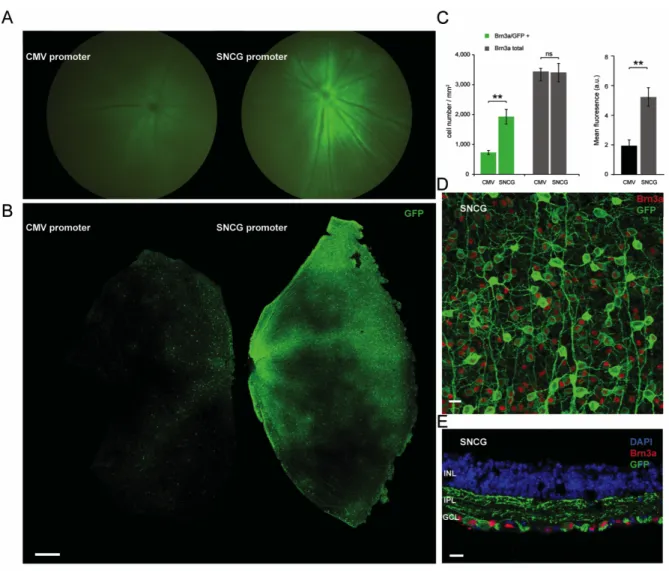
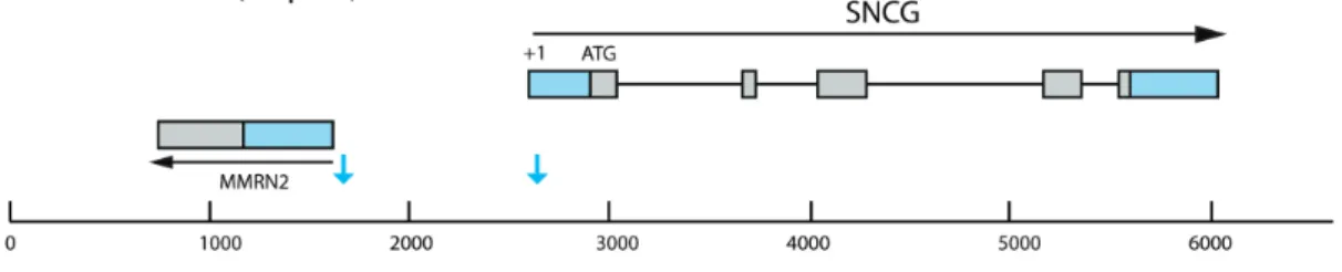
We thus amplified a human genomic DNA fragment of 1 kB between the +1 of the gamma synuclein gene and the untranslated 5’ region downstream (Supplemental Fig. 1). This amplified region contains the promoter, but that we did not attempt any additional promoter characterization within this reagion (i.e. promoter bashing). We cloned this fragment upstream of humanized CatCh (human codon-optimized channelrhodopsin bearing the L132C mutation) in an AAV backbone. AAV2 vectors were produced with either CMV or SNCG promoter driving the expression of CatCh in fusion with GFP. Five rd1 mouse eyes were intravitreally AAV-injected with either CMV or SNCG driving expression of CatCh-GFP. Fluorescent fundus images showed higher fluorescence in all eyes injected with SNCG with respect to eyes injected with CMV (Figure. 1 A). Eyes were enucleated 8 weeks after injection and retinal flat-mounts corroborated higher intensity fluorescence with SNCG promoter compared to CMV (Figure 1 B). We evaluated strength and efficacy of gene expression in RGCs by investigating localization of expression by co-labeling with Brn3a (Figure 1 C). Brn3a is specifically expressed in RGCs and antibodies against this transcription factor are considered a reliable marker to identify and quantify RGCs 35. Confocal microscopy
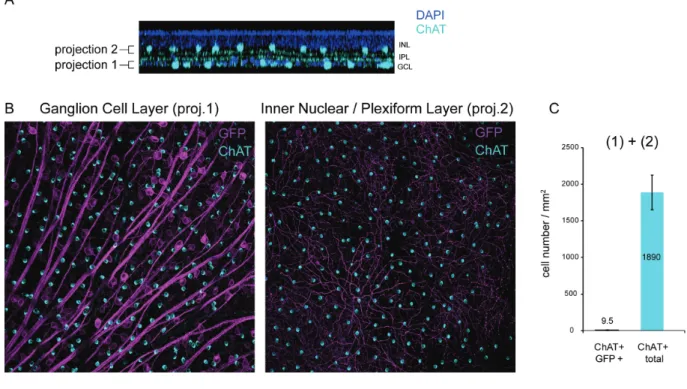
images of retinal flat-mounts (Figure 1 D) and cross-sections (Figure 1 E) from SNCG-CatCh-GFP retinas showed strong GFP expression in RGCs and this expression was highly co-localized with the Brn3a labeling. No expression was noted in deeper retinal layers and in ChAT-positive amacrine cells of the RGC layer (Supplemental Fig. 2). In cell quantification of such images, we showed that the Brn3a antibody labels an equal number of RGCs on retinas transfected with either the SNCG or CMV promoter. In SNCG retinas, 57% of these Brn3a-positive RGCs were also expressing GFP whereas this proportion decreased to 21% in the CMV retinas (Figure 1 C, left). Furthermore, the GFP intensity, as quantified by area and mean immunofluorescence (Figure 1 C, right), was higher in retinas expressing GFP under SNCG promoter. The differences between SNCG and CMV retinas were statistically significant (p<0.01 for both Brn3a/GFP+ co-labeling and mean fluorescence values, unpaired t-tests). The greater number of GFP-expressing Brn3a-positive RGCs and the intensity of GFP expression demonstrated the efficacy of SNCG promoter to drive high-level gene expression in mouse RGCs (Figure 1 D-E). The minor percentage of Brn3a negative but GFP positive cells might belong to a class of RGCs of the accessory optic, pretectal, or hypothalamic pathways which do not stain with Brn3a 35.
Retinal and cortical responses following CatCh expression in RGCs of the rd1 retina under the SNCG promoter
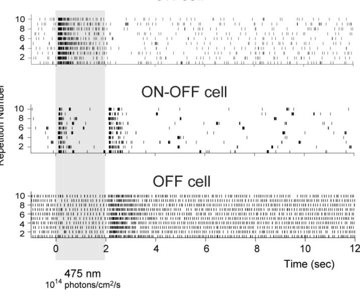
To demonstrate that selective RGC targeting of CatCh-GFP can restore visual function in blind rd1 retinas (age > 12 weeks), we recorded spiking activity from RGCs using a multi-electrode array (252 electrodes MEA), 8-12 weeks after injection (Figure 2 A-E). Rd1 mice were injected at 4-8 weeks after birth with 3 doses of AAV2 encoding CatCh-GFP either under CMV or SNCG promoter. Wild-type (c57bl6) mice and uninjected rd1 mice served as controls. All rd1 retinas were recorded in presence of LAP-4. Only the wild-type controls were recorded without LAP-4. A variety of response profiles were observed in wild-type retinal ganglion cells (Supplemental Fig. 3) at an intensity of 1014
photons/cm2/s. Ganglion cells from rd1 mice expressing CatCh displayed only
ON-responses, between 1014 to 1017 photons/cm2/s. Light evoked spiking activity was
observed with 2 second full-field flashes (Figure 2 A-E), whereas control rd1 retinas did not show any light-elicited increase in spiking activity (data not shown). Percentage of cells responding to light was dependent on viral dose (Figure 2 A-B). Greater numbers of recorded cells responded in rd1 animals injected with the SNCG promoter at the highest AAV dose at 1014 photons/cm2/s (Figure 2 A, p<0.001, two-way ANOVA followed by
Holm-Sidak multiple comparisons). This improved sensitivity corroborates that SNCG drives higher-level CatCh expression in RGCs ensuring light-responses with lower viral load. Firing rate frequency was intensity dependent, giving rise to robust light-responses at 1014 photons/cm2/s for CatCh expressed under SNCG promoter at a viral
dose of 5x10⁹ vg per eye (Figure 2 A). The normalized firing rate increased with rising
light intensities (Figure 2 C-D) with a representative raster plot and peri-stimulus time histogram showing increase in spike frequency during full field flashes at 480 nm in a retina expressing CatCh under control of the SNCG promoter (Figure 2 E). To demonstrate the functional efficacy in live animals, we recorded local field potentials and spiking activity in the visual cortex in vivo. For this experiment, another series of rd1 mice were injected with AAV2-SNCG-hCatCh-GFP, electrophysiological recordings were done in the visual cortex in response to increasing light intensities (Figure 2 F-J). VEPs were recorded on the contralateral hemisphere when stimulating the treated eye with 200 ms pulses of blue light (with light intensities up to 1.7 1017 photons/cm2/s)
percentage of recorded RGCs in MEA experiments and some of the cortical responses could be mediated by ipRGCs36 depolarized with the same wavelength. No VEPs were
visible (flat traces) on recordings from untreated rd1 mice. CatCh-driven VEPs had shorter latencies compared to VEPs measured on wild-type mice (Figure 2 I) as described previously 12,13. A shorter latency is expected because the phototransduction
cascade and subsequent retinal computation done by the circuit are bypassed. Figure 2 F-H and J illustrate the intensity dependence of the cortical spiking responses showing that sizeable light responses are observed at light intensities as low as 1015
photons/cm2/s. The latency of the light-elicited spikes in CatCh-treated rd1 mice was
shorter (10.1 +/-2ms, n=3) than the mean ON latency in wild-type mice (52.98 +/-3.83 ms, n=3) as expected. These functional results clearly demonstrate that AAV-mediated expression of CatCh under the SNCG promoter can activate RGCs in the blind mouse retina and restore light sensitivity up to the visual cortex.
In vivo inflammatory responses in NHP eyes
In mouse studies, we used doses ranging from 5x10⁷ to 5x10⁹ vg/eye. We observed that
using low light intensities required for minimizing phototoxicity in humans (1014-1015
photons/cm2/s range), the particle numbers necessary to obtain light responses on MEA
and at the visual cortex were in the 5x10⁸ to 5x10⁹ vg/eye range. Since the volume of the
macaque vitreous is about 100 times greater than the vitreous volume of a mouse, we decided to use the pharmacological equivalent of this dose range in our non-human primate experiments. Ten non-human primates were chosen based on absence of neutralizing antibody titers against AAV2 in their blood sera37. One primate was injected
intravitreally with two vector doses (one eye with 1x10¹¹ and the other with 5x10¹¹ vg),
with CatCh expressed under SNCG in fusion with GFP to monitor gene expression in vivo (NHP1). As GFP can be immunogenic 24, we used CatCh without fluorescent tag the
remaining nine primates. 18 macaque eyes were injected intravitreally with either 1x10¹¹ (n=5 eyes) or 5x10¹¹ particles (n=5 eyes) or 1x10¹² (n=4 eyes) of AAV2 encoding
CatCh under SNCG or CMV promoter (Table 1). NHPs were followed for three to six months with regular ophthalmic exams (Figure 3 A-B). We then used Spectralis HRA-to examine the eye fundus. This instrument is built on a Confocal Scanning Laser Ophthalmoscopy (cSLO) platform that is sensitive to the fluorescence of Fluoresceine or GFP providing focused and high contrast images of the back of they eye. For the GFP
construct, green fluorescence (shown in white) detected by Spectralis HRA showed gene expression for the 5x10E11 dose starting at 1 month post-injection. Fluorescence increased up to 8 weeks post injection (Figure 3 A, middle) but was invisible for the low dose injection (Figure 3 A, left). Expression stabilized between 8-12 weeks post injection. This in vivo observation was consistent with CatCh expression in the retina for high viral vector doses- a result subsequently confirmed functionally and histologically in retinas where CatCh was expressed without GFP tag (see below). Standardized uveitis nomenclature38,39 was used to score anterior chamber flare, cells, corneal precipitates (Supplemental Fig. 4), posterior uveitis and the level of vitreal haze (Figure 3B). Between months 1 and 3, one animal in each group showed minimal to mild vitreal haze and faint to moderate posterior uveitis. All vitreal haze resolved by 4 months post-injection. Small focal vein coating resembling periphlebitis was identified in the far periphery of four animals by indirect ophthalmoscopy at two months post-injection. Fluorescent angiography was performed in these animals and showed no pathological signs (Supplemental Fig. 4 C). All of these symptoms resolved by month three after the injection. Although the number of macaques in each dosing group is too small to conclude, it seems there was no correlation between the viral dose and the inflammatory symptoms. This suggests that the inflammatory signs were in response to the procedure rather than to the viral vector or transgene product itself. Finally, none of the observed events of the inflammatory reaction throughout of five-six months observation period had influence on vision.
One eye from animal NHP3, which showed moderate posterior uveitis and minimal vitreal haze in the 5x10¹¹ vg group (eye indicated with a white arrow) was enucleated at
3 months and examined for pathological signs related to treatment. The entire eye was fixed, sectioned and observed using nanozoomer technology allowing 40x resolution anywhere within the section (Figure 3C). No structural changes indicative of inflammation (existence of lymphocytes or plasma cells in the trabecular meshwork and the irido-corneal angle, inflammatory cells in the vitreous, or perivascular lymphocytes in the retina) were noted (Figure 3C). Despite the variable levels of vitreal haze and cells observed upon indirect ophthalmoscope evaluation at 1-2 months post-injection (Figure 3 B, C); the absence of inflammatory cells and retinal damage at three months indicate that any preceding immune reaction did not lead to permanent changes in retinal
structure. Overall the retina and anterior segments of the eye were void of any signs of damage or inflammation.
Electrophysiological recordings reveal CatCh-driven responses in RGCs around the fovea at three months post-injection
To start investigating the functionality of the CatCh expression, NHP 1 injected with AAV2-SNCG-CatCh-GFP was sacrificed three months post injection. Retinas were dissected and foveal regions were carefully cut in two halves for MEA and patch-clamp recordings because the green fluorescence attributed to GFP was maximal in this area (Figure 4 A and Figure 4 G-H). In all of our experiments, endogenous ON responses were blocked using a metabotropic glutamate receptor agonist L-(+)-2-Amino-4-phosphonobutyric acid (L-AP4) at 50µM in the bath solution. We had previously validated the L-AP4 blockade of all ON responses in wild-type retinas in both mouse, macaque and human retinas 11,15.
At the single cell level, cell-attached recordings revealed ON light-responses under 1.46 1016 photons/cm2/s at 470 nm in RGCs from AAV2-SNCG-CatCh-GFP retina and in the
retinas from NHP2 infected with AAV2-SNCG-CatCh without GFP which were recorded without assistance by fluorescent label (Figure 4B left and center/top right). In all patched cells under whole-cell configuration, we observed typical channelrhodopsin-evoked photocurrents consisting of a fast transient current followed by a steady-state one at a holding potential of -60mV (Figure 4B, bottom right). To define how these photocurrents control RGC activity at the population level, retinal flatmounts were recorded with the multielectrode array (MEA) technique. Spectral tuning of the firing frequency was calculated and showed highest frequency responses to 480 nm light (Figure 4 C), which corresponds to the excitation peak of ChR2 40. Firing rate frequency
of responsive cells was intensity dependent with the maximum frequency reached at the maximum light intensity applied - 1017 photons/cm2/s (Figure 4 D). Figure 4E illustrates
raster plots from a single unit recording of NHP1 left eye (5x10¹¹ vg). Data in Figure 4F
show the distribution of light responses represented as a percentage of spontaneous activity over light intensities ranging from 1014 to 3x10¹⁷ photons/cm2/s for NHP1 and
2. Similar light responses were obtained for the right eye of NHP2 injected with 5x10¹¹
vg as well as both eyes from NHP3. 45 to 90% of electrodes detected light responses in the four retinas injected with the dose of 5x10¹¹ vg. Our results by patch clamp and MEA
indicate that the GFP tag is not required to obtain functional CatCh expression in RGCs. Eyes injected with 1011 vg dose showed either no responses or only a few cells were
responsive suggesting that 1011 particles are at the threshold where we can expect
reliable optogenetic activation of RGCs through expression of CatCh.
Afer MEA experiments retinal flat-mounts were stained with antibodies against channelrhodopsin 41 and Brn3a. Tissue from NHP1 was labeled with anti-GFP antibodies
in green and anti-Brn3a antibodies in red (Figure 4 H). This is the only retina where we could use the RGC specific marker, Brn3a, in conjunction with an antibody indicating localization of CatCh as both Brn3a and anti-channelrhodopsin antibodies are produced in the same species. In this tissue spanning a ~1 mm square from the center of the fovea, we counted Brn3a (+) and CatCh-GFP (+) cells in half a circle with a 600 µm radius around the fovea (Figure 4 G, H). In this area, around 37% of Brn3a(+) cells were also positive for GFP (523 GFP-positive cells for 1413 Brn3a-positive cells). In the tissue from NHP 2, we counted 1351 CatCh-positive cells for one half of a retina and 455 in the contralateral retina in the region spanning 600 µm from the center of the fovea (Figure 4 I). One of the retinas from the 5 x 10¹¹ -dose group was damaged following the MEA
recording process and could not be used for immunofluorescence labeling. Our results indicate that at least one fourth of peri-foveolar RGCs were labeled with CatCh after an injection with 5 x 10¹¹ vg dose.
CatCh-driven light responses at six months post-injection
The electrophysiological experiments done at 3-4 months post injection showed that SNCG driven CatCh expression is safe and functional at a viral dose of 5x10¹¹ vg. Next, we
aimed to extend the dosing range and compare SNCG to CMV promoter at a 6 months time point. This extended time point was chosen as previous studies established that after intravitreal delivery of AAV2, the transgene expression levels is stable after 6 months 29. Based on this, we first wanted to examine if the low dose (1 x 10¹¹ vg) would
lead to better functional results at 6 months. NHP5 was sacrificed at 6 months post injection and MEA was used to measure light responses as described previously. A few light responsive cells were present in the right eye of this animal injected with 1 x 10¹¹
vg. The contralateral eye was unresponsive confirming that 1011 vg dose is too low to
For the rest of the study, we focused on comparing CatCh mediated responses under CMV or SNCG at the two highest doses: 5 x 10¹¹ vg and 1012 vg. Recordings were first
performed on macaques injected with 5x10¹¹ vg of either AAV2-CMV-CatCh (n=3
retinas) or AAV2-SNCG-CatCh (n=3 retinas). First, we compared light responses with CMV-CatCh to SNCG-CatCh using single-cell recordings. 17 out of the 52 peri-foveaolar RGCs recorded in the CMV-CatCh group were responsive to light. This ratio was as high as 18 out of 24 cells for SNCG-CatCh (Figure 5A, p<0.001, Chi-square). RGCs recorded in the cell-attached configuration showed significantly higher spiking frequencies for SNCG-CatCh compared to CatCh expressed under the control of CMV (Figure 5B, see averaged response in C, p<0.001 by t-test at 2x10¹⁶ photons/cm2/s, unpaired t-test).
Response latencies were also significantly shorter with SNCG than CMV (see Figure 5B, first spike latency of 12.5 ms and 32.5 ms at 1016 photons/cm2/s, respectively, p<0.01 at
2x10¹⁶ photons/cm2/s, unpaired t-test). Next, we recorded cells expressing CatCh under
SNCG in whole-cell patch-clamp configuration, in order to measure CatCh-elicited inward currents at different light intensities. We found that photocurrents can be elicited at just over 1014 photons/cm2/s and response amplitudes increase non-linearly
with light intensity (Figure 5D). We observed photocurrents (with averageτ On ~8 ms and τOff ~16 ms at 2x10¹⁶ photons/cm2/s) characterized by a transient phase followed
by a robust steady-state phase under whole-cell configuration. These currents were converted into robust spiking activities under cell-attached configuration, lasting for the duration of the stimuli ranging from 20 ms to 4 s in our conditions (Figure 5E). These photocurrents and spiking activity were fast enough to follow up to 30 Hz light-pulses (Figure 5F). Next, the light intensity requirements at a population level were investigated over the entire macular region using MEA recordings. Light responses originated from the peri-foveolar area where GFP expression was observed on the retinal flatmount and their amplitude increased with stimulation intensity (Figure 5G). Figure 5H represents confocal stack projections of the same retinas labeled with ChR2 (green) antibodies. In agreement with our single-cell recordings, the number of light responsive cells over the total number of cells displaying spontaneous activity was significantly higher in retinas transduced with SNCG-CatCh compared to CMV-CatCh (Figure 5I, p<0.001, Chi-square). Normalized spiking frequencies across all eyes treated with SNCG (n=3) and CMV-CatCh (n=3) at a 5 x 10¹¹ vg dose are shown in Figure 5J. MEA
recordings over a larger population of RGCs in the macular region show the higher light-sensitivity of RGCs expressing CatCh under SNCG promoter compared to CMV. Firing frequencies recorded across 3 retinas for each condition indicate a clear separation in the light sensitivity of the responses obtained using the two promoters.
Lastly, we investigated the utility of increasing further the viral dose to 1012 vg using
both CMV and SNCG promoters. MEA recordings of the retina expressing CatCh under either the CMV (n=2) or the SNCG (n=2) promoters at the 1012 particle dose corroborate
previous conclusions on the better efficacy of SNCG compared to CMV. This superiority was confirmed at both the functional level (Figure 5J) and at the cell density level (Figure 5K). Using the highest dose (1012 vg) slightly increased the maximum multi-unit
spiking frequencies obtained with SNCG. The firing frequencies with SNCG were already very high (276,3 +/-17,9 Hz compared to 199,6 +/-40,6 Hz at 5 x 10¹¹ vg under 8x10¹⁵
photons/cm2/s) at 5x10¹¹ vg. Moreover, the light sensitivity curves were similar for the
two doses with the SNCG promoter as well as with the CMV promoter (Fig. 5J). These results clearly demonstrate the superiority of the SNCG promoter, which cannot be compensated by increasing the viral dose while using the CMV promoter.
Discussion:
There is great interest in developing optogenetics as a therapeutic modality that can be applied in humans with photoreceptor degeneration. In order to predict safety and feasibility in humans, gene delivery-related parameters need to be examined in non-human primates, which represent the non-human eye and immune system most closely. There are two primate models that can be used for this type of experiment, macaque and marmosets. The marmoset has a small eye with an axial length of 11 mm and AAV transduction patterns in this animal model differ from macaques in terms of cellular selectivity, spatial pattern, and depth of transduction through the retina28. Immune
responses in the marmoset are weaker than macaques although, marmosets have an advantage over macaques for functional studies as genetic neurodegeneration models can be generated in this species42. As there is no non-human primate model of retinal
degeneration available today, we decided to use macaques for our study. The success of optogenetics in vision restoration depends on our ability to express functional amount of the microbial opsin without eliciting immune responses to the gene delivery vector and the opsin itself. Finally, the light intensity requirements for functional activation need to fall in the range of radiation safety limits for the human eye. In view of these
requirements, we explored safety and feasibility of RGC activation with CatCh in the macaque retina. There are several AAVs that have been tested for retinal gene delivery via the intravitreal route, and some have evoked safety concerns that have not been fully addressed at this stage25,43. As one such example a recent study suggests that the
engineered AAV variant AAV2-7m8 might be more immunogenic when used at high doses via intravitreal injections25. Therefore, we chose AAV2 for its known safety and
efficacy in targeting RGCs in non-human primate studies 27–29 and the large body of work
using this vector in human clinical trials 20–22,44,45. High-level expression with a limited
viral dose being a major parameter in obtaining functional expression, we designed a new RGC specific promoter, driving strong transgene expression in RGCs. We show that this promoter, called SNCG drives expression in twice as many RGCs compared to the ubiquitous CMV promoter. We also found that the strength of transgene expression was qualitatively stronger with SNCG promoter compared to CMV. This observation was corroborated with single cell patch experiments where higher amplitude photocurrents were recorded from cells expressing CatCh under SNCG promoter. In macaque retinas, CatCh without GFP tag localizes to the membrane in peri-foveolar RGCs and is able to mediate light-driven action potential firing in a population of RGCs with light intensities above 1015 photons/cm2/s. As expected, RGCs responding to light are concentrated on
the foveal center in a 500-600 µm disc where AAV2 affords efficient transduction28. This
transduction pattern is expected to lead to a narrow visual field, albeit with good acuity. New AAV vectors and surgical procedures could help expand this limited area of transducion. Finally, we show for the first time that it is possible to stimulate CatCh expressing RGCs above 30 Hz ensuring high temporal resolution.
Gene therapeutic approaches that can provide treatments beyond the degeneration of photoreceptors are particularly important as most patients do not come to the clinic until the degenerative process has gone beyond the loss of rods and the start of cone degeneration 46. In this regard, the results presented here provide basis for
further clinical development of optogenetic reactivation of RGCs. Our study investigated the feasibility of optogenetic activation of RGCs in macaques and constitutes the basis for further investment in this direction towards improved application in patients. Because optogenetic activation relies on the resolution of the optical stimulation and the cell size, it is very likely to provide greater visual acuity than current electrical stimulations with retinal prostheses 6. The peri-foveolar expression pattern obtained in
the non-human primate retina indicates it will be possible to optogenetically stimulate these retinal ganglion cells, making use of human retina's innate high-acuity RGC circuit. However, with current technologies it is not yet possible to achieve peripheral vision.
We found that reliable CatCh activity is seen while stimulating at an intensity threshold around 1015 photons/cm2
/s, with response amplitudes increasing in a light-intensity-dependent manner when using the SNCG promoter, and that significant responses can be obtained up to ~500 nm. This activation threshold is just below the radiation safety limits for the human eye47. By contrast, the one log unit shift in light
sensitivity for the CMV promoter suggests that it could fail to elicit light responses in a safe dosing range. Moreover, potential long-term effects of expressing a light-driven channel (CatCh) with increased calcium permeability need to be taken into account when using this approach. In order to avoid potential blue light toxicity while stimulating the retina at high light intensities, developments in opsin engineering 48 and
discovery49,50 will help further refine optogenetic vision restoration strategies. In the
future, we anticipate developments in both viral technologies and surgical techniques will aid in obtaining better distribution of the optogenetic protein across the primate retina. Finally for patients who still have intact bipolar cell layer, new viral technologies might also grant access to the bipolar cells of the primate retina for circuit specific insertion of G protein coupled vertebrate opsins 51–53.
Our study provides evidence that the choice of an appropriate promoter is essential in obtaining high-level microbial opsin expression compatible with illumination safety. A promoter that can drive strong gene expression in designated cells is crucial in clinical development of a strategy using a protein of foreign origin, which requires high-level expression for functional activation. Materials and methods: Animals All experiments were done in accordance with the National Institutes of Health Guide for Care and Use of Laboratory Animals. The protocol was approved by the Local Animal Ethics Committees and conducted in accordance with Directive 2010/63/EU of the European Parliament. All mice used in this study were C3H/HeN (rd1 mice) or C57Bl6J mice (wild type) from Janvier Laboratories (Le Genest Saint Isle, France) or cynomolgus macaques (macaca fasicularis) from foreign origin.
AAV production
SNCG and CMV promoters were cloned into an AAV backbone plasmid containing human codon optimized CatCh sequence in fusion with or without GFP. The constructs all included WPRE and bovine growth hormone polyA. Recombinant AAVs were produced by the plasmid co-transfection method 54, and the resulting lysates were purified via
iodixanol gradient ultracentrifugation as previously described. Briefly 40% iodixanol fraction was concentrated and buffer exchanged using Amicon Ultra-15 Centrifugal Filter Units. Vector stocks were then tittered for DNase-resistant vector genomes by real time PCR relative to a standard 55.
Injections
Mice were anesthetized with ketamine (50 mg/kg) xylazine (10 mg/kg Rompum). Injection of 1 µl stock containing 107 to 109 particles of AAV was made with direct
observation of the needle in the center of the vitreous cavity. Primates were anesthetized with 10∶1 mg/kg ketamine:xylazine. 100 µL of viral vector containing either 1 - 5 x 10¹¹ or 1012 viral particles were injected into the vitreous. Ophthalmic
steroid and antibiotic ointment was applied to the corneas post-injection.
Immunohistochemistry
Transduced mouse retinas were dissected and fixed in 4% paraformaldehyde for 30 min at room temperature Retinas were incubated in PBS with 1% Triton X-100, 0.5% Tween 20 and 5% BSA blocking buffer for 1 h at room temperature (RT). Retinas were incubated overnight at 4°C with polyclonal antibodies directed against GFP (Life Technologies; 1:2000) and ChAT (Millipore; 1:400); and monoclonal anti-Brn3a antibody (Millipore Chemicon; 1:100) in half diluted blocking buffer. Secondary anti-rabbit IgG, anti-mouse IgG and anti goat IgG conjugated with Alexa TM594 , Alexa TM488 and Alexa TM647 respectively (Molecular Probes; 1:500) were applied for 1 h at RT. Primate retinas were labelled with antibodies directed against Brn3a, GFP and Channelrhodopsin in similar conditions. Cell nuclei were revealed with 4’,6-diamidino-2-phenylindole (Sigma-Aldrich; 10 µg/mL). Retinas were rinsed and flat-mounted in mounting medium. Cryosections of the labeled flatmounts were obtained by unmounting, cryopreserving and embedding in OCT before cryosections (15 μm).
Confocal Microscopy
Olympus FV1000 laser-scanning confocal microscope was used to acquire images sequentially, line-by-line. Step size was defined according to the Nyquist-Shannon sampling theorem. Twelve bit Images were then processed with FIJI, Z-sections were projected on a single plane using maximum intensity under Z-project function and finally converted to 8-bit RGB color mode. Efficiency of transduction was assessed by counting Brn3a (+) cells transduced with CatCh-GFP in mice and in foveal area in primate retinas. Confocal stacks through the RGC layer were acquired using the 20x objective, in four adjacent regions; above, below, to the right and left of the optic-nerve.
MEA Recordings of isolated retinas
Mice were anesthetized and sacrificed by quick cervical dislocation. Eyeballs were removed and placed in Ames medium (Sigma Aldrich A1420) bubbled with 95% O2 and
5% CO2 at room temperature. Eyecups were obtained under red light using binoculars
by cutting around sclera, below ora serrata. Cornea and leans were then removed, and retinas were isolated and flattened. Primate eyeballs were transfered in CO2-independent medium (Life Technologies, Carlsbad, CA) after sacrifice with a lethal dose of pentobarbital. Retinas were isolated upon arrival, and the perifoveal area was cut into squared shaped pieces (approximately 4 to 5 mm) and cultured in Neurobasal medium complemented with B27 serum-free supplement (Life Technologies, Carlsbad, CA) on Transwell permeable culture support (Corning Inc., Corning, NY) as previously described (15). Primate retinal explants were left to rest in incubator for one to two days prior to MEA recordings. Isolated retinas were placed on a cellulose membrane and gently pressed against an MEA (MEA256 100/30 iR-ITO, Multichannel systems, Germany), with the RGCs facing the electrodes. The retina was continuously perfused with bubbled Ames medium at 34ºC at a rate of 1-2 ml/min during experiments. Metabotropic glutamate receptor agonist L-(+)-2-Amino-4-phosphonobutyric acid (LAP-4, Tocris Bioscience, cat No. 0103) and Glutamate antagonists 6-Cyano-7-nitroquinoxaline-2,3-dione disodium (CNQX disodium salt, Tocris Bioscience, cat No. 1045) and 3-((R)-2-Carboxypiperazin-4-yl)-propyl-1-phosphonic acid (CPP, Tocris Bioscience, cat No. 0247) were freshly diluted to concentrations of 50 µM, 25µM and 10 µM respectively and were bath-applied through the perfusion system 30 minutes prior
to recordings. Retinas were dark-adapted one hour prior to recordings. Full-field light stimuli were applied with a Polychrome V monochromator (Olympus) driven by a STG2008 stimulus generator (MCS). Output light intensities were calibrated to range from 1.1014 photons/cm2/s to 1.1017 photons/cm2/s. Stimuli were presented for two
seconds, with ten-second intervals. Wavelength sensitivity of responses was determined by stimulating ten times, from 400 nm to 650 nm, with 10 nm steps. The order of the tested wavelengths was randomized in order to prevent any adaptation of the retina. Raw extracellular RGC activity was amplified and sampled at 20kHz. Resulting data was stored and filtered with a 200 Hz high pass filter for subsequent offline analysis using Spike2 software v.7 (CED Co, UK). Single unit raster plots were obtained using a combination of template matching and cluster grouping based on principal component analysis of the waveforms. In our population analysis, significant responses were determined based on a z-score analysis. We estimated the mean and standard deviation of the activity prior to stimulus and considered that a response was detected if the activity exceeded the mean by more than four times the standard deviation in the 2 s after the onset or the offset of the stimulus (for a bin size of 50 ms). Error bars were calculated over the different experiments. For the responses to light at different wavelengths, we measured the response to each flash in a 1 s window after the stimulus. We then normalized the response of each cell by its maximum firing rate response. For the responses to light at different intensities, we estimated the error bars by bootstrapping over the set of recorded cells.
Two-Photon Imaging and electrophysiological recordings
A custom-made two-photon microscope equipped with a 25x water immersion objective (XLPLN25xWMP/NA1.05, Olympus) with a pulsed femto-second laser (InSight™ DeepSee™ - Newport Corporation) was used for imaging CatCh-GFP-positive retinal ganglion cells. AAV-treated retinas from rd1 mice were isolated in oxygenized (95% O2,
5% CO2) Ames medium (Sigma-Aldrich). For live two-photon imaging, retinas were
placed in the recording chamber of the microscope, and z-stacks were acquired using the excitation laser at a wavelength of 930 nm. Images were processed offline using ImageJ. During imaging, the retina was superfused with oxygenized Ames medium. We used an Axon Multiclamp 700B amplifier for whole-cell patch-clamp and cell-
attached recordings. Patch-clamp electrodes were made from borosilicate glass (BF100-50-10, Sutter Instruments) and pulled to 8-10 MΩ. Pipettes were filled with 112.5 mM CsMeSO4, 1 mM Mg SO4, 7.8 × 10⁻³ mM CaCl2, 0.5 mM BAPTA, 10 mM HEPES, 4 mM
ATP-Na2, 0.5 mM GTP-Na3, 5 mM lidocaine N-ethyl bromide (QX314-Br) (pH 7.2). Cells were clamped at a potential of -60 mV to isolate excitatory currents. Cell-attached recordings were obtained in current-clamp configuration (current zero) with electrodes filled with AMES solution. LAP-4 was added to Ames medium during all electrophysiological recordings. Retinas were dark-adapted one hour prior to recordings.
In-vivo recordings in the visual cortex
Mice were sedated with a low dose of ketamine-xylazine injection (ketamine: 100 mg/kg and xylazine: 10 mg/kg) and then anesthetized with urethane (1.0 g/kg, 10% w/v in saline). Animals were placed in a stereotaxic holder. The temperature was maintained at 37ºC and a coverslip covered with vitamine A (Allergan) was placed on both eyes to prevent corneal dehydration. A craniotomy (1 mm2) above V1 in the contralateral
hemisphere to the treated eye, was centered 3 mm lateral and 0.5 mm rostral from the lambda point. The dura was removed and an electrode was inserted using a 3-axis micromanipulator (Sutter Instruments) with a 30° angle to the cortical surface. It was advanced 800 µm and the exposed surface was covered with agarose (1.2% in cortex buffer).
Visual stimuli were generated by a 470 nm collimated LED (model M470L3, Thorlabs) placed at 1 cm from the eye. An isolating cone ensured that the illumination was restricted to the stimulated eye. Linear multisite silicon microprobes (sixteen electrodes at 50 μm intervals) were used for recordings. For each acquisition, after averaging over the 200 trials, the electrode showing the VEP with maximal peak amplitude was selected for quantification. The stimulation consisted of 200 ms pulses of blue light repeated 200 times at 1 Hz triggered by a Digidata (Axon). Signals were analysed in Matlab using custom scripts. For local field potentials, signals were low pass filtered at 300 Hz and averaged over the 200 trials.
In vivo imaging and ophthalmic exams in non-human primates
Fluorescent images of GFP (Fundus Autofluorescence mode: excitation wavelength of 488 nm and barrier filter of 500 nm) and infrared pictures of eye fundus and OCT
images were acquired using an Spectralis HRA+OCT system (Spectralis HRA+OCT; Heidelberg Engineering, Heidelberg, Germany) after pupil dilation. Ophthalmic exams consisting of slit lamp biomicroscopy (Portable Slit Lamp model SL-14, Kowa) and indirect ophthalmoscopy (Indirect Binocular Ophthalmoscope model HK 150-1 uno, Heine) were performed on all macaques before dosing, at 2 weeks and then on monthly basis.
Histopathological studies on macaque retina
The eye from a NHP injected with the 5 x 10¹¹
dose was enucleated at 3 months post-injection a needle was inserted and 0.15 - 0.3 ml of fixative was injected, until the eyeball became turgid. Eye was immersion fixed in fixative overnight and processed for making horizontal cross-sections across the entire structure. A retinal cross-section presenting all of the desired ocular structures was then imaged on a Nanozoomer (Hamamatsu, Japan).
Acknowledgements:
The authors thank the Sanofi Optovision team, Xavier Palazzi, Claire-Maelle Fovet, Valérie Fradot, Florian Senlaub for technical help and scientific advice. This work was supported by Sanofi, the Institut National de la Santé et de la Recherche Médicale (INSERM), Pierre et Marie Curie University (UPMC), the Centre National de la Recherche Scientifique (CNRS), FRM (Fondation pour la Recherche Medicale), Agence Nationale pour la Recherche Investissements d’Avenir Recherche Hospitalo-Univesritaire en santé (RHU- Light4Deaf) and (ANR: OPTIMA), the E-Rare Project (OPTOREMODE), The Foundation Fighting Blindness (Wynn-Gund translational research award), the Fédération des Aveugles de France, the city of Paris, the Regional Council of Ile-de-France, the French State program “Investissements d'Avenir” managed by the Agence Nationale de la Recherche [LIFESENSES: ANR-10-LABX-65], the ERC Starting Grant (OptogenRet, Grant Number 309776), the Deutsche Forschungsgemeinschaft (DFG) SFB 807, the Max-Planck Society and the National Institute of Health under Award Number U01NS090501. References: 1. Bessant, DAR, Ali, RR and Bhattacharya, SS (2001). Molecular genetics and prospects for therapy of the inherited retinal dystrophies. Current Opinion in
Genetics and Development 11: 307–316. 2. Wright, AF, Chakarova, CF, Abd El-Aziz, MM and Bhattacharya, SS (2010). Photoreceptor degeneration: genetic and mechanistic dissection of a complex trait. Nature reviews. Genetics 11: 273–84. 3. Humayun, MS, Prince, M, de Juan, E, Barron, Y, Moskowitz, M, Klock, IB, et al. (1999). Morphometric analysis of the extramacular retina from postmortem eyes with retinitis pigmentosa. Investigative ophthalmology & visual science 40: 143–8. 4. Humayun, MS, Dorn, JD, da Cruz, L, Dagnelie, G, Sahel, J, Stanga, PE, et al. (2012). Interim results from the international trial of Second Sight’s visual prosthesis. Ophthalmology 119: 779–88. 5. Jacobson, SG, Sumaroka, A, Luo, X and Cideciyan, A V (2013). Retinal optogenetic therapies: clinical criteria for candidacy. Clinical genetics 84: 175–82. 6. Lorach, H, Goetz, G, Smith, R, Lei, X, Mandel, Y, Kamins, T, et al. (2015). Photovoltaic restoration of sight with high visual acuity. Nature medicine 21: 476– 82. 7. Bi, A, Cui, J, Ma, Y-P, Olshevskaya, E, Pu, M, Dizhoor, AM, et al. (2006). Ectopic expression of a microbial-type rhodopsin restores visual responses in mice with photoreceptor degeneration. Neuron 50: 23–33. 8. Lagali, PS, Balya, D, Awatramani, GB, Münch, T a, Kim, DS, Busskamp, V, et al. (2008). Light-activated channels targeted to ON bipolar cells restore visual function in retinal degeneration. Nature neuroscience 11: 667–75. 9. Greenberg, KP, Pham, A and Werblin, FS (2011). Differential targeting of optical neuromodulators to ganglion cell soma and dendrites allows dynamic control of center-surround antagonism. Neuron 69: 713–20. 10. Pan, Z-H, Ganjawala, TH, Lu, Q, Ivanova, E and Zhang, Z (2014). ChR2 Mutants at L132 and T159 with Improved Operational Light Sensitivity for Vision Restoration. PloS one 9: e98924. 11. Busskamp, V, Duebel, J, Balya, D, Fradot, M, Viney, TJ, Siegert, S, et al. (2010). Genetic Reactivation of Cone Photoreceptors Restores Visual Responses in Retinitis Pigmentosa. Science 329: 413–417. 12. Tomita, H, Sugano, E, Isago, H, Hiroi, T, Wang, Z, Ohta, E, et al. (2009). Channelrhodopsin-2 gene transduced into retinal ganglion cells restores functional vision in genetically blind rats. Experimental Eye Research 90: 429–436. 13. Caporale, N, Kolstad, KD, Lee, T, Tochitsky, I, Dalkara, D, Trauner, D, et al. (2011). LiGluR restores visual responses in rodent models of inherited blindness. Molecular therapy 19: 1212–9. 14. Gaub, BM, Berry, MH, Holt, AE, Reiner, A, Kienzler, M a., Dolgova, N, et al. (2014). Restoration of visual function by expression of a light-gated mammalian ion channel in retinal ganglion cells or ON-bipolar cells. Proceedings of the National Academy of Sciences of the United States of America 111: E5574-83. 15. Sengupta, A, Chaffiol, A, Macé, E, Caplette, R, Desrosiers, M, Lampič, M, et al. (2016). Red-shifted channelrhodopsin stimulation restores light responses in blind mice, macaque retina, and human retina. EMBO molecular medicine 8: 1248– 1264. 16. Doroudchi, MM, Greenberg, KP, Liu, J, Silka, K a, Boyden, ES, Lockridge, J a, et al. (2011). Virally delivered channelrhodopsin-2 safely and effectively restores visual function in multiple mouse models of blindness. Molecular therapy 19: 1220–9. 17. Tomita, H, Sugano, E, Murayama, N, Ozaki, T, Nishiyama, F, Tabata, K, et al. (2014). Restoration of the Majority of the Visual Spectrum by Using Modified Volvox
Channelrhodopsin-1. Molecular Therapy 22: 1434–1440. 18. Macé, E, Caplette, R, Marre, O, Sengupta, A, Chaffiol, A, Barbe, P, et al. (2015). Targeting channelrhodopsin-2 to ON-bipolar cells with vitreally administered AAV Restores ON and OFF visual responses in blind mice. Molecular therapy : the journal of the American Society of Gene Therapy 23: 7–16. 19. Busskamp, V and Roska, B (2011). Optogenetic approaches to restoring visual function in retinitis pigmentosa. Current opinion in neurobiology 21: 942–6. 20. Bainbridge, JWB, Smith, AJ, Barker, SS, Robbie, S, Henderson, R, Balaggan, K, et al. (2008). Effect of gene therapy on visual function in Leber’s congenital amaurosis. The New England journal of medicine 358: 2231–9. 21. Cideciyan, A V, Aleman, TS, Boye, SL, Schwartz, SB, Kaushal, S, Roman, AJ, et al. (2008). Human gene therapy for RPE65 isomerase deficiency activates the retinoid cycle of vision but with slow rod kinetics. Proceedings of the National Academy of Sciences of the United States of America 105: 15112–7. 22. Maguire, AM, Simonelli, F, Pierce, EA, Pugh, EN, Mingozzi, F, Bennicelli, J, et al. (2008). Safety and efficacy of gene transfer for Leber’s congenital amaurosis. The New England journal of medicine 358: 2240–8. 23. MacLaren, RE, Groppe, M, Barnard, AR, Cottriall, CL, Tolmachova, T, Seymour, L, et al. (2014). Retinal gene therapy in patients with choroideremia: initial findings from a phase 1/2 clinical trial. The Lancet 383: 1129–1137. 24. Vandenberghe, LH, Bell, P, Maguire, AM, Cearley, CN, Xiao, R, Calcedo, R, et al. (2011). Dosage thresholds for AAV2 and AAV8 photoreceptor gene therapy in monkey. Science translational medicine 3: 88ra54. 25. Ramachandran, PS, Lee, V, Wei, Z, Song, JY, Casal, G, Cronin, T, et al. (2016). Evaluation of Dose and Safety of AAV7m8 and AAV8BP2 in the Non-Human Primate Retina. Human gene therapydoi:10.1089/hum.2016.111. 26. Sugano, E, Isago, H, Wang, Z, Murayama, N, Tamai, M and Tomita, H (2011). Immune responses to adeno-associated virus type 2 encoding channelrhodopsin-2 in a genetically blind rat model for gene therapy. Gene therapy 18: 266–274. 27. Ivanova, E, Hwang, G-S, Pan, Z-H and Troilo, D (2010). Evaluation of AAV-mediated expression of Chop2-GFP in the marmoset retina. Investigative ophthalmology & visual science 51: 5288–96. 28. Yin, L, Greenberg, K, Hunter, JJ, Dalkara, D, Kolstad, KD, Masella, BD, et al. (2011). Intravitreal injection of AAV2 transduces macaque inner retina. Investigative ophthalmology & visual science 52: 2775–83. 29. Maclachlan, TK, Lukason, M, Collins, M, Munger, R, Isenberger, E, Rogers, C, et al. (2011). Preclinical safety evaluation of AAV2-sFLT01- a gene therapy for age-related macular degeneration. Molecular therapy 19: 326–34. 30. Bennett, J, Ashtari, M, Wellman, J, Marshall, KA, Cyckowski, LL, Chung, DC, et al. (2012). AAV2 Gene Therapy Readministration in Three Adults with Congenital Blindness. Science Translational Medicine 4: 120ra15-120ra15. 31. Kleinlogel, S, Feldbauer, K, Dempski, RE, Fotis, H, Wood, PG, Bamann, C, et al. (2011). Ultra light-sensitive and fast neuronal activation with the Ca2+-permeable channelrhodopsin CatCh. Nature neuroscience 14: 513–8. 32. Yin, L, Masella, B, Dalkara, D, Zhang, J, Flannery, JG, Schaffer, D V, et al. (2014). Imaging light responses of foveal ganglion cells in the living macaque eye. The Journal of neuroscience : the official journal of the Society for Neuroscience 34: 6596–605. 33. Feuer, WJ, Schiffman, JC, Davis, JL, Porciatti, V, Gonzalez, P, Koilkonda, RD, et al.
(2016). Gene Therapy for Leber Hereditary Optic Neuropathy. Ophthalmology 123: 558–570. 34. Surgucheva, I, Weisman, AD, Goldberg, JL, Shnyra, A and Surguchov, A (2008). Gamma-synuclein as a marker of retinal ganglion cells. Molecular vision 14: 1540– 1548. 35. Quina, LA, Pak, W, Lanier, J, Banwait, P, Gratwick, K, Liu, Y, et al. (2005). Brn3a-expressing retinal ganglion cells project specifically to thalamocortical and collicular visual pathways. The Journal of neuroscience : the official journal of the Society for Neuroscience 25: 11595–11604. 36. Allen, AE, Storchi, R, Martial, FP, Bedford, RA and Lucas, RJ (2017). Melanopsin Contributions to the Representation of Images in the Early Visual System. Current Biology 27: 1623–1632.e4. 37. Kotterman, MA, Yin, L, Strazzeri, JM, Flannery, JG, Merigan, WH and Schaffer, D V (2015). Antibody neutralization poses a barrier to intravitreal adeno-associated viral vector gene delivery to non-human primates. Gene therapy 22: 1–11. 38. Nussenblatt, RB, Palestine, AG, Chan, CC and Roberge, F (1985). Standardization of vitreal inflammatory activity in intermediate and posterior uveitis. Ophthalmology 92: 467–471. 39. Jabs, DA, Nussenblatt, RB and Rosenbaum, JT (2005). Standardization of uveitis nomenclature for reporting clinical data. Results of the First International Workshop. American journal of ophthalmology 140: 509–516. 40. Nagel, G, Szellas, T, Huhn, W, Kateriya, S, Adeishvili, N, Berthold, P, et al. (2003). Channelrhodopsin-2, a directly light-gated cation-selective membrane channel. Proceedings of the National Academy of Sciences of the United States of America 100: 13940–5. 41. Kleinlogel, S, Terpitz, U, Legrum, B, Gökbuget, D, Boyden, ES, Bamann, C, et al. (2011). A gene-fusion strategy for stoichiometric and co-localized expression of light-gated membrane proteins. Nature Methods 8: 1083–1088. 42. Belmonte, JCI, Callaway, EM, Churchland, P, Caddick, SJ, Feng, G, Homanics, GE, et al. (2015). Brains, Genes, and Primates. Neuron 86: 617–631. 43. Dalkara, D, Byrne, LC, Klimczak, RR, Visel, M, Yin, L, Merigan, WH, et al. (2013). In vivo-directed evolution of a new adeno-associated virus for therapeutic outer retinal gene delivery from the vitreous. Science translational medicine 5: 189ra76. 44. MacLaren, RE, Groppe, M, Barnard, AR, Cottriall, CL, Tolmachova, T, Seymour, L, et al. (2014). Retinal gene therapy in patients with choroideremia: Initial fi ndings from a phase 1/2 clinical trial. The Lancet 383: 1129–1137. 45. Bennett, J, Wellman, J, Marshall, KA, Mccague, S, Ashtari, M, Distefano-Pappas, J, et al. (2016). Safety and durability of eff ect of contralateral-eye administration of AAV2 gene therapy in patients with childhood-onset blindness caused by RPE65 mutations: a follow-on phase 1 trial. The Lancet 388: 661–672. 46. Byrne, LC, Dalkara, D, Luna, G, Fisher, SK, Clérin, E, Sahel, J, et al. (2015). Viral-mediated RdCVF and RdCVFL expression protects cone and rod photoreceptors in retinal degeneration. Journal of Clinical Investigation 125: 105–116. 47. Wu, J, Seregard, S and Algvere, P V. Photochemical damage of the retina. Survey of ophthalmology 51: 461–81. 48. Lin, JY, Knutsen, PM, Muller, A, Kleinfeld, D and Tsien, RY (2013). ReaChR: a red-shifted variant of channelrhodopsin enables deep transcranial optogenetic excitation. Nature neuroscience 16: 1499–508. 49. Klapoetke, NC, Murata, Y, Kim, SS, Pulver, SR, Birdsey-Benson, A, Cho, YK, et al.
(2014). Independent optical excitation of distinct neural populations. Nature methods 11: 338–46. 50. Chuong, AS, Miri, ML, Busskamp, V, Matthews, G a C, Acker, LC, Sørensen, AT, et al. (2014). Noninvasive optical inhibition with a red-shifted microbial rhodopsin. Nature neuroscience 17: 1123–1129. 51. van Wyk, M, Pielecka-Fortuna, J, Löwel, S and Kleinlogel, S (2015). Restoring the ON Switch in Blind Retinas: Opto-mGluR6, a Next-Generation, Cell-Tailored Optogenetic Tool. PLOS Biology 13: e1002143. 52. Cehajic-Kapetanovic, J, Eleftheriou, C, Allen, AE, Milosavljevic, N, Pienaar, A, Bedford, R, et al. (2015). Restoration of Vision with Ectopic Expression of Human Rod Opsin. Current Biology: 1–12doi:10.1016/j.cub.2015.07.029. 53. Gaub, BM, Berry, MH, Holt, AE, Isacoff, EY and Flannery, JG (2015). Optogenetic Vision Restoration Using Rhodopsin for Enhanced Sensitivity. Molecular therapy : the journal of the American Society of Gene Therapy 23: 1562–71. 54. Choi, VW, Asokan, A, Haberman, R a and Samulski, RJ (2007). Production of recombinant adeno-associated viral vectors. Current protocols in human genetics / editorial board, Jonathan L. Haines ... [et al.] Chapter 12: Unit 12.9. 55. Aurnhammer, C, Haase, M, Muether, N, Hausl, M, Rauschhuber, C, Huber, I, et al. (2012). Universal real-time PCR for the detection and quantification of adeno-associated virus serotype 2-derived inverted terminal repeat sequences. Human gene therapy methods 23: 18–28.
Figure legends:
Figure 1- SNCG promoter drives higher-level hCatCh-GFP expression than CMV promoter in mouse RGCs
A) Fundus image of representative rd1 mouse retinae injected with 5x10⁹ vg of either
AAV2-CMV-hCatCh-GFP (left) or AAV2-SNCG-hCatCh-GFP (right). B) Retinal flat-mounts obtained from the same injection series showing CatCh-GFP fluorescence obtained under the CMV promoter (left) and the SNCG promoter (right). Scale bar: 300 µm. C) Quantification of Brn3a-positive, GFP-positive and double labeled cells in rd1 mouse retina injected with either AAV2-SNCG-hCatCh-GFP or AAV2-CMV-hCatCh-GFP. Confocal stack projections across the ganglion cell layer for cell counts over chosen fields in the central and in peripheral regions of the retina. Regions were chosen in each quadrant and cell-counts were averaged to obtain Brn3a-positive, GFP-positive and co-labeled
cells per mm2. Mean fluorescence values were measured over the same areas using the
same number of z-stacks. Error bars represent SEM. D) Representative confocal stack projection across the RGC layer of rd1 mouse retina transduced with SNCG-CatCh-GFP, co-labeled with Brn3a (red) and anti-GFP (green) antibodies. E) Cross-sections obtained from one representative retinal flat-mount in the SNCG-CatCh-GFP injected retinas co-labeled with Bnr3a (red) and anti-GFP (green) and nuclei were labeled with DAPI (blue). Scale bars (D, E): 10 µm.
Figure 2- Functional CatCh responses in the retinas and cortex of rd1 mice
Percentages of cells with a spontaneous activity showing a light response at 480nm under either A) 1014 photons/cm2/s or B) 1017 photons/cm2/s in retinas expressing
CatCh under control of CMV (black) or SNCG (grey) at 5x10⁷, 5x10⁸ and 5x10⁹ vg per eye
(n=4). C) Response amplitude (normalized to the response obtained at maximum luminance) as a function of light intensity in retinas expressing CatCh under control of CMV promoter at 5x10⁹ viral particle dose (n=4, 155 cells) D) Response amplitude
(normalized to the response obtained at maximum luminance) as a function of light intensity in retinas expressing CatCh under control of SNCG promoter at 5x10⁷ viral
particle dose (light grey, n=4 retinas, 158 cells), 5x10⁸ (dark grey, n=4 retinas, 221 cells)
and 5x10⁹ (black, n=4 retinas, 261 cells) viral particles (vg) per eye. E) Raster plots and
peri-stimulus time histogram (PSTH) showing the light response or increase in spike frequency during full field flashes at 480 nm in a retina expressing CatCh under control of the SNCG promoter. Note that with the SNCG promoter that the curve is reaching a plateau at the maximum AAV vector dose. (F, G, H) PSTHs (top) and corresponding raster plots (bottom) of visual cortex neurons of rd1 mice expressing SNCG-CatCh in response to 475 nm full field flashes at 3 different increasing light intensities (1015, 1016
and 1017 photons/cm2/s). I) Comparison of Visually Evoked potentials (VEPs) recorded
in SNCG-CatCh treated rd1 retinas with the 5x10⁹ vg dose, compared to untreated rd1
retinas and wild-type mouse retinas. J) Normalized cortical activity (spikes and VEP) as a function of light intensities at 475 nm (n=3 mice). Error bars represent SEM.
Figure 3- In vivo imaging, ophthalmic exams and histopathology in AAV2-CatCh injected NHPs
A) Fluorescence fundus images of NHP1 injected with low (left) and high dose (center) AAV2-SNCG-CatCh-GFP respectively. OCT scan of high dose eye. Images were obtained at two months post-injection. Fluorescence around the fovea (white arrow) is only visible in the high dose injected left eye. Scale bars: 500 µm. B) Uveitis and level of vitreous haze scores for eyes of all 10 NHPs injected with low (1011 vg), mid-dose (5x10¹¹ vg) and
high dose (1012 vg) injections of AAV2 encoding CatCh under SNCG or CMV promoters.
C) Histo-pathological examination of the eye from a NHP3 with mid-dose injection of AAV2-SNCG-CatCh at three months post-injection (eye indicated with a white arrow in B) Retinal slice across the vertical meridian of the eye imaged at a resolution of 40x. Absence of detectable lymphocytes, macrophages or damage to ocular structures in the trabecular meshwork, ciliary body, optic nerve, iris and retina on magnified areas.
Figure 4- Characteristics of CatCh-mediated light responses at three months post-injection
A) Two-photon images of the peri-foveolar region of NHP 1, injected with AAV2-hCatCh-GFP displaying the high density of transfected cells with membrane bound expression. The white dotted square on the left indicates the area displayed at higher magnification on the right. B) Retinal slice showing the peri-foveolar region of NHP 2, injected with AAV2-hCatCh (no GFP tag), where light-responsive ganglion cells were recorded. Recordings in the cell-attached mode show spikes and their increase in frequency during light stimulation. Typical spiking response and photocurrent of cells recorded in cell-attached or whole-cell patch-clamp configuration, respectively. Scale bars (A, B): 20 µm. C) Average spectral tuning at 1017 photons/cm2/s after application of L-AP4 in
primate retinas expressing hCatCh. D) Average normalized response to different stimulus intensities. E) Raster plot and peri-stimulus time histogram of ganglion cell responses to full field flashes at 480nm, after application of L-AP4 in primate retina injected with AAV2-SNCG-hCatCh-GFP. F) Gray scale maps based on firing rates of responding neurons (expressed as a percentage of their spontaneous activity) at increasing light intensities in primate retinas injected with AAV2-SNCG-hCatCh-GFP (top) and AAV2-SNCG-hCatCh (bottom) at a 5x10¹¹ vg per eye dose. The macular area is
indicated by dashed green line and the black dots represent the locations of the MEA recording electrodes. Scale bar: 200 µm. G) The macular region of NHP1 prior to dissection showing CatCh expression in the peri-foveolar ring. H) Half of the foveal ring as indicated by the white rectangle in G), after MEA recordings and RGC immunolabelling. Retinal flat-mount has been stained with Brn3a (red) and GFP antibodies (green). I) The foveal region of retina from NHP2 (shown in lower right panel of F) after labeling with antibodies against channelrhodopsin (green). Nuclei have been stained with DAPI. Scale bars (H, I): 50 µm. Error bars represent SEM.