HAL Id: tel-02177211
https://tel.archives-ouvertes.fr/tel-02177211
Submitted on 8 Jul 2019HAL is a multi-disciplinary open access archive for the deposit and dissemination of sci-entific research documents, whether they are pub-lished or not. The documents may come from teaching and research institutions in France or abroad, or from public or private research centers.
L’archive ouverte pluridisciplinaire HAL, est destinée au dépôt et à la diffusion de documents scientifiques de niveau recherche, publiés ou non, émanant des établissements d’enseignement et de recherche français ou étrangers, des laboratoires publics ou privés.
Probing transcriptional specificities and fate potential of
postnatal neural progenitors in the mouse forebrain
Guillaume Marcy
To cite this version:
Guillaume Marcy. Probing transcriptional specificities and fate potential of postnatal neural progen-itors in the mouse forebrain. Neurons and Cognition [q-bio.NC]. Université Paris sciences et lettres, 2018. English. �NNT : 2018PSLEP070�. �tel-02177211�
Etude des Spécificités Transcriptionnelles et de la Compétence des
Progéniteurs Neuraux Postnataux du Cerveau Antérieur chez la Souris
Soutenue par :
Guillaume MARCY
le 19 décembre 2018
École doctorale de l’EPHE – ED 472
Spécialité : NEUROSCIENCESTHÈSE DE DOCTORAT
de l’Université de recherche Paris Sciences et Lettres
PSL Research University
Préparée à l’École Pratique des Hautes Études
COMPOSITION DU JURY :
M. Giovanni STEVANIN Université PSL EPHE Paris Directeur
M. Olivier RAINETEAU Université Lyon 1 Co-directeur
Mme Ana MARTIN-VILLALBA German Cancer Research Center (DKFZ) Rapporteure
M. Marc-André MOUTHON Université Paris XI
Rapporteur
Mme Nadine MESTRE-FRANCES Université de Montpellier
Présidente du Jury
Dirigée par :
Giovanni STEVANIN
Olivier RAINETEAU
I
Probing Transcriptional Specificities and Fate Potential
of Postnatal Neural Progenitors in the Mouse Forebrain
Guillaume MARCY
PhD Thesis
Thesis Director: Dr. Giovanni STEVANIN
Thesis Co-Director: Dr. Olivier RAINETEAU
University Paris Sciences et Lettres
Doctoral School of EPHE – ED 472Stem Cell and Brain Research Institute, Bron, France
Laboratory unit Inserm U1208 Team Postnatal Forebrain Development and Plasticity Ecole Pratique des Hautes Etudes Neurogenetics Department
Public Defense on Wednesday, December 19th 2018
Reviewer: Pr. Ana MARTIN-VILLALBA Reviewer: Dr. Marc-André MOUTHON President: Dr Nadine MESTRE-FRANCES Supervisor: Dr. Giovanni STEVANIN Co Supervisor: Dr. Olivier RAINTEAU
III
Résumé
Pendant le développement, la coordination remarquable d’évènements moléculaires et cellulaires mène à la production du cortex cérébral qui orchestre les fonctions sensori-motrices et cognitives. Son développement s’effectue par étapes : les cellules gliales radiaires (RGs) – les cellules souches neurales (NSCs) du cerveau en développement – et les cellules progénitrices de la zone ventriculaire (VZ) et de la zone sous ventriculaire (SVZ) génèrent séquentiellement des vagues distinctes de nouveaux neurones qui formeront les différentes couches corticales. Autour de la naissance, les RGs changent de devenir et produisent des cellules gliales. Cependant, une fraction d’entre elles persiste tout au long de la vie dans la SVZ qui borde le ventricule, perdant au passage leur morphologie radiale. Ces NSCs produisent ensuite les différents sous types d’interneurones du bulbe olfactif ainsi que des cellules gliales en fonction de leur emplacement d’origine et de leur localisation dans la SVZ périnatale.
Ces observations soulèvent d’importantes questions non résolues sur 1) le codage transcriptionnel régulant la régionalisation de la SVZ, 2) le potentiel des NSCs postnatales à la régénération cellulaire et à la réparation cérébrale, et 3) la relation de lignage et les spécificités transcriptionnelles entre les NSCs et leur descendants.
Mon travail de doctorat s’est construit à partir d’une étude transcriptionnelle des différents microdomaines de la SVZ postnatale. Cette étude surlignait le haut degré d’hétérogénéité transcriptionnelle entre les NSCs et les progéniteurs et identifiait des régulateurs transcriptionnels clés soutenant la régionalisation de la SVZ. J’ai développé des approches bio-informatiques pour explorer ces banques de données et connecté l’expression de facteurs de transcription avec la génération régionale de lignages neuraux distincts. J’ai ensuite développé un modèle d’ablation ciblée qui peut être utilisé pour étudier le potentiel régénératif des progéniteurs postnataux dans divers contextes. Finalement, j’ai participé au développement d’une procédure pour explorer et comparer des populations sélectionnées de progéniteurs pré et postnataux à l’échelle de la cellule unique.
Objectif 1 : Des expériences de transcriptomique ainsi que de cartographie ont été réalisées pour étudier la relation entre l’expression régionale de facteurs de transcription par les NSCs et l’acquisition de leur devenir dans des lignages neuraux distincts. Nos résultats suggèrent un engagement précoce des NSCs à produire des types cellulaires définis selon leur localisation spatiale dans la SVZ et identifient la protéine HOPX comme un marqueur d’une sous population biaisé à générer des astrocytes.
IV
Objectif 2 : J’ai mis au point un modèle de lésion corticale qui permet l’ablation ciblée de neurones de couches corticales définies pour étudier la capacité régénérative et la spécification appropriée des progéniteurs corticaux postnataux. Une analyse quantitative des régions adjacentes, incluant la partie dorsale de la SVZ, a révélé une réponse transitoire de populations de progéniteurs définis.
Objectif 3 : Nous avons développé une lignée de souris transgénique nommée Neurog2CreERT2Ai14, qui permet le marquage de façon conditionnelle de cohortes de progéniteurs glutamatergiques et de leurs descendants. Nous avons utilisé des approches de cartographie et montré qu’une large fraction de ces progéniteurs persiste dans le cerveau antérieur postnatal après la fermeture de la période de neurogénèse corticale. Ils ne s’accumulent pas pendant le développement embryonnaire mais sont produits par des RGs qui persistent après la naissance dans la SVZ et qui continuent de générer des neurones corticaux, bien que l’efficacité soit faible. Le séquençage d’ARN sur cellule unique a révélé une dérégulation transcriptionnelle qui corrèle avec le déclin progressif observé in vivo de la neurogénèse corticale.
Ensemble, ces résultats soulignent le potentiel des études transcriptomiques à résoudre mais aussi à soulever des questions fondamentales comme les changements trancriptionnels intervenant dans une population de progéniteurs au cours du temps et participant aux changements de leur destinée. Cette connaissance sera la clé du développement d’approches novatrices pour recruter et promouvoir la génération de types cellulaires spécifiques, incluant les sous-types neuronaux dans un contexte pathologique.
V
Abstract
During development, a remarkable coordination of molecular and cellular events leads to the generation of the cortex, which orchestrates most sensorimotor and cognitive functions. Cortex development occurs in a stepwise manner: radial glia cells (RGs) - the neural stem cells (NSCs) of the developing brain - and progenitor cells from the ventricular zone (VZ) and the subventricular zone (SVZ) sequentially give rise to distinct waves of nascent neurons that form cortical layers in an inside-out manner. Around birth, RGs switch fate to produce glial cells. A fraction of neurogenic RGs that lose their radial morphology however persists throughout postnatal life in the subventricular zone that lines the lateral ventricles. These NSCs give rise to different subtypes of olfactory bulb interneurons and glial cells, according to their spatial origin and location within the postnatal SVZ.
These observations raise important unresolved questions on 1) the transcriptional coding of postnatal SVZ regionalization, 2) the potential of postnatal NSCs for cellular regeneration and forebrain repair, and 3) the lineage relationship and transcriptional specificities of postnatal NSCs and of their progenies.
My PhD work built upon a previously published comparative transcriptional study of defined microdomains of the postnatal SVZ. This study highlighted a high degree of transcriptional heterogeneity within NSCs and progenitors and revealed transcriptional regulators as major hallmarks sustaining postnatal SVZ regionalization. I developed bioinformatics approaches to explore these datasets further and relate expression of defined transcription factors (TFs) to the regional generation of distinct neural lineages. I then developed a model of targeted ablation that can be used to investigate the regenerative potential of postnatal progenitors in various contexts. Finally, I participated to the development of a pipeline for exploring and comparing select populations of pre- and postnatal progenitors at the single cell level.
Objective 1: Transcriptomic as well as fate mapping were used to investigate the relationship between regional expression of TFs by NSCs and their acquisition of distinct neural lineage fates. Our results supported an early priming of NSCs to produce defined cell types depending of their spatial location in the SVZ and identified HOPX as a marker of a subpopulation biased to generate astrocytes.
Objective 2: I established a cortical lesion model, which allowed the targeted ablation of neurons of defined cortical layers to investigate the regenerative capacity and appropriate
VI
specification of postnatal cortical progenitors. Quantitative assessment of surrounding brain regions, including the dorsal SVZ, revealed a transient response of defined progenitor populations.
Objective 3: We developed a transgenic mouse line, i.e. Neurog2CreERT2Ai14, which allowed the conditional labeling of birth-dated cohorts of glutamatergic progenitors and their progeny. We used fate-mapping approaches to show that a large fraction of Glu progenitors persist in the postnatal forebrain after closure of the cortical neurogenesis period. Postnatal Glu progenitors do not accumulate during embryonal development but are produced by embryonal RGs that persist after birth in the dorsal SVZ and continue to give rise to cortical neurons, although with low efficiency. Single-cell RNA sequencing revealed a dysregulation of transcriptional programs, which correlates with the gradual decline in cortical neurogenesis observed in vivo.
Altogether, these data highlight the potential of transcriptomic studies to unravel but also to approach fundamental questions such as transcriptional changes occurring in a population of progenitors over time and participating to changes in their fate potential. This knowledge will be key in developing innovative approaches to recruit and promote the generation of selected cell types, including neuronal subtypes in pathologies.
VII
Acknowledgments
Here, I would like to share my thanks to all the people who have participated one way or
another to this PhD work. Basic Research is so psychologically demanding because of Up
and Downs that team work, supports, encouraging words, kindness are necessary and I
wouldn’t have enough words to thank all of you enough for that. It wouldn’t have been
possible without you!
This work was performed at the SBRI between October 2014 and December 2018 and my
first thanks are to Oliver Raineteau. Thank you for recruiting me in 2013! It was a pleasure
to help setting up the lab. My sincere gratitude to you for accepting me up into the PhD
program despite the high concentration of PhD students at the same time. You were not
obliged to do that. I will always be thankful for that. I have to apologize for the extra work
that this created to you, especially from my “pathology” of starting everything at the last
minute… I, on the personal side, really appreciated working with you, discussing, sharing
and the relationship based on trust. I am, on the work side, grateful for your supervision,
advices, patience and general support and will always be impressed by your . Hope this
will continue as long as possible. Many thanks!
Then, I can’t be grateful enough as well to Giovanni Stevanin. You understood my
curriculum and accepted to defend my PhD application and supervise me always in a kind
way. It won’t have been possible without you and was lucky to meet you. Thank you for
pushing me up, your optimism and advices, and to give me the chance to explore new
opportunities in teaching. Many thanks!
I would like to express my thankfulness to the jury members, Ana Martin-Villalba,
Nadine Mestre-Frances and Marc-André Mouthon. I know that the end of the year is
always a heavy period so thank you so much for accepting this invitation and sacrificing
your time in evaluating my work. I really appreciate it and am honored by your presence.
Thanks.
I also have a thought to Denis Jabaudon who participated to my yearly committee. Thank
you for your support, inputs and encouragements. And thank you for sharing protocols and
expertise.
VIII
My gratitude goes to all the past and current lab members. Thank you for the great
atmosphere you created, your friendship and the funny moments we’ve shared. I still think
that you all were a bit crazy (me being normal) but it is good to be crazy. Wish you all, all
the best! It was good to leave “abroad in France” (Switzerland, Holland, Vendée and France
origins! What a good mix and good image for Europe!) I have a special thanks to Stefan
Zweifel and Vanessa Donega, because we have shared projects and all the up and downs
that go with it. Finally we made it through, congrats! Thank you Dr. Stefan for your
friendship and your refreshing personality. I was impressed by your creativity, your
meticulous way of work and the way you managed all the things during your last months.
I wish I could be as good as you regarding that. Didn’t honor my drinking evening with
you. Hope that in the near future I can honor my promise. Geil! (I think I understood it
means awesome…). Dr. Vanessa, well, I really appreciated working with you. Thanks for
all the help, you were always available and enthusiastic. I could always count on you.
Thanks for the discussions we had on everything, it was really nice to share things of life
in general with you. I may apologize for the 2 fingers you lost while I was cutting the mouse
tails. But the vision declines when you get older you know… And also for the StarWars
spoilers. May the force be with you. Thank you Dr. Elodie Gaborieau for you refreshing
and enthusiastic personality which brought fun in the office. Thanks for the easy way it was
to work with you. I must say you impressed me the way you finished up the thesis and the
papers. It was really an amazing job so wish you to find the right direction that make you
happy now. I can’t forget Quentin Lo Giudice who basically saved my life with
bioinformatics (ha the geeks…). I don’t really have words to express my gratitude for the
contribution you made. And I don’t forget that you saved my computer!! You showed me
so many things and it was so pleasant to work with such a great person. Can’t wait to assist
to your thesis defense in Geneva! I also would like to thank Charlotte Verrier, who was a
very great technician student, for the quality work you made, Pierre Marijon for
establishing bioinformatics tools and Diane Angonin. My last words will go to Louis
Foucault, our new PhD student. I wish you all the best and am really glad to continue
working with you. Easy going, nice and always showing the desire to learn. It’s impressive!
Good luck! I may teach you how to ifly in the future because it’s definitely your
weakness… I also have thoughts to Kaz, a previous postdoc in Olivier’s former lab who
teached me and provided me with a lot informations and advices at the beginning of the
different projects.
IX
I would like to thank all the different people that I knew before or those I learnt to know
over the last years at the SBRI who helped me and encouraged me. I have worked here
during 6 years from 2003 to 2009 so it’s with a particular emotion that I am turning the
page of this part of my life here. It is great to work with you and can’t cite and thank all of
you here. Still, my thankfulness to Colette Dehay and Henry Kennedy for their support,
Chandara Nay and Jennifer Beneyton for their administrative support and to all the
animal facility technicians for their help. I would like to thank some people outside SBRI
for their technical assistance, expertise and contribution: people from ProfilExpert and
Isabelle Durand.
The last but not the least, I have no doubt when I am saying that you helped me to hold
when things are not working and that it would have been so much more difficult without
you. You are always here to give me more strength and self-confidence and to push me
forward. Thank you for being in my life and thank you for the sacrifice you made,
professionally and personally, to allow me to concentrate to finish this up. My forever thank
to you Irène. And of course, how can I not say a word on my two little monsters. I wasn’t
sure if I would thank you kids because you didn’t really help me to rest and relax!! but only
you can transform the worse day ever into peanuts by a simple look or smile which make
me realizing what’s really important in life. Love you and hope you will be proud of me
because it’s a bit for you that I was doing it.
XI
Long Résumé
Une activité germinale persiste dans le cerveau postnatal des mammifères dans des niches spécialisées, à savoir le gyrus denté (DG) de l'hippocampe et la zone sous-ventriculaire (SVZ) qui entoure le ventricule latéral (LV). Les cellules souches neuronales (NSCs) de la SVZ postnatale se divisent et donnent naissance à des progéniteurs transitoires (TAPs) qui génèrent des neuroblastes migrant via la voie de migration rostrale (RMS) vers le bulbe olfactif (OB), où ils se différencient en neurones. La SVZ génère en outre des progéniteurs gliaux qui envahissent le parenchyme local. Récemment, de plus en plus de preuves ont mis en évidence la nature hétérogène de la SVZ postnatale en ce qui concerne ses différents microdomaines générant des lignages neuraux distincts. Par exemple, les progéniteurs des neurones GABAergiques sont principalement dérivés de la SVZ latérale (lSVZ), tandis que la production des progéniteurs des neurones glutamatergiques est limitée à la SVZ dorsale (dSVZ). De plus, des oligodendrocytes postnataux sont générés à partir de la dSVZ.
Cette hétérogénéité a pour origine le développement embryonnaire précoce et est intrinsèquement codée par l'expression de facteurs de transcription (TFs) spécifiques. Ainsi, l’expression régionalisée de TFs enrichis dans des régions du cerveau antérieur embryonnaire persiste dans les microdomaines correspondants de la SVZ postnatale. Nous avons récemment décrit les hétérogénéités transcriptionnelles des différentes populations de cellules contenues dans la SVZ postnatale. Ainsi, un nombre élevé et inattendu de transcrits (1900) étaient exprimés de manière différentielle dans des NSCs et TAPs isolés à partir de microdomaines définis de la SVZ. Nous avions observé que l'hétérogénéité transcriptionnelle observée entre les NSCs latérales et dorsales (dNSCs et lNSCs) était due à l'expression de facteurs de transcription. Notamment, la protéine HOPX a été identifiée avec une expression abondante spécifiquement dans les dNSCs. HOPX est une petite protéine atypique (73 acides aminés) de la famille homeobox dépourvue de sites de liaison à l'ADN. L'expression d’Hopx est minimale au jour embryonnaire 14,5 (E14,5) et atteint un pic autour d’E16,5 avec un gradient rostromédial à caudolatéral. La protéine HOPX est exprimée dans les cellules souches du gyrus denté de l’hippocampe chez l’adulte, alors qu'elle est décrite comme étant systématiquement absente de la SVZ adulte. De plus, HOPX a récemment fait l'objet d'une attention croissante en raison de son expression dans les NSCs quiescentes, dans les astrocytes matures du DG de la souris adulte (Li et al., 2015), ainsi que dans les cellules de la glie radiaire externe (oRG) du cerveau humain en développement (Pollen et al., 2015; Thomsen et al., 2015a).
XII
Dans mon premier chapitre expérimental, des approches transcriptomiques et de cartographie du devenir cellulaire ont été utilisées pour explorer la relation entre l'expression régionale de facteurs de transcription par des cellules souches neurales (NSCs) et leur spécification dans des lignages neuraux définis. En particulier, nous avons caractérisé le lignage et le profil d’expression spatio-temporelle d’HOPX dans la SVZ postnatale. J'ai développé en collaboration avec Q. Lo Giudice un outil bio-informatique appelé Heatmap Generator qui vise à combiner et à comparer des ensembles de données de transcription publiés afin de réaliser une méta-analyse. J'ai également effectué des expériences histologiques ainsi qu'une méta-analyse d'une expérience de séquençage d'ARN sur cellule unique récemment publiée. Nos résultats démontrent un amorçage précoce des NSCs pour la genèse de types cellualires définis en fonction de leurs localisations spatiales dans la SVZ et identifient HOPX comme marqueur d'une sous-population amorcée vers un destin astrocytaire.
Dans cette étude, les TFs qui régulent des lignages neuronaux distincts ont été caractérisées pour leur enrichissement dans des microdomaines spécifiques de la SVZ. En sélectionnant l'un de ces transcrits, nous montrons que les NSCs se distinguent spatialement et qu'elles sont amorcées dans la différenciation vers des destins neuronaux spécifiques. Nos résultats identifient Hopx comme un gène révélant une hétérogénéité accrue de la SVZ dorsale (dSVZ) régulant certains aspects de l’astrogenèse.
La diversité des sous-types de neurones générés par les NSCs de la SVZ après la naissance est beaucoup plus grande qu'on ne le pensait. Le concept de régionalisation de la SVZ, dans lequel la genèse de lignées neuronales distinctes est régulée spatialement et temporellement, est de plus en plus étudié. Les NSCs situées dans les microdomaines SVZ proviennent de régions spécifiques du cerveau antérieur en développement (Fuentealba et al., 2015a) et génèrent une grande diversité de cellules neurales, y compris des sous-types neuronaux, en fonction de l'expression de programmes de transcription spécifiques. En conséquence, l'expression des TFs est directement corrélée à l'acquisition de destins neuronaux définis, un concept qui a été exploré dans cette première étude.
Nous avons tiré parti du transcriptome du génome entier des NSCs postnatales spécifiques aux différentes régions de la SVZ, récemment décrit (Azim et al., 2015). Une méta-analyse des TFs exprimés dans les NSCs latérales et dorsales avec des jeux de données de sous-populations neuronales et gliales isolées à partir du cerveau antérieur (Cahoy et al., 2008) a mis en évidence des réseaux transcriptionels correspondant aux
XIII
lignages dérivés des NSCs de chaque micro-domaines. Nous démontrons ainsi que l’expression précoce de TFs spécifiques amorcent les NSCs dans un destin spécifique. Une tel amorçage précoce est corroboré par un séquençage de l’ARN en cellule unique des NSCs de la SVZ adulte (Llorens-Bobadilla et al., 2015). L'identification de marqueurs de NSCs régionalisées, tels que HOPX, facilitera grandement l'exploration de l'hétérogénéité spatiale et de la nature restreinte des NSCs lors de la génération de lignées neuronales spécifiques. En utilisant deux approches distinctes, nous démontrons que l'expression d’HOPX est limitée à une sous-population de NScs dorsales (dNSCs) alors que son expression est minimale dans les NSCs latérales (lNSCs). De plus, nos résultats impliquent une association d’HOPX dans le lignage astroglial. De même, au stade embryonnaire 17.5 (E17.5), l'ARNm d’Hopx est confiné (20%) à une sous-population de cellules gliales radiaires (RGCs) – les cellules souches du cerveau en développement – (20%) caractérisée par un enrichissement en marqueurs astrocytaires issus des astrocytes de la SVZ adulte. Ceci est en corrélation avec la cartographie récente réalisée de manière clonale, du destin des RGCs suggérant une transition neurogénique à gliogénique en grande partie incomplète, dans la mesure où seulement une fraction des RGCs (estimée à environ 1/6) produit des cellules gliales. Il est important de noter, bien que l'expression d’HOPX soit observée dans une sous-population de NSCs produisant principalement des astrocytes, HOPX ne peut pas être considérée comme un marqueur astrocytaire général. En effet, l'expression d’HOPX est limitée dans l'espace et est donc susceptible d'être associée à la génération d'une sous-population d'astrocytes. Des études de cartographie de lignage ont révélé que les astrocytes sont localement produits selon le site embryonnaire d’origine dans la zone ventriculaire (Tsai et al., 2012). En outre, l'analyse transcriptomique d'astrocytes isolés de différentes régions du cerveau révèle une expression hétérogène de plusieurs marqueurs astrocytaires. Par exemple, HOPX s'est avéré être enrichi dans les astrocytes du cerveau antérieur dorsal (cortex et hippocampe) et faiblement exprimé dans les astrocytes des régions sous-corticales (thalamus et hypothalamus; Morel et al., 2017). L'hétérogénéité des astrocytes dans le CNS a récemment été décrite comme influençant la synaptogenèse et la maturation neuronale via la sécrétion de plusieurs protéines de la matrice extracellulaire (Eroglu et Barres, 2010). De plus, la densité d'astrocytes varie considérablement entre les régions du cerveau (Azevedo et al., 2009). Le rôle d’HOPX dans l’acquisition des propriétés et / ou la régulation des densités des astrocytes régionaux reste à explorer.
Les mécanismes par lesquels HOPX assure ses fonctions restent largement inconnus. HOPX est un TF atypique qui ne se lie pas directement à l'ADN, mais module d'autres TFs et / ou effecteurs de voies de signalisation au niveau post-transcriptionnel. Une interaction
XIV
d’HOPX avec SRF a été démontrée au cours du développement cardiaque (Shin et al., 2002), mais il est peu probable qu'elle se produise dans la SVZ où l'expression de SRF reste faible (données non présentées). Une fonction plus probable d’HOPX est la modulation des voies de signalisation dorsalement actives, telles que les voies BMP et WNT (voir aussi Azim et al. 2014; Azim et al. 2017), qui ont été démontrées comme raffinant l'astrogenèse avec la neurogenèse au cours de corticogenèse (Gross et al., 1996; Takizawa et al., 2001; Tiberi et al., 2012). Une action réciproque entre BMP et WNT a été rapportée dans plusieurs populations de progéniteurs (He et al., 2004; Plikus et al., 2008; Kandyba et al., 2013; Genander et al., 2014; Song et al., 2014). et peut être intégrée par l'expression dHOPX, comme récemment démontré dans les cardiomyoblastes (Jain et al., 2015). Des études futures visant à manipuler l'activité de ces deux voies de signalisation chez les animaux HOPX KO pourraient nous permettre d'aborder ces questions et d'étudier le rôle de l'intégration du signal extrinsèque dans la spécification du lignage des populations de NSCs voisines.
Il est intéressant de penser que d'autres voies de signalisation peuvent influencer le profil d’expression d’HOPX et pourraient être impliquées dans son évolution chez les primates. Curieusement, l'expression d’HOPX dans la SVZ de souris suit la maturation spatio-temporelle des cellules épendymaires, ce qui peut limiter progressivement le contact des RGCs avec le liquide céphalorachidien (Mery et al., 2010). Ceci, combiné à l'expression d’HOPX dans les oRGCs, qui manquent de processus apicaux chez les primates, suggère qu'un signal inconnu sécrété par dans le liquide céphalo-rachidien pourrait réguler l'expression d’HOPX. En résumé, nos travaux démontrent que la dSVZ est beaucoup plus hétérogène qu'on ne le pensait auparavant en termes de ségrégation spatiale et d’amorçage précoce des NSCs lors de la génération de lignages neuronaux spécifiques. L'expression abondante du TF HOPX contribue à l'hétérogénéité intrarégionale de la dSVZ chez les rongeurs.
Dans mon deuxième chapitre expérimental, des approches transcriptomiques et de cartographie du devenir cellulaire ont été utilisées pour étudier l’origine, les spécificités transcriptionnelles et la compétence des progéniteurs glutamatergiques (Glu) postnataux. J'ai établi le pipeline complet de l'isolement des progéniteurs Glu des souris Ng2Creert2Ai14 - après optimisation des injections de tamoxifène - à l'analyse bioinformatique du séquençage d'ARN en cellule unique à l'aide du logiciel Seurat sous R. J'ai également effectué des manipulations génétiques in vivo sur des embryons ainsi que sur des animaux nouveau-nés pour aborder la question de l'origine et de la compétence des progéniteurs Glu. Nos résultats ont montré qu’une grande partie des progéniteurs Glu
XV
persistaient dans le cerveau antérieur postnatal après la fermeture de la période de neurogenèse corticale. Les progéniteurs postnataux Glu ne s'accumulent pas au cours du développement embryonnaire mais sont produits par des cellules gliales radiales embryonnaires qui persistent après la naissance dans la zone sous-ventriculaire dorsale (dSVZ) et continuent à donner naissance à des neurones corticaux, bien que l’efficacité soit faible. Le séquençage d'ARN sur cellule unique révèle une dérégulation des programmes transcriptionels corrélée au déclin progressif de la neurogenèse corticale observé in vivo. Des manipulations génétiques et pharmacologiques montrent que les progéniteurs postnataux sont partiellement permissifs.
Au cours du développement cortical, les neurones glutamatergiques naissent de progéniteurs Glu situés dans la zone ventriculaire (VZ) et la zone sous-ventriculaire (SVZ) et s'assemblent pour former les circuits sous-jacents aux fonctions cognitives. Comme décrit précédemment, il est généralement admis que la période de neurogenèse corticale s’achève autour du jour embryonnaire (E) 17,5 chez la souris, les progéniteurs neuronaux changeant alors de destin pour produire des astrocytes (Li et al., 2012). Cependant, une fraction importante des progéniteurs neuraux ne change pas de destin. Par exemple, une population de progéniteurs subsistant dans la SVZ postnatale contribue à la neurogenèse du bulbe olfactif et à la gliogenèse parenchymateuse tout au long de la vie (Doetsch et al., 1999b). Certains au moins de ces progéniteurs proviennent de cellules de la glie radiaire (RGCs) embryonnaires, cyclant lentement ou en quiescence, qui se divisent entre E13.5 et E15.5 (Fuentealba et al., 2015b; Furutachi et al., 2015). L'analyse de la cartographie de leur destinée a montré qu'elles donnaient lieu à des lignages neuronaux et / ou gliaux distincts, en fonction de leur localisation dans la SVZ (Fiorelli et al., 2015). Étonnamment, plusieurs études suggèrent la persistance des progéniteurs Glu dans la SVZ dorsale (dSVZ) jusqu'au début de l'âge adulte (Brill et al., 2009; Winpenny et al., 2011). Nous avons utilisé des souris Neurog2Creert2 / tdTom pour marquer de manière permanente et
spécifique des cohortes synchrones de progéniteurs prénatux et postnataux Glutamatergiques afin d'étudier leurs relations de lignage et leurs spécificités transcriptionnelles. Nos résultats montrent que les progéniteurs Glu continuent à être produits après la fermeture de la période de neurogenèse corticale. Le séquençage d'ARN sur cellule unique (scRNA-seq) révèle que les progéniteurs postnataux Glu présentent une dérégulation des gènes impliqués dans le métabolisme, la différenciation et la migration, parallèlement à un déclin rapide de leur capacité à migrer et à se différencier. Nos données suggèrent que cette dérégulation de la transcription chez les progéniteurs de Glu postnataux pourrait résulter d'une diminution de la méthylation de la N6-méthyladénosine (m6A) de certains gènes proneuronaux. Néanmoins, les progéniteurs postnataux Glu
XVI
restent partiellement permissifs aux manipulations pharmacologiques et génétiques ce qui suggère qu'ils pourraient être recrutés pour une réparation corticale.
La période périnatale a longtemps été considérée comme une période de gliogenèse corticale exclusive associée à la maturation des neurones nés pendant le développement embryonnaire et là leur intégration dans des circuits fonctionnels. Cette vision est actuellement contestée par la démonstration de la neurogenèse dans des régions corticales spécifiques. Ainsi, les progéniteurs GABAergiques s'accumulent dans la substance blanche postnatale précoce et donnent naissance à une sous-population d'interneurones interstitiels corticaux (Frazer et al., 2017). De plus, il a été démontré que la migration des interneurones dans le cortex frontal persistait tôt après la naissance chez les rongeurs (Inta et al., 2008; Le Magueresse et al., 2012), ainsi que chez les bébés humains (Nogueira et al., 2003). 2017).
Nos résultats révèlent que cette neurogenèse persistante ne se limite pas à la lignée GABAergique mais inclut également les neurones de la lignée glutamatergique. En effet, notre travail identifie une petite population de neurones corticaux Satb2/Cux1+ générés à
la naissance. Les neurones survivants développent des épines et des projections axonales intracorticales, favorisant ainsi leur intégration dans les réseaux corticaux. Nos résultats indiquent en outre que ces neurones proviennent d'une large population palliale de RGCs qui ne basculent pas vers l'astrogenèse et persistent dans la dSVZ. Ces résultats sont en accord avec des expériences MADM suggérant que seulement 1 cellule neurogénique radiaire sur 6 produit des cellules gliales (Gao et al., 2014a). Nos résultats soulignent un déclin rapide de la capacité des progéniteurs Glu à se différencier et à migrer, contribuant ainsi à la fermeture de la période de neurogenèse corticale.
Nos données de scRNAseq ont mis en lumière les mécanismes à l'origine de cette perte progressive du pouvoir neurogène. Les changements épitranscriptomiques apparaissent comme des mécanismes clés dans la médiation du contrôle temporel sur la progression du lignage. La modification de l'ARNm m6A est la modification la plus répandue dans les cellules eucaryotes (Desrosiers et al., 1974) et a récemment été suggérée dans la régulation de la corticogenèse (Yoon et al., 2017). Nos résultats identifient cette méthylation comme un mécanisme possible conduisant à la dérégulation de la transcription que nous observons chez les progéniteurs Glu postnataux. Outre ces modifications épitranscriptomiques, l'analyse KEGG met en évidence les cahngements de plusieurs voies de signalisation clés, telles que celles impliquées dans l'astrogenèse (comme les voies de signalisation Jak-Stat et Notch; Rowitch & Kriegstein 2010),
XVII
suggérant qu'elles peuvent affecter simultanément le potentiel de différenciation des progéniteurs de Glu. Une autre voie de signalisation dérégulée est la voie de signalisation Wnt. Ceci est en accord avec une étude précédente décrivant une augmentation progressive de l'activité de GSK3β à partir de E15.5 et se traduisant par la phosphorylation de Neurog2, effecteur du lignage neuronal glutamatergique, affectant ainsi son activité (Li et al., 2012). Parallèlement à une activité transcriptionelle réduite de Neurog2, 64% de ses gènes cibles (Gohlke et al., 2008) sont régulés négativement à P2, alors que 2% seulement sont régulés à la hausse, malgré la persistance de l'expression de Neurog2. Il est important de noter que les progéniteurs postnataux Glu semblent toujours être permissifs à la manipulation intrinsèque/extrinsèque. Nous montrons que leur prolifération et migration Glu peuvent être favorisées par des manipulations génétiques ou pharmacologiques. Nos expériences révèlent toutefois que ces manipulations ne sont pas suffisantes pour favoriser la survie à long terme des neurones, ce qui suggère que le cortex n'est pas permissif à l'intégration de ces neurones nouveau-nés dans des conditions physiologiques. Cependant, des observations récentes suggèrent que la permissivité de l'environnement pourrait être accrue après une lésion, comme après une hypoxie chronique néonatale, où une neurogenèse corticale de novo a été observée (Fagel et al., 2009; Bi et al., 2011; Falkner et al., 2011). , 2016; Azim et al., 2017). Il est probable que nos résultats fourniront des informations importantes pour orienter les recherches futures dans ce contexte.
Les régions germinales ne sont pas homogènes, mais très hétérogènes, avec des différences de transcription conduisant à la production de lignées cellulaires divergentes. Récemment, notre laboratoire a contribué à résoudre cette hétérogénéité en démontrant un niveau inattendu d'hétérogénéité transcriptionnelle entre la zone sous-ventriculaire dorsale et latérale du cerveau antérieur de la souris postnatale, ainsi que dans leurs cellules souches neurales et leurs progéniteurs (Azim et al. 2015). Dans le premier chapitre de ma thèse, mes résultats ont contribué à décrire et à discuter un nouveau niveau d'hétérogénéité régionale et spécifique à la lignée dans la SVZ dorsale basée sur l'expression de Hopx (chapitre 1). Ces travaux soutiennent la coexistence de NSCs biaisés vers un lignage, les NSCs voisines exprimant des facteurs de transcription distincts qui influencent leurs comportements respectifs et les guident tout au long de l'acquisition de différents destins.
Dans le deuxième chapitre de ma thèse, j'ai poursuivi l'exploration de l'hétérogénéité régionale et spécifique à la lignée de la SVZ dorsale en caractérisant les progéniteurs des
XVIII
neurones glutamatergiques corticaux (c.-à-d. Les progéniteurs Glu) dont il a déjà été montré qu'ils persistaient après la naissance (Donega et al., 2018a). On pense généralement que les progéniteurs de neurones glutamatergiques corticaux changent de destin avant la naissance pour produire des astrocytes. Mes résultats montrent que ce changement est en grande partie incomplet et qu’une grande partie des progéniteurs Glu continue à être produite dans le cerveau antérieur postnatal après la fermeture de la période de neurogenèse corticale. Le séquençage d'ARN sur cellule unique révèle toutefois une dérégulation transcriptionnelle profonde aux stades postnataux, ce qui est en corrélation avec le déclin progressif de la neurogenèse corticale observé in vivo (chapitre 2).
Pris ensemble, mon travail de thèse illustre l’existence d’une profonde hétérogénéité des NSCs dans la SVZ postnatale, qui peut être explorée du niveau régional au niveau cellulaire. Cela met également en évidence l'intérêt des études transcriptionnelles pour explorer la diversité de l'hétérogénéité des NSCs à ces différentes échelles. Il démontre enfin la nécessité de l'analyse clonale en histologie et de la transcriptomique unicellulaire pour éclairer de manière plus approfondie notre compréhension de l'activité germinative du cerveau antérieur aux moments pré et postnataux. Dans la discussion générale de ce manuscrit de thèse, je passe en revue la contribution récente des approches unicellulaires, y compris les travaux décrits dans cette thèse, sur la progression du lignage et des compétences des progéniteurs neuraux tout au long du développement pré et postnatal (revue concise soumise et en cours de révision). Enfin, je conclus en examinant l’implication de ces résultats dans la réparation du CNS et en proposant un nouveau modèle expérimental pour étudier la compétence des progéniteurs neuronaux postnataux dans la régénération de sous-types neuronaux définis à la suite d’une lésion cérébrale.
XIX
List of Abbreviations
AC anterior commissure
AH anti Hem
Aldh1l1 aldehyd dehydrogenase
ANR anterior neural ridge
BLBP brain lipid-binding protein
BMP bone morphogenetic protein
BrdU 5-bromo-2'-deoxyuridin
CB calbindin
CC corpus callosum
CGE caudal ganglionic eminence
CH cortical hem
CNS central nervous system
CoPNs commissural PNs CP cortical plate CPNs callosal PNs CR calretinin CSF cerebrospinal fluid CTPNs corticotectal PNs Ctrl. control DCX doublecortin
dEPO dorsal electroporation
DG dentate gyrus
dlEPO dorso-lateral electroporation dlNSC dorso-lateral neural stem cell
dlSVZ dorso-lateral subventricular zone microdomain dmEPO dorso-medial electroporation
dmNSC dorso-medial neural stem cell
dmSVZ dorso-medial subventricular zone microdomain
dNSC dorsal neural stem cell
dpe days post electroporation
dSVZ dorsal subventricular zone microdomain
DT diphtheria toxin
DTR diphtheria toxin receptor
EGFR epidermal growth factor receptor
EPO electroporation
FACS fluorescence activated cell sorting FGF fibroblast growth factors
GFAP glial fibrillary acidic protein
GFP green fluoresent protein
GL glomerular layer
GLAST glutamate aspartate transporter
Glu glutamatergic
IFC integrated fluidic circuits
XX IPC intermediate progenitor cell IUE in utero electroporation
LGE lateral ganglionic eminence
LIF leukemia inhibitory factor
lNSC lateral neural stem cell
lSVZ lateral subventricular zone microdomain
LV lateral ventricle
MADM mosaic analysis with double markers MAP2 microtubule-associated protein-2
MGE medial ganglionic eminence
mSVZ medial subventricular zone microdomain NECs neuroepithelial stem cells
NeuN neuronal nuclei
NP neural progenitor
NSC neural stem cell
OB olfactory bulb
Olig2 oligodendrocyte transcription factor 2 OPC oligodendrocyte precursor cell
oRG outre radial glia
PCA principal Component Analysis
PN projection Neuron
POA preoptic area
PPI proteinprotein interaction
Prog progenitor
PSA-NCAM polysialylated-neural cell adhesion molecule
PV parvalbumin
qNSCs quiescent NSCs
qPCR quantitative polymerase chain reaction
RG radial glia
RMS rostral migratory stream
S100β s100 calcium-binding protein B SCDE single-cell differential expression scRNA-seq Single cell RNA sequencing
SCZ subcallosal zone SGZ subgranular zone Shh sonic hedgog SST somatostatin St striatum SVZ subventricular zone Tam tamoxifen
TAP transient amplifying progenitor
Tbr2 T-box brain protein 2
TF transcription factor
TH tyrosine hydroxylase
t-SNE distributed stochastic neighbour embedding
XXI
List of Figures
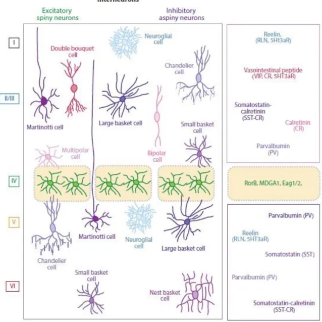
1.1.1. Figure 1: Cerebral cortex development and morphogens expression 1 1.1.1. Figure 2: Neural progenitor subtypes and neurogenic phases during
cerebral cortex development in mice
3
1.1.2. Figure 3: The genetics of early pallial patterning 5 1.1.3. Figure 4: Schematic representation of the major neocortical interneuron
subtypes
7
1.1.4. Figure 5: Schematic illustration of the six horizontal layers characteristic of the cerebral neocortex
9
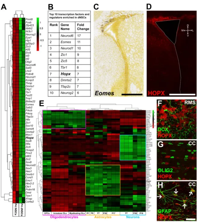
1.2.1. Figure 6: The SVZ replenishes olfactory interneurons throughout life 12 1.2.2. Figure 7: Cellular composition of the ventricular–subventricular zone (SVZ) 14 2.3. Figure 8. A Meta-Analysis of TFs Enrichment in dNSCs Highlights Their
Association with Distinct Neural Lineages
39
2.3. Figure 9. HOPX Exhibits a Complex Spatial and Temporal Expression Pattern within the SVZ Where it Labels a Subpopulation of dNSCs
41
2.3. Figure 10. Lineage-Specific Markers Highlight Acquisition of Divergent Cell Fates by NSCs Located in Different dSVZ Subdomains
43
2.3. Figure 11. Conditional Fate Mapping Reveals HOPX-Expressing NSCs Are Biased to Generate Astrocytes
45
2.3. Figure 12. Single-cell RNA Sequencing Meta-analysis Reveals Hopx-expressing RGCs Share Transcriptional Features with Adult Astrocytes
47
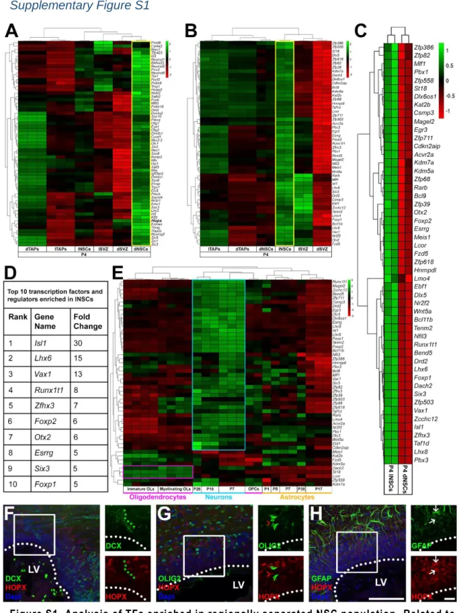
2.5. Figure S1. Analysis of TFs Enriched in Regionally Separated NSC Population. Related to Figure 8
51
2.5. Figure S2. Spatial Heterogeneity of Hopx Expression at P4. Related to Figure 9
52
2.5. Figure S3. Confirmation of Lineage Specificity of Selected Transcripts using the Dataset from the Barres Group. Related to Figure 10
53
2.5. Figure S4. Hopx and Astrocytic Markers Identified by Zywitza et al. Identify a Subpopulation of “Astrogenic” RGCs at E17.5. Related to figure 12
54
3.3. Figure 13. Neurog2Creert2/tdTom Mice Allow Fate Mapping of Birth-Dated
Cohorts of Glu Progenitors and Reveal Their Persistence at Postnatal Stages
62
3.3. Figure 14. Postnatal Glu Progenitors Originate from a Pool of Slow-Cycling RGCs that Accumulate in the SVZ during Corticogenesis
65
3.3. Figure 15. Dysregulation of Neurogenic Transcriptional Coding in Postnatal Glu Progenitors
XXII
3.3. Figure 16. Partial Rescue of Impaired Migration and Differentiation of Postnatal Glu Progenitors by Pharmacological and Genetic Manipulations
69
3.4. Figure S5. Related to Figure 13. Neurog2Creert2/tdTom transgenic mouse line allows detailed analyses of cell morphology
72
3.4. Figure S6. Related to Figure 13. Postnatal Glu progenitors mainly give rise to glutamatergic projection neurons of sensory cortical regions
73
3.4. Figure S7. Related to Figure 13. Neurog2Creert2/tdTom mice reveal a late wave of corticogenesis early postnatally
74
3.4. Figure S8. Related to Figure13. Postnatally born glutamatergic neurons develop intracortical projections and dendritic spines
75
4.2. Figure 17. The periventricular germinal niche at pre- and postnatal timepoints 85 4.2. Figure 18. A unifying scheme of pre- and postnatal germinal activity,
integrating single cell findings discussed in this review
91
4.3. Figure 19: Specific Targeting of Layer IV Cortical Neurons induces fast and efficient neuronal ablation
96
4.3. Figure 20: Targeted cortical ablation induces progenitor responses within the dSVZ
XXIII
Table of Contents
1. Regional Heterogeneity and Competence of Neural Stem Cells Throughout Development and Postnatal Life ... 1
1.1. The Developing Forebrain Generates Neuronal Diversity ... 1 1.1.1 Diversity of the Progenitors during Cortical Neurogenesis ... 1 1.1.2 Regionalization of the Developing Brain ... 4 1.1.3. Basic Principles of Cortical Organization ... 6 1.1.4. Molecular Diversity of Anatomically Defined PNs ... 8 1.2. Germinal Activity Persists in the Postnatal SVZ ... 11
1.2.1. The Postnatal Niche ... 11 1.2.2. The Radial Glia Origin of Type B1 Cells: ... 13 1.2.3. Type B1 Cells: A Specialized Astrocyte ... 13 1.2.4. Regionalization and heterogeneity of Postnatal SVZ NSCs ... 15 1.2.5. Regionalization also applied to gliogenesis ... 17 1.2.6. Signals from the CSF and the niche act in concert to regulate NSCs activity and regionalization throughout pre- and postnatal life ... 18 1.3. Transcriptional correlates of SVZ NSCs regionalization ... 25 1.4. Objectives of the PhD thesis ... 27 2. Experimental Chapter 1: HOPX Defines Heterogeneity of Postnatal Subventricular Zone Neural Stem Cells ... 31
2.1. Introduction ... 32 2.2. Experimental procedures ... 33 2.3. Results... 38
Hopx Is Enriched in NSCs of the dSVZ and in Cells of the Astrocytic Lineage. ... 38 HOPX Expression Reveals Intraregional Heterogeneity within the dSVZ ... 40 The dSVZ Is Defined by Microdomains with Distinct Lineage Outputs ... 42 HOPX-Expressing dNSCs Are Biased to Acquire an Astroglial Fate ... 44
XXIV
Hopx-expressing RGCs are Present in the Late Developing pallium and Share Transcriptional Featrues with adult Astrocytes ... 46 2.4. Discussion ... 48 2.5. Supplementary Figures ... 51 3. Experimental Chapter 2: Transcriptional Dysregulation in Postnatal Glutamatergic Progenitors Contributes to Closure of the Cortical Neurogenic Period ... 55 3.1. Introduction ... 56 3.2. Experimental Procedures ... 56 3.3. Results ... 61
Fate Mapping of Birth-Dated Cohorts of Glutamatergic Neurons ... 61 A Large Population of Glu Progenitors Persist in the Postnatal SVZ ... 63 ScRNA-Seq Reveals Transcriptional Dysregulation in Postnatal Glu Progenitors .. 64 Postnatal Glu Progenitor Differentiation Can Be Partially Rescued ... 68 3.4. Discussion ... 69 3.4. Supplementary Figures ... 72 4. General Discussion ... 79 4.1. Summary and Opened Questions ... 79 4.2. Contributions of Single cell Approaches to Probe Neural Progenitor’s Heterogeneity and Dynamics ... 80 4.2.1.Clonal Techniques in Histology and Transcriptomic ... 80 4.2.2.Contributions of Single Cell Approaches to Probe Spatial Identity Specification ... 82 4.2.3. Contributions of Single cell Approaches to Probe Temporal Identity Specification ... 86 4.2.4. Contributions of Single cell Approaches to Probe Adult NSCs Origin and Biology ... 88 4.3. Implication for Brain Repair... 92
4.3.1 Are embryonic and Postnatal/adult NSCs Representing Distinct Populations in term of Diversity and Competence? ... 92
XXV
4.3.2. Targeted Neuronal Ablation as a Model to Study Competence of Postnatal NSCs for Cortical Repair ... 93 5. Annexes ... 101 6. References ... 124 7. CV ... 143 8. List of Publications ... 144 9. Additional Publications ... 145
XXVII
1
1. Regional Heterogeneity and Competence of Neural
Stem Cells Throughout Development and Postnatal Life
The central nervous is a tissue of exquisite complexity. It forms in a stepwise manner during embryonic development from a population of specialized cells within the ectoderm. Here I describe its gradual development and complexification over time, with a special emphasis on the forebrain and of the cortex, the regions of interest of this PhD work. Germinal activity does not sharply stop at birth, but persists within the subventricular zone of the lateral ventricle. In a second part of the introduction, I describe the transformation of this germinal region at postnatal stages. I finally describe its specificities and similarities compared to its embryonic counterpart, both in term of cellular and regional organization, which were both explored further during my PhD.
1.1. The Developing Forebrain Generates Neuronal Diversity
The neocortex is by far one of the most complex regions of the mammalian brain, characterized by a remarkably diversity of neuronal and non-neuronal cell types, whose spatial and temporal coordinated development and function guarantee the execution of high-order cognitive, sensory, and motor behaviors. Decoding its heterogeneity have been at the core of neuroscientists’ work for decades. In this first part of the introduction I would like to introduce some of the key aspects of its organization and development.
1.1.1 Diversity of the Progenitors during Cortical Neurogenesis
Once neurulation is completed, the neural tube forms three primary vesicles, namely the forebrain, the midbrain and the hindbrain (Figure 1A). The forebrain rapidly evolves in further subdivisions to form secondary vesicles including the telencephalon. Its dorsal part becomes the pallium where the cerebral cortex derives.
2
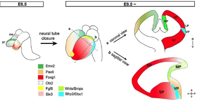
Figure 1: Cerebral cortex development and morphogens expression. (A) The rostral part of the neural tube evolves rostrally into the primary forebrain (FB), midbrain (MB) and hindbrain (HB) vesicles while the caudal part gives the spinal cord (SC). The secondary vesicles telencephalon (Tel), diencephalon (Di), metencephalon (Met) and myelencephalon (Myel) develop from the primary vesicles. (B) The anterior neural ridge (ANR), cortical hem (CH) and anti-hem (AH) are secreting centers. ANR secretes FGFs, CH secretes Wnts/BMPs and AH secretes FGF15 morphogens in the telencephalon. Shh is mainly present in the ventral part of the telencephalon, where ganglionic eminences are specified and generate interneurons that follow dorsal tangential path to invade the developing cortex. All morphogens are also found in the CSF secreted by choroid plexus (CP) and filling t he lateral ventricles (LV). (C) Morphogens gradients in the dorsal telencephalon. Illustration from Agirman et al., 2017.
At the end of the neural tube closure, the neuroepithelium is composed of neuroepithelial stem cells (NECs), the earliest progenitors of the cortex. They are organized in a pseudostratified neuroepithelium because their nuclei migrate up and down the apical– basal axis during the cell cycle. NECs show typical epithelial features and are highly polarized along their apical–basal axis. These cells initially expand their pool by successive symmetric proliferative divisions and later divide asymmetrically to generate radial glial cells (RGCs) that sit in the ventricular zone (VZ) of the dorsal part of the telencephalon, the most apical cell layer that lines the ventricles. During this process, NECs lose some of their epithelial traits to acquire astroglial hallmarks. For example, in contrast to NECs, RGCs show several astroglial properties such as the presence of glycogen granules and the expression of astrocytic proteins like GLAST, S100β, GFAP, Vimentin and BLBP. This transition marks the onset of neurogenesis in mice and occurs throughout most of the brain between embryonic days 10 (E10.5) and E12.5.
RGCs maintain a bipolar morphology with apical and basal processes (Figure 2A) and produce most of the neurons in the brain, either directly or indirectly (Haubensak et al., 2004; Miyata, 2004; Noctor et al., 2004). Cortical neurogenesis which extends from E10.5 to E18.5 in mice is followed by the gliogenesis period, which terminates after birth. At early stage of corticogenesis, RGCs in the pallium divide asymmetrically to self-renew and generate a projecting neuron. This process is defined as direct neurogenesis. As corticogenesis proceeds, RGCs give rise to intermediate progenitors (IPs) that delaminate from the apical surface to invade the subventricular zone (SVZ) (Figure 2B). Contrary to RGCs, IPs divide symmetrically to give birth to two identical neurons following further round of cell division. This process is defined as indirect neurogenesis and is critical to increase the number of neurons that are generated from a given number of RGCs (Haubensak et al., 2004). IPs differ from RGCs in terms of genes they express. For
3
example, they specifically express the genes that encode the transcription factor Tbr2 (Englund, 2005). Newborn neurons generated directly or indirectly from RGCs migrate to reach the cortical plate (CP). Early born neurons move basally through somal translocation and integrate the deep layers of the cortex (Tabata et al., 2009). In contrast, later born neurons transit through multiple morphologies and use the processes of RGCs as a scaffold for their locomotion towards the upper cortical layers (Evsyukova et al., 2013; Hippenmeyer, 2014). Cortical layering occurs in an ‘inside-out’ fashion whereby earlier born neurons populate deep layers and later born neurons progressively occupy upper layers (Angevine and Sidman, 1961). These successive waves produce six layers in the neocortex.
4
Figure 2: Neural progenitor subtypes and neurogenic phases during cerebral cortex development in mice. (A) RGCs maintain a bipolar morphology with apical and basal processes. (B) Sequential steps of neurogenesis in the mouse: neuroepithelial cells (NECs, grey) self-renew by symmetric division, then turn into apical radial glial cells (RGCs, pink) that divide either symmetrically to self-renew, or asymmetrically to give rise first to primary neurons including Cajal–Retzius cells (dark green) (direct neurogenesis) which migrate to the cortical surface to form the marginal zone (MZ), and to intermediate progenitor cells (I PC) at E12.5 and onwards; IPCs populate the subventricular zone (SVZ) and generate cortical layer neurons (dark to light blue), which migrate along the basal process of RGCs through the intermediate zone or subplate (IZ/SP) towards their destined layer. At later stages, some RGCs can undergo final symmetric divisions generating two neurons or switch to gliogenesis. Illustration adapted from Jiang and Nardelli, 2015.
1.1.2 Regionalization of the Developing Brain
Within the telencephalon, a dorso-ventral polarity rapidly develops leading to the emergence of the pallium at its dorsal part. This is due to the reciprocal action of transcription factors and morphogens secreted by three signaling centers, the cortical hem (CH) in the mediodorsal region, the anti-hem (AH) in the lateral aspect, and the anterior neural ridge (ANR) in the anteromedial region (Figure 1B and 1B). These patterning molecules diffuse in complementary gradients and induce expression of regionally defined transcription factors (TFs). Some of the main TFs involved in the regionalization of the embryonic pallium are presented here. Some of these morphogens and their role in neural stem cell activity are covered in more details in part 1.3.5. of this introduction.
The early forebrain patterning in mouse embryo is established around E8.5. Otx2 first establishes the forebrain and midbrain territories (Inoue et al., 2012). Then, Emx2 and Pax6 functions redundantly to establish the caudal forebrain, which contributes to the medial pallium, ventral pallium and diencephalon while Six3 establishes the rostral forebrain domain (Lagutin et al., 2003) (Figure 3).The expression of Fgf8 in the anterior neural ridge induces Foxg1 in the Six3-expressing domain and establishes the dorsal pallium and subpallium (Kobayashi et al., 2002; Lagutin et al., 2003). The boundary between the pallium and subpallium is regulated by cross-repression between Gli3 and Shh, and Fgf8 expression (Gutin, 2006). Through these interactions, the border between the Pax6+ pallium and the Nkx2.1+ subpallium is first established around E9.5 (Shimamura and Rubenstein, 1997), which is subsequently replaced by a Pax6/Gsx2 boundary by E12.5 (Yun et al., 2001). After this, the entire pallium is defined by Pax6 expression in the progenitors and Tbr1 expression in the postmitotic neurons (Puelles et al., 2000), although Tbr1 is downregulated in many of the layers II–V neurons (Han et al., 2011). Emx1 further delineates an expression boundary between the dorsal and ventral pallium (Puelles et al.,
5
2000). Thus, excitatory glutamatergic projection neurons arise from progenitor cells of the Emx1-positive dorsal pallial area (Louvi et al., 2007), whereas the subpallium which includes the ganglionic eminences (GE) produces inhibitory interneurons (Hébert and Fishell, 2008; Moreno et al., 2009).
Following E9.5 several transcription factors have a clear regionalized expression pattern. This is particularly clear for markers of the pallium and the subpallium. Members of the Dlx family (Distal-less), namely homeobox genes Dlx1, Dlx2 (Bulfone et al., 1993), Dlx5 and Dlx6 (Simeone et al., 1994) are enriched in the ganglionic eminences of the subpallium and consistently absent from the pallial domain. Other ventrally enriched genes are Nkx2, Nkx6.2, Gsx1 and Gsx2 (Zhong et al., 2008; Zhong and Holland, 2011).
On the other hand, homeobox genes Emx1, Emx2 and Pax6 show restricted expression to the pallium as well as the T-box transcription factors Tbr1 and Tbr2 (also referred as Eomes) (Bulfone et al., 1995; Bulfone et al., 1999), as well as the bHLH transcription factor Neurog2 (reviewed in Lee, 1997. Expression of these TFs antagonize a subpallial expression of the bHLH transcription factor Mash1 (Casarosa et al., 1999).
Figure 3: The genetics of early pallial patterning. Coronal view indicates spatial subdivisions of the pallium, where dorsal pallium lies between the medial and lateral/ventral pallium. In sagittal view, the medial and lateral/ventral pallium connects at caudal levels. DP, dorsal pallium; LP, lateral pallium; me, mesencephalon; MP, medial pallium; pr, prosencephalon; SP, subpallium; VP, ventral pallium. Adapted from (Kumamoto and Hanashima, 2014).