Directed assembly of living Pseudomonas
aeruginosa on PEI patterns fabricated by
nanoxerography for statistical AFM bio-experiments
Eric Jauvert 1, 2, 3, Etienne Palleau1, Etienne Dague2, 3 and Laurence Ressier1*
1 Université de Toulouse; LPCNO ; INSA, CNRS, UPS ; 135 avenue de Rangueil, 31077
Toulouse, France
2 CNRS ; LAAS ; 7 av du colonel Roche, 31077 Toulouse cedex 4, France
3 CNRS ; ITAV-UMS 3039 ;31106 Toulouse, France
Keywords: directed assembly, Pseudomonas aeruginosa, PEI, atomic force microscopy, assembly, nanoxerography
Abstract
Immobilization of living micro-organisms on pre-defined areas of substrates is a major prerequisite of their characterizations by Atomic Force Microscopy (AFM) in culture media. It remains challenging since micro-organisms should not be denatured but attached strongly enough to be scanned with an AFM tip in liquid. Here, we propose a novel approach where biological objects of interest are electrostatically assembled on 2 nm thick Polyethylenimine (PEI) patterns fabricated by nanoxerography. This nanoxerography process consists in electrostatic trapping of PEI chains on negatively charged patterns written on electret thin films by AFM or electrical micro-contact printing. The great potentialities of this approach were demonstrated using a common biological system, Pseudomonas aeruginosa bacteria. Such bacteria, negatively charged, were selectively assembled on large scale arrays of PEI patterns. The number of bacteria grafted on each pattern was finely controlled either by tuning the surface potential or the lateral size of charge patterns. Contrary to uncontroled PEI films commonly used for cell anchoring, these ultra-thin PEI patterns strongly grafted on the surface are shown to not denature assembled Pseudomonas aeruginosa bacteria. AFM characterizations in culture media of large populations of individual living bacteria can thus easily be performed using this approach, offering the opportunity of representative statistical data analysis. These opportunities can be opened up to any micro-organism negatively charged in solution.
Introduction
Atomic force microscope (AFM) is a powerful tool in microbiology since it allows topographical, mechanical and chemical characterizations at the nanoscale of biological objects of interest, still evolving in their culture media. However, working on immobilized living micro-organisms is one of the main prerequisites of AFM measurements in liquid. Nowadays, realizing this selective assembly is still challenging since the strength of the grafting has to respect a certain balance: it has to be strong enough to maintain biological objects which can be altered or moved by the AFM tip interaction while scanning the surface; but not too aggressive to prevent them from possible denaturation.
Ideally, a technique that allows the immobilization of living biological objects of interest, on pre-defined periodic arrays over large areas of a substrate will help gaining time on the localization of the zone of observation by AFM and collecting statistical data on heterogeneous population in their culture media. For this purpose, several approaches for cell assembly have been developed and can be divided into two main groups: (i) mechanical trapping in topographically patterned or soft substrates (based on a gel matrix1,2, with patterns obtained by lithography3, through porous membranes4–6, assisted by convective self-assembly7) (ii) immobilization using localized functionalization of a substrate (based on electrostatic interaction
8–10
, covalent bonds11–13, antibody-antigen linking14, assisted by convective self-assembly15). Nevertheless, all these techniques present a least one negative side effect: complexity of implementation, lack of selectivity in assembling, non-versatility or denaturation of assembled micro-organisms. Over the range of available assembly techniques, electrostatic immobilization is very convenient for electrostatically charged microorganism. Most of them are negatively charged and therefore polycations were used t create positively charged substrates. Among them,
Poly L Lysine and Polyethylenimine (PEI) films are probably the most largely employed ones since they provide an efficient grafting and are very simple to implement. However, this technique suffers from two main drawbacks: (i) it generates random deposition of studied micro-organisms, (ii) polycation chains poorly grafted to the substrate are released in the solution and contaminate the culture environnement16.
In the present study, we propose a novel approach where biological objects of interest are electrostatically assembled on Polyethylenimine (PEI) patterns pre-fabricated by nanoxerography (EAPPFN). The nanoxerography process consists in electrostatic trapping of PEI chains on negatively charged patterns written on electret thin films by AFM or electrical micro-contact printing17 (e-µCP). The potentialities of this approach are evaluated, by applying it to the medical relevant bacterium, Pseudomonas aeruginosa. This bacteria are, indeed, responsible for hospital acquired infections, and able to acquire resistance to all the known antibiotics. In previous studies we studied the effect of antibiotics on the cell wall of this micro organisms but were limitated by the random deposition of the cells. Here, we have investigated if AFM characterizations in culture media can be carried out of bacteria arrays obtained by EAPPFN, checked the viability of assembled bacteria and studied how to control finely the number of bacteria immobilized on each PEI pattern. Such developments are of first interest to generate statistically relevant AFM data, in a reasonable time scale.
Results and discussion
Fabrication of PEI patterns by nanoxerography and characterizations.
polymethylmethacrylate (PMMA) spin-coated on 1016 cm-3 p-doped silicon substrates, by applying negative voltage pulses either to a highly n-doped silicon AFM tip or a micro-structured gold-metallized PolyDiMethylSiloxane (PDMS) stamp (so-called electrical micro-contact printing (e-µCP) process) (Fig. 1a). These two kinds of charge writings are complementary: AFM charge writing is versatile since AFM allows to draw charge patterns of complex geometry in a few minutes and to quantify their surface potential by its derived electrical mode of Kelvin Force Microscopy (KFM). Charge writing by e-µCP is very useful to fabricate, in parallel, hundreds of thousands of charge patterns covering large areas of substrates or to perform repetitive charge writings with the same stamp on various samples, a necessity for future scaling up and automation of the protocol. In the second phase (Fig. 1b), a 10 µl droplet of 0.2% concentrated solution of aqueous Poly(ethyleneimine) (PEI) was incubated on the electrostatically patterned substrates for 90s. The substrates were subsequently immersed in absolute ethanol for 30s, rinsed with pure water and dried under nitrogen, following the two-step protocol previously developed by our group, for aqueous colloidal suspensions18. This second phase led to the selective electrostatic trapping of the cationic PEI polymer chains on charge patterns of opposite polarity thanks to electrophoretic forces.
Figure 1. Schematics of the fabrication of PEI patterns by nanoxerography: charge writing by AFM or electrical micro-contact printing (e-µCP), (b) development by incubation in an aqueous PEI solution.
Figure 2 shows examples of PEI patterns fabricated by AFM and e-µCP nanoxerography. The first one, consisting of a 17 µm large Occitane cross, illustrates the flexibility in the pattern design offered by AFM nanoxerography (Fig. 2a). The KFM image on the left panel reveals that this charge pattern, written using -30 V voltage pulses, presents a surface potential of -1.5 V. After development in the PEI solution, the AFM topographical image in the middle panel shows that a 1.5 nm thick PEI layer is selectively grafted on the charge pattern, reproducing it very accurately. As expected, this PEI pattern presents a positive surface potential, of +80 mV in this case (right panel). Figure 2b shows similar results for another pattern geometry obtained by e-µCP nanoxerography. In that case, hundreds of 5 µm wide PEI squares were fabricated on a 1 cm² zone. Each PEI square exhibits a 2 nm mean thickness and a surface potential of about +80 mV.
In both cases, it is important to note that PEI patterns fabricated by nanoxerography present a similar surface potential as observed on regular thicker PEI continuous films. This indicates that such PEI patterns, even composed of a limited number of PEI chains, have the same electrostatic trapping capability than thicker PEI films.
Figure 2. (a) Occitane cross-shaped PEI pattern fabricated by AFM nanoxerography and (b) array of 5 µm square PEI patterns fabricated by e-µCP nanoxerography. In each case, KFM surface potential images of the charge patterns written on a PMMA thin film are presented on the left panel, AFM height images and KFM images of the PEI patterns obtained after development are shown on the middle and the right panels respectively. Representative sections are systematically shown under each image. Note that a larger view by optical microscopy of the array of PEI squares is provided in inset of the AFM height image presented on the middle panel of (b).
Directed assembly of Pseudomonas aeruginosa bacteria on PEI patterns and characterizations.
The directed assembly of negatively charged Pseudomonas aeruginosa on positively charged PEI patterns was performed by incubation a 10 µl droplet of the aqueous bacteria suspension for 10 min, on arrays of PEI patterns similar to the one depicted in Figure 2b, followed by a rinsing in deionized water. The samples were then transferred in a 10 mM phosphate buffered saline (PBS) buffer to avoid denaturating bacteria immobilized on PEI patterns. It is worth noting that it would not be possible to fix directly the negatively charged bacteria on positively charged patterns written on PMMA because the required immersion of bacteria in ethanol would denature them.
Figure 3a presents typical AFM images in buffer medium of Pseudomonas aeruginosa assembled on an array of 5 µm square PEI patterns. Ten bacteria are immobilized on each PEI square, demonstrating the repeatability of the process. Firmly fixed on the patterns, the bacteria can be imaged in contact monde in buffer medium several hours without revealing any damaging or displacement. Figure 3b shows a zoom-in AFM deflection image of bacteria assembled on the PEI pattern marked by a dashed square in Fig 3a. It evidences ten Pseudomonas aeruginosa immobilized on the 5 µm PEI pattern. The mean height of 500 nm measured on the cross-sections of the AFM height images is very typical of living Pseudomonas aeruginosa19,20. Force
volume mapping experiments performed in buffer medium evidence heterogeneities of Young’s modulus within the bacteria (Fig. 3c). Force volume mappings on 200 nm scan in the middle of the five bacteria labelled in Figure 3b allow us to extract, using the Sneddon model21, the distribution of Young’s modulus for each bacterium. The elasticity values vary from 740 kPa to 967 kPa, in agreement with values usually reported in the literature19,20. These heterogeneities of elasticity from one bacterium to another pointed out by these distributions show the absolute
necessity to perform statistical analysis on a large population of micro-organisms to obtain accurate mechanical characteristics.
Figure 3. (a) AFM height image (left) and deflection error image (right) in buffer medium of
Pseudomonas aeruginosa assembled on an array of 5µm square PEI patterns similar to the one
presented in Fig. 2b, (b) deflection error image of ten Pseudomonas aeruginosa assembled on the square PEI pattern marked by a dashed square on (a), with height profiles of the five bacteria labelled on the deflection error image, (c) Young’s modulus force volume mapping in buffer medium of the area observed in (b) with Young’s modulus histograms of the five bacteria labelled in (b).
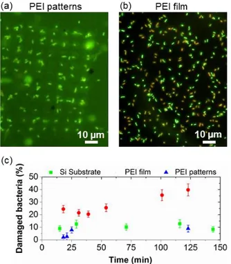
To check if the cell cytoplasmonic membrane of the immobilized bacteria was not damaged by PEI patterns fabricated by nanoxerography, a Livedead® kit was employed (see Materials and methods section for details). Pseudomonas aeruginosa tagged with the LIVEDEAD® cell viability assays, were immobilized by incubation for 30s on three different silicon substrates: one covered by 5 µm square PEI patterns fabricated by e-µCP nanoxerography, one covered by a PEI thick film and a bare one. Figures 4a and 4b present typical fluorescence optical microscopy images taken in buffer medium on assemblies of bacteria on the PEI patterns and the PEI film respectively. These images recorded after 20 minutes of incubation, show that a strong proportion of bacteria assembled randomly on the thick PEI film are colored in red, meaning that their cell wall are permeable to Propidium Iodide and thus damaged. On the contrary, all the bacteria selectively assembled on the PEI patterns fabricated by nanoxerography are colored in green confirming that their cell wall is not denatured. Figure 4c quantified the percentage of damaged bacteria as a function of time grafted in the case of the three tested substrates. The rate of damaged bacteria is about 25% after only 25 minutes and strongly increases with time in the case of bacteria immobilized on PEI films. One hour after their immobilization on the continuous PEI film, 40% of the bacteria are damaged. As reported in the literature, PEI films are toxic for bacteria because PEI chains are released back in the solution16. The rate of damaged bacteria assembled on a silicon wafer, around 10%, is weak and stable with time. It probably corresponds to the naturally dead bacteria in the starting solution. Similar results are obtained in the case of bacteria assembled on PEI patterns, indicating that PEI patterns are not toxic for bacteria. This observation points out a major advantage of PEI patterns fabricated by nanoxerography: PEI patterns are constituted of a limited amount of PEI chains selectively attached on charge patterns (a negligible amount of PEI is deposited outside the
charge pattern), strongly grafted on the substrate by electrostatic interactions, preventing the culture medium from poisoning.
Figure 4. (a) Fluorescence microscopy image in buffer medium of Pseudomonas aeruginosa tagged with the Livedead® kit 20 minutes after their assembly on an array of 5 µm square PEI patterns similar to the one presented in Figure 2b (left) and on a continuous thick PEI film (right), (b) Percentage of damaged bacteria as a function of time when assembled on a bare silicon (Si) substrate, a PEI film and PEI patterns.
To control the number of bacteria immobilized on each PEI pattern, two main experimental parameters in AFM nanoxerography experiments were varied: the surface potential of charge patterns and their lateral size.
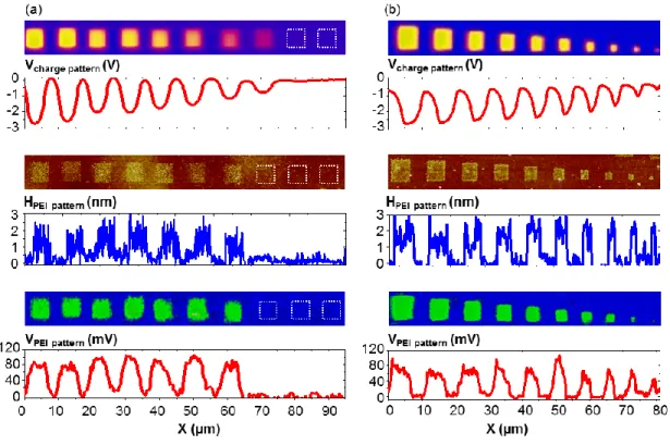
Figure 5a presents a first serie of ten PEI patterns fabricated from 5 µm square charge patterns written with decreasing voltage pulses (-80 V, -75 V, -70V, -65 V, -60 V, -50 V, -40 V, -30 V, -20 V, -10 V from the left to the right). The surface potential of such charge patterns varies from -3 V down to -500 mV (from the left to the right). No charge pattern was visible
between -20 V and -30 V, in agreement with previous results22. The development by incubation of a PEI solution conducts to 2 nm thick PEI patterns, independently of the surface potential of charge patterns (between -3 V and -1.15 V). All these PEI patterns present a surface potential of +80 mV. No PEI grafting was observed on the -0.770 V charge pattern revealing that the electric field generated by this charge pattern was not strong enough to trap PEI chains. This evidences the existence of a threshold of surface potential of charge patterns (around -1 V) for effective PEI grafting. A more precise analysis of topographical AFM images of PEI patterns reveals a significant decrease of the roughness of PEI patterns with increasing in absolute value the surface potential of charge patterns. This indicates that the density of grafted PEI chains increases with the surface potential of charge patterns in absolute value. Similar results were reported on directed assembly of charged colloidal nanoparticles by AFM nanoxerography23,24 where an increase of the surface potential led to an increase of NP density on charge patterns. Here, the increasing surface potential in absolute value, i.e. increasing electric field, generated by charge patterns allows more PEI chains to be assembled on charge patterns while a second layer of PEI molecules cannot be grafted on the first one because of induced electrostatic repulsion between each layer.
Figure 5b illustrates a second serie of ten PEI patterns fabricated from square charge patterns of various sizes ranging from 5 µm down to 500 nm (from the left to the right) by 500 nm step. As expected, whatever the initial dimension of the charge pattern, the thicknesses of obtained PEI patterns are homogeneous and are about 2 nm. Their surface potentials are constant around +80 mV; the observed smaller values for the 1 µm and 500 nm pattern come from the average effect artefact inherent to KFM measurements25–27.
Figure 5. Fabrication of PEI patterns by AFM nanoxerography using charge patterns of (a) decreasing surface potentials and (b) decreasing lateral sizes (from the left to the right). Data reported are KFM surface potential images and associated sections of the charge patterns (top), AFM height images and associated sections of the PEI patterns after development (middle) and KFM images and associated sections of the PEI patterns (bottom).
Pseudomonas aeruginosa bacteria were then assembled on twenty series of PEI patterns
similar to the ones depicted in Figure 5. Figure 6 reports the mean number of bacteria immobilized per PEI pattern while varying independently the surface potential of charge patterns (red square symbols) and their lateral size (blue disk symbols). The number of bacteria fixed per pattern increases with the surface potential in absolute value of the charge patterns. As previously mentioned, the increasing surface potential in absolute value of the charge patterns leads to an increasing density of PEI chains grafted on charge patterns which creates more
anchoring points for bacteria. On the other side, we observed that the mean number of bacteria assembled per pattern increases with the lateral size of the PEI patterns. The reason for this fine control of the bacteria number per pattern is purely geometrical: once grafted, a single micron sized bacteria do not leave sufficient room for others to be grafted on the same pattern. The fine tuning of any of these two parameters allows adjusting the mean number of trapped bacteria immobilized per PEI pattern down to one single bacteria, as illustrated in the inset of Figure 6.
Figure 6. Evolution of the mean number of bacteria assembled per PEI pattern with the lateral size (blue disk symbols) or the absolute value of the surface potential of the charge patterns (red square symbols). An AFM deflection image in buffer medium of an array of single Pseudomonas
aeruginosa bacteria immobilized on 1 µm PEI patterns is shown in the inset.
Conclusion
We presented a novel approach to tackle a major prerequisite of AFM characterizations of living micro-organisms in their culture media: their immobilization on pre-defined areas of a substrate. This approach consists in electrostatic immobilization of biological objects of interest on PEI patterns fabricated by electrostatic trapping of PEI chains on charge patterns written on
electret thin films by AFM or e-µCP. As a proof of concept, we validated it using a common opportunistic pathogen, Pseudomonas aeruginosa. The flexibility of nanoxerography allowed us to design PEI patterns with various geometry, size and density of PEI chains. Obtained PEI patterns, only 2 nm thick, present a constant positive surface potential similar to that of continuous PEI thick films. But, contrary to these ones, the strong and selective grafting of a limited amount of PEI chains prevents micro-organisms immobilized on these PEI patterns from poisoning. We demonstrated that the number of bacteria immobilized on each PEI pattern can be controlled down to unity by either tuning the lateral size or the surface potential of charge patterns. This approach allows user-friendly AFM characterizations in buffer medium on large populations of bacteria without any denaturation of these ones. Most of all, it opens a generic route for electrostatically assembling any biological objects of interest and statistically characterize them by AFM in culture media without denaturation.
Materials and methods
Charge writing.
Charge writing was performed under ambient conditions by applying 1 ms negative voltage pulses at a frequency of 50 Hz to a highly n-doped silicon AFM tip (AFM charge writing) or a 1 cm × 1 cm micro-structured gold-metallized PDMS stamp (e-µCP charge writing), while the substrates were grounded. The amplitude of the pulses was varied from -20 V to -80 V. These
specific writing conditions are reliable and reproducible, causing no tip, no stamp and/or sample damage at the high voltages used, as demonstrated previously22.After the charge writing step, the surface potential of the charge patterns was systematically measured in air by the electrical derived mode of amplitude modulation Kelvin Force Microscopy (KFM).
Growth of Pseudomonas aeruginosa bacteria.
The Pseudomonas aeruginosa ATCC 27853 used in this work were kindly provided by Raphael Duval from SRSMC, Nancy. Pseudomonas aeruginosa cultures were conducted at 35°C in Mueller Hinton media (difco, 275730-500 g) during 24 hours in static conditions. Bacteria at their stationary phase of growth were used. 1mL of media at a 108 cells/mL concentration was collected and centrifuged at 5000 rpm during 10 min. Supernatant was then removed and replaced by 1 mL of deionized water. This washing step was repeated 3 times.
AFM characterizations.
AFM images and force spectroscopy on the bacteria assembled on PEI patterns were all carried out in buffer medium in contact mode using an ICON from Bruker, equipped with a Nanoscope V controller. Si3N4 AFM probes (MLCT manufactured by Veeco Instruments), presenting a
pyramidal tip, with an opening angle of 35° were used for all the experiments. Their cantilever spring constants were systematically measured using the thermal tune method. They ranged from 10 to 30 pN/nm. The maximum force applied to the cells was limited to 1 nN in order to probe the cell wall elasticity (Young’s Modulus) and not the cell turgor pressure that is made at higher loading forces28,29. The topography and surface potential of PEI patterns were determined by AFM imaging in tapping mode and KFM mapping under ambient conditions.
Fluorescence optical microscopy characterizations using a Livedead® assay
In order to test the viability of Pseudomonas aeruginosa bacteria immobilized on PEI patterns fabricated by nanoxerography, PEI thick film and bare silicon substrate, the bacteria were tagged using a Livedead® kit, purchased from life Technologies XXX. This kit was composed of two fluorophores: (i) Cyto®9 (green) which enters the cytoplasmic membrane of any bacteria and (ii) propidium iodide (red) which penetrates the cytoplasmic membrane only if bacteria cannot control anymore osmotic flows. Observations in buffer medium by fluorescence optical microscopy of bacteria immobilized on these three substrates allowed us to quantify the percentage of damaged (red colored) and non-damaged (green colored) bacteria.
References
1. De, T., Chettoor, A. M., Agarwal, P., Salapaka, M. V. & Nettikadan, S. Immobilization method of yeast cells for intermittent contact mode imaging using the atomic force microscope.
Ultramicroscopy 110, 254–258 (2010).
2. Gad, M. & Ikai, A. Method for immobilizing microbial cells on gel surface for dynamic AFM studies. Biophys. J. 69, 2226–2233 (1995).
3. Kailas, L. et al. Immobilizing live bacteria for AFM imaging of cellular processes.
Ultramicroscopy 109, 775–780 (2009).
4. Kasas, S. & Ikai, A. A method for anchoring round shaped cells for atomic force microscope imaging. Biophys. J. 68, 1678–1680 (1995).
5. Dufrêne, Y. F., Boonaert, C. J. P., Gerin, P. A., Asther, M. & Rouxhet, P. G. Direct Probing of the Surface Ultrastructure and Molecular Interactions of Dormant and Germinating Spores ofPhanerochaete chrysosporium. J. Bacteriol. 181, 5350–5354 (1999).
6. Francius, G. et al. Detection, Localization, and Conformational Analysis of Single Polysaccharide Molecules on Live Bacteria. ACS Nano 2, 1921–1929 (2008).
7. Dague, E. et al. Assembly of live micro-organisms on microstructured PDMS stamps by convective/capillary deposition for AFM bio-experiments. Nanotechnology 22, 395102 (2011). 8. Lower, S. K., Tadanier, C. J. & Hochella Jr., M. F. Measuring interfacial and adhesion forces between bacteria and mineral surfaces with biological force microscopy. Geochim.
Cosmochim. Acta 64, 3133–3139 (2000).
9. Bolshakova, A. V. et al. Comparative studies of bacteria with an atomic force microscopy operating in different modes. Ultramicroscopy 86, 121–128 (2001).
10. Yoshida, T. & Nagasawa, T. ε-Poly-l-lysine: microbial production, biodegradation and application potential. Appl. Microbiol. Biotechnol. 62, 21–26 (2003).
11. Camesano, T. A., Natan, M. J. & Logan, B. E. Observation of Changes in Bacterial Cell Morphology Using Tapping Mode Atomic Force Microscopy. Langmuir 16, 4563–4572 (2000). 12. Cerf, A., Cau, J.-C., Vieu, C. & Dague, E. Nanomechanical Properties of Dead or Alive Single-Patterned Bacteria. Langmuir 25, 5731–5736 (2009).
13. Razatos, A., Ong, Y.-L., Sharma, M. M. & Georgiou, G. Molecular determinants of bacterial adhesion monitored by atomic force microscopy. Proc. Natl. Acad. Sci. 95, 11059– 11064 (1998).
14. Rozhok, S. et al. Methods for Fabricating Microarrays of Motile Bacteria. Small 1, 445– 451 (2005).
15. Ressier, L. et al. Combining Convective/Capillary Deposition and AFM Oxidation Lithography for Close-Packed Directed Assembly of Colloids. Langmuir 24, 13254–13257 (2008).
16. Xia, B. et al. Preparation and characterization of chemically-crosslinked polyethyleneimine films on hydroxylated surfaces for stable bactericidal coatings. Thin Solid
Films 520, 1120–1124 (2011).
17. Jacobs, H. O. & Whitesides, G. M. Submicrometer Patterning of Charge in Thin-Film Electrets. Science 291, 1763–1766 (2001).
18. Palleau, E., Sangeetha, N. M., Viau, G., Marty, J.-D. & Ressier, L. Coulomb Force Directed Single and Binary Assembly of Nanoparticles from Aqueous Dispersions by AFM Nanoxerography. ACS Nano 5, 4228–4235 (2011).
19. Formosa, C. et al. Nanoscale analysis of the effects of antibiotics and CX1 on a Pseudomonas aeruginosa multidrug-resistant strain. Sci. Rep. 2, (2012).
20. Formosa, C., Grare, M., Duval, R. E. & Dague, E. Nanoscale effects of antibiotics on P. aeruginosa. Nanomedicine Nanotechnol. Biol. Med. 8, 12–16 (2012).
21. Sneddon, I. N. The relation between load and penetration in the axisymmetric boussinesq problem for a punch of arbitrary profile. Int. J. Eng. Sci. 3, 47–57 (1965).
22. Ressier, L. & Nader, V. L. Electrostatic nanopatterning of PMMA by AFM charge writing for directed nano-assembly. Nanotechnology 19, 135301 (2008).
23. Ressier, L., Palleau, E., Garcia, C., Viau, G. & Viallet, B. How to Control AFM Nanoxerography for the Templated Monolayered Assembly of 2 nm Colloidal Gold Nanoparticles. Nanotechnol. IEEE Trans. On 8, 487–491 (2009).
24. Palleau, E., Sangeetha, N. M. & Ressier, L. Quantification of the electrostatic forces involved in the directed assembly of colloidal nanoparticles by AFM nanoxerography.
Nanotechnology 22, 325603 (2011).
25. Charrier, D. S. H., Kemerink, M., Smalbrugge, B. E., de Vries, T. & Janssen, R. A. J. Real versus Measured Surface Potentials in Scanning Kelvin Probe Microscopy. ACS Nano 2, 622–626 (2008).
26. Jacobs, H. O., Leuchtmann, P., Homan, O. J. & Stemmer, A. Resolution and contrast in Kelvin probe force microscopy. J. Appl. Phys. 84, 1168–1173 (1998).
27. Palleau, E., Ressier, L., Borowik, ł & Mélin, T. Numerical simulations for a quantitative analysis of AFM electrostatic nanopatterning on PMMA by Kelvin force microscopy.
Nanotechnology 21, 225706 (2010).
28. Arnoldi, M. et al. Bacterial turgor pressure can be measured by atomic force microscopy.
29. Yao, X. et al. Atomic force microscopy and theoretical considerations of surface properties and turgor pressures of bacteria. Colloids Surf. B Biointerfaces 23, 213–230 (2002).