Therapeutic impact of
2-[fluorine-18]fluoro-2-deoxy-D-glucose positron emission tomography in the pre- and
postoperative staging of patients with clinically
intermediate or high-risk breast cancer
B. Klaeser
1,4*, O. Wiederkehr
2, D. Koeberle
3, A. Mueller
2,3, B. Bubeck
1& B. Thuerlimann
2,3 1Department of Nuclear Medicine,2
Senology Center of Eastern Switzerland and3
Department of Internal Medicine, Division Oncology-Hematology, Kantonsspital, St Gallen;4
Department of Nuclear Medicine, Inselspital, Bern, Switzerland
Received 20 December 2006; revised 25 March 2007; accepted 26 March 2007
Background:
Positron emission tomography with 2-[fluorine-18]fluoro-2-deoxy-D-glucose (FDG–PET) is an accurate imaging modality for the staging of breast cancer. The aim of this study was to determine the potential therapeutic impact of pre- and postoperative FDG–PET in patients with clinically intermediate or high-risk breast cancer.Patients and methods:
One hundred and fourteen patients with newly diagnosed breast cancer were examined before (73) or after (41) surgery. Patient data were translated into three scoring sheets corresponding to information available before positron emission tomography (PET), after PET and after further diagnostic tests. Three medical oncologists independently reviewed the retrospectively acquired patient data and prospectively made decisions on the theoretically planed treatment for each time point, according to the recommendations of St Gallen Consensus Guidelines 2005.Results:
FDG–PET changed the planed treatment in 32% of 114 patients. In 20% of cases, therapeutic intention (curative versus palliative) was modified. Radiation treatment planning was changed in 27%, surgical planning in 9%, chemotherapy in 11% and intended therapy with bisphosphonates in 13% of all patients.Conclusion:
Based on current treatment guidelines, FDG–PET, as a staging procedure in patients with newly diagnosed clinically intermediate or high-risk breast cancer examined pre- and postoperatively, may have a substantial therapeutic impact on treatment planning.Key words:
breast cancer, FDG, patient management, positron emission tomography, primary staging, therapeutic impactintroduction
Breast cancer is the most frequently diagnosed malignant
tumor among women in Europe, with 370 000 new cases
estimated in Europe in 2004 [1]. Although there have been
recent decline in breast cancer mortality rates in several
European countries [2], breast cancer still ranks first among
cancer deaths in women in Europe, with an estimated 129 900
cancer deaths in 2004 [1].
Primary staging of breast cancer most commonly consists of
a chest X-ray, liver ultrasound and bone scan, and sentinel
lymph node biopsy or axillary lymph node dissection for
evaluation of lymph node involvement. Suspicious findings
may be further investigated by computed tomography (CT)
and magnetic resonance imaging (MRI).
2-[fluorine-18]fluoro-2-deoxy-D-glucose Positron emission
tomography (FDG–PET) has proven to be an accurate imaging
modality for the staging of breast cancer with sensitivities and
specificities of 93% and 75% for primary staging [3] and in the
range of 89%–100% and 72%–88%, respectively [4–8], for
detection and restaging of recurrent breast cancer. Some studies
have already addressed the impact of FDG–PET on the
management of patients with recurrent or metastatic breast
cancer and reported changes of the original therapeutic plans in
overall 32% to 44% of cases [9–11]. However, data regarding
the clinical impact of positron emission tomography (PET) in
the primary staging of breast cancer are rare [12].
The purpose of our study therefore was to assess the potential
clinical impact of FDG–PET in the pre- and postoperative
staging of patients with newly diagnosed clinically intermediate
or high-risk breast cancer based on current treatment
recommendations [13].
patients and methods
patients
From 2002 to 2004, a total of 269 patients with breast cancer underwent an FDG–PET scan at our institution. All patients and PET scans were
original
article
*Correspondence to: Dr B. Klaeser, Klinik und Poliklinik fu¨r Nuklearmedizin, Inselspital, 3010 Bern, Switzerland. Tel:+41-31-6323655; Fax: +41-31-6323137; E-mail: [email protected]
prospectively registered in a database according to national law. Inclusion criteria were clinically intermediate or high-risk breast cancer, whole body staging with FDG–PET pre- or postoperatively and availability of medical records. Two hundred and twenty-three patients assigned by a Department of Kantonsspital St Gallen or associated oncologists were evaluated. Forty-six patients did not meet the inclusion criteria because of nonavailability of medical records. A total of 114 patients (73 staged with FDG–PET before surgery, hereafter pre-OP group and 41 with FDG–PET before adjuvant therapy, hereafter post-OP group) fulfilled the inclusion criteria and were included in this study (Table 1). One hundred and eight patients scanned for suspicion or restaging of tumor relapse, or assessment of therapy response were not included. One patient with an initial diagnosis of breast caner was retrospectively excluded because further histological evaluation during follow-up revealed amelanotic melanoma.
clinical data
The clinical data of all patients were obtained through systematic review of all inpatient and outpatient medical records. Data included age, relevant
comorbidities and previous treatments, date of initial diagnosis, histology, hormone receptor status, tumor marker level at time of diagnosis, results of mammography, chest X-ray, liver ultrasound, bone scan and further imaging modalities (MRI of the breast or other parts of the body, CT of the chest and/or abdomen) employed either before or after FDG–PET.
18
F-FDG–PET
FDG–PET was done using a dedicated full-ring Scanner (GE Advance NXi) with a transaxial resolution of 4.8–6.2 mm, 1–20 cm off center (National Electrical Manufacturers Association, Rosslyn, USA-1994). Patients fasted for at least 6 h before an i.v. injection of 370 MBq 2-fluoro-2-deoxy-D-glucose (FDG) into a vein contralateral to the tumor side. Blood 2-fluoro-2-deoxy-D-glucose level was <10 mmol/l in all patients. Whole body data were obtained in supine position 45–60 min after injection in 2D-acquisition mode, with 5 min per bed position for emission and 2–3 min for transmission. In patients scanned preoperatively, and if axillary lymph node status was doubtful after whole body imaging, a second scan in prone position with elevated arms was carried out 130–140 min after injection. Image reconstruction was done with iterative 2D-OSEM algorithm. FDG–PET was interpreted by consensus of at least two experienced nuclear medicine physicians on clinical service at the time of acquisition. The mean time lag between diagnosis of breast cancer and FDG–PET was 1.9 weeks in the pre-OP group and 6.6 weeks in the post-OP group.
study design
The study was conducted as part of a quality assurance program of the Senology Center of Eastern Switzerland and the Department of Nuclear Medicine, Kantonsspital, St Gallen, Switzerland. Patient data was collected retrospectively and made anonymous. According to clinical practice with routine baseline staging, FDG–PET scanning in cases of lingering suspicion of metastases after negative conventional tests and occasionally further radiological tests (CT, MRI) in doubtful cases after PET, we categorized data as chronologically available. Diagnostic information was divided into data available before PET (time point pre-PET), after PET (time point PET) and after accomplishment of further diagnostic tests (time point post-PET). The data were finally translated into three scoring sheets using the categorization as mentioned above. The pre-PET scoring sheet included age, results of conventional diagnostic tests and resulting clinical stage grouping (Table 1), tumor–node–metastasis (TNM) classification (adapted) according to American Joint Committee on Cancer (AJCC) Cancer Staging Manual 6th edition [14], histology, hormone receptor status and previous treatments. Results of the FDG–PET scan and findings of subsequent diagnostic tests as well as updated TNM classifications were given in the PET and post-PET scoring sheets.
Three senior medical oncologists blinded to each other evaluated the scoring sheets independently and prospectively defined theoretical treatment decisions on the intention-to-treat (curative versus palliative), surgery, chemotherapy, endocrine therapy, therapy with trastuzumab, radiotherapy and bisphosphonates consecutively for each time point. Treatment decisions were made according to the recommendations of St Gallen Consensus Guidelines 2005 [13] and j scores for the concordance of treatment decisions were calculated to measure interobserver agreement. Strength of agreement was classified as slight (j = 0.20–0.40), moderate (j = 0.41–0.60), substantial (j = 0.61–0.80) or excellent (j = 0.81–1.0).
In order to prevent ambiguity in the assessment of the anonymous patient data, following definitions were used. Breast-preserving surgery is intended if medically appropriate. In case of T2 classification (large operable), neo-adjuvant chemotherapy is offered to the patient with the intention to facilitate breast-preserving surgery. Locally advanced disease (T ‡3 or N ‡2) without distant metastases (M0) is treated by mastectomy and axillary clearance after neo-adjuvant systemic therapy in curative Table 1. Patient characteristics
Pre-OP group (n = 73) Post-OP group (n = 41) All patients (n = 114) Age (years) Range 38–83 32–80 32–83 Median 60 57 59 Mean 61 57 59 Ca 15–3a(kU/l) Range 5–944 5–78 5–944 Median 20 17 18 Mean 50 20 40 Histological subtypes (%) Ductal 53 76 61 Lobular 7 2 5 Mixed ductal–lobular 4 13 7 Large cell 6 2 4 Undifferentiated 4 0 3 Other subtypes 12 5 10 Not specified 14 2 10 Thereof clinically inflammatory subtype 12 2 9
Clinical stage grouping after conventional stagingb(%)
I 8 2 6 IIA 24 17 22 IIB 14 15 14 IIIA 10 25 15 IIIB 30 2 20 IIIC 3 32 13 IV 11 7 10
Diagnostic staging procedures before PET (%)
Chest X-ray 47 81 59 Liver ultrasound 30 63 42 Bone scan 18 44 27 Computed tomography 16 7 13
aReference value <27 kU/l.
bAccording to American Joint Committee on Cancer cancer staging manual
6th edition [14].
Pre-OP, FDG–PET before surgery; Post-OP, FDG–PET before adjuvant therapy; PET, positron emission tomography.
intention. Supraclavicular and internal mammary lymph node metastases leads to an extended field of radiation therapy. Classification for distant metastases is M0 if chest X-ray, liver ultrasound and bone scan are negative, and if one or more tests have not been carried out, classification is MX. If tumor and lymph node classification is T1 N0, the absence of distant metastases (M0) is assumed even if all tests have not been carried out. In preoperatively staged patients, the detection of limited axillary lymph node involvement (N1) by FDG–PET is not assumed to affect treatment decision, as these lymph node metastases would be detected in histological work-up anyway.
results
changes of clinical stage grouping
Stage grouping according to AJCC 6th edition was changed
in 48 of 114 patients (42%) after FDG–PET and in four
patients after accomplishment of further diagnostic tests (4%).
Rates of changes after PET based on clinical stage grouping
before PET are given in Table 2.
T stage changed in two patients (2%) due to multifocal
disease. PET revealed previously unknown regional lymphatic
spread classified as N3 in 24 cases (21%), supraclavicular lymph
node metastases in 15 (13%) and internal mammary lymph
node metastases in 18 patients (16%). Distant metastases were
diagnosed by FDG–PET in 22 cases. Distant metastases in FDG–
PET were confirmed by other imaging modalities in 10 patients
or during a follow-up of 39 6 8 months in nine cases. In two
patients with exclusively distant lymphatic spread according to
PET metastases were not proved by other imaging modalities
and patients remained clinically stable after adjuvant treatment
during follow-up. In 59 patients previously classified as MX
due to equivocal results or incomplete conventional staging
procedures, PET confirmed staging as M0.
Post-PET examinations did not change T classification, but
lead to a downstaging in one case with previously suspected
lymph node metastases and in one patient with a false-positive
PET finding in the vertebral column due to posttraumatic
changes. Post-PET results defined unclear stage grouping after
PET in three patients with sarcoidosis, an ovarian breast cancer
metastases and a second primary in the lung.
impact on patient management
Results of FDG–PET changed the treatment plan in 32% of
all patients (Table 3). The intention-to-treat was modified from
a curative to a palliative approach or vice versa in overall
20% and surgical planning was revised in 9% of all patients.
Radiation treatment planning was changed in a total of 27%
of patients with omission of planed adjuvant radiation
Table 2. Changes of stage grouping and TNM classification according to conventional staging (time point pre-PET, Table 1) after FDG–PET (time point PET) or further diagnostic tests (time point post-PET)a
Time point PET Time point post-PET
Proportions (%) Proportions (%)
Up Down All Up Down All Up Down All Up Down All Stage grouping I 2/7 2/7 29 29 IIA 11/25 11/25 44 44 1/25 1/25 4 4 IIB 5/16 5/16 31 31 1/16 1/16 6 6 IIIA 6/17 6/17 35 35 1/17 1/17 6 6 IIIB 14/23 14/23 61 61 IIIC 9/15 9/15 60 60 IV 1/11 1/11 9 9 1/11 1/11 9 9 All 47/114 1/114 48/114 41 1 42 1/114 3/114 4/114 1 3 4 TNM classification Pre-OP group T stage 2/73 2/73 3 3 N stage 22/73 35/73b 1/73 23/73 36/73b 30/48b 1 32/49b M stage 12/73 0/73 40/73c 12/73 52/73c 16 0/55c 16/71c Post-OP group T stage N stage 9/41 9/41 22 22 1/41 1/41 2 M stage 9/41 1/41 20/41c 10/41 29/41c 22 2/49c 24/71c 1/41 2/41 3/41c 3/41 4/41c 2 5/7c 7/9c All patients T stage 2/114 2/114 2 2 N stage 31/11444/114b 1/114 32/114 45/114b 27/39b 1 28/40b 1/114 1/114 1 1 M stage 21/114 1/114 60/114c 22/114 81/114c 18 1/53c 19/71c 1/114 2/114 3/114c 3/114 4/114c 1 2/3c 3/4c aAccording to American Joint Committee on Cancer cancer staging manual 6th edition [14].
bIncluding nodal upstaging from N0 to N1 in 13/114 patients. cIncluding downstaging from MX to M0 in 59/114 patients.
TNM, tumor–node–metastasis; PET, positron emission tomography; FDG–PET, 2-[fluorine-18]fluoro-2-deoxy-D-glucose positron emission tomography; Pre-OP, FDG–PET before surgery; Post-OP, FDG–PET before adjuvant therapy.
treatment after diagnosis of distant metastases in 17% and
enlargement of the radiation treatment field after detection of
previously unknown internal mammary and supraclavicular
lymph node metastases in 10% of patients. Diagnosis of
internal mammary node involvement alone accounted for
changes in 6% of cases. Changes of the surgical treatment were
due to omission of planned breast and axillary surgery after
diagnosis of distant metastases, detection of breast cancer in the
breast contralateral to the known primary and diagnosis of
axillary lymph node metastases remaining in situ after axillary
dissection. Intended chemotherapy was changed in 11% of all
patients, endocrine therapy was modified in 1% and treatment
with bisphosphonates was started for previously unknown bone
metastases in 13% of patients. Diagnostic tests and surgical
exploration carried out after PET modified treatment plans
by confirmation of metastases, revealing false-positive
findings and detection of PET-negative metastases in overall
5% of cases.
Kappa analysis showed substantial or excellent concordance
of treatment decisions between three medical oncologists
except for endocrine therapy, which was changed in only one
percent of cases.
use of conventional staging procedures
The number of diagnostic tests carried out before FDG–PET
declined during the investigation period from an average 5.2
tests per patient in 2002 to 1.8 in 2004. Likewise, the number of
investigations accomplished after FDG–PET fell from an
average 1.1 tests per patient in 2002 to 0.3 in 2004.
discussion
FDG–PET is a valuable imaging modality for detection and
restaging of recurrent breast cancer, and is more accurate than
conventional staging methods in screening for lymph node
metastases [7, 15], especially for the detection of internal
mammary and mediastinal nodes [16, 17], as well as in the
detection of distant metastases [3, 18] and proved to have
a major clinical impact in this context [9–11]. In contrast, data
defining the role of PET in the primary staging of breast cancer
are rare [12]. In the absence of prospective studies it is not
entirely clear, which patients may benefit from a PET scan as
part of the primary staging and under which circumstances
staging with FDG–PET might be a cost-effective approach.
In this study, we intended to analyze the potential value of
FDG–PET in a collection of patients of a single center, which
were referred for staging clinically intermediate or high-risk
breast cancer. Focusing on patients defined as mentioned
above, we found that based on PET results, changes of the
planed treatment would be required in 32% of all patients.
This number is comparable to what was reported in studies
investigating patients with recurrent or metastatic disease
[9–11]. Eubank et al. [9] retrospectively reviewed the medical
records of 125 consecutive patients with recurrent and
advanced disease and found documented changes in the
therapeutic plan in 32% of the patients. Yap et al. [10] and
Grahek et al. [11] evaluated the impact of PET on the
management of patients referred for restaging through
questionnaires sent to the referring physician before and after
the PET study, and reported changes of treatment in 60% and
44% of the cases. Response rates of the questionnaires were
relatively low (31% and 56%, respectively), making it likely,
that a responder bias lead to an overestimation of the rate of
management changes. Van der Hoeven et al. [12] evaluated
FDG–PET in the primary staging of patients with locally
advanced breast cancer referred for participation in a trial on
chemo- and immunotherapy. The authors detected unexpected
distant metastases leading to a palliative treatment strategy in
8% of 48 patients, compared with 17% in the corresponding
pre-OP group in our trial. However, Van der Hoeven [12]
reported that five of nine patients, who were PET positive for
distant metastases, were treated as planed according to the
study protocol when conventional imaging was negative. In our
study, nearly half of the PET positive distant metastases were
also negative in the initial conventional imaging and proved to
be true positive in follow-up examinations. Carr et al. [19]
presented data of a multimodality breast imaging study
designed to evaluate multimodal local staging. The authors
reported that FDG–PET added little additional information
and detected distant metastases in only 3% of all patients. In
fact, the study population consisted of only 69% of patients
with invasive cancers and overall <10% with tumors classified
as stage IIB or higher. These results therefore do not contradict
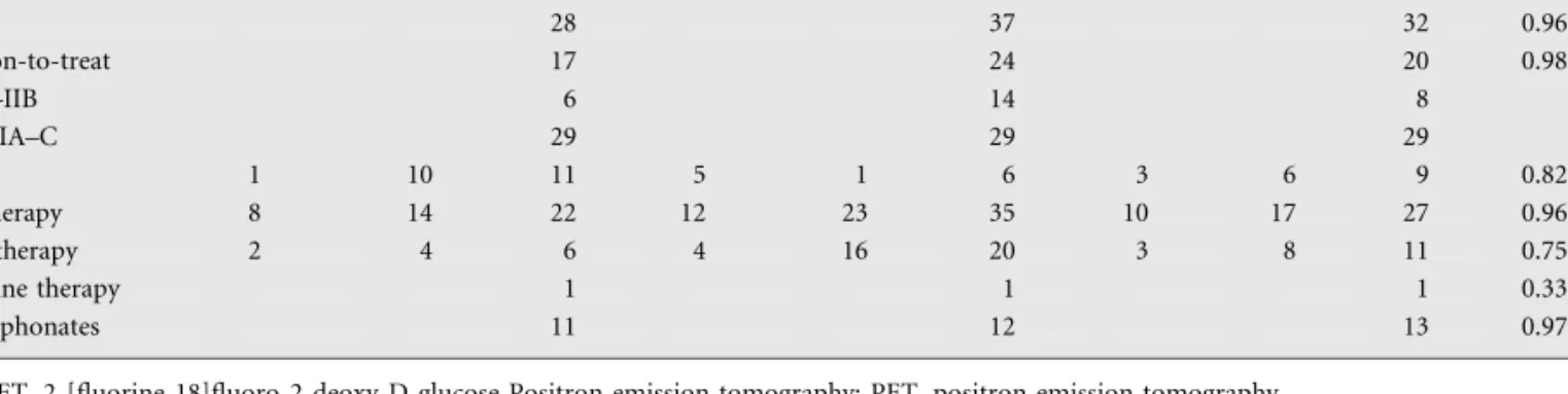
Table 3. Treatment modifications after FDG–PET and concordance of treatment decisions between three medical oncologists Changes of treatment
plans (%)
Pre-PET group Post-PET group All patients
More Less All More Less All More Less All j score
Overall 28 37 32 0.96 Intention-to-treat 17 24 20 0.98 Stage I–IIB 6 14 8 Stage IIIA–C 29 29 29 Surgery 1 10 11 5 1 6 3 6 9 0.82 Radiotherapy 8 14 22 12 23 35 10 17 27 0.96 Chemotherapy 2 4 6 4 16 20 3 8 11 0.75 Endocrine therapy 1 1 1 0.33 Bisphosphonates 11 12 13 0.97
our own findings, but highlight the importance of an
appropriate patient selection for PET staging.
Recently, Gil-Rendo et al. [20] published a prospective study
focusing on the role of FDG–PET in the axillary staging of 275
patients with stage I and stage II diseases. The authors found
a change of treatment in overall 33% of patients, when
taking into account sparing of sentinel lymph node biopsy in
cases of positive FDG uptake in the axilla.
According to our results, FDG–PET necessitated changes of
radiation treatment planning in 27% of the patients, which is
consistent with previously reported results [21]. In 6%, the
planed radiation field was extended because of unexpected
internal mammary lymph node metastases. The detection of
internal mammary metastases is of particular importance as
metastases to the internal mammary chain are associated with
a poor prognosis [16, 17, 22]. The rate of internal mammary
metastases in our study is lower than previously reported for
patients with advanced or recurrent disease, presumably due to
smaller average tumor size and lower percentage of
inflammatory breast cancer [16, 17, 23].
Surgical planning and chemotherapy were mainly revised
after detection of distant metastases, resulting in the omission
or reduction of planned treatments in 6% and 8% of patients,
respectively. Treatment with bisphosphonates was intended in
cases of multifocal bone involvement. Endocrine therapy is
indicated in all our patients with hormone receptor-positive
tumors irrespective of whether classified as intermediate or
high risk. Changes of stage grouping therefore lead to
additional chemotherapy with no major modifications of
endocrine treatment.
A certain limitation of our study is the retrospective
collection of clinical data. As a consequence, not all
conventional staging procedures were carried out as part of the
primary staging in every patient. Hence, a certain number of
metastases detected by FDG–PET possibly would also have
been detected by conventional procedures, if carried out. The
standard algorithm at a given institution for staging breast
cancer will also influence the additional value of FDG–PET
which may be less pronounced in sites where CT instead of
chest X-ray and liver ultrasound is routinely carried out. As
the exact rate of expected treatment changes and the potential
therapeutic impact and usefulness of FDG–PET depends on
the prevalence of lymphatic and distant metastatic spread, an
appropriate patient selection based on the clinical experience of
the referring physician is of particular importance. Patient
referral by experienced medical oncologists therefore might
lead to a certain selection bias in our study.
According to our experience, considering patients with
intermediate or high-risk breast cancer as candidates for PET
staging leads to a reasonable rate of treatment modifications
and showed substantial or excellent concordance of treatment
decisions between three medical oncologists. A preselection of
patients with clinically stage III disease may change the
intention-to-treat in up to one-third of all patients.
The impact of FDG–PET on patient management ultimately
depends on the confidence in the accuracy and validity of PET
among clinicians. The declining number of additional
conventional imaging procedures carried out before and after
FDG–PET during the study period in our institution reflects
the latter aspect and underlines the impact of FDG–PET in and
for daily clinical practice. The introduction of integrated PET/
CT systems leads to a further improved diagnostic accuracy
[24] and adds incremental diagnostic confidence in PET in
>50% of patients and regions with increased FDG uptake [25].
conclusions
FDG–PET in the primary staging of patients with clinically
intermediate or high-risk breast cancer has a substantial impact
on patient management. PET not only influences individual
treatment decisions but also impacts on clinical and diagnostic
workflows, as redundant staging procedures tend to be omitted
and tumor staging is more straight forward. Implementation of
FDG–PET in the staging of breast cancer leads to improved
stage grouping in a significant number of patients, hence
facilitating appropriate treatment and avoiding straining and
dispensable therapies. Our results give reason to investigate
whether modified clinical workflows with FDG–PET, when
implemented in the primary staging of advanced breast cancer,
might be a cost-effective approach under certain conditions.
acknowledgement
The authors would like to thank Rene Oettli for collecting
follow-up of patient data.
references
1. Boyle P, Ferlay J. Cancer incidence and mortality in Europe, 2004. Ann Oncol 2005; 16(3): 481–488.
2. Levi F, Bosetti C, Lucchini F et al. Monitoring the decrease in breast cancer mortality in Europe. Eur J Cancer Prev 2005; 14(6): 497–502.
3. Schirrmeister H, Kuhn T, Guhlmann A et al. Fluorine-18 2-deoxy-2-fluoro-D-glucose PET in the preoperative staging of breast cancer: comparison with the standard staging procedures. Eur J Nucl Med 2001; 28(3): 351–358. 4. Isasi CR, Moadel RM, Blaufox MD. A meta-analysis of FDG-PET for the evaluation
of breast cancer recurrence and metastases. Breast Cancer Res Treat 2005; 90(2): 105–112.
5. Weir L, Worsley D, Bernstein V. The value of FDG positron emission tomography in the management of patients with breast cancer. Breast J 2005; 11(3): 204–209.
6. Goerres GW, Michel SC, Fehr MK et al. Follow-up of women with breast cancer: comparison between MRI and FDG PET. Eur Radiol 2003; 13(7): 1635–1644.
7. Gallowitsch HJ, Kresnik E, Gasser J et al. F-18 fluorodeoxyglucose positron-emission tomography in the diagnosis of tumor recurrence and metastases in the follow-up of patients with breast carcinoma: a comparison to conventional imaging. Invest Radiol 2003; 38(5): 250–256.
8. Kim TS, Moon WK, Lee DS et al. Fluorodeoxyglucose positron emission tomography for detection of recurrent or metastatic breast cancer. World J Surg 2001; 25(7): 829–834.
9. Eubank WB, Mankoff D, Bhattacharya M et al. Impact of FDG PET on defining the extent of disease and on the treatment of patients with recurrent or metastatic breast cancer. AJR Am J Roentgenol 2004; 183(2): 479–486.
10. Yap CS, Seltzer MA, Schiepers C et al. Impact of whole-body 18F-FDG PET on staging and managing patients with breast cancer: the referring physician’s perspective. J Nucl Med 2001; 42(9): 1334–1337.
11. Grahek D, Montravers F, Kerrou K et al. [18F]FDG in recurrent breast cancer: diagnostic performances, clinical impact and relevance of induced changes in management. Eur J Nucl Med Mol Imaging 2004; 31(2): 179–188.
12. Van der Hoeven JJ, Krak NC, Hoekstra OS et al. 18F-2-fluoro-2-deoxy-d-glucose positron emission tomography in staging of locally advanced breast cancer. J Clin Oncol 2004; 22(7): 1253–1259.
13. Goldhirsch A, Glick JH, Gelber RD et al. Meeting highlights: international expert consensus on the primary therapy of early breast cancer 2005. Ann Oncol 2005; 16(10): 1569–1583.
14. Greene FL, Page DL, Fleming ID et al. (eds): AJCC Cancer Staging Manual, 6th edition. American Joint Committee on Cancer. Berlin, Heidelberg: Springer-Verlag New York 2002.
15. Bender H, Kirst J, Palmedo H et al. Value of 18fluoro-deoxyglucose positron emission tomography in the staging of recurrent breast carcinoma. Anticancer Res 1997; 17(3B): 1687–1692.
16. Eubank WB, Mankoff DA, Takasugi J et al. 18fluoro-deoxyglucose positron emission tomography to detect mediastinal or internal mammary metastases in breast cancer. J Clin Oncol 2001; 19(15): 3516–3523.
17. Bellon JR, Livingston RB, Eubank WB et al. Evaluation of the internal mammary lymph nodes by FDG-PET in locally advanced breast cancer (LABC). Am J Clin Oncol 2004; 27(4): 407–410.
18. Dose J, Bleckmann C, Bachmann S et al. Comparison of fluorodeoxyglucose positron emission tomography and ‘‘conventional diagnostic procedures’’ for the detection of distant metastases in breast cancer patients. Nucl Med Commun 2002; 23(9): 857–864.
19. Carr CE, Conant EF, Rosen MA et al. The impact of FDG PET in the staging of breast cancer [Meeting Abstracts]. J Clin Oncol 2006; 24: 530. 20. Gil-Rendo A, Zornoza G, Garcia-Velloso MJ et al. Fluorodeoxyglucose
positron emission tomography with sentinel lymph node biopsy for evaluation of axillary involvement in breast cancer. Br J Surg 2006; 93(6): 707–712.
21. Dizendorf EV, Baumert BG, von Schulthess GK et al. Impact of whole-body 18F-FDG PET on staging and managing patients for radiation therapy. J Nucl Med 2003; 44(1): 24–29.
22. Lohrisch C, Jackson J, Jones A et al. Relationship between tumor location and relapse in 6,781 women with early invasive breast cancer. J Clin Oncol 2000; 18(15): 2828–2835.
23. Danforth DN Jr, Aloj L, Carrasquillo JA et al. The role of 18F-FDG-PET in the local/regional evaluation of women with breast cancer. Breast Cancer Res Treat 2002; 75(2): 135–146.
24. Fueger BJ, Weber WA, Quon A et al. Performance of 2-deoxy-2-[F-18]fluoro-D-glucose positron emission tomography and integrated PET/CT in restaged breast cancer patients. Mol Imaging Biol 2005; 7(5): 369–376.
25. Tatsumi M, Cohade C, Mourtzikos KA et al. Initial experience with FDG-PET/CT in the evaluation of breast cancer. Eur J Nucl Med Mol Imaging 2006; 33(3): 254–262.