doi:10.1093/ejo/cjs099
Advance Access publication 14 January 2013 All rights reserved. For permissions, please email: [email protected]
Investigation of bacteremia induced by removal of orthodontic
mini-implants
Christos Livas*
,******, Konstantina Delli**, Stergios Karapsias***,
Nikolaos Pandis****
,****** and Yijin Ren*****
*Department of Orthodontics and ***Laboratory of Clinical Microbiology, 251 Hellenic Air Force VA General Hospital, Athens and ****Private Practice, Corfu, Greece, **Department of Oral Surgery and Stomatology and ******Department of Orthodontics and Dentofacial Orthopedics, University of Bern, Switzerland, *****Department of Orthodontics, University Medical Centre Groningen, University of Groningen, the Netherlands
Correspondence to: Christos Livas, Department of Orthodontics, 251 Hellenic Air Force VA General Hospital, 3 Kanellopoulou Ave., 11525 Athens, Greece. E-mail: [email protected]
SUMMARY The aim of this study was to investigate potential occurrence of bacteremia in orthodontic patients after removal of miniscrews.The study group comprised 30 healthy subjects (17 males, 13 females) with a mean age of 24.1 years treated with self-ligating fixed appliances and mini-implant anchorage. Two 20 ml venous blood samples were obtained prior to and 30–60 seconds after miniscrew explantation following an aseptic technique. Blood culturing in aerobic and anaerobic conditions was carried out by means of the BACTEC blood culture analyzer. Microbiological analysis showed that none of the pre- and post-operative samples exhibited detectable bacteremia. Future research should be focused on determin-ing the collective bacteremic effect of a sequence of orthodontic procedures includdetermin-ing miniscrew place-ment or removal, typically performed during a single treatplace-ment session.
Introduction
Transient bacteremia commonly results from dental opera-tive procedures and routine daily activities like tooth-brushing, flossing and food chewing (Wilson et al., 2007). Subsequent dissemination of microorganisms in various tar-get organs may provoke focal infections, including infective endocarditis (IE) (Tomás et al., 2012).
The considerable morbidity–mortality attributed to IE urged domestic and international expert committees to periodically analyze the available evidence and publish preventive guidelines such as antimicrobial prophylaxis. According to the newly revised statement of American Heart Association (AHA) on IE-related dental procedures, antibiotic administration should be reserved for those involving management of the gingival or periapical region of teeth or perforation of the oral mucosa. Such a prophylac-tic regimen is strictly recommended to patients with under-lying cardiac conditions associated with the highest risk of reverse outcome from IE, i.e. patients with prosthetic car-diac valve, history of IE, congenital heart disease or carcar-diac transplantation recipients that develop cardiac valvulopathy (Wilson et al., 2007). Likewise, the Working Party of the British Society for Antimicrobial Chemotherapy (Gould
et al., 2006) and the European Society of Cardiology (Habib
et al., 2009) advise prophylaxis for patients susceptible to IE undergoing dental treatment that implies dentogingival manipulation, and endodontics.
Reports of bacterial endocarditis in orthodontic patients have been so far sparse in the literature (Biancaniello and
Romero, 1991; Dajani, 1991; Hobson and Clark, 1993; Ziolkowska et al., 2010). Despite a direct relationship has not been proven, fixed appliance adjustment, likely impli-cated with mucosal injury that forewent the onset of symp-toms, might have contributed from a theoretic perspective in the appearance of IE. Since Degling first evidenced absence of bacteremia in full-banded orthodontic patients (Degling, 1972), a number of researchers attempted to elucidate the link between several orthodontic procedures and bacteremia (Table 1). Apparently, the only orthodontic procedure estab-lished to be significantly associated with bacteremia is the placement of elastic separators (Lucas et al., 2002).
The introduction of mini-implants in orthodontics (Kanomi, 1997, Costa et al., 1998) simplified maximum anchorage achievement, and enabled, due to the advanta-geous technical characteristics, clinical application on a routine basis (Livas et al., 2006). Nowadays miniscrews are being widespread used worldwide with the US num-bers estimated to approximate 83% of residency programs and 69% of private practices (Shirck et al., 2011). It is also acknowledged that oral bacteria may inhabit the peri-implant sulcus causing infection of surrounding soft and hard tissues, especially in case of poor oral hygiene after implantation (Apel et al., 2009). In particular, bacterial colonization of the implant surface within a 3 week post-placement period was confirmed in miniscrews retrieved from orthodontic patients (Apel et al., 2009; Tortamano
et al., 2012). Furthermore, vascular injuries adjacent to plaque biofilm triggered by periodontal probing, scaling,
root planning, or tooth extractions may lead to microbial seeding into the bloodstream (Forner et al., 2006). Hence, given that that mini-implant anchorage may be maintained for several months, it is conceivable to presume a compa-rable effect might take place after miniscrew explantation.
Therefore, the purpose of this study was to examine the prevalence of bacteremia in a sample of orthodontic patients following mini-implant removal.
Materials and methods
Thirty subjects (17 males, 13 females) with an average age of 24.1 years (standard deviation: 10.7) treated between January and July 2012 at the orthodontic clinic of 251 Hellenic Air Force VA General Hospital utilizing skeletal anchorage were enrolled in this study. Full fixed orthodontic appliances treatment and implantation procedures have been performed out by one experienced specialist. All patients were bearing in interradicular sites for various anchorage requirements at least one self-drilling mini-implant of 1.4 mm diameter and 10 mm length (Dual-Top® Anchor System, Jeil Medical
Corporation, Seoul, South Korea) for an average period of 0.8 years (standard deviation: 0.7)(Table 2). The exclusion criteria applied for sample selection are displayed in Table 3. From the literature and after piloting none of the patients had bacteremia before implant insertion. It was assumed that an increase in the prevalence of bacteremia to 35% from an ini-tial prevalence of 10% or less before implant insertion would be of clinical importance. The assumption of 10% bactere-mia before the placement of the implant was taken by aver-aging findings from other studies cited in the manuscript. At an alpha level of 5% and power of 80% it was calculated that 24 patients would be enough to allow us to detect a dif-ference in prevalence of at least 25% in bacteremia before
and after implant removal if such difference exists. To allow for potential losses to follow-up, it was decided to recruit 30 patients.
Ethical approval was granted by the Institutional Scientific Committee of 251 Hellenic Air Force VA General Hospital (# 035519122011). The participants and their guardians, in case of minors, were informed verbally and in writing, and a written informed consent was obtained. Food consump-tion and toothbrushing was instructed to be avoided 2 hours before the scheduled sampling session.
Blood collection and implant management preceded fixed appliances adjustment. In case of subjects with two mini-implants, blood sampling procedures were carried out for the first miniscrew determined to be removed. The right antecubital fossa of each patient was prepared with 1% povidone iodine solution and a 20G sterile plastic can-nula (Bio-Med Healthcare Products Pvt. Ltd, Haryana, India) was inserted into the antecubital vein. The can-nula was fitted with a sterile three-way valve device (B. Braun Melsungen AG, Melsungen, Germany) to facilitate intended blood sampling. Immediately before mini-implant removal, a preoperative blood sample of 20 ml was obtained through the cannula and the three-way valve device adjust-ing a 21G sterile needleless syradjust-inge (Shandong Qiaopai Group Co., Ltd, Shandong, China) following a strict asep-tic technique. When sufficient blood volume had been withdrawn, the syringe was removed, and the intact sterile 21G needle was fixed to allow blood inoculation into cul-ture bottles. Prior inoculation, the rubber seals of the bot-tles were swabbed with alcohol to ensure the asepsis of the technique. In each blood sampling two bottles were used, one for aerobic (BD BACTEC Plus Aerobic Culture Vial, Becton, Dickinson and Company, Shannon, County Clare, Ireland) and one for anaerobic culture (BD BACTEC Plus Table 1 Studies investigating prevalence of bacteremia (in percentages) in orthodontic patients before and after therapeutic and
preventive procedures ([-]: no preoperative blood sample withdrawn).
Procedures Studies Burden et al., 2004 Chung et al., 1986 Degling 1972 Erverdi et al., 1999 Erverdi et al., 2000 Erverdi et al., 2001 Gürel et al., 2009 Lucas et al. 2002 Lucas et al., 2007 McLaughlin et al., 1996 Rosa et al.,
2005 Schlein et al., 1991 Uysal et al., 2010 Archwire adjustment 33–19.4 Banding [-]–0 0–7.5 36–44 3.3–10 Debanding [-]–0 6.6–6.6 19–26 Banding/Chlorhexidine rinsing 0–2.5 Debanding/Chlorhexidine rinsing 2.5–2.5 Debonding/Debanding 3–13
Bonded RME appliance removal 0–32
Haas palatal expander removal 0–50
Gold chain adjustment 57–57
Mini-implant insertion 0–2.5
Separator placement 27–36
Toothbrushing 66.7–20 0–25
Anaerobic Culture Vial, Becton, Dickinson and Company, Shannon, County Clare, Ireland). Each culture bottle was inoculated with 10 ml of blood. After mini-implant was unscrewed with the corresponding manufacturer’s screw-driver, 20 ml of blood was collected by the abovementioned protocol, and finally inoculated into aerobic and anaerobic culture bottles. Post-operative sample was taken between 30
and 60 seconds after miniscrew manipulation. Blood cultur-ing was achieved uscultur-ing the BACTEC blood culture analyzer (Becton–Dickinson Diagnostic Instrument Systems, MD, USA), a device that produces a qualitative aerobic or anaer-obic culture calculating a colormetric detection algorithm. The incubation of the blood samples took place at 37oC for
seven days. When the BACTEC blood culture analyzer had provided a growth alert, the positive bottle culture was sub-cultured onto blood agar (Bio-Rad, Marnes-la-Coquette, France), blood agar with hemin and menadione (Sigma Chemical Co., St. Louis, United States), chocolate agar (Bio-Rad, Marnes-la-Coquette, France), and MacConkey agar (Bio-Rad, Marnes-la-Coquette, France) plates. The incubation of all agar plates was executed aerobically (blood agar and MacConkey agar), anaerobically (blood agar with hemin and menadione), and in a microaerophilic environment (chocolate agar) containing 5–10% carbon dioxide. Additionally, at the end of every 7 day incubation period, samples of all negative blood cultures were obtained from the respective bottles, inoculated onto the above agar plates and incubated at 37oC for another 2 days as a
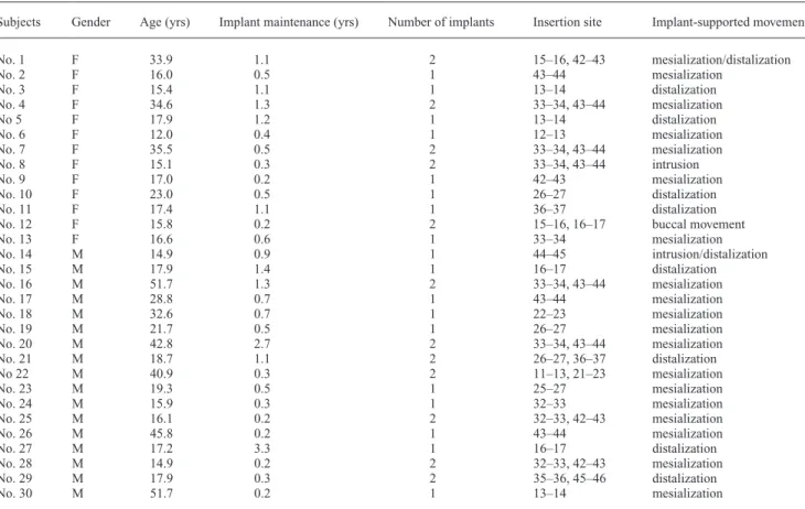
cross-check with the BACTEC blood culture analyzer. Colonies arised from any culture were undergone a Gram staining procedure as a first step of morphological identification. Species classification was designed to be accomplished by Table 2 Patient gender, age, and mini-implant placement details.
Subjects Gender Age (yrs) Implant maintenance (yrs) Number of implants Insertion site Implant-supported movement
No. 1 F 33.9 1.1 2 15–16, 42–43 mesialization/distalization No. 2 F 16.0 0.5 1 43–44 mesialization No. 3 F 15.4 1.1 1 13–14 distalization No. 4 F 34.6 1.3 2 33–34, 43–44 mesialization No 5 F 17.9 1.2 1 13–14 distalization No. 6 F 12.0 0.4 1 12–13 mesialization No. 7 F 35.5 0.5 2 33–34, 43–44 mesialization No. 8 F 15.1 0.3 2 33–34, 43–44 intrusion No. 9 F 17.0 0.2 1 42–43 mesialization No. 10 F 23.0 0.5 1 26–27 distalization No. 11 F 17.4 1.1 1 36–37 distalization
No. 12 F 15.8 0.2 2 15–16, 16–17 buccal movement
No. 13 F 16.6 0.6 1 33–34 mesialization No. 14 M 14.9 0.9 1 44–45 intrusion/distalization No. 15 M 17.9 1.4 1 16–17 distalization No. 16 M 51.7 1.3 2 33–34, 43–44 mesialization No. 17 M 28.8 0.7 1 43–44 mesialization No. 18 M 32.6 0.7 1 22–23 mesialization No. 19 M 21.7 0.5 1 26–27 mesialization No. 20 M 42.8 2.7 2 33–34, 43–44 mesialization No. 21 M 18.7 1.1 2 26–27, 36–37 distalization No 22 M 40.9 0.3 2 11–13, 21–23 mesialization No. 23 M 19.3 0.5 1 25–27 mesialization No. 24 M 15.9 0.3 1 32–33 mesialization No. 25 M 16.1 0.2 2 32–33, 42–43 mesialization No. 26 M 45.8 0.2 1 43–44 mesialization No. 27 M 17.2 3.3 1 16–17 distalization No. 28 M 14.9 0.2 2 32–33, 42–43 mesialization No. 29 M 17.9 0.3 2 35–36, 45–46 distalization No. 30 M 51.7 0.2 1 13–14 mesialization
Table 3 Exclusion criteria applied for sample selection. Exclusion criteria
Congenital heart disease History of rheumatic fever
Aortic stenosis, mitral stenosis, or both Prosthetic heart valves
History of subacute bacterial endocarditis Hypertrophic cardiomyopathy
Surgically constructed systemic-pulmonary shunts Vascular and joint prostheses
Immunosuppression Diabetes mellitus Bleeding disorder Pregnancy
Antibiotic usage within the past 3 months Regular use of antiseptic mouthwash
contemporary standard methods such as semi-automatic identification system (Microscan, Siemens Healthcare Diagnostics, Deerfield, IL, USA) (Jin et al., 2011) and manual biochemical testing techniques (API System, BioMérieux SA, Lyon, France) (http://www.biomerieux. com/servlet/srt/bio/portail/home). Susceptibility profile to a wide variety of antimicrobial agents was intended to be determined by the disk diffusion method in accordance with the current Clinical Laboratory Standards Institute guide-lines and the Minimal Inhibitory Concentration definition (Etest, BioMérieux, Marcy l’Etoile, France).
Results
The microbiological cultures of the preoperative speci-men, produced by the technique of choice, were free of aerobes and anaerobes. Negative results were also acquired from aerobic and anaerobic culturing of the blood sample received after mini-implant removal. The absence of bacte-ria was cross-checked by the supplementary 2 day cultiva-tion of the negative cultures.
Discussion
Albeit scientific consensus has not been met, the frequency and density of bacteremias are considered to be influenced by the degree of inflammation or infection at the site of the trauma (Wilson et al., 2007). Moreover, a significant association between the increase of plaque accumulation and gingival inflammation parameters and the development of bacteremia following toothbrushing has been recently substantiated (Tomás et al., 2012). Given that the implant surface roughness contributes in bacterial adhesion (Chin
et al., 2007), and prolonged plaque retention in the peri-implant gingival tissue and mucosa triggers the develop-ment of localized cell inflammation (Sebbar et al., 2012), we conducted this study to investigate whether bacteremia occurs after miniscrew explantation. Interestingly, our cul-ture-based microbiological methods confirmed absolute lack of bacteremia in all sixty blood samples. Negative results have been also obtained from the baseline samples before miniscrew installation in 40 orthodontic patients (Uysal et al., 2010). Nevertheless, in the latter investigation bacteremia was not developed in all but one individual after mini-implant insertion.
Orthodontic studies dedicated to bacteremic occurrence after miscellaneous interventions have yielded rates reach-ing up to 57% in the instance of gold chain adjustment (Lucas et al., 2007). However, a closer examination of the results reveals that in specific studies bacteremia percent-ages did not increase in post-operative samples, and either maintained (Erverdi et al., 2000, 2001; Lucas et al., 2007) or even declined (Chung et al., 1986; Lucas et al., 2002). These findings, not discussed by the authors in the respec-tive papers, pose questions about the study design and
methodology. Reviewers of microbiological studies, pub-lished between the years 1950–77 and 2006–10, discerned greater precision in detection and identification of bacterial isolates in the later papers (Tomás et al., 2012). The timing of the second blood sampling is of crucial importance for accurate determination of bacteremia. Based on robust data that the peak value of bacteremia is attained between 30 and 60 seconds after dental extraction (Roberts et al., 1992), the aforementioned period was selected as method of choice. Afterwards, the number of positive blood cultures drops rap-idly, whereas small prevalence percentages may be observed in the second half-hour post-procedure (Wilson et al., 2007). Of note, times ranging from 30 seconds (Burden et al., 2004; Lucas et al., 2002, 2007) to 15 minutes (Chung et al., 1986) have been cited in similar articles and this may stand for the different outcomes. Queries may be also raised for studies that did not provide time details (Degling, 1972; Erverdi
et al., 2001), or whether the procedure of interest can be consistently completed within limited time (Burden et al., 2004). Standard treatment performed with bonded brackets on incisors, canines, premolars and bands placed with glass-ionomer cement on first molars instigates dental plaque accumulation and gingival inflammation as well as growth of pathogenic bacteria and anaerobes (Ristic et al., 2007; Liu
et al., 2011). It is generally recognized that inflammatory reaction of gingival tissue and calculus apposition in fixed orthodontics are related to retentive surfaces around bonded attachments (Alexander, 1991). Oral hygiene status was set as inclusion criterion elsewhere (McLaughlin et al., 1996; Erverdi et al., 1999, 2000, 2001), probably with the intention to identify bacteremia directly derived from the intervention under investigation. Intuitively, such a decision might have led to erroneous research design with recruitment of not typical orthodontic patients.
Besides the evidence-based timing in blood collection and carefully elected sample, this study offers further methodo-logical advantages such as utilization of a single miniscrew type and one orthodontist involvement in treatment proce-dures. In contrast, the age of participants and mini-implant location may imply confounding. Though optimal in terms of study design, stratification of a larger study group with solid characteristics would have been impractical and ethi-cally sensitive. Nonetheless, the number of patients coin-cides or is even greater than in the majority of previous bacteremic-orthodontic investigations (Table 1). As regards the microbiological technique of the study, the advocates of molecular sequence-based methods may argue for the higher sensitivity relative to culture-based methods (Parahitiyawa
et al., 2009). Still, the lack of validation and use of molecu-lar methods in prospective clinical trials of oral bacteremia needs to be addressed beforehand (Tomás et al., 2012).
The prognostic role of bleeding for bacteremia is ambigu-ous. Substantial bacteremia may occur regardless of pres-ence of clinically discernible bleeding (Roberts, 1999). In this context, the AHA writing group (Wilson et al., 2007)
revisited antecedent recommendations that warranted anti-biotics for oral interventions for which bleeding was antici-pated (Dajani et al., 1997). On the other hand, generalized bleeding after toothbrushing was correlated with an almost eightfold increase in risk of developing bacteremia (Lockhart
et al., 2009). In the current study, notwithstanding bleeding of some extent was evident in all subjects after mini-implant had been removed, it did not affect bacteremia acquisition.
In the last years, the concept of cumulative exposure over time has been introduced to assess the risk of bacteremias arising from various activities. It has been calculated that the collective exposure to random bacteremias caused by mastication and everyday oral hygiene measures largely exceeds the duration of bacteremia related to tooth extrac-tion (Guntheroth, 1984; Roberts, 1999). With reference to orthodontics, though, the nature and frequency of visits call for attention when organizing research on the potential bac-teremic impact of treatment procedures. In effect, a regular orthodontic treatment session does not necessarily mean a single-appointment procedure. On annual basis, the number of fixed appliances controls (Fleming et al., 2010; DiBiase
et al., 2011; Johansson and Lundström, 2012) may be 7 to 10 times greater than the average attendance of dental offices (Wall and Brown, 2003).
In an era where the orthodontic armamentarium is increas-ingly upgraded with novel therapeutic systems, it is the task of the professionals to illuminate all relevant aspects encoun-tered in the clinical practice. While antimicrobial prophylactic therapy is endorsed merely for patients with predisposing to IE cardiac conditions planned to receive bands (Wilson et al., 2007), our results do not rationalize preoperative administra-tion of antibiotics for miniscrew removal. Future research design based upon the aggregated investigation of mini-implants in conjunction with other orthodontic procedures customarily performed during a single visit may advance our comprehension over orthodontics-related bacteremia.
Conclusions
Our study clearly demonstrates that none of the 30 patients presented bacteremia following removal of orthodon-tic miniscrews. Prospective clinical studies should aim to inquire the cumulative bacteremic capacity of mini-implants combined with additional orthodontic treatment techniques.
Funding
This study was supported by the research funds of 251 Hellenic Air Force VA General Hospital.
Acknowledgement
The authors thank Sgt (DA) Melpomeni Petrou, staff of the Department of Orthodontics, 251 Hellenic Air Force VA General Hospital, for her participation in blood sampling.
References
Alexander S A 1991 Effects of orthodontic attachments on the gingival health of permanent second molars. American Journal of Orthodontics and Dentofacial Orthopedics 100: 337–340
Apel S, Apel C, Morea C, Tortamano A, Dominguez G C, Conrads G 2009 Microflora associated with successful and failed orthodontic mini-implants. Clinical Oral Implants Research 20: 1186–1190
Biancaniello T M, Romero J R 1991 Bacterial endocarditis after adjust-ment of orthodontic appliances. The Journal of Pediatrics 118: 248–249 Burden D J, Coulter W A, Johnston C D, Mullally B, Stevenson M 2004
The prevalence of bacteraemia on removal of fixed orthodontic appli-ances. European Journal of Orthodontics 26: 443–447
Chin M Y, Sandham A, de Vries J, van der Mei H C, Busscher H J 2007 Biofilm formation on surface characterized micro-implants for skeletal anchorage in orthodontics. Biomaterials 28: 2032–2040
Chung A, Kudlick E M, Gregory J E, Royal G C, Reindorf C A 1986 Toothbrushing and transient bacteremia in patients undergoing ortho-dontic treatment. American Journal of Orthoortho-dontics and Dentofacial Orthopedics 90: 181–186
Costa A, Raffainl M, Melsen B 1998 Miniscrews as orthodontic anchorage: a preliminary report. The International Journal of Adult Orthodontics and Orthognathic Surgery 13: 201–209
Dajani A S 1991 Bacterial endocarditis after minor orthodontic proce-dures. The Journal of Pediatrics 119: 339–340
Dajani A S et al. 1997 Prevention of bacterial endocarditis. Recommendations by the American Heart Association. JAMA: The Journal of the American Medical Association 277: 1794–1801
Degling T E 1972 Orthodontics, bacteremia, and the heart damaged patient. The Angle Orthodontist 42: 399–402
DiBiase A T, Nasr I H, Scott P, Cobourne M T 2011 Duration of treat-ment and occlusal outcome using Damon3 self-ligated and conventional orthodontic bracket systems in extraction patients: a prospective rand-omized clinical trial. American Journal of Orthodontics and Dentofacial Orthopedics 139: e111–e116
Erverdi N, Kadir T, Ozkan H, Acar A 1999 Investigation of bacteremia after orthodontic banding. American Journal of Orthodontics and Dentofacial Orthopedics 116: 687–690
Erverdi N, Biren S, Kadir T, Acar A 2000 Investigation of bacteremia following orthodontic debanding. The Angle Orthodontist 70: 11–4; discussion 15
Erverdi N, Acar A, Işgüden B, Kadir T 2001 Investigation of bacteremia after orthodontic banding and debanding following chlorhexidine mouth wash application. The Angle Orthodontist 71: 190–194
Fleming P S, DiBiase A T, Lee R T 2010 Randomized clinical trial of orthodontic treatment efficiency with self-ligating and conventional fixed orthodontic appliances. American Journal of Orthodontics and Dentofacial Orthopedics 137: 738–742
Forner L, Larsen T, Kilian M, Holmstrup P 2006 Incidence of bacteremia after chewing, tooth brushing and scaling in individuals with periodontal inflammation. Journal of Clinical Periodontology 33: 401–407 Gould F K et al. 2006 Guidelines for the prevention of endocarditis:
report of the Working Party of the British Society for Antimicrobial Chemotherapy. The Journal of Antimicrobial Chemotherapy 57: 1035–1042
Guntheroth W G 1984 How important are dental procedures as a cause of infective endocarditis? The American Journal of Cardiology 54: 797–801
Gürel H G, Basciftci F A, Arslan U 2009 Transient bacteremia after removal of a bonded maxillary expansion appliance. American Journal of Orthodontics and Dentofacial Orthopedics 135: 190–193
Habib G et al.; ESC Committee for Practice Guidelines 2009 Guidelines on the prevention, diagnosis, and treatment of infective endocardi-tis (new version 2009): the Task Force on the Prevention, Diagnosis, and Treatment of Infective Endocarditis of the European Society of Cardiology (ESC). Endorsed by the European Society of Clinical Microbiology and Infectious Diseases (ESCMID) and the International
Society of Chemotherapy (ISC) for Infection and Cancer. European Heart Journal 30: 2369–2413
Hobson R S, Clark J D 1993 Infective endocarditis associated with ortho-dontic treatment: a case report. British Journal of Orthoortho-dontics 20: 241–244
Jin W Y et al. 2011 Evaluation of VITEK 2, MicroScan, and Phoenix for identification of clinical isolates and reference strains. Diagnostic Microbiology and Infectious Disease 70: 442–447
Johansson K, Lundström F 2012 Orthodontic treatment efficiency with self-ligating and conventional edgewise twin brackets: a prospective randomized clinical trial. The Angle Orthodontist 82: 929–934 Jorgensen J H, Ferraro M J 2009 Antimicrobial susceptibility testing: a
review of general principles and contemporary practices. Clinical Infectious Diseases: An Official Publication of the Infectious Diseases Society of America 49: 1749–1755
Kanomi R 1997 Mini-implant for orthodontic anchorage. Journal of Clinical Orthodontics: JCO 31: 763–767
Liu H et al. 2011 Periodontal health and relative quantity of subgingival Porphyromonas gingivalis during orthodontic treatment. The Angle Orthodontist 81: 609–615
Livas C, Renkema A M, Kiliaridis S, Katsaros C 2006 [Bone anchorage in orthodontics. A review]. Nederlands Tijdschrift Voor Tandheelkunde 113: 96–100
Lockhart P B et al. 2009 Poor oral hygiene as a risk factor for infec-tive endocarditis-related bacteremia. Journal of the American Dental Association (1939) 140: 1238–1244
Lucas V S, Omar J, Vieira A, Roberts G J 2002 The relationship between odontogenic bacteraemia and orthodontic treatment procedures. European Journal of Orthodontics 24: 293–301
Lucas V S, Kyriazidou A, Gelbier M, Roberts G J 2007 Bacteraemia fol-lowing debanding and gold chain adjustment. European Journal of Orthodontics 29: 161–165
McLaughlin J O, Coulter W A, Coffey A, Burden D J 1996 The inci-dence of bacteremia after orthodontic banding. American Journal of Orthodontics and Dentofacial Orthopedics 109: 639–644
Parahitiyawa N B, Jin L J, Leung W K, Yam W C, Samaranayake L P 2009 Microbiology of odontogenic bacteremia: beyond endocarditis. Clinical Microbiology Reviews 22: 46–64
Roberts G J, Gardner P, Simmons N A 1992 Optimum sampling time for detection of dental bacteraemia in children. International Journal of Cardiology 35: 311–315
Ristic M, Vlahovic Svabic M, Sasic M, Zelic O 2007 Clinical and micro-biological effects of fixed orthodontic appliances on periodontal tissues in adolescents. Orthodontics & Craniofacial Research 10: 187–195 Roberts G J 1999 Dentists are innocent! “Everyday” bacteremia is the real
culprit: a review and assessment of the evidence that dental surgical
procedures are a principal cause of bacterial endocarditis in children. Pediatric Cardiology 20: 317–325
Rosa E A, Rached R N, Tanaka O, Fronza F, Fronza F, Araújo Assad R 2005 Preliminary investigation of bacteremia incidence after removal of the Haas palatal expander. American Journal of Orthodontics and Dentofacial Orthopedics 127: 64–66
Schlein R A, Kudlick E M, Reindorf C A, Gregory J, Royal G C 1991 Toothbrushing and transient bacteremia in patients undergoing ortho-dontic treatment. American Journal of Orthoortho-dontics and Dentofacial Orthopedics 99: 466–472
Sebbar M, Bourzgui F, Badre L, El Quars F 2012 Anchorage miniscrews: a histologic study of peri-implant soft tissue. International Orthodontics / Collège Européen D’orthodontie 10: 85–95
Shirck J M, Firestone A R, Beck F M, Vig K W, Huja S S 2011 Temporary anchorage device utilization: comparison of usage in orthodontic programs and private practice. Orthodontics: The Art and Practice of Dentofacial Enhancement 12: 222–231
Tomás I, Diz P, Tobías A, Scully C, Donos N 2012 Periodontal health sta-tus and bacteraemia from daily oral activities: systematic review/meta-analysis. Journal of Clinical Periodontology 39: 213–228
Tortamano A, Dominguez G C, Haddad A C, Nunes F D, Nacao M, Morea C 2012 Periodontopathogens around the surface of mini-implants removed from orthodontic patients. The Angle Orthodontist 82: 591–595
Wall T P, Brown L J 2003 Recent trends in dental visits and private dental insurance, 1989 and 1999. Journal of the American Dental Association (1939) 134: 621–627
Wilson W et al.; American Heart Association Rheumatic Fever, Endocarditis and Kawasaki Disease Committee, Council on Cardiovascular Disease in the Young; Council on Clinical Cardiology; Council on Cardiovascular Surgery and Anesthesia; Quality of Care and Outcomes Research Interdisciplinary Working Group; American Dental Association 2007 Prevention of infective endocarditis: guidelines from the American Heart Association: a guideline from the American Heart Association Rheumatic Fever, Endocarditis and Kawasaki Disease Committee, Council on Cardiovascular Disease in the Young, and the Council on Clinical Cardiology, Council on Cardiovascular Surgery and Anesthesia, and the Quality of Care and Outcomes Research Interdisciplinary Working Group. Journal of the American Dental Association (1939) 138: 739–45, 747
Uysal T, Yagci A, Esel D, Ramoglu S I, Kilinc A 2010 Investigation of bacteremia following insertion of orthodontic mini-implants. World Journal of Orthodontics 11: 357–361
Ziolkowska L, Olczak-Kowalczyk D, Kawalec W, Turska-Kmiec A 2010 Fixed orthodontic appliance and infective endocarditis. The Pediatric Infectious Disease Journal 29: 1155–1156