i Université de Sherbrooke
GIMAP5 influence la survie des cellules T naïves en participant à la régulation du calcium emmagasiné dans les organites
Par Daniel Serrano Programme d’immunologie
Thèse présentée à la Faculté de médecine et des sciences de la santé en vue de l’obtention du grade de Philosophiae Doctor (Ph.D.) en immunologie
Sherbrooke, Québec, Canada Mars 2017
Membres du jury d’évaluation
Sheela Ramanathan, Programme d’immunologie, Université de Sherbrooke Christine Lavoie, Département de pharmacologie, Université de Sherbrooke Patrick McDonald, Programme d’immunologie, Université de Sherbrooke Mercedes Rincon, Department of Immunobiology, University of Vermont
ii Live so that when your children think of fairness, caring, and integrity, they think of you.
iii
R
ÉSUMÉGIMAP5 influence la survie des cellules T naïves en participant à la régulation du calcium emmagasiné dans les organites
Par Daniel Serrano Programme d’immunologie
Thèse présentée à la Faculté de médecine et des sciences de la santé en vue de l’obtention du diplôme de Philosophiae Doctor (Ph.D.) en Immunologie, Faculté de médecine et des sciences de
la santé, Université de Sherbrooke, Sherbrooke, Québec, Canada, J1H 5N4
La survie des cellules T naïves est essentielle au bon fonctionnement du système immunitaire à long terme. Les rats BBDP (Bio-breeding Diabetes prone) sont caractérisés par une haute prédisposition au développement du diabète ainsi que par une diminution significative du nombre de cellules T naïves. Ces rats comportent une mutation de type décalage de lecture dans le gène codant pour «GTPase Immunity-Associated Protein 5» (Gimap5) ce qui entraine l’apoptose des lymphocytes T. Le mécanisme par lequel la déficience de la protéine GIMAP5 conduit les cellules T à la mort est actuellement méconnu. GIMAP5 a également été associée à différentes maladies auto-immunes, ce qui suggère son influence dans l'homéostasie des lymphocytes T. Des résultats antérieurs de notre groupe de recherche ont montré que l'absence de GIMAP5 entraîne une diminution du flux de Ca2+ ainsi qu’une réduction de la capacité mitochondriale à emmagasiner du Ca2+ suite à la stimulation du TCR. Cependant, GIMAP5 n'est pas une protéine mitochondriale. Afin de mieux comprendre le rôle de GIMAP5 dans la biologie des cellules T, au cours de mes études doctorales, je me suis concentré sur la localisation cellulaire de la protéine ainsi que sur son rôle dans l'homéostasie du Ca2+. Comme modèle d’étude, j'ai établi des lignées cellulaires HEK293T stables pour l’expression de GIMAP5, ainsi que pour différents mutants et variantes de la protéine. Ceci m’a permis d’élucider l'importance du domaine transmembranaire (TM) pour la localisation et le rôle physiologique de GIMAP5 ainsi que la différence entre les deux variantes de cette protéine.
Mes résultats ont permis de montrer que l'expression de Gimap5 ne semble pas être nécessaire après l’activation des lymphocytes T. En parallèle, j'ai confirmé nos observations antérieures qui démontrent l’influence de GIMAP5 dans l'homéostasie du Ca2+ et sa colocalization avec les microtubules. En outre, j'ai montré que GIMAP5 se trouve dans des structures de type vésiculaire, particulièrement dans la membrane lysosomale où son domaine TM est essentiel à son bon fonctionnement et localisation. Mes résultats suggèrent que les mitochondries exhibent un défaut dans leur capacité à emmagasiner du Ca2+ au niveau basal, ainsi que suite à l’activation du TCR. Enfin, j'ai démontré pour la première fois, que l'influence de GIMAP5 sur le stockage de Ca2+ lysosomal peut avoir un impact sur la survie des lymphocytes T. D’après ces observations, une des fonctions probables de GIMAP5 serait d’empêcher la fermeture prématurée des canaux de relâche calcique. Finalement, GIMAP5 pourrait être engagé dans des mécanismes visant à prolonger et raffiner la signalisation du Ca2+ dans les cellules T. Bref, la régulation du Ca2+ lysosomal médié par GIMAP5 est essentielle à la survie de cellules T naïves.
Mots Clés: GIMAP5, survie des cellules T, homéostasie du Ca2+, Ca2+ lysosomal, Ca2+ mitochondrial, RE, cytosquelette.
iv
S
UMMARYGIMAP5 influences naïve T cell survival through organelle calcium storage regulation. By
Daniel Serrano Immunology Program
Thesis presented at the Faculty of medicine and health sciences for the obtention of Doctor Degree diploma Philosophiae Doctor (Ph.D.) in Immunology, Faculty of Medicine and health
sciences, Université de Sherbrooke, Sherbrooke, Québec, Canada, J1H 5N4
Healthy and long-term survival of naïve T cells is essential for proper functioning of the immune system. In bio-breeding diabetes prone (BBDP) rats, there is a critical decrease in the number of naïve T cells. In these rats, a recessive frameshift mutation in the GTPase of Immune-Associated Protein 5 (Gimap5) gene induces lymphocytes to undergo spontaneous apoptosis. The death of T cells driven by a deficiency of the GIMAP5 is currently not fully understood. Interestingly, different autoimmune diseases have shown an association with perturbations in the Gimap5 gene, which further suggests its influence in basal lymphocyte homeostasis. Previous findings by our group have shown that the absence of GIMAP5 results in a decrease calcium flux following TCR stimulation and an impaired capacity of the mitochondria to buffer calcium entry. However, GIMAP5 is not a mitochondrial protein. During my Ph.D. studies, I focused on clarifying the cellular localization of GIMAP5 as well as its function in Ca2+ homeostasis in order to further understand its role in T cell biology. As a model, I established HEK293T cells stable for the expression of the different mutants and variants of the GIMAP5 protein. Where I uncovered the importance of the transmembrane domain (TM) for GIMAP5 localization and physiological role, as well as the differences between the two variants of GIMAP5.
The results obtained show that the expression of Gimap5 is no longer needed after T cells activation. Moreover, our previous observations were confirmed and expanded upon regarding GIMAP5’s influence on Ca2+ homeostasis and colocalization with the cytoskeleton. It was also shown that GIMAP5 localizes to vesicular-like structures, particularly to the lysosomal membrane, where its TM domain is critical for proper functioning and localization. My results suggest that the mitochondria might be impaired to uptake as well as retain Ca2+ at their full capacity in the absence of GIMAP5. Finally, I observed for the first time that GIMAP5’s influence on lysosomal Ca2+ storage could impact lymphocyte survival. These results suggest that GIMAP5 may work as a backup mechanism to prevent premature closure of Ca2+ channels and Ca2+ influx or as a mechanism to prolong and refine Ca2+ signaling in T cells.
Keywords: GIMAP5, Naïve T cell survival, Ca2+ homeostasis, Lysosomal Ca2+, Mitochondrial Ca2+, ER, Cytoskeleto
v
T
ABLE OF CONTENTSRésumé ... iii
Summary ... iv
Table of contents ... v
List of figures ... vii
List of Tables ... ix
List of Abbreviations ... x
1. Introduction ... 1
1.1. Naïve T cell homeostasis ... 2
1.1.1. Interleukin 7 in naïve T cell survival ... 4
1.1.2. T cell receptor in naïve T cell survival ... 7
1.1.3. Metabolic signals that regulate T cell survival ... 10
1.2. Ca2+ homeostasis in T cell survival ... 11
1.2.1. The endoplasmic reticulum as the main Ca2+ store ... 12
1.2.2. The role of mitochondria in Ca2+ homeostasis ... 14
1.2.3. Lysosomes as new players in Ca2+ regulation ... 17
1.2.4. The Cytoskeleton network influences Ca2+ homeostasis via organelle movement regulation ... 19
1.3. GIMAP family of proteins ... 22
1.3.1. GIMAP5 ... 27
2. Rationale and hypothesis ... 35
2.1. Rationale ... 35
2.2. Hypothesis ... 36
2.2.1. Aims ... 36
3. Materials And Methods ... 37
3.1. Constructions and Cell lines ... 37
vi
3.3. Animals ... 38
3.4. Isolation of T cell subsets ... 39
3.5. Immunoprecipitation and western blot ... 39
3.6. Quantitative PCR ... 41
3.7. Confocal Microscopy ... 41
3.8. Calcium measurements ... 42
3.9. Statistical analysis ... 42
4. Results ... 43
4.1. GIMAP5 protein constructs ... 43
4.2. T cells express predominantly the shorter variant of GIMAP5 ... 44
4.3. GIMAP5 variants co-localize similarly with cytoskeletal elements ... 45
4.4. GIMAP5v2, but not GIMAP5v1, regulates cellular Ca2+ ... 51
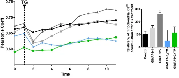
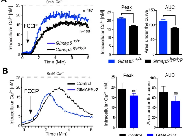
4.5. GIMAP5 influences mitochondrial Ca2+ uptake ... 52
4.6. GIMAP5v2 influence Mitochondrial Ca2+ content ... 54
4.7. GPN-induced Ca2+ release is increased in the absence of GIMAP5v2 ... 62
4.8. Nocodazole treatment increases the Ca2+ influx following GPN treatment ... 65
4.9. Lysosomes accumulate Ca2+ following cross-linking of TCR ... 66
4.10. GIMAP5 variants interact with each other ... 67
5. Discussion ... 69
5.1. Gimap5 expression and variants ... 70
5.2. GIMAP5 protein localizes in different vesicle-like structures ... 72
5.3. GIMAP5 as a mediator of organelle Ca2+ regulation ... 73
5.4. GIMAP5 protein influences mitochondrial Ca2+ homeostasis ... 74
5.5. The intermediary role of GIMAP5v1 ... 78
Perspectives ... 80
Acknowledgments ... 81
References ... 83
Annexes ... 105
vii
L
IST OF FIGURESFigure 1-1: T cell development. ... 3
Figure 1-2: TCR and IL-7 signaling. ... 6
Figure 1-3: ER, mitochondria and lysosome Ca2+ release and uptake channels. ... 15
Figure 1-4: The cytoskeleton and the motor proteins. ... 21
Figure 1-5: Phylogenetic tree of IAN/GIMAP proteins expression in Arabidopsis thaliana, Homo sapiens, Mus musculus and Rattus norvegicus... 24
Figure 1-6: GIMAP5 secondary structure in rats, mice, and humans. ... 29
Figure 4- 1: Cloning of GIMAP5 variants. ………...44
Figure 4- 2: Gene expression of Gimap5 variants in rat T lymphocytes. ... 45
Figure 4- 3: Co-localization of GIMAP5 variants with microtubules... 46
Figure 4- 4: Co-localization of GIMAP5 variants with actin filaments. ... 47
Figure 4- 5: GIMAP5v2ΔTM diffuse cellular localization. ... 48
Figure 4- 6: Co-localization of GIMAP5 variants with kinesin. ... 49
Figure 4- 7: Co-localization of GIMAP5 variants with dynein. ... 50
Figure 4- 8: Absence of co-localization of GIMAP5 with myosin. ... 50
Figure 4- 9: GIMAP5v2 expressing vesicles move along microtubules. ... 51
Figure 4- 10: Full-length GIMAP5v2 regulates intracellular Ca2+ homeostasis. ... 52
Figure 4- 11: GIMAP5v2 promotes mitochondrial Ca2+ accumulation. ... 54
Figure 4- 12: Influence of GIMAP5v2 on Ca2+ release from mitochondria. ... 55
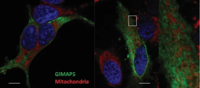
Figure 4- 13: GIMAP5v2 does not colocalize with mitochondria. ... 56
Figure 4- 14: Co-localization of GIMAP5v2 with Lysotracker Red. ... 57
Figure 4- 15: Movement of GIMAP5v2 and LAMP1 expressing vesicles in HEK293T stable cells. ... 58
Figure 4- 16: GIMAP5 variants are anchored on the lysosomal membrane through the C-terminal transmembrane domain. ... 59
Figure 4- 17: GIMAP5 colocalizes with the lysosomal Ca2+ channel TPC2. ... 59
Figure 4- 18: GIMAP5 colocalizes with different vesicles of the endo-lysosomal system. .. 61
viii Figure 4- 20: GPN-induced lysosomes osmotic lysis is similar for Control and GIMAP5 expressing cells. ... 64 Figure 4- 21: Nocodazole treatment increases the Ca2+ influx following GPN treatment. .. 65 Figure 4- 22: Lysosomes accumulate Ca2+ following activation through T cell receptor. .... 67 Figure 4- 23: GIMAP5 variants interact with each other. ... 68 Figure 5- 1: Schematic representation of other the different deletion mutants from GIMAP5v1. 105
Figure 5- 2: Protein expression of the different deletion mutants of GIMAP5. ... 106 Figure 5- 3: Stable HEK293T cells stable for the expression of different mutants of the GIMAP5 protein. ... 107
ix
L
IST OFT
ABLESx
L
IST OFA
BBREVIATIONSADPR Cyclic ADP-ribose
AIG1 AvrRpt2-Induced Gene1
Arp2/3 Actin-related protein 2/3
ATP Adenosine triphosphate
Bcl10 B-cell lymphoma 10
Ca2+ Calcium
cADPR Cyclic ADP-ribose
CaM Calmodulin
CARMA1 Caspase recruitment domain-containing membrane-associated guanylate kinase protein 1
CHOP C/EBP-homologous protein
CN Calcineurin
CPA Cyclopiazonic acid
CRAC Ca2+ release-activated Ca2+ channel
DAG Diacylglycerol
DGKs Diacylglycerol kinases
DP Double positive
ERMES ER-mitochondria encounter structure
FCCP Carbonyl cyanide 4-(trifluoromethoxy)phenylhydrazone GIMAP GTPase immunity-associated protein
GPN Gly-Phe β-naphthylamide
GSK3 Kinase glycogen synthase kinase 3
IkB IkB kinase
IMM Inner mitochondrial membrane
IP3 Inositol trisphosphate
IP3R Inositol triphosphate receptors
ITAMs Immunoreceptor tyrosin-based activation motifs
KIF5 Kinesin-1 family
LAT Linker for the activation of T cells Lck Leukocyte-specific tyrosine kinase
LKB1 Liver kinase B1
LSDs Lysosomal storage disorders
MALT1 Mucosa-associated lymphoid tissue lymphoma translocation gene 1
MAMs Mitochondria associated membranes
MAPK Mitogen-activated protein kinase MCU Mitochondrial Ca2+ uniporter
MFN2 GTPase Mitofusin 2
MHC Major Histocompatibility complex
xi mTOR Mechanistic target of rapamycin
NAADP Nicotinic acid adenine dinucleotide phosphate NFAT Nuclear factor of activated T cells
NF-kB Nuclear factor kappa-light-chain-enhancer of activated B cells
NK Natural killer
OMM Outer mitochondrial membrane
PA Phosphatidic acid
PIP2 Phosphatidylinositol bisphosphate
PKA Protein kinase A
PKCθ Protein kinase C-θ
PLCγ Phospholipase C-γ
RasGRP RAS guanyl nucleotide-releasing protein
RyR Ryanodine receptors
SERCA Sarcoplasmic/endoplasmic reticulum calcium ATPase
SHP1 Tyrosine phosphatase SH2-domain-containing phosphatase 1 Slp76 SH2-domain-containing leukocyte protein of 76 kDa
SOCE Store-operated Ca2+ entry
SP Simple positive
TCA Tricarboxylic acid
TCR T cell receptor
TFEB Master transcriptional regulator of lysosomal biogenesis and autophagy
TG Thapsigargin
TPC2 Two-pore channels
TRAK Adaptor protein trafficking kinesin protein
TRPML Mucolipin TRPs
TSC Tumor suppressor tuberous sclerosis complex VDAC Voltage-dependent anion channel
1
1. I
NTRODUCTIONEvery minute, thousands of events and interactions occur within the human body at the cellular and molecular level. Physiological balance and homeostasis is essential in remaining alive and healthy. As living organisms, we are constantly interacting with our environment, the nutrients we consume through our diet, as well as to the pathogens we can be exposed, can directly impact our health. During evolution, defense mechanisms have developed to keep our body, as a whole, under constant surveillance to maintain balance and health during our lifespan.
To pursue this goal, the immune system has taken charge of eliminating potentially cancerous cells and fighting against external pathogens that could impact our health. During the last decades, our knowledge regarding the immune system and its functions has advanced, which has led to the eradication of certain dangerous diseases through the use of vaccines. Although the increase in knowledge has helped improve our general health significantly, there is still much left to be understood.
Studies on the immune system have enabled us to progress from learning the existence of white cells to understanding, albeit incompletely, complex systems derived from two main branches: the innate and adaptive. Furthermore, several subtypes of immune cells have been uncovered, each with specific characteristics and roles. Although we rely on the immune system to protect us, it is also understood that an unbalanced immune system can lead to undesirable consequences, such as autoimmune diseases. In addition, a perturbed immune system can significantly aggravate an ongoing illness, therefore reminding us of the importance to improve our understanding of how immune cells interact with each other and their environment, and how they respond to pathogenic conditions.
2 In the context of my doctoral studies, I worked on an animal model where the basal survival of T cells at their naïve stage was impaired. This condition led to a significant decrease in the number of naïve T cells in the periphery of bio-breeding diabetes prone rats, which will be describe later. Specifically, I focused on uncovering the mechanisms of how a frameshift mutation in the GTPase Immunity-associated protein 5 (GIMAP5) can significantly affect naïve T cell homeostasis, driving T cells to apoptosis. The findings obtained during my studies will help shed light on new factors that were previously not taken into account while considering the maintenance and survival of naïve T cells in the periphery.
1.1. Naïve T cell homeostasis
Healthy and mature naïve T cells are crucial in maintaining a long-term balanced and functional immune system. However, T cells need to undergo a selection process before becoming functional cells. Once hematopoietic cells from a lymphoid progenitor exit the bone marrow, they populate the thymus and initiate the generation of immature thymocytes. Thymocytes, at this stage, are double negative (DN) for CD4 and CD8. Next, by somatic DNA rearrangement, they generate a β-TCR that will bind to the α-TCR to form the pre-αβTCR (Fink, 2012). Successful pairs will stop the β chain rearrangement and proceed to initiate proliferation of thymocytes that express CD4 and CD8 molecules on their surface. These thymocytes will then become double positive (DP). In the cortex of the thymus, DP thymocytes will engage different self-peptides presented by major histocompatibility complexes (MHC) I and II, which are found in epithelial cells of the thymus cortex (Germain, 2002).
3 Figure 1-1: T cell development.
Hematopoietic cells give rise to lymphoid progenitors that migrate to the thymus. In the thymus, they turn into DN thymocytes that lack the expression of TCR, CD4 or CD8 receptor. During their maturation process, DN cells progress from DN1 to the DN4 stage where they first rearrange the TCR beta chain and then the TCR alpha chain. After correct TCR chain rearrangement, they become DP thymocytes and can express CD4 and CD8 receptors. Cells are selected positively for their capacity to recognize MHCI or MHCII. The TCR signaling force of SP CD4 or CD8 will then negatively select cells with high affinity against self-peptides. Finally, naïve T cells are exported from the thymus medulla to the peripheral lymphoid organs.
During this positive selection process, DP thymocytes that can recognize the MHC molecules will be selected and migrate to the medulla as CD4 or CD8 thymocytes, depending on whether they were selected by MHC class II or I (Swain, 1983). The thymocytes that do not interact with the MHC will go to death by neglect. Once in the medulla, thymic epithelial cells, specifically the (thymic dendritic cells that can express self-antigens from all tissues), will present different MHCI or MHCII self-peptides, to the single positive (SP) CD8 or CD4 thymocytes (Figure 1-1). SP thymocytes, whose interaction is too strong, will receive an apoptotic signal that results in cell death. Even if the vast majority of thymocytes die after their migration and selection through the thymus, this process is nevertheless necessary to acquire a polyclonal repertoire of naïve lymphocytes that would normally not overreact against self-peptides (Robey and Fowlkes, 1994).
After migration through the thymus, thymocytes undergo positive and negative selection processes prior to becoming mature naïve single positive T cells (Starr et al., 2002). Once thymocytes exit the thymus, they become mature naïve T cells that migrate to secondary
4 peripheral organs, where they become part of the long-term survival pool of naïve T cells (Jameson, 2002; Marrack et al., 2000; Surh and Sprent, 2008). In order to remain in a quiescent state, naïve T cells require both extracellular signals and internal pathways (Hua and Thompson, 2001). Even though the mechanisms are not entirely understood, it is known that naïve T cell homeostasis primarily requires two signals. These signals are interleukin 7 receptor stimulation by IL-7, and basal self-peptide-MHC complex engagement by the T cell receptor (TCR) (Krogsgaard et al., 2007; Park et al., 2007; Surh and Sprent, 2008). These signals help to maintain the pool of naïve CD4 and CD8 T cells in the periphery.
1.1.1. Interleukin 7 in naïve T cell survival
IL-7 was first discovered as a cytokine for T cell development in the thymus and then for its role in the development and survival of other peripheral lymphocyte populations (von Freeden-Jeffry et al., 1995; Peschon et al., 1994). In the periphery, the importance of IL-7 signaling in T cell homeostasis and survival was established by T cell-restricted deletion of the α subunit of the IL-7 receptor (CD127) in mice. After approximately three days, there was a 30% decrease in the number of CD4 and CD8 T cells in the periphery of these mice, which confirmed the essential role of IL-7 signaling in peripheral T cell survival (Jacobs et al., 2010).
IL-7 is a cytokine that is mainly produced by stromal cells in the thymus and the bone marrow, as well as in the periphery by fibroblastic reticular cells in secondary lymphoid organs. In the periphery, other sources of IL-7 can be keratinocytes, hepatocytes, as well as other cells in different organs such as the lung and the intestine (Kim et al., 2011; Mazzucchelli et al., 2009; Shalapour et al., 2010). IL-7 binds to its receptor IL-7R, which is composed of an α (CD127) and a γ subunit (CD132). The latter CD132 is shared by other cytokine receptors of the common γ chain family, which includes interleukin receptors 2, 4, 9, 15 and 21 (Rochman et al., 2009).
5 Once IL-7 binds the IL-7R receptor, CD127 and CD132 chains, which are associated with association promotes Jak1 and Jak3, to phosphorylate themselves. Once phosphorylated, activation of the transcription factor STAT5 through the JAK/STAT pathway will occur, thereby inducing the translocation of STAT5 to the nucleus. This process will result in the expression of survival genes, such as MCL1 and BCL2 (Jiang et al., 2004; Kimura et al., 2013; Takada and Jameson, 2009). Mice deficient in STAT5, CD127, or Jak3 have shown severe combined immunodeficiency phenotypes (Yao et al., 2006), which confirms the importance of these signaling events in the survival and proper function of lymphocytes. In this way, the expression of the anti apoptotic genes Bcl-2 and Mcl-1 counterbalances the induction of apoptosis by the mitochondrial pathway (Dzhagalov et al., 2008). Similarly, pro-apoptotic proteins, Bad and Bax, can induce the mitochondrial apoptotic pathway after IL-7 withdrawal (Khaled et al., 1999; Li et al., 2004). (Figure 1-2)
In addition to its role in regulating anti-apoptotic and pro-apoptotic signals, IL-7 can also influence T cell survival by affecting cell metabolism through regulation of the glucose transporter GLUT1 (Wofford et al., 2008).
Different mechanisms are in place to regulate CD127 expression on the cell. IL-7 stimulation downregulates CD127 cell surface expression on the surface of the cells. The IL-7 receptor is not constantly expressed on the surface in the absence of IL-7 stimulation. Therefore, CD127 is mainly internilized through the endo-lysosomal pathway via clathrin-coated vesicles, and only a relatively small number will not be recycled but will be sorted to the lysosomal and proteasomal pathways for degradation (Henriques et al., 2010). Transcription factors, like Foxo1, can also regulate the expression of CD127 through the downstream PI3/Akt signaling pathway following TCR activation (Fabre et al., 2008). It has been shown that the survival of naïve CD4 and CD8 T cells in the periphery, as well as their ability to populate peripheral lymph organs, can be impaired when they are deficient in Foxo1 gene expression (Gubbels Bupp et al., 2009; Ouyang et al., 2009).
6 Even if both CD4 and CD8 cells are dependent on IL-7 stimulation for survival, it has been shown that there are certain differences in their sensitivity threshold as CD8 T cells are more dependent on IL-7 than CD4 T cells (Carrette and Surh, 2012; Tan et al., 2002). Indeed, CD8 expression in T cells can be upregulated with IL-7 in vivo IL-7 treatment, whereas IL-7 deficient mice showed a decreased CD8 expression. In fact, IL-7 signaling can regulate its receptor in a process known as “Coreceptor tuning.” For example, in the case of naïve CD8+ T cells, this process will modulate the quantity of CD8+ to fine-tune TCR engagement in a continuous feedback loop. Cytokine signaling can then upregulate CD8, whereas TCR will induce a reduction in CD8 expression. In contrast, CD4 receptor does not seem to be regulated by IL-7 stimulation (Park et al., 2007).
Figure 1-2: TCR and IL-7 signaling.
Principal signaling events for naïve T cell survival. On the left part of the panel, MHC-peptide presentation triggers TCR downstream signaling. After the engagement, LCK and ZAP phosphorylate LAT, which recruits SLP-76. PLCγ is recruited to this complex and hydrolyze PIP2 into IP3 and DAG near the membrane. IP3 will finally induce an increase in [Ca2+]i, which will activate CaM and CN to enable NFAT dephosphorylation and translocation to the nucleus. In parallel, DAG will induce AP-1 through the RAS, MAPK pathway and NF-κβ through PKCθ to activate survival and proliferation genes. On the right part of the panel, IL7 stimulates its receptor, engaging the phosphorylation of JAKs which will induce STAT5 translocation to the nucleus. IL7 can also induce the activation and
7 phosphorylation of the PI3K/AKT pathway, which can promote survival and antiapoptotic gene expression.
1.1.2. T cell receptor in naïve T cell survival
In addition to cytokine stimuli by IL-7, once mature naïve CD4 or CD8 T cells are in the periphery, engagement with self-peptides on MHC is essential to induce pro-survival and proliferation signals through the T cell receptor (Takeda et al., 1996; Tanchot et al., 1997). In fact, naïve CD4 and CD8 T cells are a very rich population of cells in the context of their polyclonal expression of TCR with different affinities to a panoply of exogenous and self-peptides (Mahajan et al., 2005; Qi et al., 2014).
The TCR on each mature naïve T cell is prepared to specifically interact with a defined peptide that will transform its naïve state into an activated state and thereby initiate an immune response. Normally, T cell activation results in a strong signal from the TCR in response to an exogenous antigen/peptide presented by the MHC. Nevertheless, TCR is not constantly triggered strongly, and therefore most of the time the vast majority of naïve T cells remain in a naïve quiescent state. An important point to keep in mind is that mature naïve T cells are not dormant cells, but active physiological cells that require constant signaling to remain physiologically active in a naïve state, yet ready to respond to possible threats. In fact, self-peptide-MHC complexes are constantly weakly stimulating naïve T cells through their TCRs, initiating a set of signaling events that will end in the expression of survival and proliferation genes (Surh and Sprent, 2008). Various experiments have shown the impaired survival of T cells in the absence of TCR expression (Polic et al., 2001). Similarly, when naïve CD4 or CD8 T cells are restricted from their respective MHCI or MHCII ligands, there is a decrease in their survival rates (Labrecque et al., 2001; Tanchot et al., 1997).
Once the TCR is engaged by recognition of a peptide presented by the MHC, it initiates conformational changes in the activation motifs of the CD3 tails. Assisted by the CD4 and CD8 coreceptors, leukocyte-specific tyrosine kinase (Lck) will phosphorylate the immunoreceptor tyrosine-based activation motifs (ITAMs) of the CD3, which will lead to the
8 recruitment of the Zeta-activated protein 70 kDa (Zap70) (Malissen and Bongrand, 2015). Next, Zap70 can recruit and phosphorylate the linker for the activation of T cells (LAT), which is a membrane-associated protein that serves as a scaffold of multiple protein complexes (Samelson, 2002). The phosphorylation of LAT then enables the recruitment of SH2-domain-containing leukocyte protein of 76 kDa (Slp76), which binds to LAT via the Grb2-related adaptor protein 2 (Koretzky et al., 2006). Zap70 can then phosphorylate Slp76 resulting in a recruitment platform formed by LAT/SLP76, which in turn will serve for the recruitment of other signaling effectors like the phospholipase C-γ (PLCγ), which interacts directly with both of them. PLCγ activation will hydrolyze phosphatidylinositol bisphosphate (PIP2) into diacylglycerol (DAG), and inositol trisphosphate (IP3). On one side, DAG will recruit downstream proteins, which include protein kinase C-θ (PKCθ) and RAS guanyl nucleotide-releasing protein (RasGRP). RasGRP can then activate the small GTPase Ras, which in turn activates the mitogen-activated protein kinase (MAPK) signaling pathways. In parallel, phosphorylated Slp76 can recruit the (Guanine nucleotide exchange factor) GEF Vav, which activates the small GTPases Rac and Cdc42.
On the other side, DAG and IP3, IP3 initiates a cascade of events that are calcium dependent and essential for the expression of several genes involved in T cell survival and proliferation. This pathway will be described in greater detail in subsequent sections of this introduction. As for DAG, two signaling pathways are initiated which are, the Ras pathway and PKCθ pathway (Carrasco and Merida, 2004). Ras will activate, through an MAPK/Erk signaling pathway, the transcription factor Fos. As for PKCθ, it will activate the IkB kinase (IkB) through the mucosa-associated lymphoid tissue lymphoma translocation gene 1 (MALT1), the caspase recruitment domain-containing membrane-associated guanylate kinase protein 1 (CARMA1) and the B-cell lymphoma 10 (Bcl10) (Wegener et al., 2006). These will induce the phosphorylation of IkB, leading to its degradation by ubiquitination. Next, NF-kB, which was sequestered in the cytoplasm, translocates to the nucleus and induce the expression of proliferation genes like IL-2. The actin-related protein 2/3 (Arp2/3), which is
9 triggered by the LAT-Slp76 signalosome activation of Cdc42 and Rac through Vav, induces a burst of actin polymerization at the immunological synapse.
In parallel to DAG, IP3 will stimulate the opening of the Ca2+ permeable channels in the ER, known as the IP3 receptors. This process will initiate the depletion of Ca2+ from the ER (Lewis, 2001). Decreased [Ca2+]
ER are sensed by the ER membrane proteins sensors stromal interaction molecule1 (STIM1) and STIM2, which will then oligomerize and translocate to interact with the plasma membrane proteins (olfactory receptor class A related 1 ) ORA1, forming the Ca2+ release-activated Ca2+ channel (CRAC) (Hoth and Penner, 1992; Zweifach and Lewis, 1993). The opening of the CRAC channels will induce entry of Ca2+ from the extracellular milieu and therefore result in a rise of the intracellular Ca2+ concentration [Ca2+]
i. STIM1 and STIM2 both detect different thresholds of a Ca2+ decrease in the ER lumen. On the one hand, STIM1 responds to greater changes in [Ca2+]
ER. In contrast, STIM2 responds to small Ca2+ fluctuations and may be important to sustain and capacitative Ca2+ entry under basal conditions as well as to improve STIM1 recruitment to enhance Ca2+ signaling (López et al., 2012; Ong et al., 2015; Thiel et al., 2013). This increase in Ca2+ in the cytoplasm will enable the nuclear factor of activated T cells (NFAT) to translocate to the nucleus. In cooperation with other transcriptional partners, it will then induce the expression of survival and proliferation genes (Macian, 2005). In fact, NFAT which is dependent on Ca2+ concentration is sequestered in the cytoplasm when Ca2+ concentrations are low. The kinase glycogen synthase kinase 3 (GSK3) phosphorylation of NFAT induces its nuclear export. In contrast, once Ca2+ concentration increases following TCR stimulation, the Ca2+ binding protein calmodulin (CaM) will associate and activate the calcineurin (CN) phosphatase. This, in turn, will dephosphorylate NFAT and enable its translocation to the nucleus where, in conjunction with other factors from the DAG signaling, will activate survival and proliferation genes, like IL-2 (Oh-hora and Rao, 2008). (Figure 1-2)
Regulation following TCR stimulation occurs at different levels. Lck and Zap70 are dephosphorylated by the tyrosine phosphatase SH2-domain-containing phosphatase 1
10 (SHP1). Lck, Zap70, as well as Vav can also be the targets of the E3 ubiquitin ligase, which causes them to undergo proteasomal degradation (Duan et al., 2004). For its part, diacylglycerol kinases (DGKs) will phosphorylate DAG to obtain phosphatidic acid (PA) (Zhong et al., 2008).
Costimulatory signals, which are essential for T cell activation, will not be discussed as the focus of the thesis is mainly the understanding of the quiescent naïve state of T cells.
1.1.3. Metabolic signals that regulate T cell survival
As previously mentioned, IL-7 and TCR are the two most important extracellular signals that maintain the pool of naïve T cells. However, another important characteristic of naïve T cells is their metabolic status. In fact, naïve T cells remain in a quiescent, yet active catabolic metabolic state by favoring energetic and efficient processes, such as oxidative phosphorylation (OXPHOS) vs. aerobic glycolysis, which are used during the activated state of T cells (Wang and Green, 2012). By doing this via the tricarboxylic acid (TCA) cycle, they can generate around 30 to 32 molecules of ATP per molecule of glucose. Several factors are in charge of the maintenance and regulation of this metabolic state. The mechanistic target of rapamycin (mTOR) has a decisive role in T cell metabolism, and early activation of mTOR can impair T cell survival (Chi, 2012; Yang and Chi, 2013, 2012). In cell metabolism, mTOR works as a sensor of nutrients, amino acids, and oxygen availability, as well as a modulator of extracellular signals that will impact the metabolic state of the cell (Heikamp and Powell, 2012).
In naïve T cells, the protein kinase that regulates the differences in the energy status through the AMP and ATP ratio is AMPK. As mTOR activation initiates an anabolic metabolism, mTOR activity is minimal in naïve T cells. In a catabolic state, high levels of AMP versus ATP will induce AMPK to activate the tumor suppressor tuberous sclerosis complex (TSC1 and TSC2) (Fingar and Blenis, 2004). Indeed, mTOR activation is negatively regulated by activated TSC1 and TSC2 complex, which in naïve T cells serves to maintain their catabolic homeostatic state, which is essential for their survival. In fact, mice KO for the expression
11 of TSC1, have shown a less restricted mTOR. As a result, the basal metabolic state that is key to naïve T cell quiescence and survival is no longer maintained. As a consequence, TSC1/2’s negative regulation of mTOR is essential to maintain a quiescent state and prevent naïve T cells from undergoing apoptosis (Qi et al., 2011; Xie et al., 2012; Yang et al., 2013, 2011; Zhang et al., 2011). In addition, TCR stimulation contributes to an increase in the expression of AMPK by the liver kinase B1 (LKB1) and by an increase of the intracellular [Ca2+] (Fracchia et al., 2013). Thus, modulation of naïve T cell metabolism by maintaining a catabolic status is as important to their long-term survival as the homeostatic extracellular signals that induce the expression of pro-survival genes.
1.2. Ca2+ homeostasis in T cell survival
Ca2+ influx is essential for the regulation of up to 75% genes implicated in survival and proliferation (Feske et al., 2001). Ca2+ is one of the main second messengers. Once cells receive and input through receptors on the membrane, this can lead to an increase of intracellular Ca2 concentrations. Store-operated Ca2+ entry (SOCE) through Ca2+ release-activated calcium (CRAC) channels is the principal mechanism of Ca2+ extracellular influx in lymphocytes after TCR stimulation (Lewis, 2001). An increase in intracellular Ca2 is essential for the activation of different enzymes as well as for the transcription of a panoply of genes implicated in cell survival and proliferation.
Whereas CRAC channels are the principal gates for extracellular Ca2+ flux, other channels can also contribute, to a lesser extent, to the intracellular Ca2+ concentration. Transient receptor potential channels TRPM7 and TRPM2, even if not fully understood, have been shown to play a role in T cell Ca2+ homeostasis. They can be activated by different agonists such as NAAD, ADPR, and cADPR, likely produced as a result of TCR stimulation (Beck et al., 2006; Guse et al., 1999). In T cells, some members of the voltage-gated Ca2+ channels have been reported, but their contribution to Ca2+ homeostasis remains under study (Hogan et al., 2010). L-type “voltage-dependent” Ca2+ channel Ca
V1.4 has been shown to play a major role in the survival of naïve T cells by modulating intracellular Ca2+ storage and TCR
12 downstream Ca2+ dependent signaling events (Omilusik et al., 2009). Similarly, a recent study showed that Cav1.1 is expressed in naïve T cells and upregulated in activated T cells upon TCR stimulation (Matza et al., 2016). Another group of receptors that contribute to the influx of extracellular Ca2+ during T cell activation are the purinergic receptors (Schenk et al., 2008; Yip et al., 2009). In T cells, three purinergic receptors P2X1, P2X4, and P2X7 have been reported to influence T cell Ca2+ flux. Indeed, ATP activation of P2X7 opens the channel to Ca2+ influx which activates calcineurin, resulting in IL-2 production and T cell proliferation (Feske, 2013; Woehrle et al., 2010). Interestingly, ATP release by mitochondria near the immune synapse has been shown to play a major role in activating P2X1 and P2X4, and by doing so, prolonging Ca2+ influx.
Nevertheless, Ca2+ is not constantly available in the cytoplasm but stored by different organelles, which will release it for specific needs, even in the absence of a major extracellular flux of Ca2+. The most well-known organelle is the endoplasmic reticulum, which has been recognized for a long time for its capacity to store, regulate and trigger Ca2+ fluxes. Even if the ER remains the leading Ca2+ store of the cell, other organelles can also store Ca2+ in a significant manner and therefore influence Ca2+ dependent signaling pathways. Contribution from other organelles, such as mitochondria and lysosomes, to the regulation of Ca2+ signaling dependent pathways, is necessary to better regulate the different Ca2+ thresholds, as well as particular needs in specific regions of the cell (Raffaello et al., 2016; Srikanth and Gwack, 2013).
1.2.1. The endoplasmic reticulum as the main Ca2+ store
The ER is an organelle with a multitude of functions, ranging from drug detoxification, lipid metabolism to post-transcriptional modifications and protein synthesis (Görlach et al., 2006). Moreover, it also serves as the primary Ca2+ storage compartment. ER lumen Ca2+ concentration [Ca2+]
ER can vary from cell to cell, going from 0.3 to 0.7mM (Montero et al., 1997). The ER contributes importantly to cellular Ca2+ homeostasis and signaling. Calcium uptake is done by the Sarcoplasmic/endoplasmic reticulum calcium ATPase (SERCA) pump
13 and by the release channels IP3 and ryanodine receptors (Aulestia et al., 2011; Golovina and Blaustein, 1997) (Figure 1-3). In fact, mammals express three different types of SERCA proteins. The isoform SERCA2b is expressed ubiquitously. As a protein, the SERCA pump has a molecular weight of 110 kDa and can transport two Ca2+ ions for each molecule of ATP hydrolyzed (Zampese and Pizzo, 2012). SERCA activity can be regulated both physiologically and pharmacologically. Within the cells, the phospholamban protein can inhibit the SERCA pump, and its phosphorylation by the protein kinase A (PKA) stops this inhibition (MacLennan and Kranias, 2003). Artificially, three main pharmacological drugs can inhibit the SERCA pumps in a reversible and non-reversible way. Thapsigargin (TG), whose action is irreversible, is the most used SERCA inhibitor. Cyclopiazonic acid (CPA) and 2,5-di (t-butyl) hydroquinone are reversible blockers and have less affinity than TG for the SERCA pump (Pinton et al., 1998).
In order to release Ca2+ into the intracellular medium, the ER relies on two ion channels: the inositol trisphosphate receptors (IP3R) and the ryanodine receptors (RyR) (Foskett et al., 2007). The IP3R family includes three isoforms, whose expression varies among tissues, but all of them have been detected in T cells (Akimzhanov and Boehning, 2012). To regulate different Ca2+ signaling events, the IP
3R can form homo and heterodimers that are primarily controlled and modulated via second messengers molecules, such as IP3, Ca2+ and ATP (Chandrasekhar et al., 2016; Raffaello et al., 2016). The other important Ca2+ releasing channels are the RyR. Similarly, as IP3R, RyR have three different isoforms whose expression varies within tissues. Working as homotetramers, Ca2+ release from the RyR isoforms can be induced in various ways by PKA, calmodulin, Ca2+, voltage-gated Ca2+ channels and cyclic ADP-ribose (ADPR) (Raffaello et al., 2016; Zampese and Pizzo, 2012).
Recently, it has been suggested that Nicotinic Acid Adenine Dinucleotide Phosphate (NAADP) can also trigger RyR1 receptors and synthetic inhibition by adding NAADP to cells can attenuate survival and proliferation signals in T cells (Dammermann et al., 2009). This process, which occurs within the first milliseconds upon T cell stimulation and Ca2+ signaling,
14 may induce Ca2+ microdomains near the ER that in turn will serve as coactivators of IP
3R channels that may later impact the final intracellular Ca2+ levels (Guse and Wolf, 2016; Wolf et al., 2015).
1.2.2. The role of mitochondria in Ca2+ homeostasis
Mitochondria are commonly known as the powerhouses of the cell. One of their main roles is to ensure the production of adenosine triphosphate (ATP) generated by aerobic respiration and the Krebs cycle. Mitochondria consist of an outer mitochondrial membrane (OMM) that interacts with the cytosol and of an inner mitochondrial membrane (IMM) that forms invaginations known as cristae, that helps to increase the total membrane surface area of the mitochondria. Mitochondrial enzymes and DNA reside in the mitochondrial matrix, which is enclosed in the IMM (Heath-Engel and Shore, 2006). Mitochondria are also key players in cell death and survival, by activating apoptosis via the intrinsic pathway. However, several antiapoptotic proteins from the Bcl-2 family in the OMM prevent apoptosis by initiating the release of other proapoptotic proteins, such as cytochrome c, which reside in the IMM matrix, to the cytosol (Tait and Green, 2010; Wang and Youle, 2009).
Although mitochondria are commonly viewed as rigid organelles, they are continuously undergoing morphological changes. These changes can differ depending on the cell type, making it possible to depict single mitochondrion units in neurons, or the establishment of a vast interconnected and more rigid mitochondrial network, commonly seen on myocytes. Several proteins that ensure active status are implicated in fission and fusion processes to shape mitochondrial morphology according to cellular physiology and needs. The incorrect function of these mechanisms is linked to problems in immune cell signaling, neurodegeneration, cell lifespan and Ca2+ signaling by impairing basal physiology and cellular homeostasis (Campello and Scorrano, 2010; Sheridan and Martin, 2010).
15 Figure 1-3: ER, mitochondria and lysosome Ca2+ release and uptake channels.
The three most important organelles that can importantly store Ca2+ are ER (in gray), mitochondria (in amber), and lysosomes (in light red). The ER is the primary Ca2+ store, storing in its lumen around 0.3 to 0.7 mM of Ca2+. The main Ca2+ entry goes through the SERCA pump, and the release is assured by the IP3R and RyR receptors. Mitochondria storage capacity ranges between 0.1 and 0.5 mM depending on the cell type and extent of the mitochondrial network. Ca2+ uptake is done on the outer membrane by the VDAC channel and more specifically to the matrix by the inner MCU channel. It has been proposed that mitochondrial Ca2+ release is done by the Na/Ca2+ exchanger. Lysosomes, which are less known for their Ca2+ storage capacity, can store around 0.5 mM of Ca2+ in their lumen. TRPML1 and TPC2 have been proposed to release lysosomal Ca2+. Lysosomal Ca2+ uptake is done by an H+ gradient through an H+/Ca2+ exchanger. Mitochondria and ER crosstalk and exchange Ca2+ in rich Ca2+ microdomains between their membranes. Proteins like MFN2 and GRP75, help to link both organelles in this mitochondrial associated membrane sites (MAMs). Multiple contact sites (MCS) have also been seen in lysosomes and the ER, primarily for lipid exchange. Lysosomal Ca2+ management and homeostasis is a research topic under development.
A less commonly associated role of mitochondria is their capacity to serve as Ca2+ storage organelles. Although their ability to buffer Ca2+was first uncovered in the 1960s (Deluca and Engstrom, 1961), only a few studies regarding mitochondrial Ca2+ were performed until the 2000s. Once they regained attention, the molecular components of mitochondrial Ca2+ uptake started to be elucidated. Indeed, mitochondrial calcium concentration [Ca2+]
M in the mitochondrial matrix can range between 0.1 to 0.5mM (Arnaudeau et al., 2001; Montero et al., 2000; Suzuki et al., 2014). In lymphocytes, mitochondria play a fundamental role in sustaining Ca2+ flux through the CRAC channels following TCR stimulation and downstream signaling. In fact, CRAC channels can close prematurely, through a negative regulatory
16 feedback mechanism, after high Ca2+ concentrations are achieved beneath the plasma membrane. Different proteins and signaling pathways depend on a sustained Ca2+ flux to fulfill their functions. To prevent this early closure of CRAC channels, mitochondria can move near the CRAC channels and buffer some of the incoming Ca2+ . Mitochondria buffer Ca2+ through their voltage-dependent anion channel (VDAC) on the OMM and more selectively, through their mitochondrial Ca2+ uniporter (MCU) located on the IMM (Kirichok et al., 2004; Rizzuto et al., 2000) (Figure 1-3). Surprisingly, the affinity of the MCU for Ca2+ is not high. However, mitochondria can move in a [Ca2+] dependent manner to rich Ca2+ microdomains beneath the plasma membrane, where the Ca2+ flux through the CRAC channel entered the cell. This enables mitochondria to buffer the Ca2+ that is rapidly entering the cell in a nonlinear manner without saturation (Rizzuto et al., 1993). The mitochondria will then slowly release Ca2+ in the intracellular milieu via the Na+/Ca2+ exchanger (Griffiths, 1999; Santo-Domingo and Demaurex, 2010). This mechanism prevents premature closure of CRAC channels by Ca2+ feedback inhibition and assures a constant and adequate [Ca2+]
i (Oh-hora, 2009; Parekh, 2008; Quintana et al., 2006).
Mitochondrial capacity to detect different [Ca2+] is due to the mitochondrial outer membrane receptor protein, Rho-GTPases MIRO-1, which contains four Ca2+ binding EF-hand motifs (Fransson et al., 2006; Frederick et al., 2004; Klosowiak et al., 2013). Nevertheless, MIRO1 by itself cannot move mitochondria towards rich Ca2+ regions. As a consequence, MIRO1 is linked to the kinesin-1 family (KIF5) by the adaptor protein trafficking kinesin (TRAK). Both proteins form an adaptor/motor complex which is essential for mitochondrial motility (Fransson et al., 2003; Glater et al., 2006; Stowers et al., 2002; Wang and Schwarz, 2009). In fact, the mitochondrial movement to Ca2+ rich microdomains is ceased when elevated levels of Ca2+ stop or disassemble the MIRO1-TRAK-KIF5 complex (Macaskill et al., 2009; Wang and Schwarz, 2009). Another mechanism stopping the ceasing of mitochondrial transport is the Ca2+ induced competition for kinesin of TRAK versus syntaphilin, a microtubule docking protein (Chen and Sheng, 2013). Recently, MIRO1 has been shown to be able to associate with the dynein complex, a process that will help to
17 better understand mitochondrial bidirectional transport (Morlino et al., 2014). Moreover, mitochondrial movement near Ca2+ rich domains will not only contribute to sustaining longer Ca2+ fluxes after TCR stimulation by buffering Ca2+ but also to producing ATP, which will stimulate P2X purinergic receptors that work as ATP-gated Ca2+ channels (Ledderose et al., 2014).
Furthermore, mitochondria can also influence Ca2+ flux through its interaction and crosstalk with the ER. A constitutive Ca2+ transfer from the ER to the mitochondria is necessary to maintain ATP production and other organelle functions (Cárdenas et al., 2010). These interactions between the ER and the mitochondria are referred to as mitochondria-associated membranes (MAMs) (Pizzo and Pozzan, 2007; Rusiñol et al., 1994) (Figure 1-3). Different proteins, such as the outer mitochondrial membrane GTPase Mitofusin 2 (MFN2), which is expressed in both organelles’ membranes, as well as the chaperone GRP75, which links VDAC on the OMM to the IP3R, are involved in maintaining those tethering sites. Currently, their specific roles are still under debate. (Bakowski et al., 2012; de Brito and Scorrano, 2008; Lionetti et al., 2013; Singaravelu et al., 2011). Moreover, MFN2, which assists with mitochondrial tethering to ER, can also decrease STIM1 trafficking to open CRAC channels when mitochondria are depolarized, possibly to prevent overload when mitochondrial Ca2+ uptake capacity is compromised (Singaravelu et al., 2011).
1.2.3. Lysosomes as new players in Ca2+ regulation
Cell requirements for Ca2+ are not always equal but differ in time, concentration, and intensity. Although the mitochondria and ER play essential roles in Ca2+ storage and signaling, other compartments have recently garnered increased attention for their role in Ca2+ regulation. The endo-lysosomal system, particularly the acidic compartments such as the lysosomes, have been established as important compartments for Ca2+ storage and regulation (Docampo and Moreno, 2011; Patel and Docampo, 2010).
18 Lysosomes are catabolic membrane-delimited organelles whose primary function is the degradation of cell components and breakdown of proteins. It is these main catabolic capacities that have given lysosomes their reputation as the trash cans or recycle bins of the cell. To fulfill its primary role, lysosomes use glycosidases, lipases, and proteases; containing over 60 different kinds of hydrolases (Kolter and Sandhoff, 2005). Cell nutrient status and signaling regulate the degradation catabolic pathways (Luzio et al., 2009). The cell uses two main routes for the degradation of components by the lysosomes. The extracellular components are delivered to the lysosomes via internalization to the endo- lysosomal network (Luzio et al., 2009). Other intracellular components can be targeted to the lysosomes via autophagy (Singh and Cuervo, 2011), a process that involves the formation of an autophagosome and a posterior fusion with lysosomes. Lysosomes have a very acidic lumen (pH around 4.5 to 5) that is crucial for its internal hydrolytic functions. An H+ pump in their membrane, known as the v-ATPase, constantly pumps H+ inside the lysosome to preserve its acidic environment, which is necessary for the lysosome’s catalytic functions (Forgac, 2007). In contrast, malfunction of the v-ATPase pump impairs cellular waste degradation, cargo sorting and can misbalance cellular metabolism (Saftig and Klumperman, 2009). In parallel, the acidic environment of the lysosomal compartment has been associated with the capacity of lysosomes to store higher concentrations of Ca2+, when compared with other vesicles of the endolysosomal pathway (Docampo and Moreno, 2011). Indeed, lysosomes can store up to 0.5mM of Ca2+ in their lumen (Lloyd-Evans et al., 2008; Morgan et al., 2011). The lysosomal acidic environment is one of the plausible explanations for why lysosomes can accumulate more Ca2+. Recently, one group has shown that for rapid lysosomal Ca2+ refilling, it is the ER’s close contact with lysosomes that is potentially playing an essential role for rapid lysosomal Ca2+ refilling (Garrity et al., 2016).
The three most important Ca2+ mobilizing second messengers, IP
3, cyclic ADP-ribose (cADPR), and NAADP, the latter is the most potent Ca2+ release messenger (Bootman et al., 2002; Churchill and Galione, 2001; Lee and Aarhus, 2000). Interestingly, NAADP is produced after TCR engagement and is involved in the release of Ca2+ from acidic compartments
19 through the recently characterized two-pore channels (TPC) (Brailoiu et al., 2009; Calcraft et al., 2009; Dammermann and Guse, 2005). In addition to TPC2, another important Ca2+ release channel in lysosomes is the Mucolipin TRPs (TRPML1-3)(Cheng et al., 2010; Dong et al., 2010; Grimm et al., 2012) (Figure 1-3). Mutations in the Mucolipin1 gene lead to diseases associated with lysosomal storage (Sun, 2000). It has been shown by a patch-clamp technique that TRPML1 activation goes through a localized phosphatidylinositol biphosphate PI (3,5)P2 in the endo-lysosomal compartment (Cheng et al., 2010). Moreover, the role of lysosomal Ca2+ in the overall cellular Ca2+ homeostasis has been demonstrated in experiments where the release of Ca2+ from the lysosomes can initiate further ER Ca2+ release into the cytoplasm (Penny et al., 2015). Similarly, ER Ca2+ release has been shown to impact lysosomal Ca2+ concentration, which demonstrates that bidirectional communication is a mechanism for Ca2+ homeostasis that is constantly taken place intracellularly (López-Sanjurjo et al., 2013; Morgan et al., 2013; Ronco et al., 2015). Furthermore, lysosomal Ca2+ is known to play a role in endocytic vesicles fusion as well as in the induction of expression of genes associated with the lysosomal/autophagy pathway (Medina et al., 2015; Settembre et al., 2013). In addition, well-characterized proteins STIM1 and STIM2 have been reported to be expressed in acidic organelles (Zbidi et al., 2011).
1.2.4. The Cytoskeleton network influences Ca2+ homeostasis via organelle
movement regulation
In addition to provide structural support to the cell, the cytoskeleton provides a significant transit network inside the cell where many types of cargoes can transit to fulfill cellular demands. The cytoskeleton network is mainly composed of actin filaments, intermediary filaments, and microtubules, which are all complementary to each other, but with different localization and organization within the cell (Huber et al., 2015). Actin filaments are found in the periphery of the cells and are composed of G-actin monomers that can agglomerate to form F-actin microfilaments. Two parallel F-actin microfilaments can then assemble to form the actin microfilaments from the cytoskeleton (Korn et al., 1987). As microtubules are more robust than actin filaments, they are present everywhere in the cytoplasm to
20 provide structural support as well as to help with the communication between the cell’s interior and its periphery by facilitating intracellular transport (Figure 1-4).
Microtubules are composed of α-tubulin and β-tubulin dimers that bind GTP in order to produce polymerized tubules with a positive and negative orientation, being negative the microtubule organization center near the nucleus and positive near the plasma membrane. This characteristic is key for other proteins that interact and move along microtubules (Carlier et al., 1984; Mitchison and Kirschner, 1984; Ohi and Zanic, 2016). The main forming units of the cytoskeleton network, specifically G-actin, α-tubulin, and β-tubulin, are abundant cellular proteins that have been highly conserved during evolution across species (Erickson, 2007). This particular characteristic facilitates their study among different cell types, but could potentially make results difficult to interpret due to their abundance.
Actin filaments and microtubules are constantly used by different cargoes for transport across the cell (Kolomeisky and Fisher, 2007). The nature of these cargoes, ranging from protein complexes to whole organelles, makes this activity complex and subjected to regulation. In fact, cargoes by themselves cannot move along the different filaments that compose the cytoskeletal network. Therefore, a group of protein complexes, known as molecular motors, are in charge of this task. By hydrolyzing ATP, motor proteins can transform chemical energy into a mechanical workforce, thus generating a movement of cargoes along the filaments (Hirokawa and Takemura, 2005; Ross et al., 2008).
Myosin is the main molecular motor that takes charge of the movement of cargoes over actin filaments. Myosins are divided into two main groups, conventional myosin-II and unconventional myosins (Berg et al., 2001). While conventional myosin-II are in charge of contraction in muscle cells, unconventional myosins, such as myosin-V and myosin VI, are in charge of vesicular trafficking along actin filaments (Batters and Veigel, 2016; Lu et al., 2014; Maravillas-Montero and Santos-Argumedo, 2012) (Figure 1-4). Due to the fact that actin filaments, as well as microtubules, have a polarity, cargoes travel toward positive or
21 negative ends of the polymer structure. In actin filaments, cargoes can associate with myosin-V to move toward the plus-end of a filament or with myosin VI to travel towards the opposite direction. Both myosin V and VI seem to have a monomeric conformation that gives the molecular motors the capacity to tether vesicles, but they require the formation of dimers to mechanically move cargos over actin filaments (Akhmanova and Hammer, 2010).
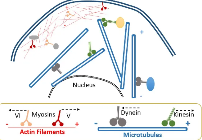
Figure 1-4: The cytoskeleton and the motor proteins.
The cytoskeleton is principally composed of actin and microtubule filaments. Actin filaments are thinner than microtubules and are mainly localized at the periphery of the cell. On the other hand, microtubules are all over the cytoplasm. Both filaments shape the cell structure but also serve as a network to transport various cargos trough the cell. For actin filaments, myosin is the main motor protein in charge of cargo trafficking. As for microtubules, kinesin and dynein move cargoes in a specific direction. Kinesin is responsible for anterograde movement, while dynein performs the retrograde movement.
In contrast to actin filaments, which principally support short movements, microtubules can support short and long movements that are regulated by the interaction of cargoes with the molecular motors, kinesin and dynein (Sheetz, 1996). In general, kinesin will take charge
+
-
+
22 of anterograde movements, which means toward the periphery of the cell, while dynein will control retrograde trafficking, toward the MTOC or nucleus of the cell (Mallik and Gross, 2004). To note, movement of cargoes over filaments required more than one motor protein, depending on the size of the cargo as well as to the distance to cover. Several kinesin motors can work together to accomplish the movement of cargo from point a to point b. In parallel, the tug of war model, an existing theory, suggests that motor proteins with different polarities, like kinesin and dynein, can compete to establish where the cargo or organelle will move (Müller et al., 2008). Even if several molecular motors with different direction affinities compete, the motor that loses the battle remains attached to the organelle to regulate further movement and to help the cargo overcome possible obstacles over the microtubule track (Bryantseva and Zhapparova, 2012).
Other proteins, called adaptor proteins, also play a vital role in determining which direction cargoes may travel. It has been shown that MIRO1), an adaptor protein, has some calcium binding EF motifs, thus depending on the intracellular Ca2+ concentration can dictate the movement of mitochondria according to the intracellular Ca2+ concentration. MIRO1 in collaboration with another adaptor protein MILTON interacts with kinesin as well as with mitochondria. This leads to the formation of a mitochondrial transport machinery that can better dictate where mitochondria need to move (Wang and Schwarz, 2009). Several other adaptor proteins interact with both the organelles and motor proteins to further regulate and coordinate movement of cargoes all over the cytoplasm (Mallik and Gross, 2004).
1.3. GIMAP family of proteins
GTPases of immune associated proteins (GIMAP) are coded by a set of Gimap genes located in tight clusters (Krücken et al., 2004; Nitta and Takahama, 2007). Interestingly, Gimap genes share a conserved region with the AvrRpt2-Induced Gene1 (AIG1), a gene in charge of the immune response against pathogens in plants (Liu et al., 2008). In fact, Gimap genes are present in vertebrates and plants, which shows their conserved role among species in immune responses. Gimap genes are expressed principally in immune tissues, where they
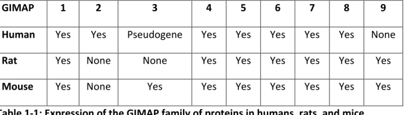
23 all seem to play a role at different stages of thymocyte maturation and T cell survival (Ciucci and Bosselut, 2014; Dion et al., 2005; Filén and Lahesmaa, 2010). Among the ten genes that compose the GIMAP family of proteins, humans express seven genes clustered on chromosome 7. In the same way, rats express seven genes clustered on chromosome 4, and mice express eight genes localized on chromosome 6 (Table-1) (Dion et al., 2005; Krücken et al., 2005, 2004).
GIMAP 1 2 3 4 5 6 7 8 9
Human Yes Yes Pseudogene Yes Yes Yes Yes Yes None
Rat Yes None None Yes Yes Yes Yes Yes Yes
Mouse Yes None Yes Yes Yes Yes Yes Yes Yes
Table 1-1: Expression of the GIMAP family of proteins in humans, rats, and mice.
In addition to plants, where the immune nucleotide associated genes have been studied for their importance as a defense mechanism against pathogens. In mammals, the most evolved members of the family of Gimap proteins have been investigated, principally, in rats, mice, and humans. Phylogenetical tree comparison between the IAN/GIMAP proteins in higher plants, humans, mice, and rats, show how different family members are most closely related to them, than with others and may explain why some members have been associated with similar physiological roles (Figure 1-5).
24 Figure 1-5: Phylogenetic tree of IAN/GIMAP proteins expression in Arabidopsis thaliana, Homo sapiens, Mus musculus and Rattus norvegicus.
A phylogenetic tree is showing the evolutionary relationships among the first IAN conserved protein in plants with the GIMAP family of proteins in the three principal organisms where the proteins have been studied: humans, mice, and rats. The phylogenetic tree was generated after protein alignment of the different members of the IAN/GIMAP family of proteins with Clustal Omega bioinformatics tool (Sievers et al., 2011).
All GIMAP proteins have a molecular mass between 33 and 38 kDa, except for GIMAP8, whose molecular weight is around 75 kDa. Similarly, they contain a conserved AIG1 domain at the N-terminal, where five GTP/GDP binding motifs, G1, G2, G3, G4, and G5 have been identified (Nitta and Takahama, 2007). Despite being small GTPases, they are not categorized in the known small GTPases families, RAS, RHO or Rab, making them a new subclass of small GTPases. Although they do not classify in the regular groups of GTPases, phylogenetical studies have shown that they conserved a canonical fold on their G domain
25 structure, which relates GIMAP proteins to the septin and dynamin GTPases (Schwefel et al., 2010). With regards to their GTPase activity, few members have been studied for their capacity to hydrolyse or bind to GDP and GTP. Among the first members to be explored were mouse GIMAP3, which showed GTP binding activity (Dahéron et al., 2001) and human GIMAP4, which showed a capacity to bind GDP and GTP as well as to exhibit an intrinsic GTPase activity (Cambot et al., 2002). More detail and structural studies performed on human GIMAP2 and GIMAP7 have shown that GIMAP2 can also bind GTP. Interestingly, this group demonstrated that GIMAP7 was able to stimulate its own GTPase activity, and that of GIMAP2 by dimerization (Schwefel et al., 2013). Moreover, Schwefel et al. suggested that the ability of the GIMAP family to dimerize and form oligomers may constitute an important mechanism to activate their GTPase activity. In parallel, ongoing unpublished data by the same group attributes human GIMAP5 the capacity to bind to GTP. In parallel, ongoing unpublished data by the same group attributes human GIMAP5 the capacity to bind to GTP, in a similar way as GIMAP2 (Schwefel and Daumke, 2011).
Besides the AIG1 conserved domain, all GIMAP family members contain a coiled-coiled domain, which may serve for protein-protein interactions. GIMAP proteins 1, 3 and 5 contain one hydrophobic transmembrane region at the C-terminal and GIMAP2 contains two hydrophobic C-terminal regions, which play a major role in anchoring them to different cellular compartments. (Dion et al., 2005; Krücken et al., 2004; Nitta et al., 2006). All GIMAPs are cytoplasmic intracellular proteins localized to distinct subcellular compartments, and they seem to have principally a physiological role in the development and maintenance of different cells that belong to the immune system (Filén and Lahesmaa, 2010).
GIMAP1 localizes to the ER and is primarily expressed in the lymph nodes tissues and spleen (Stamm et al., 2002). Upregulation of Gimap1 mRNA has been observed in thymocytes development from DN to DP state, (Dion et al., 2005; Nitta et al., 2006) while it has been observed to be differently regulated in T helper cell differentiation (Filén et al., 2009).